Cell-Free In vitro Protein Synthesis of Polyketide Synthase Proteins for Production of Natural Products
←
→
Page content transcription
If your browser does not render page correctly, please read the page content below
Abstract   1

   Cell-Free In vitro Protein Synthesis of
Polyketide Synthase Proteins for Production
            of Natural Products

            CHRISTOPHER ANTONIO SARMALES-MURGA

              Thesis submitted to the University of Ottawa
             in partial Fulfillment of the requirements for the
                           Master in Chemistry

           Department of Chemistry and Biomolecular Sciences
                            Faculty of Science
                           University of Ottawa

      © Christopher Antonio Sarmales-Murga, Ottawa, Canada, 2020
Abstract    ii

Abstract
Heterologous expression of multigene biosynthetic pathways is an essential tool in the study of

natural product biosynthesis. Due to its in vivo nature, this process is often limited by, for example,

toxicity of the encoded natural product or its biosynthetic proteins, or competition of the

biosynthetic proteins with other cellular enzymes for key small molecule building blocks. Cell-free

in vitro transcription and translation can overcome some of these limitations. Natural product

toxicity is rendered moot in cell-free systems since they are not alive and contain only the necessary

proteins, rRNAs, cofactors, substrates, and energy sources for transcription and translation of

proteins. As exogenous chemicals can be easily added to the system, building blocks supply issues

can be readily solved. We thus investigated using cell-free protein synthesis (CFPS) to biochemically

reconstitute the biosynthetic pathway for the fungal polyketide natural product monocillin

II. Significant optimization enabled cell-free expression of the full-length monocillin II polyketide

synthase (PKS) proteins Rdc5 and Rdc1 directly from plasmids containing their genes under control

of the T7 promoter. Correct post-translational modification of the apo-acyl carrier protein domain

of the PKS proteins was confirmed by SFP-mediated transfer of a fluorescently modified

phosphopantetheinyl group from a chemically modified CoA analog. Unfortunately, treatment of

the CFPS produced holo-PKS proteins with their native substrates, malonyl-CoA and NADPH, did not

lead to the expected production of detectable levels of monocillin II. Our work suggests that while

the CFPS system can generate full length PKS proteins that are sufficiently folded to be recognized,

and post-translationally modified by SFP, one or more of the required catalytic domains on these

large multidomain proteins is in an inactive state, preventing production of the final product.

Identifying non-functional domains, and addressing the issue, may make CFPS an appealing strategy

for characterizing PKS biosynthetic gene clusters and prototyping engineered PKS systems.
Acknowledgements        iii

Acknowledgements
I would like to primary thank Dr. Christopher N. Boddy for giving me the opportunity to

continue my studies, and to allow me to continue pursuing my interest and passion of research.

I’ve learned a significant amount of new techniques and skills which make me a better

researcher and student than I was when I first started my Masters.

It’s been a long and arduous endeavor with lots of up and downs, mostly downs, which is why I

would like to also thank the rest of The Boddy Lab members. They took me in as one of their

own and I’ve grown fond of them all, and definitely enjoyed my time with them. They’ve helped

me tremendously in term of answering any questions I would have had and/or helped me

improve any skill or techniques to be more efficient and/or to conduct experiments better.

Additionally, they’ve helped pass the time and keep me sane when things would go array or

when nothing would seem to be working. I respect them all and hope they succeed in whatever

endeavours they pursue.

Through the combined effort of Dr. Christopher Boddy, and the rest of the lab, I’ve been able to

complete my master’s study and to complete my research project. When I move onto my PhD, I

will definitely take the knowledge and teachings everyone has bestowed upon me and use it to

continue to grow and succeed as a researcher, while bestowing knowledge, teaching and

helping those around me.
Table Contents               iv

Table Contents

Table of Contents
         Abstract ........................................................................................................................................ii
         Acknowledgements.....................................................................................................................iii
         Table Contents ............................................................................................................................iv
         Table of Figures ...........................................................................................................................vi
         Table of Tables .......................................................................................................................... viii
         Table of Abbreviations ................................................................................................................ ix
Chapter 1: Heterologous Expression of Proteins .......................................................................... 1
         1.1…Importance of Heterologous Expression ............................................................................ 1
         1.2…Challenges of Heterologous Expression .............................................................................. 4
         1.3…Strategies for Heterologous Expression of BGCs ................................................................ 6
         1.4…Cell-Free In vitro Protein Synthesis ..................................................................................... 9
         1.5…Goals of the Project ........................................................................................................... 13
Chapter 2: Precursor Biosynthetic Pathway of Radicicol ........................................................... 16
2.1…Introduction .......................................................................................................................... 16
2.2…Results ................................................................................................................................... 19
         2.2.1…Expression of Rdc1 Thioesterase as CFPS proof of concept .......................................... 19
         2.2.2…Cell-Free Production of Fungal Monocillin II Biosynthetic Proteins .............................. 25
         2.2.3…Visualization of proteins synthesis through incorporation of BODIPY-Lysine ............... 28
         2.2.4…Phase I Monocillin II Production Assays ......................................................................... 30
         2.2.5…Visualization of the modification of the ACPs with BODIPY-CoA and SFP ..................... 31
         2.2.6…Supplementing PURExpression to increase protein production ................................... 34
         2.2.7…Production of an authentic Monocillin II standard in S. cerevisiae ............................... 37
         2.2.8… Phase II Monocillin II Production Assays ....................................................................... 38
         2.2.9…Combination experiments using purified and Cell-Free multi-domain proteins........... 42
         2.2.10…Production of TAGless Rdc5 and Rdc1 expression plasmids for Phase I Production
         assays of Monocillin II using TAGless Rdc5 and Rdc1 ............................................................... 44
         2.2.11… Production of Monocillin II within E. coli strain BAP1 ................................................ 46
2.3…Discussion and Final Thoughts .............................................................................................. 50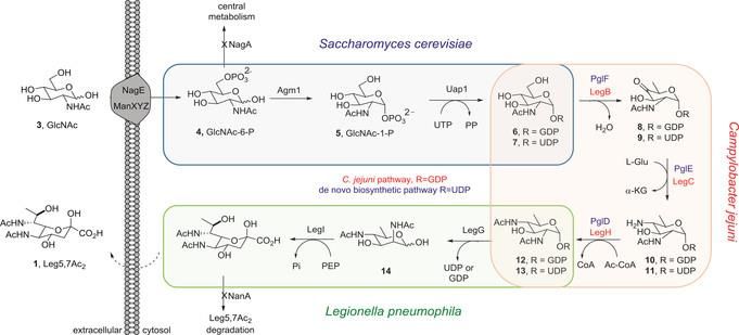
Table Contents                v

2.4…Methods ................................................................................................................................ 53
   Expression of Rdc1 Thioesterase as CFPS proof of concept ..................................................... 53
   Cell-Free Monocillin II Production ............................................................................................ 56
   Visualization of proteins synthesis through incorporation of BODIPY-Lysine.......................... 57
   Phase I Monocillin II Production Assays ................................................................................... 58
   Visualization of the modification of the ACPs with BODIPY-CoA and SFP................................ 60
   Phase II Monocillin II Production Assays .................................................................................. 63
   Supplementing PURExpression to increase protein production .............................................. 65
   Production of an authentic Monocillin II standard in S. cerevisiae .......................................... 66
   Phase III Monocillin II Production Assays ................................................................................. 68
   Combination experiments using purified multi-domain with Cell-Free multi-domain proteins
   ................................................................................................................................................... 71
   Production of TAGless Rdc5 and Rdc1 expression plasmids for Phase I Production assays of
   Monocillin II using TAGless Rdc5 and Rdc1 .............................................................................. 76
   Phase II Monocillin II Production Assays using TAGless proteins ............................................. 78
   Production of Monocillin II within E. coli strain BAP1 .............................................................. 80
References .................................................................................................................................... 85
Appendix ....................................................................................................................................... 88
   Primer List ................................................................................................................................. 88
   PCR Product Maps..................................................................................................................... 88
   Vector Maps .............................................................................................................................. 89
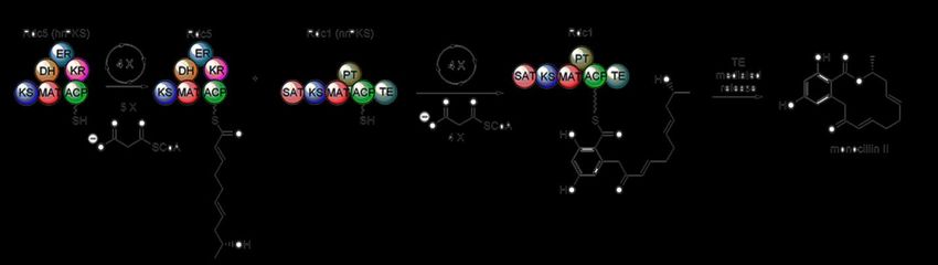
Table of Figures               vi

Table of Figures
Figure 1: Genetic Overview of Demethoxyviridin3 ......................................................................... 2
Figure 2: Congener discovery using eSNaPD5 ................................................................................. 3
Figure 3: De novo biosynthetic pathway of Leg5,7Ac24 ................................................................. 7
Figure 4: SYBR-green based qPCR analysis shows that transcription limits heterologous
production of oxytetracycline in E. coli.16....................................................................................... 9
Figure 5: Complex versus Defined CFPS systems.......................................................................... 10
Figure 6: Example of a multi-domain PKS protein ........................................................................ 13
Figure 8: Anti-HIS Tag antibody Western Blot of BAP1 extracts .................................................. 17
Figure 9: Biosynthetic Pathway of Monocillin II ........................................................................... 17
Figure 10: Acyl carrier proteins (ACP) are post-translationally modified by
phosphopantetheinylation. .......................................................................................................... 18
Figure 11: General overview of the study .................................................................................... 19
Figure 12: Schematic diagram of protein synthesis by PURExpress24 .......................................... 20
Figure 13: SDS-PAGE analysis of proteins of various sizes produced by the PURExpress® In vitro
Protein Synthesis Kit.24.................................................................................................................. 20
Figure 14: In vitro Biochemical assay to evaluate recombinant purified Rdc TE
enzymatic activity. ........................................................................................................................ 21
Figure 15: SDS-PAGE of cell free preparations of Rdc1TE. ........................................................... 22
Figure 16: Enzymatic assay HPLC chromatogram of Rdc1 TE with Substrate .............................. 23
Figure 17: Negative Control Assay HPLC Chromatogram of Substrate Assay ............................. 24
Figure 18: Initial Visualization of PURExpressed Radicicol biosynthetic pathway proteins via SDS-
PAGE .............................................................................................................................................. 26
Figure 19: Coomassie stain visualization of PURExpressed Radicicol biosynthetic pathway
proteins via SDS-PAGE .................................................................................................................. 27
Figure 20: Expression, and in vitro, incubation temperature manipulation studies with Rdc1 ... 28
Figure 21: Visualization of BODIPY-Lysine incorporated PURExpressed proteins........................ 29
Figure 22: Initial Monocillin II Production Assays HPLC Chromatograms .................................... 30
Figure 23: Overview of the modification of ACP .......................................................................... 32
Figure 24: Visualization of Post-Translational modification of Rdc5 and Rdc1 ACPs using SFP and
BODIPY-CoA .................................................................................................................................. 33
Table of Figures               vii

Figure 25: Coomassie staining of PURExpressed Rdc1 with Supplementary Solutions of
Substrates ..................................................................................................................................... 35
Figure 26: HPLC Chromatograms of Monocillin II standards ........................................................ 38
Figure 27: LC-MS Chromatograms of Yeast Extracts containing Monocillin II ............................. 39
Figure 28: LC -MS Positive Scan Chromatogram of Initial Monocillin II In vitro production assay
....................................................................................................................................................... 40
Figure 29: LC-MS Positive Scan Chromatograms for In vitro Experiments with different
supplementary buffers ................................................................................................................. 41
Figure 30: LC-MS Positive Scan Chromatogram for combination assays with PURExpressed Rdc5
& Rdc1 with heterologously expressed, and purified Rdc5 and Rdc1 .......................................... 43
Figure 32: Coomassie staining of PURExpressed Rdc1 and Rdc5 to verify integrity of TAGless
plasmids ........................................................................................................................................ 45
Figure 31: LC-MS Negative Scan Chromatogram of in vitro assays using TAGless Rdc5 and
TAGless Rdc1 ................................................................................................................................. 45
Figure 33: Coomassie staining of PURExpressed Rdc1 and Rdc5 to verify integrity of plasmid
switching ....................................................................................................................................... 47
Figure 34: Agarose Gel Electrophoresis separation of PCR amplified reversely transcribed
extracted RNA from BAP1 expressing Rdc5 and Rdc1 .................................................................. 48
Figure 35: Western detection of HIS tagged proteins within soluble and insoluble fractions of
BAP1 harboring pFM50 and pCAS04 ............................................................................................ 49
Figure 36: Brief Overview on the experiment .............................................................................. 50
Figure 37: HPLC Method of Separation for TE substrate assay .................................................... 55
Figure 38: HPLC separation conditions for scouting on Monocillin II........................................... 59
Figure 39: Initial separation method of compounds in Shimadzu UFLC for Mass Spectroscopy in
LCMS-2020 .................................................................................................................................... 69
Figure 40 Updated separation method of compounds in Shimadzu UFLC for Mass Spectroscopy
in LCMS-2020 ................................................................................................................................ 69
Table of Tables              viii

Table of Tables
Table 1: Reaction conditions for PURExpress reactions along with ½ fraction and ¼ fraction .. 22
Table 2: Amino Acid Compositions and kDa of Rdc5, Rdc1 and SFP ............................................ 25
Table 3: Modern Standard PURExpress conditions ...................................................................... 33
Table 4: PURExpress conditions for Supplemental Reactions ...................................................... 34
Table 5: Standardized PURExpression Composition moving forward .......................................... 36
Table 6: Reaction protocol for CFPS production of proteins. ....................................................... 53
Table 7: Content volumes of reactions in 2.7.3.B ......................................................................... 60
Table 8: Composition matrix of Supplements in Supplementary Solution for Corresponding Lane
in Figure 12a.................................................................................................................................. 65
Table 9: Composition matrix of Supplements in Supplementary Solution for Corresponding Lane
in Figure 12b ................................................................................................................................. 66
Table 10: Composition of investigation of supplemented buffer on production of Monocillin II 71
Table 11: Standard PCR concentrations used ............................................................................... 72
Table 12: Standard PCR method with 2 Variable Temperatures .................................................. 72
Table 13: Standard imidazole fractions used for protein purification.......................................... 74
Table of Abbreviations       ix

      Table of Abbreviations

      ADP    Adenosine diphosphate                               PKS Polyketide Synthase

      ATP    Adenosine triphosphate                             hrPKS Highly Reducing PKS

       CE    Carboxylesterase                                   nrPKS Non-Reducing PKS

      kDa    kilodalton                                            KS Ketosynthase

       EPI   Epoxyketone proteasome inhibitors                  M/AT Malonyl/ Acyl Transferase

     GDH     Glucose dehydrogenase                                ER Enoyl Reductase

       HIS   Hexahistidine                                        DH Dehydratase

     HPLC    High Pressure Liquid Chromatography                  KR Keto Reductase

      Kan    Kanamycin                                           ACP Acyl Carrier Protein

   LC - MS   Liquid Chromatography - Mass Spectroscopy           CoA    Coenzyme A

    mRNA     Messenger Ribonucleic Acid                           TE    Thioesterase

   NADPH     Nicotinamide Adenine Diphosphate Reduced           CFPS Cell-Free Protein Synthesis
                                                                         PURExpress® In vitro Protein
     NMR     Nuclear Magnetic Resonance                    PURExpress
                                                                        Synthesis Kit
    rNTPs    Ribonucleotide triphosphate                         BGC    Biosynthetic Gene Cluster

  TAGless    A protein lacking a tag

     tRNA    Transfer Ribonucleic Acid

       UV Ultraviolet

   PPTase Phosphopanetheinyl Transferase

    RNAP RNA Polymerase
             Sodium dodecyl sulfate - Polyacrylamide Gel
SDS - PAGE
             Electrophoresis
1.1…Importance of Heterologous Expression 1 Chapter 1: Heterologous Expression of Proteins 1.1…Importance of Heterologous Expression Natural product biosynthesis occurs in all organisms, producing secondary metabolites for specific uses and functions within the organisms. These compounds have a wide range of biological activity, including antibiotic and anticancer activity. Their potent biological function coupled with their diverse and complex structures makes their biosynthesis particularly interesting.1 An essential tool in understanding biosynthesis is the heterologous expression of a biosynthetic gene clusters (BGC), which enables production of the biosynthetic proteins and thus the secondary metabolite in a new (heterologous) host. Heterologous expression is an essential tool in the arsenal of natural product biosynthesis researchers. It is the most effective experiment for confirming that a proposed BGC is responsible for production of a specific natural product. For example, heterologous production of the natural product epothilone in Streptomyces coelicolor was used to unambiguously confirm that the gene cluster identified from the deltaproteobacteria Sorangium cellulosum was in fact the epothilone BGC.2 Epothilone, with a derivative called Ixabepilone as an approved anticancer drug, is very therapeutically important and an example of why it’s important to investigate biosynthetic pathways, as they can give rise to novel therapeutics such as epothilone.
1.1…Importance of Heterologous Expression                               2

Heterologous expression also plays a key role in deciphering how a biosynthetic pathway works

and allows assigning of function to the various genes in the pathway. Heterologous expression

enables genetic manipulation of the gene cluster, either through knockout or knockdown

experiments, especially when the native producer lacks molecular biology tools such as the

ability to be transformed, or to readily undergo homologous recombination. For example, Abe

and coworkers used heterologous expression in Aspergillus oryzae with demethoxyviridin

biosynthetic genes to characterize the enzymes that are responsible for the unusual pregnane

side-chain cleavage in this biosynthetic pathway (Figure 2).3

Figure 1: Genetic Overview of Demethoxyviridin3
Representative furanosteroids and biosynthetic gene cluster of demethoxyviridin (1). a Structures of wortmannin, viridin, and demethoxyviridin
(1). b Gene map of the demethoxyviridin biosynthetic gene cluster from Nodulisporium sp. (no. 65-12-7-1), consisting of 19 genes from vidA
(g3266) to vidS (g3284). The arrow indicates the direction from the start to the stop codon. Different types of genes are indicated by different
colors and among them red arrows indicate six CYP genes. Image republished from open access article “Biosynthetic pathway for
furanosteroid demethoxyviridin and identification of an unusual pregnane side-chain cleavage”, Wang et al., published in
Nature Communications, 2018.

It’s also possible to heterologously express individual proteins of a biosynthetic pathway and

combine them in an enzymatic assay to determine if they work sequentially to produce the next
1.1…Importance of Heterologous Expression                               3

precursor of the biosynthetic pathway. An example of this is the production of Radicicol

through heterologous expression, isolation and purification of it’s biosynthetic proteins to

produce each precursor in vitro until Radicicol is ultimately produced.4

Perhaps one of the more exciting examples of the power of heterologous expression is the

recent use of this tool to discover new compounds from the metagenome. By incorporating

fragments of the metagenome from soil samples into heterologous hosts it is now becoming

possible to produce and characterize novel natural products and/or biosynthetic pathways. An

example is the discovery of epoxyketone proteasome inhibitors (EPI) natural products from the

soil metagenome. EPIs inhibit the 20s proteasome through irreversible binding, which leads to a

toxic accumulation of polyubiquitinated proteins in the cell.5

Figure 2: Congener discovery using eSNaPD5
(i) eDNA is extracted from samples collected around the globe; these can be archived as large insert libraries if desired. (ii) NPSTs are then
generated by sequencing PCR amplicons amplified from eDNA templates with degenerate primers that target conserved biosynthetic motifs.
(iii) Analysis of NPST data using eSNaPD identifies NPSTs that derive from biosynthetic gene clusters of interest; these are mapped to collection
locations or positions within arrayed libraries using position information incorporated in the PCR primers. (iv) Biosynthetic gene clusters of
interest are then recovered from arrayed libraries and sequenced. Bioinformatics analysis of annotated eDNA gene clusters is then used to
prioritize clusters for heterologous expression studies. (v) Prioritized gene clusters are transferred to a laboratory-friendly host for
heterologous expression. (vi) LCMS and/or biological activity profiles of strains harboring eDNA clusters are compared with a vector control
strain to identify new metabolites for purification, structure elucidation, and bioactivity studies. Copyright 2015 National Academy of
Sciences

The authors amplified ~1 x 10⁶ unique environment sequences acquired from soil using

degenerate primers that target adenylation domains, a common biosynthetic motif of non

ribosomal peptides, and ketosynthase domains that are involved in polyketide biosynthesis.
1.2…Challenges of Heterologous Expression      4

Following amplification, the EPI biosynthetic pathways underwent bioinformatics evaluation

using multiple tools, such as antiSMASH, to predict the structures of the compounds encoded

by these pathways. From the many analyzed sequences, two were selected for heterologous

expression, with Streptomyces as the host, and two novel potent 20S proteasome inhibitors,

AR412 and AR456, were discovered (Figure 2).5

As can be seen from these examples, heterologous expression has become a well-established

technique in natural product biosynthesis. It has enabled the discovery of new natural products,

biosynthetic pathways, and biosynthetic enzymes. While highly effective, heterologous

expression does have significant challenges that often limit its utility.

1.2…Challenges of Heterologous Expression
Not surprisingly, many of the challenges of heterologous expression can be attributed to

selection of the host organism. In many cases, the heterologous host may not be able to

express one or more of the biosynthetic genes in a functional state. For example, rabbit liver

carboxylesterase (CE) was expressed within E. coli, S. cerevisiae and other expression hosts to

compare the abilities of the host to produce folded, active CE.6 This study showed that although

significant amounts of the proteins were detected in E. coli, the recombinant protein had poor

solubility with very low levels of enzyme activity. Within S. cerevisiae no protein was detected.

Functionally active protein was only detected when the CE was heterologously expressed in

Pichia pastoris.6 Thus, while protein may be active and functional when expressed in one host,

it may not be expressed, or expressed in an active form, in another host.
1.2…Challenges of Heterologous Expression   5

A challenge unique to heterologous biosynthetic pathway expression is that the building blocks

required to produce its natural product may not be present in the heterologous host.

Coenzymes and substrates used in secondary metabolite biosynthesis do not necessarily have

broad distribution across the kingdom of life, and as such they may or may not be present

within a particular host. Furthermore, the enzymatic machinery for correct post-translational

modification of biosynthetic proteins often is highly selective to the producing organism and

related hosts. As such, the heterologous host may not be able to correctly post-translationally

modify biosynthetic proteins. A clear example occurs with acyl carrier proteins (ACPs) from

polyketide biosynthetic pathways. ACPs are important polyketide synthase enzymes and

require a phosphopantetheinyl group for activity, which is added post-translationally, to hold

the growing polyketide intermediate. While all bacteria possess phosphopantetheinyl

transferase (PPTase) enzymes required for adding the phosphopantetheinyl group from

coenzyme A (CoA) onto fatty acid synthase ACPs, these enzymes typically do not recognize

polyketide synthase ACPs.7 As such, many heterologous hosts, such as E. coli, are unable to

post-translationally modify polyketide synthase ACPS and thus cannot produce polyketide

natural products heterologously. To address this serious problem, E. coli has been genetically

engineered to produce the Bacillus subtilis PPTase SFP, which has broad substrate tolerance for

polyketide synthase ACP domains.8 While the solution is effective for E. coli expression of ACP

containing biosynthetic pathways, other heterologous hosts suffer from a lack of effective post-

translational modification of biosynthetic proteins.

Toxicity is also a significant issue when dealing with heterologous expression. Many

heterologously expressed proteins can be highly toxic to the producing organism. For example,
1.3…Strategies for Heterologous Expression of BGCs      6

the human antimicrobial protein CAP37, encoded by the gene AZU1, cannot be heterologously

expressed in bacteria in native form. In the context of heterologous expression of biosynthetic

pathways, toxicity is further complicated by the inherent biological activity of the natural

products and often of its intermediates.9 These compounds, and intermediates, can hinder the

viability of the host.1 Thus, when the biosynthetic pathway for the antibiotic natural product

kanamycin (Kan), is introduced into a bacteria lacking the resistance mechanism for Kan, the

expression of the pathway will simply kill the host.10

In addition, the misfolding of proteins, along with toxicity of the encoded proteins and

biosynthetic products, and heterologous expression of biosynthetic pathways suffers from

additional challenges. A significant challenge is ensuring the heterologous host effectively

transcribes all of the biosynthetic genes. Lacking effective transcription of a single gene can

prevent an entire pathway from functioning, thus amplifying the challenge of a problem that

can typically be readily solved for a single gene system. As such, this is a significant problem,

with many of the common strategies for heterologous expression being based on specific

solutions to this fundamental challenge.

1.3…Strategies for Heterologous Expression of BGCs
Perhaps the most common heterologous expression strategy employed is introduction a

biosynthetic gene cluster into a very related host.11 As many bioactive natural products are

produced by actinobacteria, the model actinobacteria Streptomyces coelicolor and the related

strain Streptomyces lividans are commonly used12. A major advantage of this strategy is that

native BCGs can be used for heterologous expression, as typically the promoters used in

actinobacterial BGCs are recognized and expressed by these two streptomyces. The ability to
1.3…Strategies for Heterologous Expression of BGCs                                7

use native biosynthetic pathways is particularly appealing since these pathways typically

contain six or more genes which span, in many cases, 25-35kb in size. Thus, capturing the native

BGC from a genomic library13 or via the more modern transformation assisted recombination14

can readily lead to the required vector. This strategy for heterologous expression has been

highly successful in producing a wide number of polyketide natural products. It, however, can

be challenging to implement large BGCs (>35 kb), such as those encoding very large modular

polyketide synthases and non-ribosomal peptide synthetases.

Figure 3: De novo biosynthetic pathway of Leg5,7Ac24
Production in E. coli. Enzymes listed in blue are from the engineered UDP-linked pathway and those in red from the native C. jejuni GDP-linked
biosynthetic pathway. Used with permission© 2016 WILEY‐VCH Verlag GmbH & Co. KGaA, Weinheim

An alternative strategy for heterologous expression relies on re-cloning each gene of a BGC

under the control of a promoter known to function in the native host. E. coli has proved to be a

good host for this approach as strategies for ensuring transcription are well studied and

documented. For example, the introduction of multiple biosynthetic genes, each under the
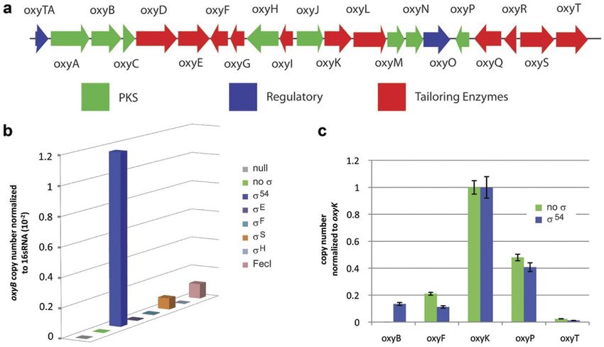
1.3…Strategies for Heterologous Expression of BGCs       8

control of the T7 promoter, into E. coli enabled it to produce legionaminic acid (Figure 3).

Legionaminic acid, a nonulosonic acid, is found on cellular surfaces of some bacteria with its

presence being correlated with virulence in humans. While there have been total synthesis

methods to produce legionaminic acid, they’re highly demanding and very low yielding.

Heterologous expression made it possible to produce significant quantities of Legionaminic acid

(121 mgL-1 of cell culture).4

One of the newest strategies for heterologous expression, has been to activate transcription of

an otherwise silent unexpressed BGC by expression of a transcriptional factor (Figure 4). An

excellent example of this can be seen in the heterologous production of the polyketide

oxytetracycline in E. coli. Direct introduction of the Streptomyces rimosus oxytetracycline gene

cluster into E. coli produced no oxytetracycline. This was attributed to the lack of transcription

of one of the key operons. Sigma (σ) factors control the specificity of gene transcription by

binding to RNA polymerase (RNAP) and recruiting it to promoter sequences upstream of the

gene to be transcribed.15 Within the context of this example (Figure 4b), these sigma factors

were heterologously expressed in the presence of the oxytetracycline BGC and transcription of

the key oxyB containing operon was quantified. Expression of σ54 significantly increased the

transcript of the oxyB containing transcript, and enabled production of oxytetracycline in E.

coli.16 Ultimately, the productivity of this system was limited by the inherent antibacterial

activity of the oxytetracycline produced from the BGC.

While these examples showcase the diverse strategies for heterologous expression, it is clear

that they do not address all the challenges associated heterologous production of natural

products. As all are cell based, they invariably suffer from toxicity of either proteins from the
1.4…Cell-Free In vitro Protein Synthesis                       9

biosynthetic pathway or the encoded natural product. Moreover, they all require the

generation of complex vectors to introduce the BGC into the host. Lastly, they are dependent

on the production, and presence, of the natural product building blocks by the heterologous

hosts. This is frequently rate limiting, as was recently shown in the heterologous production of

cannabinoids in yeast.17 A number of these challenges can potentially be addressed by the use

of a cell-free in vitro transcription, and translation, approach to heterologous expression.

Figure 4: SYBR-green based qPCR analysis shows that transcription limits heterologous production of oxytetracycline in E. coli. 16
(a) The 32 kb oxytetracycline biosynthetic gene cluster is shown. Five putative operons, oxyABCDE, oxyIHGF, oxyJKLMNO, oxyRQP, and oxyST
are predicted for this gene cluster (b) qPCR analysis shows that over-expression of the alternative sigma factors σ54, σS and FecI enable
detectable levels of the oxyB transcript to be produced. Over-expression of no sigma factor, σE, σF and σH do not lead to detectable levels of
the oxyB transcript. (c) qPCR analysis shows that over-expression of the alternative sigma factor σ54 lead to detectable levels of transcripts for
all five putative operons in the oxytetracycline biosynthetic pathway. In the absence of σ54 over-expression, the oxyB transcript cannot be
detected. Used under open access permission.

1.4…Cell-Free In vitro Protein Synthesis
The first successful attempt to use cell-free extracts was conducted by Eduard Buchner in 1897,

where he used the extracts from yeast to convert sugar to ethanol and carbon dioxide. He was

awarded the Chemistry Nobel Prize in 1907. He demonstrated that it is possible to use the
1.4…Cell-Free In vitro Protein Synthesis                        10

      extracts, absent of live cells, to conduct in vitro experiments 18. This was taken a step further in

      1961 with Nirenberg and Matthaei when they used cellular extracts to decipher the 64 triplet

      codons in the genetic code by using nucleic acid homo polymers to translate specific amino

      acids.19,20 Since then, these cellular extracts have been used as bioreactors to make proteins21,

      and refined to develop easy to use protocols and commercial kits for the production of high

      purity cell-free recombinant proteins.

      There are two strategies for the preparing cell-free in vitro protein synthesis (CFPS) systems, the

      complex and the defined systems. The complex variety entails direct lyses of the cells supplying

      the machinery for in vitro transcription and translation in S30 buffer. S30 contains dithiothreitol

Figure 5: Complex versus Defined CFPS systems.
Complex CFPS systems are generated by direct cell lysis and contain all the soluble cellular components from the original cells. Defined systems contain only
purified biochemical proteins, ribosomes, and metabolites needed to effect transcription and translation.
1.4…Cell-Free In vitro Protein Synthesis 11 (DTT) and it requires addition of only a few reagents, such as nucleotides, amino acids, a source of ATP, and double stranded DNA containing a gene with the appropriate promoter to initiate in vitro transcription, and subsequent translation, of the protein of interest. Complex systems have several advantages and disadvantages. The simplicity of the preparation is the major advantage to this strategy. It’s main disadvantage is the fact that along with the components essential for expression of genetic material, the mixture is composed of many non- essential proteins, cofactors and substrates that can not only influence the expression of desired protein(s), but can potentially impact the ability of the protein products to function correctly. In addition, there can be a noticeable batch-to-batch variability for complex CFPS mixtures that can impact reproducibility, particularly with targets that push the limits of in vitro transcription and translation. These disadvantages are relevant to heterologous production of biosynthetic gene clusters. In addition, the complex mixtures generated from these CFPS systems add to the challenge of detecting and quantifying natural product produced from cell- free biosynthesis. Thus, while the complex form of cell-free in vitro protein synthesis is a versatile tool for protein expression, it is not ideally suited for heterologous production of biosynthetic gene clusters.22 The second strategy for CFPS has a defined set of components, making the system much less complex when compared to the complex strategy. The defined form, which can be purchased as a kit, requires addition of all the recombinantly expressed and purified proteins essential for transcription, translation and ATP production to power these activities.23 In addition to these purified proteins, purified ribosomes are also added to the system, thus providing all the components needed for in vitro transcription and translation
1.4…Cell-Free In vitro Protein Synthesis   12

A clear advantage of the defined kit is the well-defined set of concentrations of required

proteins, cofactors, building blocks as well as the lack of extraneous protein components, which

results in a much cleaner in vitro assay. In addition, to facilitate isolation of the cell-free

produced proteins, typically a tag is incorporated into the proteins used for transcription and

translation, enabling their removal by affinity chromatography.22 The primary drawback of this

strategy is the cost. An example of the defined system is the PURExpress® In vitro Protein

Synthesis Kit, from New England Biolabs. In this kit, recombinant proteins for transcription,

translation and ATP production were individually expressed and purified through use of a

hexahistidine (HIS) tag. Thus, all protein components of the kit contain a HIS tag. Transcription

is performed by the T7 polymerase, thus genes possessing an upstream T7 promoter can be

readily transcribed by this system.23,24

Cell-free heterologous expression of biosynthetic pathways is under-explored in the literatures

though it has a number of potential advantages. As the cell-free system lacks the vast majority

of cellular processes, it is less likely to be impacted by the toxicity of the biosynthetic proteins

or the natural products they encode. Moreover, as the cell-free systems lacks encapsulation by

a membrane, the addition of target DNA, natural products, building blocks and atypical

cofactors is simplified. Furthermore, as no other cellular processes are competing for resources,

it is possible to express a significant amount of proteins in very short time frames.

While there are examples of producing simple natural products by cell-free heterologous

expression,25 there are very limiting examples of producing natural products encoded by

complex multi-domain proteins like polyketide synthases or non-ribosomal peptide synthases.

The complex biosynthetic pathways produce some of the more potent and biologically relevant
1.5…Goals of the Project     13

       natural products known. Cell-free protein synthesis was used to investigate the expression of

       two non-ribosomal peptide synthase biosynthetic proteins, GrsA and GrsB1, from the

       gramicidin S biosynthetic pathway.26 Together GrsA and GrsB1 are known to produce a d-Phe-l-

       Pro diketopiperazine product. Cyclic dipeptide products like this are produced across a wide

       range of organisms including bacteria, fungi and plants.27 Using a complex cell-free production

       system, the authors were able to produce detectable quantities of diketopiperazine from their

       cell-free experiment, suggesting production of complex biosynthetic pathways was feasible.

       1.5…Goals of the Project
       While heterologous expression is an essential tool for biosynthetic pathway expression, it is still

       plagued by problems with toxicity of the biosynthetic proteins, the natural products they

                                                      produce, as well as challenges related to cloning of the BGCs.

                                                      Cell-free protein synthesis possesses a number of advantages for

                                                      biosynthetic pathway expression, including reduced impact of

                                                      protein and metabolite toxicity, simplified cloning requirements,
Figure 6: Example of a multi-domain PKS protein
Rdc5, a large (260 kDa) highly reducing PKS           as well as in the case of the defined cell-free protein expression
protein from Radicicol Biosynthetic Pathway.
Composed of keto-synthase (KS), malonyl/acyl
transferase (MAT), dehydratase (DH), enoyl            systems, a background matrix of reduced complexity facilitating
reductase (ER), keto reductase (KR), and acyl
carrier protein (ACP). Each have individual
activities that together produce a natural product.   metabolite detection. Preliminary data with simple biosynthetic

       pathways, such as the violacein pathway,28 and more complex pathways such as a subsection of

       the gramicidin biosynthetic gene cluster29 shows that cell-free biosynthesis can produce the

       encoded natural product. In this project, I aim to determine if cell-free protein synthesis can be

       used to express a polyketide biosynthetic pathway. If successful, this would represent the first

       example of producing function polyketide synthase proteins by cell-free methods.
1.5…Goals of the Project 14 Polyketide Synthase are the key proteins responsible for the production of polyketide natural products. These pathways can exist in many different architectures, including type I, type II, and type III polyketide synthases. The work in this thesis focuses on type I polyketide synthases. These are large, multi-domain containing proteins (Figure 6). The core catalytic domains common to all type I polyketide synthases are ketosynthase (KS) domain, acyltransferase (AT or MAT) domain, and an acyl carrier protein (ACP). Additionally, polyketide synthase proteins can possess up to three reductive processing domains, including a ketoreductase (KR), dehydratase (DH) and enoylreductase (ER). These catalytic domains function similar to fatty acid synthases to add an acetate unit from malonyl-CoA onto a growing polyketide chain through a decarboxylative Claisen condensation. As they work together to affect this biological chemistry, this group of catalytic domains is often referred to as a module. For bacterial polyketide biosynthetic pathways, there are typical one module worth of catalytic domains for each acetate unit added to the growing polyletide, thus the biosynthetic pathways can be extremely large. For example, the pathway that produce erythromycin possesses six unique modules with over 25 unique domains. Because of the size and complexity of bacterial polyketide pathways, I chose to tackle a more simplified fungal polyketide synthase system.1,30 In fungal polyketide biosynthesis, typically only one or two modules of catalytic domains are used. These catalytic domains are used iteratively, delivering multiple equivalents of acetate from malonyl-CoA to the growing polyletide. Typically, there is a highly reducing polyketide synthase (hrPKS), which contains the three reductive processing domains as well as the KS, MAT, and ACP, as well as a non-reducing polyketide synthase (nrPKS) that does not possess these reductive catalytic domains.1,30 These two proteins work in concert to generate complex
1.5…Goals of the Project     15

natural products from a very streamlined set of proteins. As such, these fungal polyketide

pathways appeared to be an appealing starting point for investigating cell-free polyketide

biosynthesis. In the following chapter, I thus describe my work to produce the fungal polyketide

monocillin II through cell-free biosynthesis.
2.1…Introduction    16

Chapter 2: Precursor Biosynthetic Pathway of Radicicol

2.1…Introduction
2.1…I ntrodu ction

The key goal of this study is to produce for the first time a PKS biosynthetic pathway via a cell-

free expression system. The fungal polyketide synthase pathway encoding the natural product

radicicol, and its precursor Monocillin II31 was selected as the target pathway due to its

relatively simple biosynthetic pathway. It requires only two polyketide synthase proteins to

make this complex bioactive compound that competes with ATP for binding in the ADP/ATP

binding pocket of Hsp90.31 This biological activity is significant as Hsp90 plays an important role

in many cellular processes with activities involved in regulation of cell cycle, cell growth, cell

survival, apoptosis, angiogenesis and lastly oncogenesis.32 By affecting these cellular processes,

the compound, and by extension its biosynthetic pathway, is a significant subject for further

characterization.

Radicicol is biosynthesized by four key proteins known as Rdc5, Rdc1, Rdc2 and Rdc4. Rdc5 and

Rdc1 are type I, iterative PKS proteins. Rdc2 is a FADH dependant halogenase and Rdc4 is an

oxidase. Of the PKS proteins, Rdc5 is a highly reducing PKS, meaning that it will is reduce a

significant portion of the elongated polyketide chain prior to transferring it to Rdc1. Rdc1 is a

non-reducing PKS, that further elongates this chain and ultimately cyclizes it to form monocillin

II, a precursor of Radicicol. To convert monocillin II to radicicol, monocillin II is chlorinated by

Rdc2 to make Pochonin D, followed by epoxide formation via Rdc4, generating radicicol.31

The biosynthetic gene cluster responsible for formation of radicicol and monocillin II is well

characterized.31 The cluster was unambiguously identified by heterologous expression in S.
2.1…Introduction         17

cerevisiae and monocillin II has also been produced via in vitro enzymatic assay using

recombinant Rdc5 and Rdc1, supplemented with 2 mM Malonyl-CoA and NADPH.31 Malonyl-

CoA is the source of all the carbons in monocillin II and NADPH supplies hydride required for

reductions by the KR and ER domains. Thus, previous work had clearly identified the

biosynthetic genes and showed they could function as recombinant proteins in an in vitro

setting. This highly supported our proposed cell-free protein synthesis of monocillin II(Figure 7).

Figure 7: Biosynthetic Pathway of Monocillin II
Displayed are the proteins involved in the production of Monocillin II. Rdc5, a highly reducing PKS, and Rdc1, a non-reducing PKS, work in
conjunction to produce Monocillin II using Malonyl-CoA and NADPH.

A clear challenge for this project however was the recombinant production of the monocillin II

producing PKS proteins in E. coli did not produce soluble proteins. For example, expression of

                                  kDa              M                  Soluble              Insoluble
                                   250

                                   150

                                  100

                                   75

 Figure 8: Anti-HIS Tag antibody Western Blot of BAP1 extracts
 Anti-hexahistidine (HIS) antibody probed Western Blot of Rdc5 and Rdc1 expression in E. coli BAP1 cells. The expected full-length
 protein bands for Rdc5 and Rdc1 were not observed in the soluble fraction at 260 kDa and 228 kDa respectively. Denaturing lysis
 (insoluble fraction) shows significant His tagged protein is present suggesting Rdc5 and Rdc1 undergo aggregation and degradation.
2.1…Introduction 18
Rdc5 and Rdc1 under the control of the T7 promoter in the E. coli strain optimized for
polyketide synthase expression, BAP1, did not show any soluble full length Rdc5 and/or Rdc1.
(Figure 8). Unfortunately, all the proteins are produced as an aggregated, partially proteolyzed,
insoluble material.
Once soluble polyketide synthase proteins are produced, their acyl carrier proteins must be
post-translationally modified. This post-translational modification is catalyzed by a
phosphopantetheinyl transferase, such as the 26 kDa protein SFP from Bacillus subtilis.33 SFP
uses coenzyme A (CoA) as a substrate and adds the phosphopantetheinyl arm of CoA onto a
seryl side chain of ACP (Figure 9). This phosphopantetheinyl arm is essential as it is the covalent
attachment site for the growing polyketide product on the PKS enzyme.7
Coenzyme A 3’,5’ - PAP
Figure 9: Acyl carrier proteins (ACP) are post-translationally modified by phosphopantetheinylation.
The B. subtilis enzyme SFP is a broadly substrate tolerant phosphopantetheinyl transferase which transfers the phosphopantatheinyl group from
Coenzyme A on to the active site Ser of an ACP domain.
As the cellular hosts used to produce these cell-free expression systems do not naturally
contain SFP, it would need to be exogenously added, along with CoA, to insure that Rdc5 and
Rdc1 are correctly modified.2.2.1…Expression of Rdc1 Thioesterase as CFPS proof of concept 19 Once the biosynthetic Rdc5 and Rdc1 domains have been expressed in cell-free, and post- translationally modified using SFP and CoA, the final step would be to assay the enzymes for in vitro production of monocillin II by treatment with malonyl-CoA and NADPH. Thus, the general overview for this project is visualized in Figure 10. Figure 10: General overview of the study 2.2…Results 2.2.1…Expression of Rdc1 Thioesterase as CFPS proof of concept As the defined form of CFPS is cleaner, simpler and more consistent, compared to the complex form, it was chosen for cell-free expression. The PURExpress® In vitro Protein Synthesis Kit, from New England Biolabs, was used due to its high quality and simplicity of use. The reaction is conducted by mixing two solutions, one containing the purified ribosomes and the other containing the recombinant proteins required for transcription, translation and energy production with the genes under T7 control encoding the desired proteins for expression.24 (Figure 11). The manufacturer notes that sufficient amounts of proteins can be synthesized, and visualized, post 2 hours of incubation at 37˚C24 (Figure 12).
2.2.1…Expression of Rdc1 Thioesterase as CFPS proof of concept                         20

   Figure 11: Schematic diagram of protein synthesis by PURExpress24

As can be seen in the “no DNA” lane of Figure 12, many prominent bands are visualized by SDS-

PAGE. These bands correspond to the various components of the PURExpress kit such as T7

polymerase and creatine phosphatase, for example. The following lanes show successful

expression of proteins of various sizes using the PURExpress® In vitro Protein Synthesis Kit. The

desired proteins are indicated with a red dot. This figure shows that proteins between 20-100

kDa can be successfully expressed in only 2 hours.

       Figure 12: SDS-PAGE analysis of proteins of various sizes produced by the PURExpress® In vitro Protein Synthesis Kit.24
2.2.1…Expression of Rdc1 Thioesterase as CFPS proof of concept 21
To evaluate our ability to monitor the enzymatic function of proteins produced via cell-free
systems, we tested a single catalytic domain from the monocillin II pathway, the C-terminal
thioesterase domain. Previous work had shown that the standalone C-terminal thioesterase
domain, RadTE, could effect macrocyclization of a synthetic thioester activated substrate34
(Figure 13).
A B
52 kDa
Figure 13: In vitro Biochemical assay to evaluate recombinant purified Rdc TE enzymatic activity.
Substrate (A) was synthesized and provided by Dr. Heberlig.
As the excised RadTE is significantly smaller than Rdc1, 52 kDa vs 228kDa, and as RadTE had
been successfully expressed in E.coli in a functional form, we were confident this experiment
would enable us to determine if cell-free expression conditions produced sufficient protein to
perform and detect biosynthetic reactions.
Some of the questions that will also be answered with the proof of concept will be:
1. Can a PKS domain, be expressed using CFPS?
2. Is the recombinant protein product enzymatically active?
3. Are there unexpected challenges in preparing and conducting the enzymatic assays?
Results from this proof of concept experiment are expected to be transferable to and inform
the more complex full length PKS proteins.2.2.1…Expression of Rdc1 Thioesterase as CFPS proof of concept 22
The components, and respective volumes, of each PURExpress® In vitro Protein Synthesis
reaction, and some examples of scaled down reactions, can be seen in Table 1. ½ and ¼ volume
scale of the PURExpress® In vitro Protein Synthesis Kit protocol would enable us to determine if
scaling down the reaction would produce not only detectable protein, but enough protein to
catalyze formation of a biochemical product that can then be detected by HPLC. For the
remainder of this thesis, PURExpress® In vitro Protein Synthesis will be referred to as
“PURExpress” and the proteins produced using this method will be referred to as
“PURExpressed” proteins.
Table 1: Reaction conditions for PURExpress reactions along with 1/2 fraction and 1/4 fraction
PURExpress ½ PURE ¼ PURE
Solution A 10.0 5.00 2.500
Solution B 07.5 3.75 1.875
DNA XX.X X.XX X.XXX
H2O XX.X X.XX X.XXX
Total 25.00 µL 12.50 µL 6.25 µL
Using PURExpression kit, RadTE could be readily expressed. SDS-PAGE analysis of PURExpressed
RadTE at ½ scale and ¼ scale (Figure 14B) showed the expected band at 52 kDa. In comparison,
the negative control, where no DNA encoding RadTE was added to the PURExpress system,
showed no detectable band for the protein.
B
Lane 1 Lane 2 Lane 3
A
Figure 14: SDS-PAGE of cell free preparations of Rdc1TE.
(A) The PURExpressed proteins were separated by SDS-PAGE and imaged using the Biorad stain free in gel fluorescence system. (B) An expansion of the
Rdc1TE bands. Lane 1: 1/2 PURE protocol (pMRH08); Lane 2: 1/4 Pure Protocol (pMRH08); Lane 3: Negative Control (Empty pET28).You can also read