Mycobacterial cell wall biosynthesis: a multifaceted antibiotic target
←
→
Page content transcription
If your browser does not render page correctly, please read the page content below
SPECIAL ISSUE REVIEW                                                                                                                                        116

                     Mycobacterial cell wall biosynthesis: a multifaceted
                     antibiotic target

                     KATHERINE A. ABRAHAMS and GURDYAL S. BESRA*
                     Institute of Microbiology and Infection, School of Biosciences, University of Birmingham, Edgbaston, Birmingham B15
                     2TT, UK

                     (Received 7 September 2016; revised 2 November 2016; accepted 8 November 2016; first published online 15 December 2016)

                     SUMMARY

                     Mycobacterium tuberculosis (Mtb), the etiological agent of tuberculosis (TB), is recognized as a global health emergency as
                     promoted by the World Health Organization. Over 1 million deaths per year, along with the emergence of multi- and
                     extensively-drug resistant strains of Mtb, have triggered intensive research into the pathogenicity and biochemistry of
                     this microorganism, guiding the development of anti-TB chemotherapeutic agents. The essential mycobacterial cell
                     wall, sharing some common features with all bacteria, represents an apparent ‘Achilles heel’ that has been targeted by
                     TB chemotherapy since the advent of TB treatment. This complex structure composed of three distinct layers, peptido-
                     glycan, arabinogalactan and mycolic acids, is vital in supporting cell growth, virulence and providing a barrier to antibio-
                     tics. The fundamental nature of cell wall synthesis and assembly has rendered the mycobacterial cell wall as the most
                     widely exploited target of anti-TB drugs. This review provides an overview of the biosynthesis of the prominent cell
                     wall components, highlighting the inhibitory mechanisms of existing clinical drugs and illustrating the potential of
                     other unexploited enzymes as future drug targets.

                     Key words: tuberculosis, cell wall, peptidoglycan, arabinogalactan, mycolic acids, antibiotics.

                     INTRODUCTION                                                                        enzymatic and phenotypic perspective) directed
                                                                                                         against the synthesis of this unique macromolecule
                     Mycobacterium tuberculosis (Mtb), the causative
                                                                                                         structure in Mtb. The Mtb cell envelope is an expan-
                     agent of tuberculosis (TB), is regarded as the
                                                                                                         sive structure and is summarized in Fig. 1. The inner
                     world’s most successful pathogen (Hingley-Wilson
                                                                                                         membrane phospholipid bilayer contains glycolipids
                     et al. 2003). Responsible for an estimated 1·4
                                                                                                         that extend into the periplasmic space. The essential
                     million deaths and 10·4 million new cases of TB,
                                                                                                         core cell wall structure is composed of three main
                     including 480 000 new cases of multi-drug resistant
                                                                                                         components: a cross-linked polymer of peptidogly-
                     (MDR)-TB in 2015 (World Health Organization,
                                                                                                         can, a highly branched arabinogalactan polysacchar-
                     2016), Mtb remains a global health emergency as
                                                                                                         ide, and long-chain mycolic acids. Intercalated into
                     declared by the World Health Organization
                                                                                                         the mycolate layer are solvent-extractable lipids
                     (WHO) (World Health Organization, 2014). New
                                                                                                         including non-covalently linked glycophospholipids
                     chemotherapeutic agents to complement or replace
                                                                                                         and inert waxes, forming the outer membrane. The
                     existing front-line treatment regimens are urgently
                                                                                                         capsule forms the outermost layer and is mainly com-
                     required to reduce treatment time (currently
                                                                                                         posed of proteins and polysaccharides. The lipid- and
                     6-month course) and to combat the increasing
                                                                                                         carbohydrate-rich layers of the cell wall serve not only
                     threat by this microorganism.
                                                                                                         as a permeability barrier, providing protection against
                        The distinguishing feature of mycobacteria, the
                                                                                                         hydrophilic compounds, but also are critical in patho-
                     complex cell wall, is a well-recognized drug target.
                                                                                                         genesis and survival. It is these traits that make the
                     The cell wall is common to all bacteria, both Gram-
                                                                                                         biosynthesis and assembly of the cell wall components
                     positive and Gram-negative, but can have vast differ-
                                                                                                         attractive drug targets. This review focuses on the
                     ences in terms of the biochemical and structural
                                                                                                         synthesis of the key cell wall components, highlight-
                     features. Over the past decade, extensive research
                                                                                                         ing previously validated targets and the ongoing
                     into cell wall assembly, aided by whole-genome
                                                                                                         drug discovery efforts to inhibit other essential
                     sequencing, has led to an increased understanding of
                                                                                                         enzymes in mycobacterial cell wall biosynthesis.
                     mycobacterial cell wall biosynthesis. This has pro-
                     moted further exploration into the discovery and
                     development of chemotherapeutic agents (from an                                     PEPTIDOGLYCAN

                                                                                                         Peptidoglycan is a major component of the cell wall
                     * Corresponding author: Institute of Microbiology and
                     Infection, School of Biosciences, University of                                     of both Gram-positive and Gram-negative bacteria
                     Birmingham, Edgbaston, Birmingham B15 2TT, UK.                                      (Vollmer et al. 2008). It is a polymer of alternating
                     E-mail: g.besra@bham.ac.uk                                                          N-acetylglucosamine and N-acetylmuramic acid

                   Parasitology (2018), 145, 116–133. © Cambridge University Press 2016. This is an Open Access article, distributed under the terms of
                   the Creative Commons Attribution licence (http://creativecommons.org/licenses/by/4.0/), which permits unrestricted re-use, distribu-
                   tion, and reproduction in any medium, provided the original work is properly cited.
                   doi:10.1017/S0031182016002377
Downloaded from https://www.cambridge.org/core. IP address: 46.4.80.155, on 26 Nov 2021 at 10:33:58, subject to the Cambridge Core terms of use, available at https://www.cambridge.org/core/terms.
https://doi.org/10.1017/S0031182016002377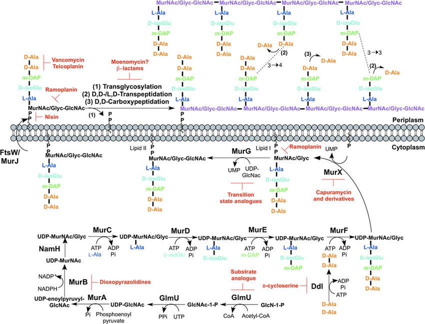
Mycobacterial cell wall biosynthesis                                                                                                                        117

                Fig. 1. The mycobacterial cell wall. A schematic representation of the mycobacterial cell wall, depicting the prominent
                features, including the glycolipids (PIMs, phosphatidyl-myo-inositol mannosides; LM, lipomannan; LAM,
                lipoarabinomannan; ManLAM, mannosylated lipoarabinomannan), peptidoglycan, arabinogalactan and mycolic acids.
                Intercalated into the mycolate layer are the acyl lipids (including TMM, trehalose monomycolate; TDM, trehalose
                dimycolate; DAT, diacyltrehalose; PAT, polyacyltrehalose; PDIM, phthiocerol dimycocerosate; SGL, sulfoglycolipid).
                The capsular material is not illustrated.

                residues via β(1 → 4) linkages with side chains of                                 acetyl-CoA is transferred to glucosamine-1-phosphate
                amino acids cross-linked by transpeptide bridges                                   (GlcN-1-P) to produce N-acetylglucosamine-1-
                (Brennan and Nikaido, 1995). Mycobacterial pep-                                    phosphate (GlcNAc-1-P). Secondly, uridine-5′-mono-
                tidoglycan has a number of unique features that                                    phosphate from UTP is transferred to GlcNAc-1-P to
                diversifies the cell wall from the typical structure                                yield UDP-GlcNAc (Zhang et al. 2009). The abun-
                including N-glycolyl- and N-acetyl-muramic acid                                    dance of GlcNAc-1-P in eukaryotes (Mio et al. 1998)
                residues (Mahapatra et al. 2005a), amidation of the                                and the functional similarity of the GlmU uridyltrans-
                carboxylic acids in the peptide stems (Mahapatra                                   ferase with human enzymes (Peneff et al. 2001) makes
                et al. 2005b) and additional glycine or serine residues                            this domain an unsuitable drug target (Rani and Khan,
                (Vollmer et al. 2008). The function of peptidoglycan                               2016). However, the absence of GlcN-1-P from
                is not only to provide shape and rigidity, but it is                               humans makes the acetyltransferase domain a potential
                responsible for counteracting turgor pressure and                                  target (Mio et al. 1998). Efforts to identify inhibitors of
                hence it is essential for growth and survival                                      this domain are underway (Tran et al. 2013). A sub-
                (Vollmer et al. 2008). Peptidoglycan is unique to                                  strate analogue of GlcN-1-P has been designed and
                bacterial cells, and it is this property that has led to                           exhibits inhibitory effect against GlmU, providing a
                numerous enzymes involved in its synthesis to be                                   candidate for further optimization (Li et al. 2011).
                targeted by potent antibiotics, with others represent-                                The next step involves the generation of the UDP-N-
                ing attractive targets in the development of future                                acetylmuramic acid (UDP-MurNAc)-pentapeptide,
                antibiotics.                                                                       which is synthesized in a sequential pathway catalysed
                                                                                                   by the Mur ligases A–F (Barreteau et al. 2008),
                                                                                                   whereby most of the Mtb genes have been found
                PEPTIDOGLYCAN BIOSYNTHESIS
                                                                                                   through homology. MurA, a UDP-N-acetylglucosa-
                The biosynthesis of peptidoglycan is summarized in                                 mine 1-carboxyvinyltransferase, and MurB, a UDP-
                Fig. 2. The first committed step is the generation of                               N-acetylenolpyruvoylglucosamine reductase, are
                uridine diphosphate-N-acetylglucosamine (UDP-                                      involved in generating UDP-MurNAc from UDP-
                GlcNAc). This is catalysed by the acetyltransferase                                GlcNAc, by first the addition of the enoylpyruvyl
                and uridyltransferase activities of GlmU (Zhang                                    moiety of PEP, followed by reduction to a lactoyl
                et al. 2009), where first the acetyl group from                                     ether moiety via NADPH. At this point, NamH, a

Downloaded from https://www.cambridge.org/core. IP address: 46.4.80.155, on 26 Nov 2021 at 10:33:58, subject to the Cambridge Core terms of use, available at https://www.cambridge.org/core/terms.
https://doi.org/10.1017/S0031182016002377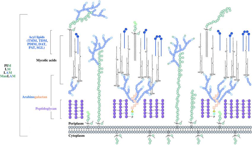
Katherine A. Abrahams and Gurdyal S. Besra                                                                                                                  118

                     Fig. 2. Inhibitors targeting peptidoglycan biosynthesis. The roles of the key enzymes involved in peptidoglycan
                     biosynthesis are illustrated. Reported inhibitors are shown in red.

                     UDP-N-acetylmuramic acid hydroxylase, hydroxy-                                      Mycobacterium smegmatis, and therefore is not condu-
                     lates UDP-MurNAc to UDP-N-glycolylmuramic                                           sive to a characteristic target property. However, gene
                     acid (UDP-MurNGlyc), providing both types of                                        deletion results in a strain hypersusceptible to β-
                     UDP-muramyl substrates; Mtb cell walls are domi-                                    lactam antibiotics and lysozyme and therefore inhibitors
                     nated by the latter (Mahapatra et al. 2005a). This struc-                           of NamH could potentiate the effect of β-lactams
                     tural modification is unique to mycobacteria (and                                    (Raymond et al. 2005).
                     closely related genera) and is considered to increase                                  The pentapeptide chain is incorporated onto the
                     the intrinsic strength of peptidoglycan, by potentially                             UDP-MurNAc/Glyc substrates by the successive
                     alleviating susceptibility to lysozyme and providing                                addition of amino acid residues L-alanine, D-isogluta-
                     sites for additional hydrogen bonding (Raymond et al.                               mate, meso-diaminopimelate (m-DAP) and D-alanyl-
                     2005). Inhibitors of Mtb MurA and MurB are yet to                                   D-alanine [generated by the D-Ala: D-Ala ligase
                     be discovered. Whilst the natural product, broad spec-                              (Ddl)] by the ATP-dependent Mur ligases C-F
                     trum antibiotic, fosfomycin, targets Gram-negative                                  respectively (Munshi et al. 2013). This results in
                     MurA, the critical residue for inhibition is absent in                              the muramyl-pentapeptide product, UDP-MurNAc/
                     Mtb, providing intrinsic resistance against this anti-                              Glyc-L-Ala-D-isoGlu-m-DAP-D-Ala-D-Ala, also
                     biotic (Kim et al. 1996). Consequently, an inhibitor                                known as Park’s nucleotide (Kurosu et al. 2007).
                     with a new mode of action is required to target Mtb                                 Despite the different amino acid specificities, the
                     MurA. A limited number of inhibitors have been                                      four ligases share common properties: the reaction
                     reported against MurB. Molecular dynamics and                                       mechanism; six invariant ‘Mur’ residues; an ATP-
                     docking studies of existing MurB inhibitors (3,5-dioxo-                             binding consensus; three-dimensional structural
                     pyrazolidine derivatives) onto the Mtb MurB structure                               domains (Barreteau et al. 2008). Due to these similar-
                     reveal the potential potent activity of these compounds,                            ities, it is plausible that a single inhibitor could target
                     which can be used to guide future structure-based drug                              more than one Mur ligase and such inhibitors have
                     design (Kumar et al. 2011). Inhibitors of NamH have                                 been reported in the literature (Tomasic et al. 2010).
                     not been documented; namH is not essential in                                       Numerous small molecule inhibitors of the Mur

Downloaded from https://www.cambridge.org/core. IP address: 46.4.80.155, on 26 Nov 2021 at 10:33:58, subject to the Cambridge Core terms of use, available at https://www.cambridge.org/core/terms.
https://doi.org/10.1017/S0031182016002377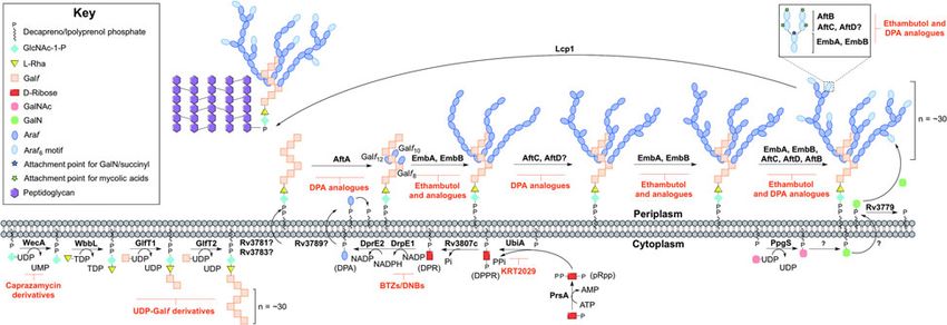
Mycobacterial cell wall biosynthesis                                                                                                                        119

                ligases have been discovered and are the subject of an                             (PBPs) (Sauvage et al. 2008). Bifunctional PBPs
                extensive review (Hrast et al. 2014). In most cases, the                           (PonA1/PBP1 and PonA2/PBP2) possess transgly-
                inhibitors were identified from high-throughput                                     cosylase and transpeptidase domains. The former
                screening (HTS) campaigns of compound libraries                                    domain is responsible for linking the disaccharide
                employing in vitro kinetic assays. These types of in                               building blocks of Lipid II to the pre-existing
                vitro screening methods are limited in use against                                 glycan chains (with the concomitant release of deca-
                Mtb Mur ligases given that only MurC and MurE                                      prenyl pyrophosphate), whereas the latter domain
                have been biochemically characterized (Mahapatra                                   catalyses the formation of the classical (3 → 4) cross-
                et al. 2000; Li et al. 2011). This dictates the next                               links, between m-DAP and D-Ala of the adjacent
                rational step towards the target-based discovery of                                pentapeptide chains, with the cleavage of the terminal
                Mur ligase inhibitors. Ddl is the target of D-cycloser-                            D-Ala. D,D-transpeptidation and D,D-carboxypep-
                ine (Bruning et al. 2011), a second-line drug used in                              tidation is performed by the monofunctional PBPs,
                the treatment of TB, and is at the cornerstone of treat-                           both resulting in the cleavage of the terminal D-Ala
                ment for MDR and extensively drug resistant (XDR)-                                 of the peptide stem (Goffin and Ghuysen, 2002).
                TB. D-cycloserine acts as a structural analogue of D-                              Only 20% of the cross-links in Mtb peptidoglycan
                Ala, inhibiting the binding of either D-Ala to Ddl                                 are (3 → 4) (Kumar et al. 2012). The majority are
                (Prosser and de Carvalho, 2013a, b).                                               (3 → 3) links between two tetrapeptide stems, with
                   The first membrane-anchored peptidoglycan pre-                                   the release of the fourth position D-Ala (Lavollay
                cursor is generated by the translocation of Park’s                                 et al. 2008). This reaction is catalysed by the L,D-
                nucleotide to decaprenyl phosphate (C50-P), cata-                                  transpeptidases, with D,D-carboxypeptidation as a
                lysed by MurX (also known as MraY), forming                                        prerequisite activity. The L,D-transpeptidases are
                Lipid I (Kurosu et al. 2007). There are a number                                   structurally unrelated to PBPs, with different active
                of nucleoside-based complex natural products that                                  site residues (cysteine and serine, respectively)
                inhibit MurX, including muraymycin, liposidomy-                                    (Mainardi et al. 2005; Biarrotte-Sorin et al. 2006).
                cin, caprazamycin and capuramycin (Dini, 2005).                                    The β-lactam antibiotics have been used in the treat-
                Capuramycin and derivatives exhibit killing in                                     ment of bacterial infections for nearly a century, and
                vitro and in vivo and more significantly, analogues                                 gave rise to the discovery of their target, the PBPs.
                of capuramycin have been shown to kill non-                                        The L,D-transpeptidases are resistant to most β-
                replicating Mtb, a feature not common to the major-                                lactam antibiotics, except the carbapenems (Dubee
                ity of cell wall biosynthesis inhibitors (Koga et al.                              et al. 2012). Until recently, β-lactams were not con-
                2004; Reddy et al. 2008; Nikonenko et al. 2009;                                    sidered for use in the treatment of TB, due to the
                Siricilla et al. 2015). Significantly, the analogue                                 expression of a broad-spectrum β-lactamase, BlaC.
                SQ641 is in preclinical development (http://www.                                   However, it has been shown that BlaC is irreversibly
                newtbdrugs.org).                                                                   inactivated by clavulanic acid, yet hydrolyses carba-
                   The final intracellular step of peptidoglycan syn-                               penems at a low rate (Hugonnet et al. 2009).
                thesis is performed by the glycosyltransferase,                                    Combined treatment of the β-lactam with the β-lacta-
                MurG. A β(1 → 4) linkage between GlcNAc (from                                      mase inhibitor has been shown to be bactericidal
                UDP-GlcNAc) and MurNAc/Glyc of Lipid I is                                          against both replicating and non-replicating forms
                formed, leading to the generation of Lipid II, the                                 of Mtb, and combinations are now being explored
                monomeric building block of peptidoglycan                                          in clinical trials (Hugonnet et al. 2009; Rullas et al.
                (Mengin-Lecreulx et al. 1991). A library of transi-                                2015). A well-documented inhibitor of the transgly-
                tion state mimics have been designed for                                           cosylase of PBPs, moenomycin (van Heijenoort
                Escherichia coli MurG, and tested against Mtb                                      et al. 1987), a natural product glycolipid, is yet to
                MurG with partial success, one being the first                                      have proven efficacy against Mtb.
                inhibitor identified against the Mtb enzyme                                            The inhibitors discussed thus far directly target the
                (Trunkfield et al. 2010).                                                           enzymes involved in peptidoglycan biosynthesis.
                   The enzyme catalysing the translocation of Lipid                                There are, however, other antibiotics that act on the
                II across the plasma membrane has been the subject                                 peptidoglycan precursors. For example, the glyco-
                of much debate. To date, there is evidence for two                                 peptides, vancomycin and teicoplanin, bind to the
                different enzymes with ‘flippase’ activity: MurJ and                                 D-Ala-D-Ala terminus of the pentapeptide stem,
                FtsW (Ruiz, 2008, 2015; Mohammadi et al. 2011,                                     preventing polymerization reactions (Reynolds,
                2014; Sham et al. 2014). Further biochemical charac-                               1989). Members of the lantibiotic family of antibio-
                terization is required to confirm the identification of                              tics, such as nisin, interact with the pyrophosphate
                the ‘flippase’. Inhibitors against this enzyme would                                moiety of Lipid II, forming a pore in the cytoplasmic
                be expected to exhibit broad-spectrum activity, tar-                               membrane, but also inhibiting peptidoglycan biosyn-
                geting a vital activity in all bacteria.                                           thesis (Wiedemann et al. 2001). The lipoglycodepsi-
                   Following translocation across the plasma mem-                                  peptide ramoplanin inhibits the action of MurG by
                brane, Lipid II is polymerized by the monofunc-                                    binding to Lipid I. Ramoplanin also binds to Lipid
                tional and bifunctional Penicillin-binding proteins                                II, preventing its polymerization (Lo et al. 2000).

Downloaded from https://www.cambridge.org/core. IP address: 46.4.80.155, on 26 Nov 2021 at 10:33:58, subject to the Cambridge Core terms of use, available at https://www.cambridge.org/core/terms.
https://doi.org/10.1017/S0031182016002377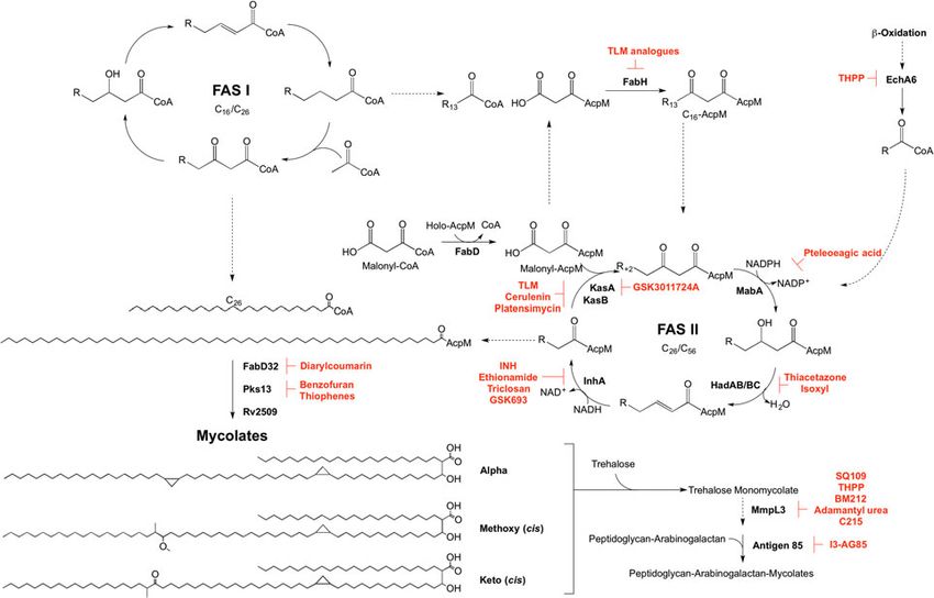
Katherine A. Abrahams and Gurdyal S. Besra                                                                                                                  120

                     Fig. 3. Inhibitors targeting arabinogalactan biosynthesis. The current understanding of the roles of enzymes involved in
                     arabinogalactan biosynthesis. Reported inhibitors are shown in red.

                     ARABINOGALACTAN                                                                     underway to characterize the enzyme via the estab-
                     The major cell wall polysaccharide, arabinogalactan                                 lishment of a microtiter plate-based assay for its
                     (Fig. 1), as the name suggests, is composed of galact-                              activity, which could be exploited in inhibitor
                     ose and arabinose sugar residues, in the furanose (f)                               library screening (Grzegorzewicz et al. 2008).
                     ring form (Galf) (McNeil et al. 1987).                                                 The linker unit provides an attachment point for
                     Arabinogalactan is attached to peptidoglycan via a                                  the polymerization of the galactan chain. This
                     single linker unit (McNeil et al. 1990). The galactan                               process also occurs in the cytoplasm. The bifunc-
                     component is a linear chain of approximately 30                                     tional galactofuranosyltransferases (GlfT1 and
                     alternating 5- and 6-linked β-D-Galf residues                                       GlfT2) (Alderwick et al. 2008) are responsible for
                     (Daffe et al. 1990). Three highly branched arabinan                                  the synthesis of the linear galactan chain. Initially,
                     chains, consisting of approximately 30 Araf residues,                               GlfT1 transfers Galf from UDP-Galf to the C-4
                     are attached to the galactan chain (Besra et al. 1995).                             position of L-Rha, and then adds a second Galf
                     The non-reducing termini of the arabinan chains act                                 residue to the C-5 position of the primary Galf, gen-
                     as an attachment site for mycolic acids, succinyl and                               erating C50-P-P-GlcNAc-L-Rha-Galf2 (Mikusova
                     galactosamine (D-GalN) moieties (Draper et al.                                      et al. 2006; Alderwick et al. 2008; Belanova et al.
                     1997; Bhamidi et al. 2008).                                                         2008). GlfT2 sequentially transfers Galf residues
                                                                                                         to the growing galactan chain with alternating β
                                                                                                         (1 → 5) and β(1 → 6) glycosidic linkages (Kremer
                                                                                                         et al. 2001a; Rose et al. 2006). The galactan chains
                     ARABINOGALACTAN BIOSYNTHESIS
                                                                                                         contain ∼30 Galf residues in vivo, forming C50-P-
                     Arabinogalactan biosynthesis is illustrated in Fig. 3.                              P-GlcNAc-L-Rha-Galf30 (Daffe et al. 1990), but
                     The first committed step begins in the cytoplasm                                     the chain length determination mechanism is yet to
                     and proceeds by the formation of the linker unit con-                               be fully understood. GlfT1 and GlfT2 are suitable
                     necting peptidoglycan to arabinogalactan, which is                                  targets, as rationalized by an in silico target identifi-
                     initiated by WecA, a GlcNAc-1-P transferase (Jin                                    cation program (Raman et al. 2008). UDP-Galf deri-
                     et al. 2010). This enzyme catalyses the transfer of                                 vatives, with modifications to the C-5 and C-6
                     GlcNAc-1-P to C50-P. WbbL, a rhamnosyltransfer-                                     positions have been investigated as suitable inhibi-
                     ase catalyses the transfer of L-rhamnose (L-Rha)                                    tors of these enzymes, whereby they cause premature
                     from dTDP-L-Rha to position 3 of C50-P-P-                                           galactan chain termination (Peltier et al. 2010).
                     GlcNAc to form C50-P-P-GlcNAc-L-Rha, com-                                              The remainder of arabinogalactan synthesis occurs
                     pleting the linker unit (McNeil et al. 1990; Mills                                  on the outside of the cell. Although the transport
                     et al. 2004). WecA has been identified as the target                                 mechanism of this cell wall polysaccharide is not
                     of caprazamycin derivatives, such as CPZEN-45,                                      fully understood, Rv3781 and Rv3783, encoding an
                     with the original nucleoside antibiotic shown to                                    ABC transporter, are potential ‘flippase’ candidates
                     target MraY (Ishizaki et al. 2013). Recently, a fluor-                               (Dianiskova et al. 2011). Araf residues are transferred
                     escence-based assay for WecA activity has been                                      directly onto C50-P-P-GlcNAc-L-Rha-Galf30 from
                     developed and used to screen compound libraries                                     the lipid donor decaprenylphosphoryl-D-arabinose
                     with some success (Mitachi et al. 2016). Inhibitors                                 (DPA) (Wolucka et al. 1994). DPA is synthesized
                     targeting WbbL have yet to be identified. This                                       through a series of cytoplasmic steps, and originates
                     essential enzyme, present in all mycobacteria, is                                   exclusively from phospho-α-D-ribosyl-1-pyrophos-
                     recognized as a promising target and efforts are                                     phate (pRpp), prior to reorientation to the extracellular

Downloaded from https://www.cambridge.org/core. IP address: 46.4.80.155, on 26 Nov 2021 at 10:33:58, subject to the Cambridge Core terms of use, available at https://www.cambridge.org/core/terms.
https://doi.org/10.1017/S0031182016002377
Mycobacterial cell wall biosynthesis 121
face of the plasma membrane. The pRpp synthetase, catalyses the transfer of the terminal β(1 → 2) Araf
PrsA, catalyses the transfer of pyrophosphate from residues (Seidel et al. 2007). C-5 of the terminal β-
ATP to C-1 of ribose-5-phosphate, forming pRpp D-Araf and the penultimate 2-α-D-Araf of this
(Alderwick et al. 2011b). A decaprenyl moiety is motif act as anchoring points for mycolic acids
added, catalysed by UbiA (decaprenol-1-phosphate (McNeil et al. 1991).
5-phosphoribosyltransferase), forming decaprenol-1- The Emb arabinosyltransferases are inhibited by
monophosphate 5-phosphoribose (Alderwick et al. EMB, a well-recognized anti-TB drug, which is
2005; Huang et al. 2005, 2008). Rv3807c encodes a employed in the short-course treatment strategy of
putative phospholipid phosphatase, which catalyses TB. Efforts are focused on investigating EMB analo-
C-5 dephosphorylation, generating decaprenol-1- gues, such as SQ109 (Jia et al. 2005a, b, c; Sacksteder
phosphoribose (DPR) (Jiang et al. 2011). Finally, et al. 2012) and SQ775 (Bogatcheva et al. 2006), for
DPA is generated by an epimerization reaction of the future lead drug development. Interestingly, the
ribose C-2 hydroxyl, catalysed by a two-step oxida- other AraTs are not inhibited by EMB (Alderwick
tion/reduction activity of the decaprenylphosphori- et al. 2006; Seidel et al. 2007; Birch et al. 2008)
bose-2′-epimerase consisting of subunits DprE1 and and screening for inhibitors against these enzymes
DprE2 (Mikusova et al. 2005). is hindered due to the nature of the protein and sub-
The DPA synthetic pathway is a validated drug strate (membrane bound). However, there have been
target. The nitro-benzothiazinones (BTZs) and the reports on the development of DPA analogues for
structurally related dinitrobenzamides target the inhibition of arabinogalactan biosynthesis
DprE1 and are effective against MDR and XDR (Pathak et al. 2001; Owen et al. 2007). A recent
strains of Mtb with low toxicity (Christophe et al. study employing a cell free assay approach with
2009; Batt et al. 2012; Makarov et al. 2014, 2015). membrane preparations has determined that
The success of these compounds has led to the various DPA analogues are able to limit the incorp-
study of the other enzymes as potential drug oration of a radiolabelled DP[14C]A (Zhang et al.
targets. Conditional knockdown mutants of dprE1, 2011).
dprE2, ubiA, prsA and Rv3807c have proven the The primary structure of arabinogalactan is
essentiality of all except Rv3807c, and a target- completed by the transfer of succinyl and D-GalN
based whole-cell screen has been developed using residues to the inner arabinan units. PpgS, polypre-
these strains of reduced expression levels to identify nyl-phospho-N-acetylgalactosaminyl synthase, cat-
enzyme-specific inhibitors. Inhibitors targeting a alyses the formation of polyprenol-P-D-GalNAc
particular enzyme cause increased sensitivity and from polyprenyl-P and UDP-GalNAc, which is
this was confirmed with BTZ and KRT2029 target- then translocated across the membrane (Skovierova
ing DprE1 and UbiA, respectively, and can be the et al. 2010; Rana et al. 2012). The deacylation to
subject of future medicinal chemistry efforts (Kolly polyprenol-P-D-GalN occurs in an undetermined
et al. 2014). location and by an unknown mechanism. The glyco-
The mechanism of DPA reorientation into the syltransferase, Rv3779, transfers D-GalN to arabi-
periplasm is unknown. The ‘flippase’ was recently nogalactan at the C-2 position of 3,5-branched
considered to be Rv3789, but there is evidence that Araf residue (Scherman et al. 2009; Skovierova
this protein plays a different role: to act as an et al. 2010; Peng et al. 2012; Rana et al. 2012).
anchor protein to recruit AftA (Kolly et al. 2015). Succinylated Araf residues have also been detected
AftA is the first arabinofuranosyltransferase at this position of non-mycolated arabinan chains
(AraT), of a predicted six, to commence the addition (Bhamidi et al. 2008), but the enzyme responsible
of arabinose from DPA onto the galactan chain is currently unknown. A comprehensive mechanistic
(Alderwick et al. 2006). AftA transfers a single and functional understanding of these enzymes is
Araf residue onto C-5 of β(1 → 6) Galf residues 8, required for evaluation as suitable drug targets and
10 and 12 of C50-P-P-GlcNAc-L-Rha-Galf30 to date, there are no identified inhibitors against
(Alderwick et al. 2005). EmbA and EmbB, so these processes. The final stage is the attachment of
called because their discovery was based on the the arabinogalactan macromolecule to peptidogly-
mode of action elucidation of ethambutol (EMB), can. The enzyme responsible for this essential liga-
catalyse the addition of further α(1 → 5) Araf poly- tion has recently been elucidated to be Lcp1
merization (Alderwick et al. 2005). AftC introduces (Harrison et al. 2016).
α(1 → 3) branching (Birch et al. 2008), with AftD
having an equivalent role (Skovierova et al. 2009).
PHOSPHATIDYL-MYO-INOSITOL MANNOSIDES,
The structure terminates in a well-defined hexa-
LIPOMANNAN AND LIPOARABINOMANNAN
arabinofuranosyl (Araf6) structural motif: [β-D-Araf-
(1 → 2)-α-D-Araf]2-3,5-α-D-Araf-(1 → 5)-α-D-Araf. The glycolipids, phosphatidyl-myo-inositol manno-
This motif is generated by EmbA, EmbB, AftC, AftD sides (PIMs), and the related lipoglycans, lipoman-
and AftB (Escuyer et al. 2001; Alderwick et al. 2005; nan (LM) and lipoarabinomannan (LAM), are
Birch et al. 2008, 2010; Skovierova et al. 2009). AftB non-covalently anchored into the inner and outer
Downloaded from https://www.cambridge.org/core. IP address: 46.4.80.155, on 26 Nov 2021 at 10:33:58, subject to the Cambridge Core terms of use, available at https://www.cambridge.org/core/terms.
https://doi.org/10.1017/S0031182016002377Katherine A. Abrahams and Gurdyal S. Besra 122
Fig. 4. Inhibitors targeting the biosynthesis of phosphatidyl-myo-inositol mannosides, lipomannan and
lipoarabinomannan. The current understanding of the biosynthesis of PIMs, LM, LAM and ManLAM. Reported
inhibitors are shown in red.
membranes of the cell wall via the phosphatidyl- glycosylated through an α(1 → 2) linkage with
myo-inositol unit (Ortalo-Magne et al. 1996) ∼50–80 Araf residues (Khoo et al. 1996).
(Fig. 1). The core structure of PIM consists of an In mycobacteria, PI and PIMs contribute up to
acylated sn-glycerol-3-phospho-(1-D-myo-inositol), 56% of all phospholipids in the cell wall and 37%
the phosphatidyl inositol (PI) unit. Glycosylation in the cytoplasmic membrane (Goren, 1984).
with mannopyranose (Manp) residues at the O-2 These significant quantities indicate their import-
and O-6 positions of myo-inositol, results in the ance. Not only are they structural components,
mannosyl phosphate inositol (MPI) anchor (Ballou they also have roles in cell wall integrity, permeabil-
et al. 1963; Ballou and Lee, 1964; Nigou et al. ity and control of septation and division (Parish et al.
2004). The MPI structure is highly diverse, with 1997; Patterson et al. 2003; Fukuda et al. 2013). LM
variations in the type (commonly palmitic and and LAM are involved in Mtb pathogenicity, with
tuberculostearic chains (Pitarque et al. 2005)), evidence to suggest they are modulators of host–
number and location of acyl chains. The most preva- pathogen interactions (Schlesinger et al. 1994;
lent forms of PIMs in mycobacteria are tri- and Nigou et al. 2002; Maeda et al. 2003). These features
tetra-acylated phospho-myo-inositol di/hexamanno- of PIMs, LM and LAM make them suitable targets
sides (Ac1PIM2, Ac1PIM6, Ac2PIM2, Ac2PIM6), in anti-TB drug discovery.
where in the hexamannosides, there is one Manp
unit on the O-2 and five Manp units on the O-6 pos-
BIOSYNTHESIS OF PHOSPHATIDYL-
ition of myo-inositol (Gilleron et al. 2001).
MYO-INOSITOL MANNOSIDES, LIPOMANNAN
Extensions of mannan and arabinomannan chains
AND LIPOARABINOMANNAN
on the MPI anchor form LM and LAM, respect-
ively. In both LM and LAM, the mannan chain con- PIM biosynthesis begins in the cytoplasm (Fig. 4).
sists of approximately 21–34 α(1 → 6) linked Manp The α-mannopyranosyl transferase (ManpT),
units, decorated with single α(1 → 2)-Manp residues PimA, of the GT-A/B superfamily, transfers Manp
(Kaur et al. 2008). In LAM, the mannan chain is from the donor GDP-Manp to position O-2 of the
Downloaded from https://www.cambridge.org/core. IP address: 46.4.80.155, on 26 Nov 2021 at 10:33:58, subject to the Cambridge Core terms of use, available at https://www.cambridge.org/core/terms.
https://doi.org/10.1017/S0031182016002377Mycobacterial cell wall biosynthesis 123
myo-inositol ring to form PIM1 (Kordulakova et al. monomannose side chains via α(1 → 2) linkages,
2002; Guerin et al. 2007). A second Manp residue is forming mature LM (Kaur et al. 2008; Mishra
transferred to position O-6 of the myo-inositol ring et al. 2011). Modification of LM leads to LAM.
by PimB’ to form PIM2 (Guerin et al. 2009). Approximately 50–80 Araf residues are added
Acylation of the Manp residue of PIM1 is performed using DPA as the donor, comparable to that of the
by the acyltransferase Rv2611c before or after the arabinogalactan domain. An unidentified ArafT
addition of the second Manp residue (Kordulakova primes the mannan chain, which is further elongated
et al. 2003). The acylation of the C-3 position of the by EmbC, adding 12–16 Araf residues with α(1 → 5)
myo-inositol ring is performed by an unknown acyl- linkages (Shi et al. 2006; Alderwick et al. 2011a).
transferase. This finishes the synthesis of the MPI AftC, the same enzyme involved in arabinogalactan
anchor. Mannosylation of Ac1/Ac2PIM2 to Ac1/ synthesis, integrates α(1 → 3) Araf branches (Birch
Ac2PIM3 is performed by a ManpT, designated et al. 2008). It has also been speculated that AftD
PimC, but this enzyme is yet to be confirmed in introduces α(1 → 3) Araf, but its function is yet to
Mtb H37Rv (Kremer et al. 2002b). It is suspected be confirmed (Skovierova et al. 2009). The arabinan
that the subsequent addition of Manp to the non- domain is terminated by β(1 → 2) Araf linkages, pre-
reducing end of Ac1/Ac2PIM3 is performed by the dicted to be performed by AftB, resulting in
unidentified PimC or PimD forming Ac1/Ac2PIM4. branched hexa-arabinoside or linear tetra-arabino-
The ManpTs have been the subject of target-based side motifs. Further structural heterogeneity is
screening programs. More specifically, in vitro introduced by capping motifs. These moieties
PimA activity was screened with approximately 350 consist of a number of α(1 → 2)-linked Manp resi-
compounds. Several hit molecules exhibited signifi- dues, producing mannosylated LAM (ManLAM)
cant inhibition, but the compounds did not exhibit (Kaur et al. 2008). Using PPM, the α(1 → 5)
in vivo activity in Mtb (Sipos et al. 2015). Substrate ManpT, CapA, attaches the first Manp residue
analogues of PimA and PimB’, galactose-derived (Dinadayala et al. 2006). MptC catalyses the add-
phosphonate analogs of PI, have also been developed, ition of subsequent α(1 → 2) Manp residues
which show enzyme inhibition in a cell-free system (Kaur et al. 2008), which can be decorated with an
(Dinev et al. 2007). α(1 → 4)-linked 5-deoxy-5-methyl-thio-xylofuranose
The biosynthesis of Ac1/Ac2PIM4 marks the tran- (MTX) residue (Ludwiczak et al. 2002; Turnbull
sition towards the synthesis of higher order PIMs, et al. 2004). The enzymes involved in the addition
LM and LAM (Fig. 4). It is predicted that the syn- of MTX and succinyl residues to LAM are still to
thesis of Ac1/Ac2PIM4 occurs on the cytoplasmic be determined.
side of the membrane, and at this point, is flipped The essentiality of PPM in lipoglycan biosyn-
across the membrane by an unidentified translocase, thesis makes Ppm1 an attractive drug target.
with the remainder of the steps thought to occur in Amphomycin, a lipopeptide antibiotic, inhibits the
the periplasmic space. The integral membrane synthesis of PPM by sequestering the polyprenol
ManpTs (of the GT-C glycosyltransferase super- phosphates, and consequently inhibits the extracel-
family) are reliant on polyprenyl-phosphate-based lular ManpTs (Banerjee et al. 1981; Besra et al.
mannose donors (PPM) rather than the nucleotide- 1997). Guy et al. (2004) designed a variety of
based sugars (Berg et al. 2007). The polyprenol prenyl-based photoactivable probes. Upon photoac-
monophosphomannose synthase, Ppm1, catalyses tivation, a number of the probes exhibited inhibitory
the synthesis of PPM from GDP-Manp and poly- activity against Mtb Ppm1 and M. smegmatis
prenol phosphates (Gurcha et al. 2002). α(1 → 6) ManpTs (Guy et al. 2004). Substrate
PimE catalyses the transfer of an α(1 → 2)-linked analogues of the ManpTs have been designed to
Manp residue onto Ac1/Ac2PIM4, generating Ac1/ investigate enzyme–substrate interactions and
Ac2PIM5 (Morita et al. 2006). The transfer of the mechanisms of action (Brown et al. 2001; Tam and
last Manp residue is either performed by PimE or Lowary, 2010). These types of studies will provide
by an unidentified GT-C glycosyltransferase an invaluable insight into the interactions involved
forming Ac1/Ac2PIM6 (Morita et al. 2006). The and for the future design of inhibitors.
distal 2-linked Manp residues are not present in
the mannan core of LM or LAM; Ac1/Ac2PIM4 is
MYCOLIC ACIDS
the likely precursor for the extension of the
mannan chain. Recent evidence suggests that the The final distinctive component of the mycobacter-
putative lipoprotein LpqW channels intermediates ial cell wall is the unique fatty acids, termed the
such as Ac1/Ac2PIM4 towards either PimE (to mycolic acids (Fig. 1). These unique long chain α-
form the polar lipids) or to LM and LAM synthesis alkyl-β-hydroxy fatty acids (comprised a meromyco-
(Crellin et al. 2008). The mannosyltransferases, late chain of C42–C62 and a long saturated α-chain
MptA and MptB (Mishra et al. 2007, 2008), are C24–C26) are attached to the arabinogalactan layer,
responsible for the α(1 → 6)-linked mannan core of but also make up other outer cell envelope lipids
LM and LAM. MptC catalyses the transfer of the such as trehalose mono/di-mycolates and glucose
Downloaded from https://www.cambridge.org/core. IP address: 46.4.80.155, on 26 Nov 2021 at 10:33:58, subject to the Cambridge Core terms of use, available at https://www.cambridge.org/core/terms.
https://doi.org/10.1017/S0031182016002377Katherine A. Abrahams and Gurdyal S. Besra 124
Fig. 5. Inhibitors targeting mycolic acid biosynthesis. The enzymes involved in the mycolic acid biosynthetic pathway are
presented. Reported inhibitors are shown in red. ‘R’ represents an acyl chain of varying carbon units in length.
monomycolate. There are three subclasses of synthase) (Choi et al. 2000), forming a pivotal link
mycolic acids: α-mycolates, containing cyclopropane between the FAS I and FAS II pathways. The
rings in the cis-configuration; methoxy-mycolates C16-AcpM formed is channeled to the FAS II
and keto-mycolates containing methoxy or ketone pathway (Bhatt et al. 2007), where it undergoes a
groups, respectively, and have cyclopropane rings round of keto-reduction, dehydration and enoyl-
in the cis- or trans- configuration (Brennan and reduction, catalysed by: MabA, a β-ketoacyl-AcpM
Nikaido, 1995; Watanabe et al. 2001, 2002). reductase (Marrakchi et al. 2002); HadAB/BC, a β-
Mycolic acids contribute to the permeability of the hydroxyacyl-AcpM hydratase (Sacco et al. 2007);
cell wall, and as such are essential for cell viability, InhA, an enoyl-AcpM reductase (Banerjee et al.
and are also essential in virulence, making the bio- 1994). Successive cycles ensue, whereby the conden-
synthesis of mycolates suitable drug targets (Liu sation reaction of FabH is replaced by the activities
et al. 1996). of KasA and KasB, β-ketoacyl synthases (Schaeffer
et al. 2001; Kremer et al. 2002a). The AcpM-
bound acyl chain extends by two carbon units in
MYCOLIC ACID BIOSYNTHESIS
each cycle, forming a saturated long-chain meromy-
Mycolic acid biosynthesis occurs in the cytoplasm, colate of C42–C62, which is subject to modifications
involving two distinct pathways, termed fatty acid such as cis-/trans-cyclopropanation, and the addition
synthase types I and II (FAS I and FAS II) of methoxy and keto groups (Dubnau et al. 2000;
(Fig. 5). FAS I (Rv2524c), a multifunctional poly- Glickman et al. 2000; Glickman, 2003; Barkan
peptide, generates short-chain fatty acyl-CoA et al. 2010). FabD32, a fatty acyl-AMP ligase, acti-
esters that can either form the saturated α-branch vates the meromycolate chain (Trivedi et al. 2004)
(C24), or be extended by FAS II to form the mero- and the subsequent meromycolyl-AMP is linked
mycolate chain (Cole et al. 1998). Elongation of the with the α-alkyl-CoA ester, catalysed by Pks13, to
fatty acids is dependent on the availability of holo- generate a α-alkyl-β-keto-mycolic acid (Gande
AcpM, an acyl carrier protein, and malonyl-CoA. et al. 2004; Portevin et al. 2004). Finally a reduction
FabD, the malonyl:AcpM transacylase generates step, catalysed by Rv2509, generates a mature myco-
malonyl-AcpM (Kremer et al. 2001b). C14-CoA late (Bhatt et al. 2008). Transport of the mycolates to
primers from FAS I are condensed with malonyl- either the cell envelope or for attachment to arabino-
AcpM, catalysed by FabH (β-ketoacyl ACP galactan remains to be elucidated. It is considered that
Downloaded from https://www.cambridge.org/core. IP address: 46.4.80.155, on 26 Nov 2021 at 10:33:58, subject to the Cambridge Core terms of use, available at https://www.cambridge.org/core/terms.
https://doi.org/10.1017/S0031182016002377Mycobacterial cell wall biosynthesis 125
the mycolates are transported in the form of trehalose There has been significant interest in TLM due to
monomycolate (TMM). In the generation of TMM, its broad-spectrum activity and numerous analogues
Takayama et al. (2005) propose that a mycolyl- have been synthesized to improve on potency and
transferase transfers the mycolyl group from pharmacokinetic properties (Kremer et al. 2000;
mycolyl-Pks13 to D-mannopyranosyl-1-phospho- Senior et al. 2003, 2004; Kim et al. 2006). The
heptaprenol (Besra et al. 1994). The mycolyl group biphenyl-based 5-substituents of TLM also exhibit
of mycolyl-D-mannopyranosyl-1-phosphoheptapre- in vitro activity against FabH, but with no whole-
nol is transferred to trehalose-6-phosphate by a cell activity (Senior et al. 2003, 2004). The 2-tosyl-
second mycolyltransferase, forming TMM-phos- naphthalene-1,4-diol pharmacophore of TLM also
phate. The phosphate moiety is removed by a trehal- has in vitro activity against FabH, however, whole-
ose-6-phosphate phosphatase, and the TMM is cell data are yet to be published (Alhamadsheh et al.
immediately translocated outside of the cell using a 2008). Recently, a new anti-TB compound, an inda-
resistance-nodulation-division (RND) family of zole sulfonamide GSK3011724A, was discovered
efflux pumps, termed mycobacterial membrane from a phenotypic whole-cell HTS (Abrahams et al.
proteins large (MmpL), limiting TMM accu- 2016). The compound was shown to target KasA spe-
mulation in the cytoplasm (Takayama et al. 2005; cifically, with no discernable target engagement with
Grzegorzewicz et al. 2012; Varela et al. 2012). KasB or FabH, and is currently the focus of medi-
Finally, the mycolyltransferase Antigen 85 complex, cinal chemistry optimization (Abrahams et al. 2016).
formed of Ag85A, Ag85B and Ag85C, attaches the Due to the success of InhA as a chemotherapeutic
mycolic acid moiety from TMM to arabinogalactan target, there is a mounting interest in the other
(Jackson et al. 1999). This complex also catalyses enzymes involved in mycolic acid biosynthesis from
the formation of trehalose dimycolate, TDM, from a drug target perspective that could bypass INH
two TMM molecules with the release of trehalose resistance in MDR and XDR-TB. Formerly used in
(Takayama et al. 2005). TDM, or ‘cord factor’, is the treatment of TB, the thiocarbamide-containing
implicated in the pathogenicity of Mtb. drugs, thiacetazone and isoxyl, were shown to target
The enzymes involved in mycolic acid biosyn- mycolic acid biosynthesis and the inhibition mechan-
thesis are the targets of numerous inhibitors. In ism has recently been elucidated. Following activation
1952, shortly after its discovery, isoniazid (INH) by EthA, both drugs target the HadA subunit of the
was administered as a front-line and essential anti- HadABC dehydratase, forming a covalent interaction
biotic in the treatment of TB (Medical Research with the active site cysteine (Grzegorzewicz et al.
Council, 1952) and has only recently had the mode 2015). It has also been shown that thiacetazone inhi-
of action elucidated. Initially thought to target bits cyclopropanation of mycolic acids (Alahari et al.
KatG due to mutations in the corresponding gene 2007). MabA has been the subject of a molecular
in resistant isolates (Zhang and Young, 1994; docking study. Comparable with the control inhibi-
Rouse and Morris, 1995), INH was later revealed tory substrate isonicotinic-acyl-NADH, pteleoellagic
to be a pro-drug, with the true target being InhA acid had a high docking score with in vivo activity to
(Banerjee et al. 1994; Larsen et al. 2002). be confirmed (Shilpi et al. 2015). Through a target-
Ethionamide, a structural analogue of INH, also based screening approach linked with whole-genome
requires cellular activation via EthA, before target- sequencing of resistant mutants, a benzofuran has
ing InhA (Banerjee et al. 1994). Direct inhibitors been shown to target Pks13 (Ioerger et al. 2013).
of InhA that do not require activation are now Additionally, Pks13 is the target of thiophene
being searched for (Lu et al. 2010; Vilcheze et al. compounds (Wilson et al. 2013) including 2-
2011; Pan and Tonge, 2012; Encinas et al. 2014; aminothiophenes (Thanna et al. 2016). From a GFP
Manjunatha et al. 2015; Sink et al. 2015; Martinez- reporter-based whole-cell HTS, a diarylcoumarin
Hoyos et al. 2016). One such molecule is the exhibited potent activity against Mtb and this struc-
broad-spectrum antibiotic triclosan, which has not tural class was shown to target FadD32 by inhibiting
been adopted in TB treatment due to its sub- the acyl–acyl carrier protein synthetase activity
optimal bioavailability (Wang et al. 2004). In the (Stanley et al. 2013). The homologue of the Rv2509
last year, GlaxoSmithKline have published a set of reductase in M. smegmatis is non-essential but loss of
thiadiazole compounds, which directly target function increases susceptibility to lipophilic antibio-
InhA, with GSK693 demonstrating in vivo efficacy tics such as rifampicin. Targeting this ‘secondary’
comparable to INH (Martinez-Hoyos et al. 2016). drug target in Mtb could increase the susceptibility
Therefore, old drug targets should not be discounted of the bacilli to antibiotics (Bhatt et al. 2008). The
in the search for new anti-tubercular agents. Antigen 85 complex has been the focus of a number
The β-ketoacyl synthases, KasA and KasB, are the of inhibitor-based screening studies (Belisle et al.
targets of the natural products cerulenin (Parrish et al. 1997; Gobec et al. 2004; Sanki et al. 2008, 2009;
1999; Schaeffer et al. 2001; Kremer et al. 2002a), pla- Elamin et al. 2009; Barry et al. 2011). Recently, an
tensimycin (Brown et al. 2009), and thiolactomycin inhibitor from a compound library was shown to
(TLM) (Kremer et al. 2000; Schaeffer et al. 2001). bind to Antigen 85C, and derivatives of this compound
Downloaded from https://www.cambridge.org/core. IP address: 46.4.80.155, on 26 Nov 2021 at 10:33:58, subject to the Cambridge Core terms of use, available at https://www.cambridge.org/core/terms.
https://doi.org/10.1017/S0031182016002377Katherine A. Abrahams and Gurdyal S. Besra 126
have been synthesized, with 2-amino-6-propyl- be a very effective way to identify novel anti-TB
4,5,6,7-tetrahydro-1-benzothiphene-3-carbonitrile compounds with known modes of action, but is
(I3-AG85) exhibiting the lowest MIC in Mtb and limited by the specified target (Batt et al. 2015;
drug-resistant strains (Warrier et al. 2012). Martinez-Hoyos et al. 2016). Target assignment is
In the target identification of new anti-tubercular a fundamental step in in the drug discovery pipeline.
compounds, some targets can be regarded as promis- Without knowledge of the physiological target,
cuous, inhibited by multiple different chemical efforts can be wasted on developing compounds
scaffolds, exemplified by MmpL3 (Grzegorzewicz against an unsuitable target, such as those homolo-
et al. 2012; La Rosa et al. 2012; Stanley et al. 2012; gous in humans. Establishing the mode of action of
Tahlan et al. 2012; Lun et al. 2013; Remuinan an inhibitor is a prerequisite for facilitating medi-
et al. 2013), a predicted TMM transporter. cinal chemistry efforts to convert compounds into
Through the generation and sequencing of spontan- potential drug candidates.
eous resistant mutants, a number of inhibitors with
diverse chemical structures have been shown to
Concluding remarks
target MmpL3 (Grzegorzewicz et al. 2012; La
Rosa et al. 2012; Stanley et al. 2012; Tahlan et al. The essential mycobacterial cell wall, responsible for
2012; Lun et al. 2013; Remuinan et al. 2013). structural integrity, permeability and pathogenicity,
However, a recent chemoproteomics approach is an attractive drug target, both structurally and bio-
determined that one of the proposed inhibitor synthetically. Recent advancements in biochemical
classes of MmpL3, the tetrahydropyrazo[1,5-a]pyr- and omics-based techniques have led to the discovery
imidine-3-carboxamides (THPPs), has a novel alter- and mechanistic understanding of enzymes involved
native target, EchA6 (Cox et al. 2016). Sequence in mycobacterial cell wall synthesis and assembly.
analysis predicted EchA6 to be an enoyl-CoA hydra- Although a number of key enzymes are yet to be
tase, but it lacks the residues required for catalytic established, there are a plethora of suitable targets,
activity. Through an extensive biochemical investi- exploited not only in current treatment programmes
gation, Cox et al. (2016) predicted that EchA6 shut- but also for anti-TB drug discovery. In the current
tles fatty acyl-CoA esters from the β-oxidation TB treatment regimen, two of the front-line drugs,
pathway into FAS II, ready for the condensation INH and EMB, target mycolic acid and arabinogalac-
activities of KasA or KasB with malonyl-AcpM. tan biosynthesis, respectively, with the second-line
This research demonstrates that target identification drugs such as ethionamide and D-cycloserine also tar-
of inhibitory compounds can unveil not only a new geting cell wall production. The proven success of
biological pathway, but also an untapped area for these drugs validates the future development of inhi-
drug targets. bitors targeting the unique mycobacterial cell wall,
which remains a source of unexploited clinically rele-
vant drug targets. The continued progression in drug
DRUG DISCOVERY EFFORTS
discovery approaches and the optimization of bio-
The strategies involved in drug discovery are forever chemical techniques, will enable the rapid identifica-
evolving. Traditional enzyme screening campaigns tion of anti-TB agents, many of which are likely to
and medicinal chemistry focused on ligand-based target the biosynthesis of the so-called ‘Achilles
inhibitor designs (such as substrate or transition heel’ of Mtb.
state analogues) that once dominated drug discovery
are being superseded by phenotypic HTS. The
ACKNOWLEDGEMENTS
former approach often relies on the X-ray crystal
structure of the enzyme or biochemical understand- The authors would like to thank Jonathan Cox for his
ing, and successful inhibitors from these screens are technical support and advice.
further challenged by target engagement in vivo.
Over recent years, HTS has become the lead FINANCIAL SUPPORT
approach in drug discovery. HTS employs extensive
compound libraries of diverse chemical structures, G.S.B. acknowledges support in the form of a Personal
Research Chair from Mr James Bardrick, a Royal
and as a consequence, these methods can identify a Society Wolfson Research Merit Award, the Medical
multitude of inhibitors with novel chemical Research Council (MR/K012118/1) and the Wellcome
scaffolds. Phenotypic HTS can reveal anti-TB Trust (081569/Z/06/Z).
agents with whole-cell activity and unknown
modes of action, having the potential to unveil new
biochemical pathways (Abrahams et al. 2012, 2016; REFERENCES
Gurcha et al. 2014; Mugumbate et al. 2015). Abrahams, K. A., Cox, J. A., Spivey, V. L., Loman, N. J., Pallen, M. J.,
Alternatively, phenotypic HTS can be target- Constantinidou, C., Fernandez, R., Alemparte, C., Remuinan, M. J.,
Barros, D., Ballell, L. and Besra, G. S. (2012). Identification of novel
based, focusing on enzymes or pathways such as imidazo[1,2-a]pyridine inhibitors targeting M. tuberculosis QcrB. PLoS
those involved in cell wall biosynthesis. This can ONE 7, e52951.
Downloaded from https://www.cambridge.org/core. IP address: 46.4.80.155, on 26 Nov 2021 at 10:33:58, subject to the Cambridge Core terms of use, available at https://www.cambridge.org/core/terms.
https://doi.org/10.1017/S0031182016002377Mycobacterial cell wall biosynthesis 127
Abrahams, K. A., Chung, C. W., Ghidelli-Disse, S., Rullas, J., Belanova, M., Dianiskova, P., Brennan, P. J., Completo, G. C.,
Rebollo-Lopez, M. J., Gurcha, S. S., Cox, J. A., Mendoza, A., Rose, N. L., Lowary, T. L. and Mikusova, K. (2008). Galactosyl trans-
Jimenez-Navarro, E., Martinez-Martinez, M. S., Neu, M., ferases in mycobacterial cell wall synthesis. Journal of Bacteriology 190,
Shillings, A., Homes, P., Argyrou, A., Casanueva, R., Loman, N. J., 1141–1145.
Moynihan, P. J., Lelievre, J., Selenski, C., Axtman, M., Belisle, J. T., Vissa, V. D., Sievert, T., Takayama, K., Brennan, P. J.
Kremer, L., Bantscheff, M., Angulo-Barturen, I., Izquierdo, M. C., and Besra, G. S. (1997). Role of the major antigen of Mycobacterium tuber-
Cammack, N. C., Drewes, G., Ballell, L., Barros, D., Besra, G. S. culosis in cell wall biogenesis. Science 276, 1420–1422.
and Bates, R. H. (2016). Identification of KasA as the cellular target of Berg, S., Kaur, D., Jackson, M. and Brennan, P. J. (2007). The glycosyl-
an anti-tubercular scaffold. Nature Communications 7, 12581. transferases of Mycobacterium tuberculosis – roles in the synthesis of arabi-
Alahari, A., Trivelli, X., Guerardel, Y., Dover, L. G., Besra, G. S., nogalactan, lipoarabinomannan, and other glycoconjugates. Glycobiology
Sacchettini, J. C., Reynolds, R. C., Coxon, G. D. and Kremer, L. 17, 35–56R.
(2007). Thiacetazone, an antitubercular drug that inhibits cyclopropana- Besra, G. S., Sievert, T., Lee, R. E., Slayden, R. A., Brennan, P. J. and
tion of cell wall mycolic acids in mycobacteria. PLoS ONE 2, e1343. Takayama, K. (1994). Identification of the apparent carrier in mycolic
Alderwick, L. J., Radmacher, E., Seidel, M., Gande, R., Hitchen, P. acid synthesis. Proceedings of the National Academy of Sciences of the
G., Morris, H. R., Dell, A., Sahm, H., Eggeling, L. and Besra, G. S. United States of America 91, 12735–12739.
(2005). Deletion of Cg-emb in corynebacterianeae leads to a novel trun- Besra, G. S., Khoo, K. H., McNeil, M. R., Dell, A., Morris, H. R. and
cated cell wall arabinogalactan, whereas inactivation of Cg-ubiA results Brennan, P. J. (1995). A new interpretation of the structure of the
in an arabinan-deficient mutant with a cell wall galactan core. Journal of mycolyl-arabinogalactan complex of Mycobacterium tuberculosis as revealed
Biological Chemistry 280, 32362–32371. through characterization of oligoglycosylalditol fragments by fast-atom
Alderwick, L. J., Seidel, M., Sahm, H., Besra, G. S. and Eggeling, L. bombardment mass spectrometry and 1H nuclear magnetic resonance
(2006). Identification of a novel arabinofuranosyltransferase (AftA) spectroscopy. Biochemistry 34, 4257–4266.
involved in cell wall arabinan biosynthesis in Mycobacterium tuberculosis. Besra, G. S., Morehouse, C. B., Rittner, C. M., Waechter, C. J. and
Journal of Biological Chemistry 281, 15653–15661. Brennan, P. J. (1997). Biosynthesis of mycobacterial lipoarabinomannan.
Alderwick, L. J., Dover, L. G., Veerapen, N., Gurcha, S. S., Journal of Biological Chemistry 272, 18460–18466.
Kremer, L., Roper, D. L., Pathak, A. K., Reynolds, R. C. and Bhamidi, S., Scherman, M. S., Rithner, C. D., Prenni, J. E.,
Besra, G. S. (2008). Expression, purification and characterisation of Chatterjee, D., Khoo, K. H. and McNeil, M. R. (2008). The identifica-
soluble GlfT and the identification of a novel galactofuranosyltransferase tion and location of succinyl residues and the characterization of the inter-
Rv3782 involved in priming GlfT-mediated galactan polymerisation ior arabinan region allow for a model of the complete primary structure of
in Mycobacterium tuberculosis. Protein Expression and Purification 58, Mycobacterium tuberculosis mycolyl arabinogalactan. Journal of Biological
332–341. Chemistry 283, 12992–13000.
Alderwick, L. J., Lloyd, G. S., Ghadbane, H., May, J. W., Bhatt, A., Bhatt, A., Molle, V., Besra, G. S., Jacobs, W. R., Jr. and Kremer, L.
Eggeling, L., Futterer, K. and Besra, G. S. (2011a). The C-terminal (2007). The Mycobacterium tuberculosis FAS-II condensing enzymes:
domain of the Arabinosyltransferase Mycobacterium tuberculosis EmbC is their role in mycolic acid biosynthesis, acid-fastness, pathogenesis and in
a lectin-like carbohydrate binding module. PLoS Pathogens 7, e1001299. future drug development. Molecular Microbiology 64, 1442–1454.
Alderwick, L. J., Lloyd, G. S., Lloyd, A. J., Lovering, A. L., Bhatt, A., Brown, A. K., Singh, A., Minnikin, D. E. and Besra, G. S.
Eggeling, L. and Besra, G. S. (2011b). Biochemical characterization of (2008). Loss of a mycobacterial gene encoding a reductase leads to an
the Mycobacterium tuberculosis phosphoribosyl-1-pyrophosphate synthe- altered cell wall containing beta-oxo-mycolic acid analogs and accumula-
tase. Glycobiology 21, 410–425. tion of ketones. Chemistry and Biology 15, 930–939.
Alhamadsheh, M. M., Waters, N. C., Sachdeva, S., Lee, P. and Biarrotte-Sorin, S., Hugonnet, J. E., Delfosse, V., Mainardi, J. L.,
Reynolds, K. A. (2008). Synthesis and biological evaluation of novel sul- Gutmann, L., Arthur, M. and Mayer, C. (2006). Crystal structure of a
fonyl-naphthalene-1,4-diols as FabH inhibitors. Bioorganic and novel beta-lactam-insensitive peptidoglycan transpeptidase. Journal of
Medicinal Chemistry Letters 18, 6402–6405. Molecular Biology 359, 533–538.
Ballou, C. E. and Lee, Y. C. (1964). The structure of a myoinositol manno- Birch, H. L., Alderwick, L. J., Bhatt, A., Rittmann, D.,
side from Mycobacterium tuberculosis glycolipid. Biochemistry 3, 682–685. Krumbach, K., Singh, A., Bai, Y., Lowary, T. L., Eggeling, L. and
Ballou, C. E., Vilkas, E. and Lederer, E. (1963). Structural studies on the Besra, G. S. (2008). Biosynthesis of mycobacterial arabinogalactan: iden-
myo-inositol phospholipids of Mycobacterium tuberculosis (var. bovis, strain tification of a novel alpha(1→3) arabinofuranosyltransferase. Molecular
BCG). Journal of Biological Chemistry 238, 69–76. Microbiology 69, 1191–1206.
Banerjee, A., Dubnau, E., Quemard, A., Balasubramanian, V., Birch, H. L., Alderwick, L. J., Appelmelk, B. J., Maaskant, J.,
Um, K. S., Wilson, T., Collins, D., de Lisle, G. and Jacobs, W. R., Bhatt, A., Singh, A., Nigou, J., Eggeling, L., Geurtsen, J. and
Jr. (1994). inhA, a gene encoding a target for isoniazid and ethionamide Besra, G. S. (2010). A truncated lipoglycan from mycobacteria with
in Mycobacterium tuberculosis. Science 263, 227–230. altered immunological properties. Proceedings of the National Academy of
Banerjee, D. K., Scher, M. G. and Waechter, C. J. (1981). Sciences of the United States of America 107, 2634–2639.
Amphomycin: effect of the lipopeptide antibiotic on the glycosylation Bogatcheva, E., Hanrahan, C., Nikonenko, B., Samala, R., Chen, P.,
and extraction of dolichyl monophosphate in calf brain membranes. Gearhart, J., Barbosa, F., Einck, L., Nacy, C. A. and Protopopova, M.
Biochemistry 20, 1561–1568. (2006). Identification of new diamine scaffolds with activity against
Barkan, D., Rao, V., Sukenick, G. D. and Glickman, M. S. (2010). Mycobacterium tuberculosis. Journal of Medicinal Chemistry 49, 3045–3048.
Redundant function of cmaA2 and mmaA2 in Mycobacterium tuberculosis Brennan, P. J. and Nikaido, H. (1995). The envelope of mycobacteria.
cis cyclopropanation of oxygenated mycolates. Journal of Bacteriology Annual Review of Biochemistry 64, 29–63.
192, 3661–3668. Brown, A. K., Taylor, R. C., Bhatt, A., Futterer, K. and Besra, G. S.
Barreteau, H., Kovac, A., Boniface, A., Sova, M., Gobec, S. and (2009). Platensimycin activity against mycobacterial beta-ketoacyl-ACP
Blanot, D. (2008). Cytoplasmic steps of peptidoglycan biosynthesis. synthases. PLoS ONE 4, e6306.
FEMS Microbiology Reviews 32, 168–207. Brown, J. R., Field, R. A., Barker, A., Guy, M., Grewal, R., Khoo, K.
Barry, C. S., Backus, K. M., Barry, C. E., III and Davis, B. G. (2011). H., Brennan, P. J., Besra, G. S. and Chatterjec, D. (2001). Synthetic
ESI-MS assay of M. tuberculosis cell wall antigen 85 enzymes permits sub- mannosides act as acceptors for mycobacterial alpha1-6 mannosyltransfer-
strate profiling and design of a mechanism-based inhibitor. Journal of the ase. Bioorganic and Medicinal Chemistry 9, 815–824.
American Chemical Society 133, 13232–13235. Bruning, J. B., Murillo, A. C., Chacon, O., Barletta, R. G. and
Batt, S. M., Jabeen, T., Bhowruth, V., Quill, L., Lund, P. A., Sacchettini, J. C. (2011). Structure of the Mycobacterium tuberculosis D-
Eggeling, L., Alderwick, L. J., Futterer, K. and Besra, G. S. (2012). alanine:D-alanine ligase, a target of the antituberculosis drug D-cycloserine.
Structural basis of inhibition of Mycobacterium tuberculosis DprE1 by ben- Antimicrobial Agents and Chemotherapy 55, 291–301.
zothiazinone inhibitors. Proceedings of the National Academy of Sciences of Choi, K. H., Kremer, L., Besra, G. S. and Rock, C. O. (2000).
the United States of America 109, 11354–11359. Identification and substrate specificity of beta-ketoacyl (acyl carrier
Batt, S. M., Izquierdo, M. C., Pichel, J. C., Stubbs, C. J., Del Peral, L. protein) synthase III (mtFabH) from Mycobacterium tuberculosis. Journal
V.-G., Perez-Herran, E., Dhar, N., Mouzon, B., Rees, M., of Biological Chemistry 275, 28201–28207.
Hutchinson, J. P., Young, R. J., McKinney, J. D., Barros- Christophe, T., Jackson, M., Jeon, H. K., Fenistein, D., Contreras-
Aguirre, D., Ballell Pages, L., Besra, G. S. and Argyrou, A. (2015). Dominguez, M., Kim, J., Genovesio, A., Carralot, J. P., Ewann, F.,
Whole cell target engagement identifies novel inhibitors of Kim, E. H., Lee, S. Y., Kang, S., Seo, M. J., Park, E. J.,
Mycobacterium tuberculosis decaprenylphosphoryl-β-D-ribose oxidase. Skovierova, H., Pham, H., Riccardi, G., Nam, J. Y., Marsollier, L.,
ACS Infectious Diseases 1, 615–626. Kempf, M., Joly-Guillou, M. L., Oh, T., Shin, W. K., No, Z.,
Downloaded from https://www.cambridge.org/core. IP address: 46.4.80.155, on 26 Nov 2021 at 10:33:58, subject to the Cambridge Core terms of use, available at https://www.cambridge.org/core/terms.
https://doi.org/10.1017/S0031182016002377You can also read