RIP-seq analysis of eukaryotic Sm proteins identifies three major categories of Sm-containing ribonucleoproteins
←
→
Page content transcription
If your browser does not render page correctly, please read the page content below
Lu et al. Genome Biology 2014, 15:R7
http://genomebiology.com/2014/15/1/R7

 RESEARCH                                                                                                                                       Open Access

RIP-seq analysis of eukaryotic Sm proteins
identifies three major categories of Sm-containing
ribonucleoproteins
Zhipeng Lu1, Xiaojun Guan3,4, Casey A Schmidt2 and A Gregory Matera1,2*

  Abstract
  Background: Sm proteins are multimeric RNA-binding factors, found in all three domains of life. Eukaryotic Sm
  proteins, together with their associated RNAs, form small ribonucleoprotein (RNP) complexes important in multiple
  aspects of gene regulation. Comprehensive knowledge of the RNA components of Sm RNPs is critical for
  understanding their functions.
  Results: We developed a multi-targeting RNA-immunoprecipitation sequencing (RIP-seq) strategy to reliably identify
  Sm-associated RNAs from Drosophila ovaries and cultured human cells. Using this method, we discovered three
  major categories of Sm-associated transcripts: small nuclear (sn)RNAs, small Cajal body (sca)RNAs and mRNAs.
  Additional RIP-PCR analysis showed both ubiquitous and tissue-specific interactions. We provide evidence that the
  mRNA-Sm interactions are mediated by snRNPs, and that one of the mechanisms of interaction is via base pairing.
  Moreover, the Sm-associated mRNAs are mature, indicating a splicing-independent function for Sm RNPs.
  Conclusions: This study represents the first comprehensive analysis of eukaryotic Sm-containing RNPs, and provides
  a basis for additional functional analyses of Sm proteins and their associated snRNPs outside of the context of
  pre-mRNA splicing. Our findings expand the repertoire of eukaryotic Sm-containing RNPs and suggest new
  functions for snRNPs in mRNA metabolism.

Background                                                                            ribonucleoprotein (snRNP) particles (U1, U2, U4, U4atac,
Sm proteins are a family of highly conserved RNA-binding                              U5, U7, U11 and U12). These small RNPs carry out
proteins present in all three domains of life [1,2]. In bacteria                      important metabolic reactions such as pre-mRNA splicing
and archea, Sm homologs form either homohexameric                                     and 3′ end processing [9-13]. Lsm proteins form two
(for example, Sm2 and Hfq) or homoheptameric (Sm1)                                    distinct heteroheptameric complexes. The Lsm1-7 ring
ring-shaped complexes [3,4]. These complexes regulate                                 directly binds the 3′ end of oligoadenylated mRNAs and
the stability and translation of mRNAs by facilitating base                           is involved in regulating mRNA decay [14], while the
pairing interactions between small RNAs (sRNAs) and                                   Lsm2-8 ring binds to the 3′ oligouridine tail of U6 and
mRNAs [5-7]. In eukaryotes, more than 20 Sm protein                                   U6atac small nuclear (sn)RNAs to form RNP particles
homologs assemble into several distinct heteroheptameric                              that participate in pre-mRNA splicing [15-18]. Thus, the
rings [8]. There are two major eukaryotic Sm classes:                                 Lsm proteins, which regulate mRNA stability, are thought
the canonical Sm proteins and the Sm-like (Lsm) proteins                              to be more akin to their archaeal and bacterial brethren.
[9]. Canonical Sm proteins also form heptamers that                                     A growing body of evidence points to potential new
bind the major and minor uridine-rich small nuclear                                   roles for canonical Sm proteins and Sm class snRNPs
                                                                                      outside of the spliceosome in the processing, localization
* Correspondence: matera@unc.edu                                                      and translational control of messenger RNPs (mRNPs).
1
 Departments of Biology and Genetics, Integrative Program for Biological              In Caenorhabditis elegans, Sm proteins, but not other
and Genome Sciences, University of North Carolina, Chapel Hill, NC
27599-3280, USA                                                                       splicing factors, localize to germline P granules and are
2
 Curriculum in Genetics & Molecular Biology, University of North Carolina,            required for their integrity [19,20]. In Drosophila melanoga-
Chapel Hill, NC 27599-3280, USA                                                       ster, SmB and SmD3 are enriched at the posterior pole of
Full list of author information is available at the end of the article

                                         © 2014 Lu et al.; licensee BioMed Central Ltd. This is an open access article distributed under the terms of the Creative
                                         Commons Attribution License (http://creativecommons.org/licenses/by/2.0), which permits unrestricted use, distribution, and
                                         reproduction in any medium, provided the original work is properly cited.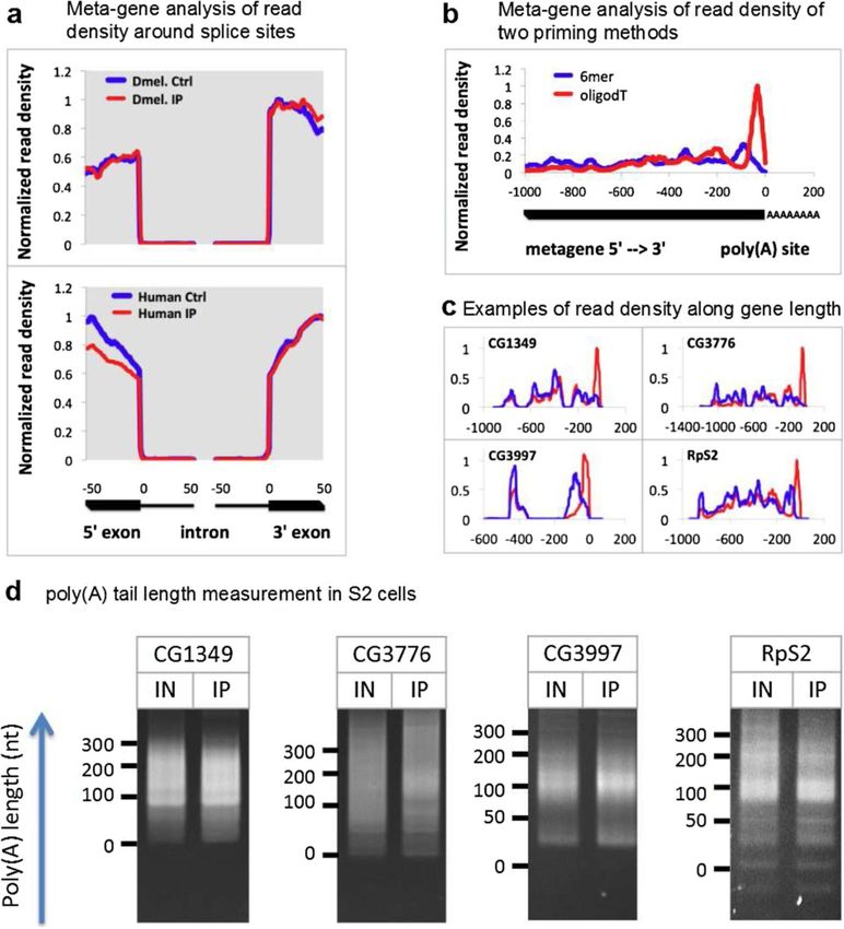
Lu et al. Genome Biology 2014, 15:R7                                                                               Page 2 of 23
http://genomebiology.com/2014/15/1/R7

developing oocytes [21,22], and a hypomorphic mutation            the mRNA-Sm association is mediated by snRNPs, and
in SmD3 causes mislocalization of oskar mRNPs and                 we show that a predicted U1 snRNP base pairing region
pronounced defects in germ cell specification that are            on an mRNA is required for interaction with this snRNP.
independent from splicing [21]. Moreover, loss of the             These mature mRNA-snRNP interactions are very stable
Sm protein methyltransferase PRMT5 results in failure to          and distinct from other previously studied interactions
specify the germline [21,23,24]. Furthermore, a genetic           (pre-mRNA splicing, ‘telescripting’ and regulation of
screen for modifiers of FMR1 (Fragile X mental retard-            promoter directionality). Taken together, the data identify
ation 1) in Drosophila identified SmD3 as a suppressor of         additional direct targets of canonical Sm proteins, and
dFMR1’s translational repression function, and SmD3 and           suggest that Sm class snRNPs may have novel, evolution-
dFMR1 were found to colocalize within neuronal mRNP               arily conserved functions in mRNA localization, stability
granules [25]. In vertebrates, Sm proteins are enriched in        and translation.
the nuage and mitochondrial cement [26,27], structures
that share many components with the invertebrate germ             Results
plasm. The U1 snRNP, in addition to its splicing role,            Identification of RNAs that co-purify with eukaryotic
protects pre-mRNA from premature polyadenylation at               Sm proteins
cryptic poly(A) signals in introns [11,12,28], and inhibits       As mentioned above, the Sm and Sm-like proteins com-
HIV RNA polyadenylation [29,30]. In addition, RNA                 prise a family of ancient evolutionary origin that functions
sequence elements complementary to the U1 5′ end                  to modulate the stability and translation of several classes
play important roles in the stabilization of promoter-            of RNA, including mRNAs [1,35]. Based on these ances-
downstream transcripts and thus contribute to promoter            tral roles, the involvement of eukaryotic Sm proteins in
directionality [31,32]. The U1 snRNP not only regulates           splicing is generally thought to be a derived function,
gene expression via RNA processing; a modified form               and additional RNA targets of Sm proteins remain to be
of U1 can also target HIV RNA to reduce viral protein             discovered.
expression [33]. Moreover, the U2 and U12 snRNPs play                To characterize the repertoire of RNA targets that are
an unexpected role in promoting U7-snRNP-dependent                associated with Sm proteins in Drosophila ovarian lysates,
processing of intronless histone mRNAs in human                   we performed RIP-seq analysis of individual subunits of
cells, and both protein-RNA interaction and RNA-RNA               the canonical Sm ring. We also performed RIP-seq on
base-pairing suffice for the activity [34]. Collectively, these   Trailer Hitch (Tral), a protein that contains an Sm domain
studies suggest additional functions for Sm proteins and          (Figure 1c). Tral is not incorporated into the canonical Sm
snRNPs in RNA metabolism; however, little is known                ring; therefore, we expected it to associate with a distinct
about the in vivo RNA targets that might be regulated by          subset of transcripts [36]. An outline of the experimental
Sm proteins/snRNPs, in these processes.                           strategy and data analysis pipeline is shown in Figure 1a.
  To systematically identify Sm protein-containing RNPs,          Immunoprecipitations (IPs) were carried out using either
we carried out RNA-immunoprecipitation (RIP) against              anti-SmB (monoclonal antibody Y12) or anti-green fluor-
multiple Sm proteins from Drosophila ovaries and HeLa             escent protein (anti-GFP) antibodies (for the GFP- and
cells, followed by high-throughput sequencing (RIP-seq)           Venus fluorescent protein (VFP)-tagged proteins). Normal
of the immunopurified RNAs. Using this robust and                 goat serum was used as control for the IP. Immunopreci-
reproducible multi-targeting RIP-seq approach, we recov-          pitated RNA was reverse transcribed to cDNA, fragmen-
ered most of the spliceosomal snRNAs. In addition, we             ted, ligated with adapters, PCR-amplified and sequenced
discovered a new Drosophila-specific snRNA, many Sm-              on an Illumina Genome Analyzer II.
associated small Cajal body-specific RNAs (scaRNAs), and             To reduce potential non-specific interactions and ar-
numerous Sm-associated mRNAs from both Drosophila                 tifacts, we carried out RIP-seq on several Sm proteins
and human cells. The new snRNA is highly conserved in             expressed from three different genomic contexts: (i)
the melanogaster group of Drosophilids, although it is not        native endogenous genes, (ii) VFP-tagged transgenes,
essential for organismal viability. Two major categories of       or (iii) a gene-trapped (GFP-tagged) endogenous gene
the Sm-associated mRNAs encode mitochondrial and                  (Figure 1c). Comparisons among this wide variety of
translation-related proteins. Using quantitative reverse          experimental conditions helps to minimize problems
transcriptase PCR (qRT-PCR), we found that some of the            associated with genetic background, transgene overexpres-
RNA-Sm interactions are tissue-specific, whereas others           sion, and antibody specificity. Four different transgenic
are more widespread. The Sm-associated mRNAs are                  lines were employed, including VFP-tagged SmD3, SmB,
properly spliced and polyadenylated, indicating that              SmD1 and SmE [21]. Transgenes were expressed using
the mRNA-Sm interactions reported here are distinct               the UAS/Gal4 system, crossed to a nanos-Gal4 driver for
from those involved in pre-mRNA splicing and Lsm1-7               germline-specific expression or, in the case of VFP-SmD1,
dependent degradation. We also provide evidence that              to a daughterless-Gal4 driver for ubiquitous expression
Lu et al. Genome Biology 2014, 15:R7                                                                                                  Page 3 of 23
http://genomebiology.com/2014/15/1/R7

 Figure 1 RIP-seq experimental analysis strategies. (a) Outline of RIP-seq analysis pipeline. See Materials and methods for details. (b) Schematic
 diagram of the canonical Sm ring. The three sub-complexes are shown separately. (c) Schematic diagram of the Sm-domain-containing proteins
 used in this study. (d) Summary of the RIP-seq and RIP-qRT-PCR experiments performed, targeting all three sub-complexes of the canonical Sm
 ring and Tral. See Table S1 in Additional file 1 for details. Ctrl, control; GFP, green fluorescent protein; IP, immunoprecipitation; RPKM
 (reads per kilobase per million reads); VFP, Venus fluorescent protein.

[37]. SmB and SmD3 form an obligate dimer (Figure 1b),                     the reads in the control IPs are not mappable (for ex-
whereas SmD1 and SmE are present in distinct sub-                          ample, rRNAs, primer/adapter dimers or even random se-
complexes within the heteroheptameric ring structure                       quences; Table S3 in Additional file 1) and those that do
[9]. Thus, IPs targeting different components of the Sm                    map to the genome typically correspond to abundant RNAs
ring further reduced potential artifacts resulting from                    that stick to the beads non-specifically Library statistics
epitope tagging, as these proteins form a complex that                     show that random hexamer priming yielded more map-
is expected to bind a similar set of RNAs. RIP-seq experi-                 pable reads than did oligo(dT)20 priming (Table S4 in
ments were performed on SmB, SmD3 and SmE, whereas                         Additional file 1). Thus, we used the random hexamer-
RIP-qRT-PCR was performed on VFP-SmD1 for identified                       primed libraries for the subsequent enrichment analyses.
targets. To broaden the scope of our study, we also                           We built a data analysis pipeline (Figure 1a) by integrating
performed RIP-seq analysis in cultured human HeLa                          previously published programs (see Materials and methods
cells, using the Y12 antibody mentioned above (Figure 1d;                  for details). Sequence reads for the Drosophila RIP-seq
see details in Table S1 in Additional file 1).                             experiments were mapped to the Drosophila expanded
                                                                           genome and quantified using ERANGE [38]. Then, for
Enrichment analysis of Sm RIP-seq experiments                              each experiment, we filtered out transcripts with read
We obtained between 8 and 28 million 35-nucleotide                         coverage less than 10. Assuming that the majority of RNA
single-end reads per Drosophila ovary RIP-seq library,                     species are not associated with Sm proteins, we normalized
and roughly 20 million 48-nucleotide paired-end reads                      the remaining transcripts against the median of all en-
per human HeLa cell RIP-seq library. All of the fly and                    richment ratios: (raw_IP + 2)/(raw_Ctrl + 2). After norma-
human sequencing data are of high quality (Figure S1                       lization, we defined the enrichment ratio as (norm_IP + 2)/
in Additional file 1). Despite differences in total read                   (norm_Ctrl + 2). The use of median-normalized raw read
numbers, the IPs consistently yielded many more mappable                   numbers is similar to the upper-quartile normalization
reads than did the controls (Table S2 in Additional file 1,                method used by others [39]. In this way, we made a
‘mapped’ and ‘%mappable’ columns). This was to be                          conservative estimate of the enrichment of RNAs in
expected; due to the low amount of input cDNA, most of                     IPs versus controls.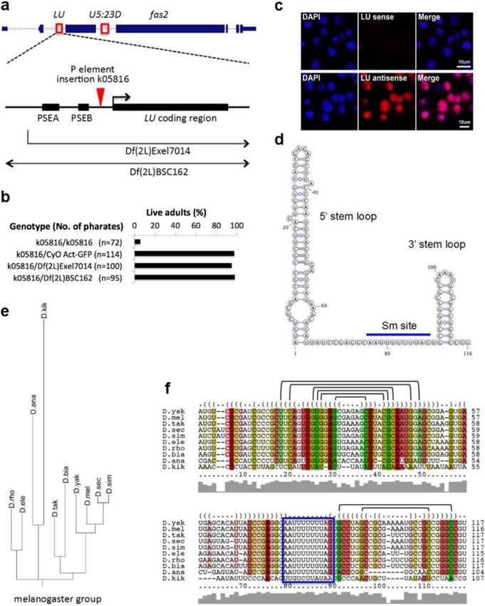
Lu et al. Genome Biology 2014, 15:R7 Page 4 of 23
http://genomebiology.com/2014/15/1/R7
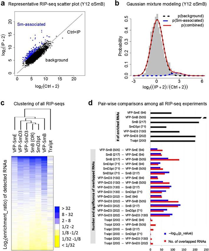
To visualize the enrichment data, scatter plots were analysis. The top of the map represents RNAs that are
constructed using the log-transformed and normalized significantly enriched (Figure 2c). As shown by the den-
read numbers. Data for the native SmB-associated RNAs drogram (Figure 2c) and consistent with expectation, the
(Oregon R, Y12 IPs) are shown in Figure 2a; data for the six canonical Sm protein RIP-seq experiments clustered
other Sm protein constructs are presented in Figure S1 together, whereas the data from the Tral IP formed an
in Additional file 1. In any co-IP experiment, there are two outgroup. The most-highly enriched transcripts among
populations of molecules: those that interact specifically the random hexamer-primed libraries from six Sm IP
with the antibody and those that stick non-specifically experiments (including one VFP-SmD3 biological replicate)
to the beads. Non-specific interaction was observed for revealed extensive overlap. Detailed analysis showed that
many transcripts, as depicted by the main cluster along 25 RNAs (9 snRNAs, 16 mRNAs) were common among
the diagonal line (Figure 2a). The dots located above the all 6 Sm protein IPs, and 52 transcripts (12 snRNAs, 40
main cluster represent the enriched RNAs. In order to mRNAs) were shared in 5 of the 6 (see Table S5 in
objectively identify Sm-associated RNAs, we employed Additional file 1 for detailed enrichment ratios). The
Gaussian mixture modeling [40], which has been used top 86 transcripts (13 snRNAs, 1 small nucleolar RNA
to analyze RIP-chip experiments [41]. Distributions of the (snoRNA), and 72 mRNAs) were shared by at least 4 of
enrichment ratios were first plotted as histograms. Next, the experiments. Since four Drosophila snRNAs (U1, U2,
we used mixtools to fit a combination of two Gaussian U4, and U5) have multiple variant paralogs, we reassigned
functions to the enrichment ratio distribution [42]. uniquely mappable reads to them and we found that all
As shown in Figure 2b, the distribution of the log- of the snRNAs with significant coverage are enriched in
transformed enrichment ratios (red line) can best be all Sm IPs (Table S6 in Additional file 1). In addition, we
explained by two different Gaussian functions, one that analyzed the consensus set of 86 Sm-associated RNAs
corresponds to the background RNAs (black dotted in the oligo(dT)20 primed libraries, and we found that
line) and one that represents the Sm-associated RNAs they are also highly enriched, despite the lower number
(blue dotted line). The cutoff between Sm-associated of mappable reads (Figure S4 in Additional file 1). Thus,
and background mRNAs was defined by the log of the our multi-targeting RIP-seq approach is robust despite the
odds (LOD) ratio between the two Gaussian functions. differences in library statistics (Table S2 in Additional
The transcripts with a LOD > 1 (that is, those that had file 1). We operationally defined the Sm-associated RNAs
a greater likelihood of being in the Sm distribution) as being those that were enriched in at least four of the six
were considered to be Sm-associated RNAs. Using this experiments.
threshold, we then mapped these assignments back Next, we carried out pair-wise comparisons among the
onto the scatter plots. As shown in Figure 2a (blue dots), seven RIP-seq experiments and performed Fisher’s exact
the enriched RNAs are clearly seen to be above the diag- test to assess the significance of any overlapping subsets
onal (black dots represent the background distribution). (Figure 2d). Interestingly, among the top 200 RNAs in
This same analysis was performed on the other Sm the Tral IP experiment, very few of them overlapped with
protein datasets, with strikingly similar results (Figure S2 any of the RNAs that associated with canonical Sm
in Additional file 1). Thus, the Gaussian mixture modeling proteins. As seen in the heat map (Figure 2c), the enrich-
procedure provides an unbiased and less arbitrary method ment ratios for the VFP-SmE IP were typically lower than
for identifying enriched RNAs [41]. Using the afore- those of the other Sm proteins. However, the pairwise
mentioned analysis pipeline, we identified roughly 200 comparisons show that SmE associates with a similar
Sm-associated RNAs in any given RIP-seq experiment, group of RNAs (see also Figure S4 in Additional file 1).
representing 0.7% of the Drosophila transcriptome, or The overlaps between the different Sm protein IPs were
4% of the significantly expressed transcripts. highly significant, as shown by their extremely small P-
values (10-32 to 10-135, plotted as negative logarithms;
A multi-targeting RIP strategy identifies highly reproducible Figure 2d). Even when all of the snRNAs were taken out
Sm-associated RNAs of the pair-wise comparisons, the P-values remained
To assess the robustness and reproducibility of the extremely small (Figure 2d; Figure S3 in Additional file 1).
Drosophila RIP-seq experiments and analysis pipeline, Despite the different experimental parameters (tagged
we visualized the log-transformed enrichment ratios for versus untagged, native versus ectopic, and so on), the
the transcripts with a read coverage greater than 10. Out lists of enriched RNAs are essentially the same. This high
of the >15,000 annotated genes in the fruitfly genome, degree of reproducibility suggests that the multi-subunit
5,296 of them showed sufficient read depth (d > 10). To targeting approach is superior to the conventional bio-
determine the relationship between the profiles of the logical replication of experiments for RNP analysis. Indeed,
seven RIP-seq experiments without prior assumptions, the variability between biological replicates was greater in
we performed an unsupervised hierarchichal clustering the case of VFP-SmD3 than it was between some of the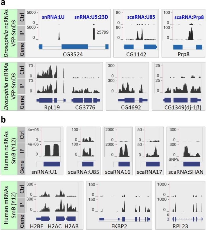
Lu et al. Genome Biology 2014, 15:R7    Page 5 of 23
http://genomebiology.com/2014/15/1/R7

 Figure 2 (See legend on next page.)
Lu et al. Genome Biology 2014, 15:R7 Page 6 of 23
http://genomebiology.com/2014/15/1/R7
(See figure on previous page.)
Figure 2 RIP-seq data analysis. (a) Scatterplot of a control (Ctrl)-IP pair of RIP-seq data (SmB IP Lu023-Lu024), where normalized and log-transformed
read numbers for each known transcript in an IP are plotted against that of Ctrl (Ctrl + 2 and IP + 2 to avoid division by zero). Black dots represent
background RNAs, while the blue dots represent enriched RNAs, as determined by Gaussian mixture modeling. Only RNAs with read coverage >10
are plotted. See Figure S1 in Additional file 1 for the rest of the scatterplots. (b) Gaussian mixture modeling of the RIP-seq data (SmB IP), where the
enrichment ratios for all the transcripts were plotted as a histogram (in gray) and fitted with a combination of two Gaussian curves. (c) Log-transformed
enrichment ratios of the 5,296 RNAs (with coverage d >10) in all 7 experiments were clustered (average linkage clustering using correlation (uncentered)
as similarity metric) and visualized as a heat map. (d) Pair-wise comparisons among all seven experiments. Numbers of enriched RNAs are listed next to
the experiment labels. Black bars, number of enriched RNAs in each experiment; red bars, number of overlapped RNAs in each pair; blue bars, negative
log10 transformed Fisher’s exact test P-values (within a superset of 5,296 RNAs). See Figure S2 in Additional file 1 for pairwise comparisons excluding
non-coding RNAs.
other RIPs (Figure 2c). Collectively, these data demon- identified in our IPs, all of which have very high enrichment
strate a high degree of specificity in the Sm protein IPs, ratios (Figure 3b).
showing that canonical Sm proteins co-precipitate with ERANGE analysis and manual inspection of the Drosoph-
essentially the same set of mRNAs. ila RIP-seq data revealed several clusters of reads that could
not be mapped to gene models. Four of them are new
genes that had not been previously annotated. During
Sm proteins associate with three major classes of RNAs
preparation of this manuscript, two transcriptomic studies
The RIP-seq experiments in both Drosophila and human
have since identified these putative new transcripts [45,46]:
cells confirmed the well-studied snRNAs as major targets
CR43708, CR43600, snoRNA:2R:9445410 (CR43574) and
of Sm proteins, and in addition indicate novel classes of
snoRNA:2R:9445205 (CR43587). Two of the four novel
Sm targets. A detailed analysis of the known and newly
transcripts, CR43708 and CR43600, showed significant
discovered RNAs from our study suggests that Sm proteins
enrichment in the IPs.
associate with three major classes of RNAs (Figures 3
We characterized the two Sm-associated ncRNAs and
and 4; Figures S4 and S6 in Additional file 1).
found that one, CR43708, has features typical of an snRNA.
CR43708 is located in the second intron of fas2 (CG3524,
RIP-seq identifies Sm class snRNAs fatty acid synthase 2), a homolog of the human fatty acid
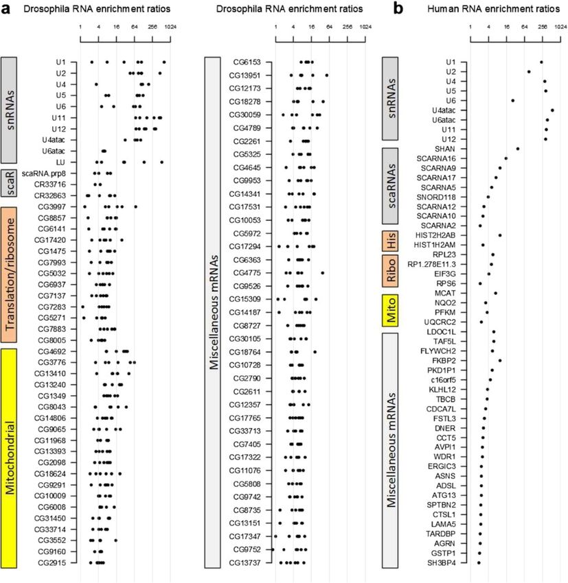
The Sm-associated transcripts and their enrichment ratios synthase gene (Figure 5a). We defined the accurate 5′ and
are listed in Figure 3. As expected, all spliceosomal snRNAs 3′ ends of CR43708, and found that this transcript is 116
were among the top-scoring transcripts in terms of their nucleotides long (ZL and AGM, unpublished). Detailed
enrichment ratios. The only missing Sm class snRNA from analysis of sequences upstream of CR43708 revealed
the list of Sm-associated RNAs is U7 snRNA, because it is conserved proximal sequence elements PSEA and PSEB,
too short (71 nucleotides in Drosophila, and 63 nucleo- highly similar to Sm-class snRNA promoters (Figure 5a;
tides in human) to be included in the size-selected cDNA Figure S7a in Additional file 1) [47,48]. To examine the
libraries (Figure 3a; Table S5 in Additional file 1) [43,44]. subcellular localization of CR43708, we carried out in situ
Other highly abundant non-coding RNAs (ncRNAs; for hybridization in Drosophila S2 cells and found that this
example, 7SK snRNA, SRP RNA, 5.8S ribosomal RNA RNA accumulates in the nucleus (Figure 5c). Using the
and so on, data not shown) were not enriched in the IPs, transcribed region and the promoter sequences, we searched
demonstrating the specificity of the approach. Multiple genome and transcriptome databases for homologs. We
distinct paralogs exist for four of the Drosophila snRNAs, recovered matches in nine species, all of which are in
U1, U2, U4 and U5, and they share long stretches of iden- the melanogaster group of the Drosophila genus, and
tical regions (Figure S5 in Additional file 1). In order to all are located within the same intron of the fas2 gene
accurately analyze each paralog without the confounding (Figure 5e,f ). Among the sequenced Drosophila species
repetitive reads, we reassigned uniquely mappable reads in the melanogaster group, the Drosophila erecta genome
to U1, U4 and U5 paralogs (Table S6 in Additional file 1). does not appear to contain CR43708, suggesting that it
We used the variant nucleotides in U2 to calculate the may have been lost. Interestingly, we found a truncated
fractions of each isoform and redistribute the total version of this gene within an intron of the Ac3 gene in
number of U2 reads among the gene paralogs. Not sur- D. melanogaster (Figure S7c in Additional file 1). The
prisingly, all snRNAs with significant read coverage are homology extends through the first 70 bp of CR43708,
enriched in the IPs (Table S6 in Additional file 1). With and lacks the promoter and the 3′ end, suggesting that
regard to the HeLa cell analysis, there are hundreds of this paralog is a pseudogene. The predicted secondary
snRNA genes in the human genome, and only a small structure of CR43708 closely resembles that of a canonical
fraction of them are properly annotated. Not surprisingly, snRNA, including the presence of 5′ and 3′ end stem
most of the annotated human spliceosomal snRNAs were loops that flank a putative Sm binding site (Figure 5c).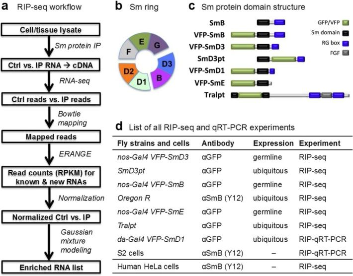
Lu et al. Genome Biology 2014, 15:R7                                                                                                      Page 7 of 23
http://genomebiology.com/2014/15/1/R7

 Figure 3 Three categories of Sm-associated RNAs in Drosophila and human. Different categories of Sm-associated RNAs are color-coded.
 (a) Drosophila Sm-associated RNAs, with enrichment ratios from all six Sm RIP-seq experiments. For snRNAs with multiple distinct paralogs (U1,
 U2, U4 and U5), all the reads were pooled for calculation of enrichment ratios. The three U6 paralogs are identical in sequence. See Table S6 in
 Additional file 1 for assignment of reads to distinct paralogs. U7 was not plotted due to low read coverage. See Table S5 in Additional file 1 for
 detailed enrichment ratios. (b) Human Sm-associated RNAs. Medians of enrichment ratios were plotted for snRNAs with multiple paralogs.
 See Table S7 in Additional file 1 for detailed enrichment ratios.

Structured sequence alignments clearly show that the                         structure (Figure 5f). Uridine-rich, Sm-class snRNAs such
putative Sm binding site (except in Drosophila kikkawai)                     as U1 and U2 are known to contain a trimethyl-guanosine
and the terminal stem loops are well conserved. In                           (TMG) 5′ cap structure that is generated upon formation
addition, we identified many covariant base pairs within                     of the Sm core RNP [9]. As expected, CR43708 was
the two stem loops, supporting the predicted secondary                       efficiently immunoprecipitated by anti-TMG antibodies
Lu et al. Genome Biology 2014, 15:R7                                                                                                Page 8 of 23
http://genomebiology.com/2014/15/1/R7

 Figure 4 Examples of the three categories of Sm-associated RNAs in Drosophila and human. For genes with multiple transcripts, the gene
 model that is most similar to the read coverage pattern is shown. The y-axis corresponds to the normalized number of reads per nucleotide. (a)
 Examples of Drosophila Sm-associated RNAs from VFP-SmD3, control (Ctrl; Lu003) and IP (Lu004). For the non-coding RNAs that are associated
 with Sm proteins, their host genes are also shown. The read coverage for U5:23D is off scale, and thus truncated. (b) Examples of human Sm-
 associated RNAs from Y12 αSmB, Ctrl (Lu045) and IP (Lu047). The histone mRNAs H2BE, H2AC and H2AB are short for HIST2H2BE, HIST2H2AC and
 HISTH2AB, respectively.

(Figure 6a). Taken together, these features led us to                     as we found a nearly invariant single-stranded region
conclude that this transcript is a novel Sm-class snRNA,                  located in the middle of LU snRNA (Figure 5d,f). Notably,
which we termed snRNA:LU (Like U).                                        we identified extensive base complementarity between
   Interestingly, the U5:23D snRNA gene is located near                   this region of LU and the 5′ end of U6 (Figure S7d in
LU, within a neighboring intron of the fas2 protein coding                Additional file 1). This putative base-pairing suggests
gene (Figure 5a). We were unable to deduce the precise                    that LU may be involved in splicing regulation. We
origin of LU; however, its juxtaposition with U5:23D                      identified four independent transposon insertions in
suggests that it could have evolved from a U5 gene duplica-               and around the LU gene locus (see Materials and methods),
tion, followed by rapid divergence. Supporting this notion,               and we confirmed that one of these insertion lines,
the 3′ end stem-loops of the LU snRNA homologs are                        fas2k05816, disrupts expression of both the fas2 host
quite similar to those of U5 snRNAs (Figure S7 in                         gene and the LU snRNA gene (Figure 5a; Figure S7e in
Additional file 1), although there is a lack of overall                   Additional file 1). Although homozygotes die around
sequence similarity between the two genes.                                eclosion; complementation analysis between fas2k05816
   To study the function of LU snRNA, we first considered                 and two other deletion lines uncovering this region
the possibility that it might base pair with other snRNAs,                suggests that neither the fas2 host gene nor the LU
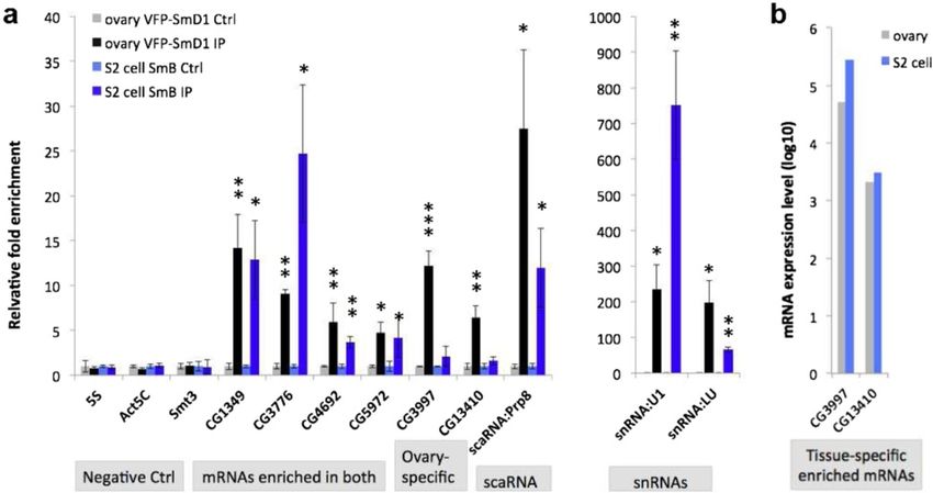
Lu et al. Genome Biology 2014, 15:R7                                                                                                          Page 9 of 23
http://genomebiology.com/2014/15/1/R7

 Figure 5 Characterization of the Like-U (LU) snRNA gene. (a) Genomic and genetic contexts of the LU snRNA locus. LU snRNA is encoded
 within the second intron of fas2; U5:23D is located in the third intron. PSEA/PSEB, proximal sequence element A/B (see Figure S7 in Additional file 1 for
 alignment of the U11 and LU promoters in Drosophilids). Locations of a P-element insertion and two deficiencies are indicated. The arrows on the
 deficiencies indicate that the regions extend beyond the displayed area. (b) Complementation analysis of LU snRNA mutations and deficiencies.
 Numbers of third instar larvae are indicated in parentheses. (c) Localization of LU snRNA in S2 cells determined by in situ hybridization using LU sense
 and antisense probes. (d) Predicted secondary structure of D. melanogaster LU snRNA. (e) Phylogeny of LU snRNA. (f) Alignment of Drosophilid LU
 snRNA orthologs using LocARNA. The blue box indicates the Sm site. Half-brackets indicate covariant base pairs.

snRNA gene are required for organismal viability                                Sm proteins associate with evolutionarily conserved and
(Figure 5b). We conclude that, although it may well                             rapidly evolving scaRNAs
contribute to organismal fitness, LU is not an essential                        scaRNAs are ncRNAs that guide methylation and pseu-
gene. This conclusion is supported by the independent                           douridylation of snRNAs, the specificity of which is deter-
loss of LU snRNA in D. erecta. Taken together, our                              mined by base-pairing with targets [49]. A previous study
RIP-seq analysis of Sm proteins reveals that a total of                         showed that in human cells, several scaRNAs specifically
11 distinct species of Sm-class snRNAs are present in                           associate with SmB and SmD3, including U85, U87, U89
Drosophila: U1, U2, U4, U5, U6, U7, U4atac, U6atac,                             and human telomerase RNA (hTR) [50]. Co-precipitation
U11, U12 and LU.                                                                of SmB/D3 with these scaRNAs was shown to require the
Lu et al. Genome Biology 2014, 15:R7                                                                                                  Page 10 of 23
http://genomebiology.com/2014/15/1/R7

 Figure 6 snRNPs associate with mature mRNAs in S2 cells. (a) Sm-associated mRNAs, as well as scaRNAs and snRNAs, can be pulled down by
 a TMG antibody in S2 cells. CG9042 (Gapdh) is used for normalization. (b) Enrichment analysis of the U1-70 K RIP-seq data in a volcano plot. The
 most highly enriched transcripts were labeled. The inset rectangular boxes highlight CG3776 and CG8108 mRNAs in the plot. Note: CG1349 and
 CG4692 could be associated with other snRNPs, and therefore not pulled down by U1-70 K. (c) CG8108 mRNA can be pulled down by TMG and
 Y12 antibodies in S2 cells. (d) CG8108 is expressed in similar levels in Drosophila ovary and S2 cells (data from FlyBase). (e) CG8108 mRNA is not
 enriched in ovary Sm RIP-seq. t-Test for significance between IP and control (Ctrl): *P < 0.05, **P < 0.01, ***P < 0.001). Error bars reflect the
 standard deviation.

conserved CAB box [50], which is essential for scaRNA                       [50], we found that two previously identified Drosophila
localization to Cajal bodies [51]. To determine whether                     scaRNAs, U85 (CR32863 or snoRNA:MeU5-C46) and
other ncRNAs co-purify with Sm proteins in Drosophila                       CR33716 (snoRNA:MeU5:U42), were enriched in the Sm
and human cells, we systematically analyzed the enrich-                     protein IPs (Figure 4a; Table S5 in Additional file 1). Inter-
ment values of snoRNAs and scaRNAs in our RIP-seq                           estingly, the new Sm-associated ncRNA identified in this
datasets. Consistent with the findings of Fu and Collins                    study (CR43600 or snoRNA:Prp8) also appears to have
Lu et al. Genome Biology 2014, 15:R7                                                                                                    Page 11 of 23
http://genomebiology.com/2014/15/1/R7

 Figure 7 RNA-Sm association is cell type-specific and not due to re-assortment. (a) RIP-qRT-PCR in da-Gal4 VFP-SmD1 fly ovary (anti-GFP)
 and S2 cells (Y12). Negative controls (Ctrl) used are 5S rRNA, Act5C and Smt3. CG9042 (Gapdh) is used as the normalization standard. snRNAs are
 shown separately due to the difference in scale. (b) mRNAs associated with Sm proteins in ovaries but not in S2 cells are expressed in S2 cells. t-
 Test for significance between IP and Ctrl: *P < 0.05, **P < 0.01, ***P < 0.001. Error bars show standard deviation.

features of box H/ACA scaRNAs. Indeed, evolutionary                          many H/ACA box scaRNAs are TMG-capped [55-58];
comparisons identify conserved H/ACA and CAB box                             consistent with these studies, we also found that scaRNA:
elements present within the detected orthologs (Figure                       Prp8 co-immunoprecipitates with anti-TMG antibodies
S6b,c in Additional file 1). snoRNA:Prp8 folds into a                        (Figure 6a).
predicted secondary structure similar to that of other                         To identify additional Sm-associated ncRNAs in HeLa
box H/ACA scaRNAs, which is further supported by the                         cells, we examined known human sno/scaRNA loci. Several
presence of multiple covariant base pairs. In support of                     of the previously reported scaRNAs, including U85, U87
the notion that snoRNA:Prp8 is an H/ACA box scaRNA,                          and U89, showed moderate but significant enrichment
we searched snRNAs for sequence complementarity to                           in Y12 IPs (Figure 4b; Table S7 in Additional file 1). In
the pseudouridylation pocket sequences, and found                            addition, we found several other scaRNAs that are highly
potential target sites in U1, U5, U7 and U11 (Figure S6d                     enriched (Figure 4b; Table S7 in Additional file 1).
in Additional file 1). Therefore, we have renamed this tran-                 However, we did not detect any significant enrichment
script scaRNA:Prp8. We detected homologs of scaRNA:                          of hTR as previously reported [50] (data not shown).
Prp8 in both Diptera (Drosophilids, Anopheles gambiae)                       We identified a novel, unannotated Sm-associated ncRNA,
and Hymenoptera (Apis mellifera), but not in Coleoptera                      which we named SHAN (Sm-associated Hybrid tRNAAsp-
(Tribolium castaneum) (Figure S6b in Additional file 1).                     containing NcRNA); its predicted secondary structure
The orthologous scaRNA:Prp8 RNAs are highly conserved,                       is shown in Figure S8c in Additional file 1. This new
suggesting their functional importance. Many scaRNA                          transcript appears to be a chimera between a tRNA
and snoRNA genes reside within introns of splicing and                       gene and an H/ACA type scaRNA gene. Supporting
translation-related genes, respectively [52]. The nested                     this hypothesis, we detected H box, ACA box and CAB
gene structures are thought to facilitate transcriptional                    box motifs in the orthologous sequences from other
co-regulation. Thus, it is not surprising that the Prp8 host                 primates (Figure S8b,c in Additional file 1). In sum-
gene encodes a splicing factor (Figure S6a in Additional                     mary, our RIP-seq analysis revealed both evolutionarily
file 1) [53,54]. Although Fu and Collins [50] reported that                  conserved and newly evolved interactions between Sm
only SmB and SmD3 co-purified with scaRNAs such as                           proteins and scaRNAs, suggesting that Sm proteins
hTR, we found that IP targeting VFP-SmD1 also pulled                         play roles in the biogenesis/function of a subset of
down snoRNA:Prp8 (Figure 7a). It has been shown that                         scaRNAs. However, we did not identify sequence/
Lu et al. Genome Biology 2014, 15:R7                                                                            Page 12 of 23
http://genomebiology.com/2014/15/1/R7

structural features that distinguish Sm-associated scaRNAs     (Figure 4b). Another histone mRNA (HIST1H2AM), was
from other scaRNAs.                                            also significantly enriched (Figure 3b). Interestingly, Steitz
                                                               and colleagues [34] previously showed that the U2 snRNP
Sm proteins associate with mRNAs encoding mitochondrial        binds to (intronless) histone pre-mRNAs and stimulates
and translation-related proteins                               3′ end processing. Our identification of histone mRNAs
Due to a relative lack of comprehensive annotation of          in Sm protein co-IPs may reflect a snRNP-mediated
Drosophila gene ontology, we manually annotated the            interaction between Sm proteins and mRNAs. However,
Sm-associated mRNAs by homolog searching, protein              none of the Drosophila replication-dependent histone
domain analysis, and literature mining. This analysis sur-     mRNAs were enriched in the Sm protein IPs (Figure
prisingly revealed two major categories of mRNAs: those        S10 in Additional file 1). Taken together, our data suggest
encoding ribosome/translation-related proteins (13/86),        that the mode of interaction between Sm proteins,
and mitochondrial proteins (including mitochondrial ribo-      snRNPs and mRNAs is conserved between vertebrates
somal proteins, 19/86). As discussed above, the enrichment     and invertebrates.
of ribosomal protein mRNAs is not simply due to high
levels of expression. Only a subset of ribosomal protein
mRNAs is enriched in the Sm protein IPs. For example,          Validation and tissue-specificity of RNA-Sm protein
mRNAs encoding RpS11 (CG8857) and RpL39 (CG3997)               interactions in Drosophila
are highly enriched in Sm protein IPs (Figure 3a; Table S5     We have shown that the B/D3 and E/F/G subcomplexes
in Additional file 1), whereas RpL19 (CG2746) and RpL4         bind essentially the same set of target RNAs. To determine
(CG5502) are not enriched at all (Figure 4a and data not       whether SmD1 (which forms heterodimers with SmD2;
shown). Anecdotally, the mRNA encoded by CG3776,               Figure 1b) also associates with the RNAs listed in Figure 3a,
which is highly enriched, is located immediately adjacent      we immunopurified ovarian RNA from daGal4, VFP-SmD1
to RpL19 in the Drosophila genome, demonstrating the           flies (using anti-GFP) and carried out qRT-PCR. Further-
high degree of specificity of our approach.                    more, to assay the observed interactions in another cell
  Two other Drosophila Sm-associated mRNAs merit               type, we also performed qRT-PCR on RNAs immunopuri-
special interest. CG4692 encodes a predicted mitochondrial     fied from S2 cells using anti-Sm antibody Y12. We chose
F1-FO ATP synthase subunit that was consistently enriched      six of the top-ranking mRNAs that were identified in the
in our IPs. We found that this mRNA localizes to the           RIP-seq experiments (targeting SmB, SmD3 and SmE),
actin-rich oocyte cortex of late-stage Drosophila egg          and found that they were all highly enriched in the
chambers (Figure S4 in Additional file 1), in a pattern that   VFP-SmD1 IPs (Figure 7a). Two snRNAs (U1 and LU)
is very similar to that of VFP-tagged Sm proteins, as          were used as positive controls, whereas three RNAs
described previously [21]. Analysis of several other           not expected to interact with Sm proteins (Act5C and
high-scoring mRNAs from Figure 3a and Figure S4 in             Smt3 mRNAs and 5S rRNA) were used as negative
Additional file 1 did not display this pattern (data not       controls (Figure 7a). In contrast to the results in ovaries,
shown), so it is not a general feature of Sm-associated        only four out of the six mRNAs we tested were signifi-
mRNAs, but was nonetheless interesting. CG1349 (dj-1beta)      cantly enriched in the S2 cell IPs (Figure 7a). Given that
encodes a Drosophila homolog of the human DJ-1/PARK7           the Sm proteins and the six mRNAs we tested all have
(Parkinson autosomal recessive, early onset 7) gene. DJ-1/     comparable expression levels in both ovaries and S2 cells
PARK7 is one of 10 genes identified to date that cause         (Figure 7b and data not shown), these findings suggest
familial Parkinson disease [59]. A subpopulation of DJ-1       that the interactions between mRNAs and Sm proteins
protein is localized to mitochondria in a regulated manner,    can be tissue-specific. A potential concern in all RIP ex-
and is required for proper mitochondrial function [60].        periments is that the co-purification of the components
Thus, it is possible that Sm proteins play a role in regu-     might be due to reassortment of complexes following
lating the localization and/or translation of associated       cell lysis [61,62]. However, the fact that CG3997 and
mRNAs.                                                         CG13410 fail to associate with Sm proteins despite the
  In contrast to the more than 70 Sm-associated mRNAs          fact that they are well expressed in S2 cells argues
in the fruitfly (Figure 3a), we identified roughly 30          strongly against this artifact.
high-scoring mRNAs in human cells (Figure 3b). The
lower number in the human dataset is potentially due           Sm proteins associate with fully spliced and
to a reduced coverage of the transcriptome. Neverthe-          polyadenylated mRNAs
less, we found that one of the replication-dependent           The identification of significantly enriched mRNAs in
histone mRNAs, HIST2H2AB, is highly enriched in the            the co-IP fractions led us to ask whether the association
IPs (Figures 3b and 4b). In contrast, two adjacent histone     between Sm proteins and mRNAs was due to the splicing
genes, HIST2H2BE and HIST2H2AC, were not enriched              reaction itself. In other words, do Sm proteins interact
Lu et al. Genome Biology 2014, 15:R7 Page 13 of 23
http://genomebiology.com/2014/15/1/R7
with partially spliced or fully mature mRNAs? A quick being the predominant crosslinked member of the hepta-
glance at Figure 3 shows that the read depth over intronic meric ring [68]. More recently, Castello et al. [69] carried
sequences is very low. Meta-gene analysis of both Drosoph- out UV- and PAR-CLIP in parallel to generate a compre-
ila and human Sm-associated intron-containing mRNAs hensive mRNA interactome in HeLa cells. As part of their
showed that the vast majority of reads map to exons, studies, they identified the Lsm1-7 proteins as mRNA
and the IPs did not pull down more pre-mRNAs than binding proteins, but the canonical Sm proteins were not
the controls did (Figure 8a). Among the few transcripts detected, again supporting the idea that Sm proteins are
that showed significant numbers of intronic reads, most not efficiently crosslinked to mRNAs.
of those were actually candidates for either new exons or However, the fact that we found all three Sm sub-
new genes (for example, scaRNA:Prp8 and snRNA:LU; complexes in association with the same set of mRNAs
Figure 4a). Thus, this analysis demonstrates that the (Figures 2 and 3) suggested interaction with a complex
mRNAs that associate with canonical Sm proteins are fully that contains an intact Sm ring. Furthermore, the pre-
spliced. Importantly, 6 of the 72 Drosophila Sm-associated viously reported binding between histone mRNAs and
mRNAs (CG6008, CG13151, CG13951, CG17531, CG11076 U2 snRNPs [34], coupled with our identification of
and CG7137), and 2 of the 30 human Sm-associated H2A mRNAs in our RIP-seq data (Figure 4) led us to
mRNAs (HIST2H2AB and HIST2H2AM) are intronless, ask whether the mRNA-Sm interaction might be indirect,
suggesting that splicing is not a prerequisite for Sm protein mediated by snRNPs. Sm-class spliceosomal snRNAs are
interaction. transcribed by a specialized form of RNA polymerase II
The highly conserved eukaryotic Lsm1-7 complex is and contain a 5′ TMG cap structure [9]. Using anti-TMG
known to bind to mRNA degradation intermediates, antibodies, we immunopurified RNPs from S2 cell lysate
preferentially those with oligoadenylated tails [14,63]. and used qRT-PCR to assess the enrichment of mRNAs.
We therefore asked whether the canonical Sm ring shares As expected, the U1 and LU snRNAs (positive controls)
this same recognition specificity. Taking advantage of the were highly enriched in the anti-TMG IPs, whereas
oligo(dT)20 and random hexamer primed RIP-seq cDNA CG7939 (RpL32) mRNA was not (Figure 6a). Notably, the
libraries, we compared the read coverage patterns for scaRNA:Prp8 transcript and all three of the Sm-associated
the various mRNAs. As shown in Figure 8b,c, there is a mRNAs we tested (CG1349, CG3776 and CG4692)
dramatic 3′ end bias in the oligo(dT)20 primed libraries were significantly enriched in the anti-TMG pulldowns
compared to the randomly primed ones. We also con- (Figure 6a). In parallel, we performed anti-TMG IPs
firmed the presence of adenylated tails of Sm-associated using purified S2 cell RNA (that is, the IP was not per-
and non-associated mRNAs by examining the unmappable formed in lysates). We detected significant enrichment of
reads in the oligo(dT)20 primed RIP-seq files (Figure S11 in U1 snRNA but not the mRNAs (Figure S12 in Additional
Additional file 1). In order to measure polyA tail lengths, file 1). Therefore, the Sm-associated mRNP complex
we performed RACE-PAT (rapid amplification of cDNA contains a TMG cap component that is structurally
ends-poly(A) tail assay) on immunopurified RNAs from S2 distinct from the mRNAs themselves, suggesting the
cells [64]. This analysis demonstrates that the poly(A) tails presence of snRNPs.
of the Sm-associated mRNAs are roughly the same length In order to test whether the interactions with mRNAs
as the input mRNAs (Figure 8d). Taken together, these data are indirectly mediated by snRNPs, we took advantage of
show that Sm and Lsm proteins have distinct specificities a database from a large-scale Drosophila S2 cell RIP-seq
and modes of mRNA interaction. analysis of 29 RNA binding proteins, including U1-70 K
[70]. The U1-70 K protein binds to U1 snRNA directly
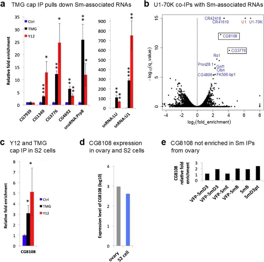
Sm protein interaction with mRNAs is mediated by snRNPs and specifically, thus allowing it to be used as an additional,
The association of snRNAs and scaRNAs with Sm independent epitope for pulldown experiments [68]. We
proteins is thought to be mediated by direct binding mined the database for RNAs that associate with U1-70 K
to Sm sites and CAB boxes, respectively [50,65,66]. We by analyzing RNAs that were enriched in IPs from U1-70 K
therefore wanted to determine whether Sm proteins associ- transfected versus non-transfected cells. The RIP-seq data
ate with mRNAs directly or indirectly. Toward that end, we were displayed on a volcano plot to identify transcripts
carried out PAR-CLIP (photoactivatable ribonucleoside- that are highly enriched in the IPs. As shown in Figure 6b,
enhanced crosslinking and immunoprecipitation) on native U1 snRNA, but not the other spliceosomal snRNAs, was
and VFP-tagged Sm complexes [67]; however, we were dramatically enriched in the IP fractions, along with a
unable to detect any significant crosslinking events in number of other ncRNAs and mRNAs. Among this latter
the precipitated RNA (data not shown). We note that category, three mRNAs were particularly noteworthy:
canonical Sm proteins are notoriously poor at crosslinking. CG3776, CG8108 and U1-70 K (CG8749) itself. Although
Even on extremely abundant targets such as U1 snRNA, U1-70 K protein may well bind to its own mRNA for
the UV crosslinking efficiency was rather low, with SmG some type of autologous feedback, one must view thisLu et al. Genome Biology 2014, 15:R7 Page 14 of 23 http://genomebiology.com/2014/15/1/R7 Figure 8 Sm proteins associate with mature mRNAs. (a) Meta-gene analysis of read density around splice sites for all Drosophila and human Sm-associated intron-containing mRNAs in all RIP-seq experiments. (b) Meta-gene analysis of read density along the gene length for all Drosophila Sm-associated mRNAs quantified from oligodT and random hexamer primed libraries. (c) Example tracks for read density along the gene length for oligodT and random hexamer primed libraries. (d) Poly(A) tail length Sm-associated mRNAs (CG3997, CG1349 and CG3776) and non-associated mRNA (RpS2) from Y12 IP in S2 cells. IN, input total RNA; IP, immunoprecipitated RNA. The labels denote the length of poly(A) tails. Oligo(dT)20 was used as the reverse primer for the reverse transcription and subsequent PCR, therefore producing the ‘smear’ of poly(A) tail. See Figure S11 in Additional file 1 for analysis of poly(A) containing reads for selected Sm-associated mRNAs. result with caution because the cells were transiently CG8108 remain good candidates. Interestingly, CG3776 transfected with U1-70 K cDNAs, artificially inflating was one of the top-ranking candidates in our ovarian expression of this transcript. However, CG3776 and RIP-seq experiments (Figures 3 and 4), but CG8108 was
Lu et al. Genome Biology 2014, 15:R7 Page 15 of 23
http://genomebiology.com/2014/15/1/R7
not identified as being enriched, even though it is serum as a control. As expected, 5S rRNA was not
expressed at similar levels in S2 cells (Figure 6d,e). Be- enriched in the IP fractions, whereas CG1349 mRNA
cause the U1-70 K data were generated from S2 cells, and U1 snRNA were both significantly enriched in the
we performed anti-TMG and anti-SmB (Y12) IPs in S2 transfections. Both endogenous and transfected CG3776wt
cells, followed by qRT-PCR. As shown in Figure 6c, we mRNAs were pulled down by the Y12 antibody, whereas
detected significant enrichment of CG8108 in both the transfected CG3776mut mRNA was not (Figure 9d). These
TMG and Sm protein IPs. These data provide additional results support two conclusions. First, splicing is not
support for the idea that the Sm-mRNA interactions are required for U1 snRNP binding, and the binding site
cell-type specific and not due to reassortment, as CG8108 for U1 snRNP is located within the CG3776 mRNA
is expressed in Drosophila ovaries (Figure 6d) but not coding sequence, since it can be efficiently pulled down
significantly enriched in Sm protein IPs (Figure 6e). by Y12 antibody. Second, the predicted U1 binding site is
In addition to CG3776, we also found other U1-70 K indeed necessary for U1 snRNP binding. Taken together,
associated RNAs that overlapped with our Sm protein our results suggest that snRNPs bind mature mRNAs, and
dataset, including CG5972 and CR32863. Although it is that at least one mechanism requires U1 snRNP base
likely that U1-70 K binds to certain RNAs in a manner pairing with target mRNAs.
that is independent of the U1 snRNP, the overlap between
our anti-Sm and anti-TMG data suggests that a cadre of
Discussion
mature mRNAs interacts with intact snRNPs outside of
We have developed an experimental and analytical pipeline
the spliceosome. Thus, we checked for sequence comple-
to identify RNAs that stably associate with Sm proteins, an
mentarity in CG3776 mRNA and found a 12 bp perfect
evolutionarily ancient group of RNA binding factors. The
duplex with the 5′ end of U1 snRNA (Figure 9a). The
targeting of multiple subunits of an RNA-binding complex
complementary region is in the middle of the second exon
in this RIP-seq approach, along with the use of different
of CG3776, far from any intron-exon boundaries and the
genetic backgrounds, ensures that the identified RNPs are
base-pairing potential is much greater than is typical
bona fide. Notably, this pipeline can be easily adapted to
for a 5′ splice site. Similarly, we found stretches of
study other RNA-binding complexes.
complementarity between U1 snRNA and exonic regions
of CG8108, CG5972 and many other transcripts (Figure
S13 in Additional file 1). Those mRNAs within our dataset Sm proteins in scaRNP complexes
that are missing from the U1-70 K pulldowns (for example, We found that subsets of scaRNAs associate with Sm
CG1349 and CG4692) are plausibly bound by other Sm proteins, in both Drosophila and human cells. These
snRNPs such as U2, U4/U6, U5, U11 and U12. A list of include the highly conserved U85 scaRNA and newly
such potential base pairing interactions was compiled by evolved and non-canonical scaRNAs, such as scaRNA:
taking known single-stranded regions from snRNAs, and Prp8 and SHAN, identified in this study. The involvement
using them to find putative binding sites on the list of Sm- of Sm proteins in scaRNP biogenesis and function has
and U1-70 K-associated mature mRNAs (Figure S13 in been shown in several previous studies. Notably, both
Additional file 1). We found many potential sites with a budding and fission yeast telomerase RNA precursors
duplex length and minimum free energy profile similar to contain canonical Sm sites and are directly bound by Sm
the ones shown in Figure 6f. Taken together with the Sm proteins [56,71]. In fission yeast, Sm binding to telomerase
and TMG IPs, these data suggest that snRNPs associate RNA stimulates spliceosome-mediated cleavage that mimics
with subsets of mature Drosophila mRNAs, in a mode that the first step of splicing [57,72]. However, none of the
is distinct from their interactions within the spliceosome. scaRNAs we found in our IPs contain readily identifiable
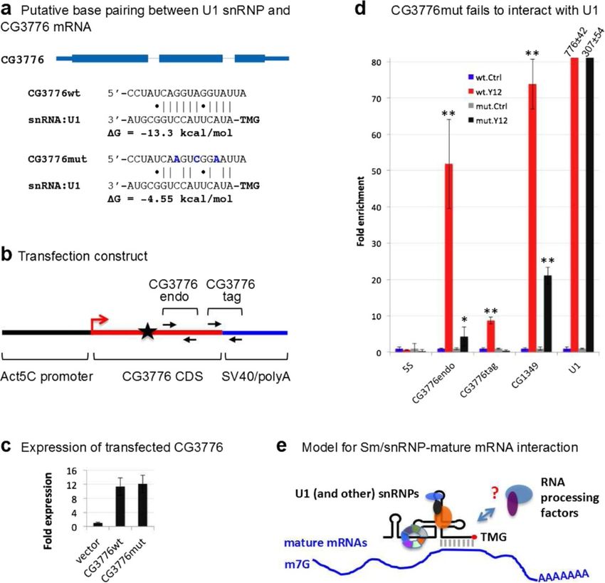
To test whether base pairing between U1 snRNP and Sm sites. Fu and Collins [50] reported that SmB and
CG3776 mRNA is responsible for their interaction, we SmD3, but not other Sm proteins, specifically associate
introduced three synonymous point mutations within with several human scaRNAs, and that this association
the twelve-nucleotide complementary region in CG3776 requires a conserved CAB box sequence. Tycowski et al.
mRNA that should completely block putative pairing [73] showed that this CAB box is bound by a protein
with U1 snRNA (Figure 9a). We then transfected both called WDR79. In our comprehensive analysis of fruit fly
wild-type and mutant CG3776 mRNA expression con- and human Sm-associated scaRNAs, we did not find
structs into S2 cells (Figure 9b). The constructs are additional sequence or structural features that distinguish
transcribed by an Act5C promoter and are terminated them. Thus, these studies suggest an evolutionarily con-
using the SV40 polyA signal and a heterologous 3′ UTR. served role for Sm proteins in scaRNA biogenesis and func-
We confirmed that both transfections produced similar tion; however, the mechanism through which scaRNAs
levels of chimeric CG3776 mRNAs (Figure 9c) and then that lack identifiable Sm sites associate with Sm proteins
performed Y12 IPs on S2 cell lysates, using normal goat is not well understood.Lu et al. Genome Biology 2014, 15:R7 Page 16 of 23 http://genomebiology.com/2014/15/1/R7 Figure 9 U1 snRNP binds mature mRNAs. (a) Putative base pairs between the 5′ end of U1 snRNA and the CG3776 mRNA coding region (upper panel). Within the putative region of base pairing, three translationally silent point mutations were introduced (bold blue letters) to disrupt the helix (lower panel). (b) Cartoon of the S2 cell transfection construct, showing the CG3776 expression unit. CG3776endo and CG3776tag indicate locations of primers for qRT-PCR. CG3776endo amplifies both endogenous and transfected CG3776 mRNAs, whereas CG3776tag amplifies transfected CG3776 mRNA only. The black star indicates the location of the putative U1 binding site. (c) pAW vector, pAW-CG3776wt and pAW-CG3776mut were transfected into S2 cells, and CG3776wt and CG3776mut expression was measured using qRT-PCR with the CG3776endo primer pair. GAPDH was used as normalization standard. (d) After pAW-CG3776wt and pAW-CG3776mut were transfected, anti-Sm (Y12) IPs were performed using S2 cell lysate. GAPDH was used as normalization standard. (e) Proposed model of snRNP-mRNA interactions. Distinct snRNPs (U1 and potentially others) associate with mature mRNAs via base pairing and/or protein-mediated interaction. Such interactions could serve as a platform to recruit RNA processing factors that act on multiple levels of RNA metabolism. t-Test for significance between IP and control (Ctrl): *P < 0.05, **P < 0.01, ***P < 0.001. Mut, mutant; wt, wild-type. Splicing-independent, evolutionarily ancient functions splicing elements, also serve as prime candidates for base for Sm-class snRNPs pairing with mature mRNAs. We propose a model whereby The available single-stranded regions of snRNPs, which Sm-class snRNPs interact with their targets via a combin- are used to identify intron-exon boundaries and intronic ation of base pairing and protein-RNA interactions, as
Lu et al. Genome Biology 2014, 15:R7                                                                               Page 17 of 23
http://genomebiology.com/2014/15/1/R7

shown in Figure 9e. Indeed, this model has precedence, as         so-called ‘sRNPs’ bind to their targets, which include ribo-
the efficacy of this combination of interactions has already      somal protein (RP) mRNAs, via a combination of base
been demonstrated. Steitz and colleagues [34] showed that         pairing and protein-RNA interactions [6,7,76-79]. Although
both RNA-RNA and protein-RNA interactions are in-                 the RP genes are not homologs of the RP mRNAs identified
dividually sufficient for function of the SF3b-hPrp43             in this study, our findings nevertheless support the hy-
subcomplex within the U2 snRNP in stimulating histone             pothesis that regulation of ribosome biogenesis is a deeply
mRNA 3′-end maturation. In the current study, we showed           conserved function of Sm proteins.
that a sequence within CG3776 mRNA that potentially                  Sequence covariation is generally considered a hallmark
base pairs with the 5′ end of U1 snRNP is required for            of conserved base-pairing interactions, underscoring func-
binding. Mutation of this sequence abrogates U1 binding.          tional importance. Not surprisingly, we found many covari-
By such a mechanism, snRNAs and/or specific proteins              ant base pairs in the stem-loops of snRNA:LU and scaRNA:
that bind to snRNPs could recruit other factors that, to-         Prp8, despite their short evolutionary histories (Figure 5;
gether, serve to regulate the processing, localization, trans-    Figures S6 and S7 in Additional file 1). However, we were
lation or degradation of target mRNAs (Figure 9e).                unable to analyze this feature in our Drosophila and human
   Recently, Berg et al. [12] proposed a function for U1          Sm/snRNP-associated mRNAs, as no clearly orthologous
snRNPs, termed ‘telescripting,’ whereby binding of U1 to          mRNA transcripts were identified. Instead, we found that
nascent transcripts acts to suppress premature cleavage           most of the targets of Sm proteins and snRNPs are different
and polyadenylation at cryptic sites. Reduction of U1             in the flies and human, with the exception of snRNAs and
snRNP levels elicited shortening of 3′ UTR length and             U85 scaRNA. This is consistent with the idea that protein-
proximal 3′ exon switching of numerous transcripts in a           RNA and RNA-RNA interaction networks rapidly rewire
dose-dependent fashion [11,12]. This process is distinct          themselves during evolution, despite the conservation of
from the interactions described here, as our data clearly         the individual components. For example, several studies on
showed snRNPs associating with mature mRNAs. More-                the RNA targets of Puf family proteins in yeast, fruit fly
over, we did not observe significant enrichment of intronic       and human suggest that even though the binding sites
regions in our RIP-seq datasets, as might have been               of the proteins are conserved, the target mRNAs are not
expected if the telescripting interactions between U1 and         [41,80,81]. Similarly, Graveley and colleagues [82] showed
post-splicing lariats were stable. Thus, the interactions         that the binding sites for PS and NOVA1/2 are highly
described here with mature mRNAs are stable, likely               conserved between insects and mammals, but the target
taking place either in the cytoplasm or just prior to             gene orthologs associated with PS and NOVA1/2 are al-
mRNA export.                                                      most entirely non-overlapping. This change of regulatory
   Furthermore, the data indicate that U1 snRNP is not            relationships in evolution has also been observed in the
the only Sm RNP that associates with mature mRNAs.                processing of minor introns and highly conserved micro-
The U2 snRNP-histone mRNA interaction [34] (and this              RNAs, such as let-7 and its targets [83,84].
work) is a case in point. We did not detect any downstream
flanking sequences in our RIP-seq data, suggesting that the       Technical considerations
U2 snRNP maintains contact with the histone mRNA long             It is likely that the Sm-associated transcriptome is larger
after 3′ end maturation, and therefore a potential function       than the one described here. Although RNA-seq is quite
downstream of 3′ end formation, for example, translational        sensitive, it may not be sensitive enough to reliably identify
control. We also identified Sm- and TMG-associated                all of the low abundance transcripts from the relatively
mRNAs in S2 cells that are not enriched in U1-70 K                minute amount of immunopurified RNAs. The spliceoso-
IPs, most prominently CG1349 and CG4692. Interestingly,           mal snRNAs comprise a majority of the immunopurified
we found that the localization pattern of Drosophila              transcripts, limiting the ability of the sequencer to identify
CG4692 within stage 10 egg chambers (Figure S9 in                 low abundance Sm-associated RNAs, especially scaRNAs
Additional file 1) mirrored that of VFP-tagged Sm proteins        and mRNAs. In addition, we employed a very stringent
[21]. Taken together, these findings suggest a general role       analysis procedure to ensure that the identified targets were
for Sm-class snRNPs in post-splicing mRNA metabolism.             not false positives. This procedure could also lead to false
   The Sm family of proteins is evolutionarily ancient.           negatives. In our normalization, we assumed that the
The eukaryotic Lsm1-7 complex regulates mRNA decap-               majority of RNAs do not associate with Sm proteins.
ping and degradation by association with oligoadenylated          This may or may not be true. There could be a very large
mRNAs [15,74,75]. The bacterial Sm orthologue, Hfq, also          number of transcripts that associate with Sm proteins with
functions to regulate the translation and stability of a num-     lower affinities than the ones identified in this study. The
ber of transcripts (for review see [76]). Similar to eukaryotic   extent to which our assumption holds true will dictate the
Sm proteins, prokaryotic Hfq forms a toroidal ring that           number of false negatives. Finally, as our qRT-PCR results
binds a class of 50- to 200-nucleotide small (s)RNAs. These       suggest, certain RNA targets associate with Sm proteins in
You can also read