MET signalling: principles and functions in development, organ regeneration and cancer
←
→
Page content transcription
If your browser does not render page correctly, please read the page content below

REVIEWS

                                     MET signalling: principles and
                                     functions in development, organ
                                     regeneration and cancer
                                     Livio Trusolino, Andrea Bertotti and Paolo M. Comoglio
                                     Abstract | The MET tyrosine kinase receptor (also known as the HGF receptor) promotes
                                     tissue remodelling, which underlies developmental morphogenesis, wound repair, organ
                                     homeostasis and cancer metastasis, by integrating growth, survival and migration cues
                                     in response to environmental stimuli or cell-autonomous perturbations. The versatility
                                     of MET-mediated biological responses is sustained by qualitative and quantitative
                                     signal modulation. Qualitative mechanisms include the engagement of dedicated signal
                                     transducers and the subcellular compartmentalization of MET signalling pathways, whereas
                                     quantitative regulation involves MET partnering with adaptor amplifiers or being degraded
                                     through the shedding of its extracellular domain or through intracellular ubiquitylation.
                                     Controlled activation of MET signalling can be exploited in regenerative medicine,
                                     whereas MET inhibition might slow down tumour progression.

                                    Throughout embryogenesis, cells bud off from develop-                   The activities — motility, survival and proliferation —
                                    ing tissues and move outwards to shape and pattern the              that occur in developing, injured and neoplastic tissues
                                    complex architecture of prospective organs1. A similar              embody a biological programme that is defined as ‘inva-
                                    process occurs in adult life during wound healing and               sive growth’8. This is triggered by extracellular stimuli
                                    tissue repair, when lingering cells migrate into injury             that regulate the activity of several transcription factors
                                    sites to recreate the pre-existing structures2. The acqui-          that, in turn, modulate the expression of a number of pro-
                                    sition of cell motility is necessary, but not sufficient, for       teins, ranging from cytoskeletal and cell–cell junctional
                                    this event. Cells that detach from their neighbours must            components to cell cycle regulators and anti-apoptotic
                                    elude anoikis, a form of apoptotic cell death that occurs           effectors9,10. One major environmental inducer of invasive
                                    when cells lose adhesion with the extracellular matrix 3.           growth is hepatocyte growth factor (HGF, also known as
                                    Moreover, migratory cells undergo extensive mitotic                 scatter factor), the ligand for the MET tyrosine kinase
                                    divisions to produce ‘founder populations’, which settle            receptor (also known as the HGF receptor)11–20 (BOX 1).
                                    in newly forming organs during development or colonize              MET function is required for various morphogenetic
                                    worn tissues during repair 4,5.                                     events in both embryonic and adult life21,22 and it drives
                                        The normal phases of embryogenesis and organ                    the malignant progression of several different types of
                                    regeneration strongly resemble the pathological process             tumours23. To do so, MET propagates an intricate system
                                    of tumour invasiveness: similarly to cells at the wound             of signalling cascades that result in a comprehensive
                                    edge, cells at the tumour’s leading front disrupt inter-            rewiring of gene expression24,25.
                                    cellular contacts and infiltrate the adjacent surroundings,             The signal transduction biochemistry of MET
Institute for Cancer Research
                                    where they resist anoikis and grow before lodging in the            includes many idiosyncratic details and only a handful of
and Treatment (IRCC),
University of Torino Medical        blood vessels for systemic dissemination6. This resem-              principles that are common to the other tyrosine kinase
School, 10060 Candiolo,             blance is not simply a biological correlate, it has a com-          receptors. Exhaustive information on the identities and
Torino, Italy.                      mon mechanistic basis: cancer cells resurrect the latent            branches of MET-dependent signalling networks can
Correspondence to L.T. and          schemes of cellular reorganization, which are usually               be found in numerous reviews22–29. Here, as well as pre-
P.M.C.
e-mails: livio.trusolino@ircc.it;
                                    confined to embryonic development and damaged                       senting some basic principles of MET signalling regu-
pcomoglio@gmail.com                 adult organs, and leverage them to become competent                 lation, we consider recent findings that have provided
doi:10.1038/nrm3012                 for metastasization7.                                               fresh knowledge on this matter at the molecular, cellular

834 | dECEMbEr 2010 | VOluME 11                                                                                               www.nature.com/reviews/molcellbio

                                                         © 2010 Macmillan Publishers Limited. All rights reserved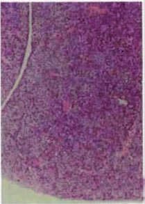
REVIEWS

 Box 1 | Domain structures of HGF and MET                                                                     and animal levels. Specifically, we discuss how the MET
                                                                                                              pathway is tuned by the functional cooperation between
 a HGF                        α-chain                                β-chain                                  various signal transducers, as well as by the receptor’s
                                                                                                              subcellular localization and trafficking. We also elabo-
          HL      K1          K2        K3      K4                     SPH
                                                                                                              rate on how MET signalling influences different organis-
 b MET            β     α                            c                                    HGF β-chain         mal functions in normal physiology and disease, a topic
                                                                                                              that deserves an overall reconceptualization. Finally,
                                                                                                              we examine the causative relationships between indi-
 Sema domain
                                                               400                                            vidual signalling inputs and specific biological outputs
                                                              382              414                            in MET-driven processes, and provide our perspective
                                               Sema                                                           on the medical implications of novel therapies that either
                       PSI                   domain         377
                                                                                                              promote or neutralize MET activity.
                       IPT1
                                                         311                                                  MET signalling: pathway components
                                                                            4 3                               HGF is secreted as a single-chain inert precursor and
                       IPT2                           301                            2
                                                                       5
                                                                         7      1                             converted into a two-chain functional heterodimer
                                                                       6          A B
                                                                                      C                       by extracellular proteases (BOX 1). This growth factor
                       IPT3                                                                D                  is widely distributed in the extracellular matrix of most
                                                                                                              tissues, where it is sequestered, mainly in its inactive
   Plasma              IPT4                                             N                                     form, by heparin-like proteoglycans30,31. Cells of mes-
   membrane                                                                                                   enchymal origin are the major source of HGF, which
                                                                                                              acts in a paracrine manner on epithelial cells that
      Ser975 P         Juxtamembrane                                                                          express the MET receptor 32. during tissue repair and
                       sequence                                              PSI domain
      Tyr1234 P
                                                                C                                             cancer invasion, several cytokines that are abundant
                       Catalytic region                                                                       in the reactive interstitial compartment — for example,
      Tyr1235 P
                                                                                                              interleukin-1 and -6, tumour necrosis factor-α and
                                                                                                              transforming growth factor-β (TGFβ) — induce trans-
      Tyr1349 P
                       Multifunctional                                                                        criptional upregulation of both HGF (in fibroblasts
      Tyr1356 P        docking site                                                                           and resident macrophages) and MET (in epithelial
                                                                                                              cells)33,34. The inflammatory and tumour stroma also
 Hepatocyte growth factor (HGF; also known as scatter  Nature   Reviews
                                                           factor)       | Molecular
                                                                   is similar        Cell Biology
                                                                              to plasminogen,  a              overexpress proteases that are involved in pro-HGF
 circulating zymogen that promotes the dissolution of fibrin blood clots in its active form as                activation, such as the plasminogen activation system
 the serine protease plasmin. Both HGF and plasminogen are synthesized as a single-chain                      and matriptase35,36. Thus, biologically competent HGF
 precursor and then converted into an active α- and β-chain heterodimer by extracellular                      is not only overproduced but also fully activated. This
 proteases. HGF consists of six domains: an amino-terminal hairpin loop (HL), four kringle                    combined transcriptional and post-translational regula-
 domains (K1–K4; each defined by three conserved disulphide bonds) and a serine
                                                                                                              tion, which leads to optimal MET activation on target
 protease homology (SPH) domain that lacks proteolytic activity (see the figure, part a).
   The HGF receptor MET is a single-pass heterodimer comprising an entirely extracellular                     cells, can be considered as part of a general mechanism
 α-subunit that is linked by a disulphide bond to a transmembrane β-subunit, which                            of physiological defence to tissue damage.
 contains the intracellular catalytic activity (see the figure, part b). The extracellular                        Following HGF binding, the kinase activity of MET
 region of MET includes three functional domains: the Sema domain (which is also                              is switched on by receptor dimerization and trans-phos-
 found in the semaphorins and plexins) spans the first 500 residues at the N terminus,                        phorylation of two ‘catalytic’ tyrosine residues (Tyr1234
 encompassing the whole α-subunit and part of the β-subunit; the PSI domain (which                            and Tyr1235) within the kinase activation loop. The sub-
 is also present in the plexins, semaphorins and integrins, hence its name) covers                            sequent step is phosphorylation of two additional ‘dock-
 approximately 50 residues and contains four conserved disulphide bonds; the residual                         ing’ tyrosines in the carboxy-terminal tail (Tyr1349 and
 400 residues connecting the PSI domain to the transmembrane helix are organized into                         Tyr1356), and when phosphorylated, these tyrosines act
 four IPT (immunoglobulin-like fold shared by plexins and transcriptional factors) domains.
                                                                                                              as a degenerate motif for the recruitment of many signal-
 The intracellular segment is composed of three portions: a juxtamembrane sequence
 that downregulates kinase activity following phosphorylation of Ser975; a catalytic                          relay molecules37 (BOX 1). MET is also a substrate for sev-
 region that positively modulates kinase activity following trans-phosphorylation of                          eral protein-tyrosine phosphatases (PTPs), including the
 Tyr1234 and Tyr1235; and a carboxy-terminal multifunctional docking site that contains                       receptor PTPs density-enhanced phosphatase 1 (dEP1;
 two docking tyrosines (Tyr1349 and Tyr1356) that are involved in the recruitment of                          also known as PTPrJ) and leukocyte common antigen
 several transducers and adaptors.                                                                            related (lAr; also known as PTPrF)38,39 and the non-
   HGF contains two MET binding sites that have different affinities. The high-affinity site                  receptor PTPs PTP1b (also known as PTPN1) and T cell
 is located in the α-chain and recognizes the IPT3 and IPT4 domains of MET independently                      PTP (TCPTP; also known as PTPN2)40. Such phosphatases
 of HGF processing and maturation155. The low-affinity site lies within the β-chain, is                       oppose MET signals by triggering dephosphorylation of
 exposed only after HGF activation and interacts with the Sema domain of MET156. This                         either the catalytic tyrosines (in the case of PTP1b and
 latter association is depicted as a ribbon representation (see the figure, part c). The HGF
                                                                                                              TCPTP)40 or the docking tyrosines (in the case of dEP1)38.
 β-chain is shown in grey and part of the extracellular region of MET is shown in a gradient
 of rainbow colours, from the N terminus (shown in blue) to the PSI domain (shown in red).                    To date, the MET tyrosine residues that are specifically
 The numbers in the centre refer to the blades, with the β-strands in blade 1 labelled                        dephosphorylated by lAr have not been identified.
 A–D. The numbers on the edge of the Sema domain represent residue numbers on each                                This is the basic signalling machinery of MET.
 side of a disordered region (dotted line). Part c modified, with permission, from REF. 156                   Further levels of complexity are provided by the inter-
 © (2004) Macmillan Publishers Ltd. All rights reserved.                                                      action of MET with different signal modifiers, including

NATurE rEViEWS | Molecular cell Biology                                                                                             VOluME 11 | dECEMbEr 2010 | 835

                                                               © 2010 Macmillan Publishers Limited. All rights reserved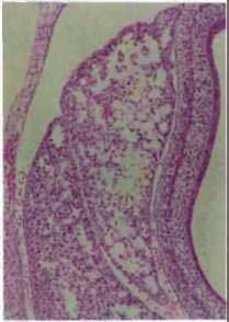
REVIEWS

                                    a                                                           b                                           c
                                                                    α6β4 integrin                                                                     Sema
                                               HGF                                               HGF                     CD44v6
                                               MET                     α β                      MET                                         MET

                                                                                                                                                          Class B
                                                                                                                                                          plexin

                                    Plasma
                                    membrane

                                              SHP2 Src                            GRB2
                                       GRB2 SHC
                                                   P PI3K                    P SHC                     P                                          P
                                        p120 PLCγ1                           P PI3K                                                                   P
                                                                                                             ERM
                                                      GRB2
                                     PLCγ1         P       GAB1              P SHP2                    P GRB2 SOS                                 P
                                                     STAT3                                                                   Actin
                                     PI3K  GAB1
                                     SHP2                                                                             Ras
                                         CRK SHC

                                   Figure 1 | Signal cooperation between MeT pathway components. The strength, duration and versatility of signals
                                   triggered by MET (also known as HGF receptor) are regulated by a network of signalling amplifiers and co-receptors that
                                   physically associate with MET. a | Adaptor proteins that have a scaffolding function, such as GRB2-associated-binding
                                   protein 1 (GAB1) and the α6β4 integrin, associate with MET and act as supplementary docking platforms for the further
                                   binding of signal transducers, thus enhancing signalling outputs. GAB1 interacts with MET both indirectly (through
                                   growth factor receptor-bound protein 2 (GRB2)) and directly. Following MET-dependent tyrosine phosphorylation,
                                   GAB1 provides additional sites for the recruitment of Src-homology-2 domain-containing transforming protein (SHC),
                                   phosphoinositide 3-kinase (PI3K), SHP2 (also known as PTPN11), CRK, phospholipase Cγ1 (PLCγ1) and p120 Ras-GTPase-
                                   activating protein (p120). Similarly, the β4 subunit of the α6β4 integrin directly associates with MET, which tyrosine-
                                   phosphorylates β4 and therefore generates extra binding sites for SHC, PI3K and SHP2. GRB2 can also associate with
                                   SHC that is bound to MET or α6β4 integrin. b | The extracellular domain of the v6 splice variant of the hyaluronan receptor
                                   CD44 (CD44v6) forms a ternary complex with MET and hepatocyte growth factor (HGF), an event that is necessary for
Paracrine                          MET activation. The intracellular portion of CD44v6 links the MET cytoplasmic domain to actin microfilaments through
Describing, or relating to, a      GRB2 and intermediate ezrin, radixin and moesin (ERM) proteins, which facilitates MET-induced activation of Ras by the
regulatory cell that secretes      guanine nucleotide exchange factor son of sevenless (SOS). c | Members of the class B plexins family associate with MET
a soluble molecule into an         and transactivate it in response to their semaphorin ligands (Sema), even in the absence of HGF, providing an alternative
intercellular space, from which
                                   way to stimulate MET-driven biological responses. Semaphorin-dependent activation of MET also results in tyrosine
it diffuses to a nearby target
cell other than the one that
                                   phosphorylation of the plexin cytoplasmic domain, but the functional consequences of this event are unknown. STAT3,
produces it.                       signal transducer and activator of transcription 3.

Reactive interstitial
compartment
A connective tissue that
                                   scaffolding adaptors, cytoskeletal connectors and                   the growth factor receptor-bound protein 2 (Grb2)
responds to neighbouring           structurally homologous co-receptors25,26,41 (FIG. 1). As           and SH2 domain-containing transforming protein
stimuli such as inflammation       a whole, this apparatus leads to efficient activation of            (SHC) adaptors37,45,46, SHP2 (also known as PTPN11;
and tumour formation or            downstream signal transduction pathways that include                an upstream activator of Src and ras) 46, phospho-
invasion.
                                   the mitogen-activated protein kinase (MAPK) cas-                    lipase Cγ1 (PlCγ1)37 and the transcription factor STAT3
Src-homology-2 domain              cades (extracellular signal-regulated kinase 1 (ErK1)               (REFS 47,48). MET also associates with Grb2-associated-
(SH2 domain). A protein            and ErK2, Jun amino-terminal kinases (JNKs) and                     binding protein 1 (GAb1), a multi-adaptor protein that,
module that recognizes and         p38), the phosphoinositide 3-kinase–Akt (Pi3K–Akt)                  upon phosphorylation by the MET receptor, provides
binds tyrosine-phosphorylated      axis, signal transducer and activator of transcription              extra binding sites for SHC, Pi3K, SHP2, CrK, PlCγ1
sequences in a sequence-
specific context and thereby
                                   proteins (STATs), and the nuclear factor-κb inhibitor-α             and p120 ras- GTPase-activating protein (p120-ras-
has a key role in relaying         (iκbα)–nuclear factor-κb (NF-κb) complex 22,25,42–44. All           GAP)43,44,49–53. The association between MET and GAb1
cascades of signal                 of these pathways positively control MET-dependent                  occurs directly, through a unique 13-amino-acid MET
transduction.                      cell proliferation, survival and migration. However,                binding site (MbS) on GAb1, and indirectly, through
                                   in defined cellular contexts, some of these signals                 MET-bound Grb2 (REFS 51,54). Hence, many trans-
GTPase-activating protein
(GAP). A protein that              can mediate a paradoxical pro-apoptotic, rather than                ducers participate in MET signalling through multiple
stimulates the intrinsic ability   anti-apoptotic, activity (BOX 2).                                   interactions with the receptor, with the GAb1 scaffolding
of a GTPase to hydrolyse                                                                               adaptor or with both (FIG. 1a).
GTP to GDP. GAPs negatively        MET and scaffolding adaptors. The promiscuous docking                   in tumours, intensification of MET signals is
regulate GTPases by converting
them from an active
                                   motif in the C-terminal tail of MET binds numerous Src-             also favoured by receptor interaction with the α6β4
(GTP-bound) to an inactive         homology-2 domain (SH2 domain)-containing effectors,                integrin. MET activation leads to phosphorylation of
(GDP-bound) state.                 such as Pi3K37, the non-receptor tyrosine kinase Src37,             the β4-subunit cytoplasmic domain (at three tyrosine

836 | dECEMbEr 2010 | VOluME 11                                                                                             www.nature.com/reviews/molcellbio

                                                        © 2010 Macmillan Publishers Limited. All rights reserved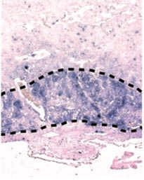
REVIEWS

                                  Box 2 | MET and apoptosis: the flip side of the coin
                                  a Protection from apoptosis          Induction of apoptosis           b Induction of apoptosis        Protection from apoptosis
                                                                                       FASL                            Stress
                                            HGF        FASL                                                            conditions
                                            MET

                                                      FAS

                                   Plasma
                                   membrane
                                                                                                                        Asp1000

                                              P                          P                              Active                             Inactive     P
                                              P                          P                              caspases                           caspases     P
                                                                                                                        Asp1374
                                                                                                        Active                             Inactive
                                                                                                        caspases                           caspases

                                  Despite acting as a prototypical survival factor, hepatocyte growth factor (HGF; also known as scatter factor) was initially
                                  named ‘tumour cytotoxic factor’ for its ability to elicit apoptotic responses in sarcomaNature
                                                                                                                           157     Reviews | Molecular
                                                                                                                               and hepatoma  158         Cell Biology
                                                                                                                                                 cells. These results
                                  were confirmed in other cell types, including liver and lung myofibroblasts133,159 and breast carcinoma cell lines160, in which
                                  HGF-induced apoptosis was attributed to the activation of protein kinase C and Jun amino-terminal kinase (JNK) or to the
                                  induction of reactive oxygen species. In ovarian cancer cells that have been treated with conventional chemotherapeutics,
                                  HGF enhances apoptosis through a p38 mitogen-activated protein kinase (MAPK)-dependent pathway73,74.
                                    The mechanism by which MET (also known as HGF receptor) activation leads to opposite outcomes — survival by
                                  default and apoptosis in restricted cellular contexts — has been only partially elucidated. The extracellular domain of
                                  MET interacts with the death receptor FAS (also known as CD95 and TNFRSF6), which prevents FAS receptor–FAS ligand
                                  (FASL) recognition and FAS self-aggregation, therefore limiting apoptosis through the extrinsic pathway161 (see the figure,
                                  part a). However, elevated HGF levels sensitize cells to FASL-mediated apoptosis161. It has been proposed that excessively
                                  high concentrations of HGF would titre out MET and liberate FAS from its association with MET, suggesting that the
                                  pro-apoptotic effects evoked by HGF in some cell lines could be related to a critical stoichiometry in the reciprocal
                                  amounts of HGF, MET, FAS and FASL.
                                    The intracellular domain of MET is also involved in the positive control of apoptosis. Under stress conditions, the MET
                                  cytoplasmic portion undergoes sequential cleavage by caspases, first at an aspartic acid residue in the carboxy-terminal
                                  end (Asp1374) and then at a second aspartic acid in the juxtamembrane portion (Asp1000) (see the figure, part b).
                                  This yields an intracellular soluble fragment, which is comprised of the kinase domain and triggers cell death162,163.
                                  Notably, if stress induction is accompanied by HGF stimulation, caspase activation is reduced and generation of the
                                  intracellular pro-apoptotic fragment is prevented162. This points to the presence of a cellular rheostat, in which life or
                                  death are ultimately dictated by the anti-apoptotic input that is generated by HGF-dependent activation of MET and
                                  the pro-apoptotic input that is generated by caspase-dependent cleavage of MET.

                                 residues that are embedded in consensus sequences)                    containing the v6 sequence participates in MET signal-
                                 and this is required for the recruitment of SHC, Pi3K                 ling by a twofold mechanism: the extracellular domain
                                 and SHP2 (REFS 55–58). by accomplishing this function,                is required for the organization of a ternary complex
                                 GAb1 and the α6β4 integrin behave as supplementary                    between MET, HGF and Cd44 and for the subsequent
                                 docking platforms for the localized recruitment of addi-              activation of MET60. The cytoplasmic tail is necessary
                                 tional transducers that synergize with those directly                 for signal transfer from active MET to ras61. Specifically,
                                 bound to MET (FIG. 1a). This mechanism allows signal                  upon MET activation, the intracellular domain of
                                 amplification at the MET-proximal level, which in turn                Cd44v6 drives the assembly of a sub-membraneous
                                 secures potent and enduring activation of downstream                  network that includes the cytoplasmic region of MET,
                                 transducers.                                                          Grb2, F-actin, ezrin, radixin and moesin (ErM) pro-
                                                                                                       teins (which connect actin microfilaments to Cd44)
                                 CD44 links MET to the cytoskeleton. Another protein                   and the guanine nucleotide exchange factor son of seven-
                                 that cooperates with MET in normal and transformed                    less (SOS). This network allows for efficient activation
                                 epithelial cells is Cd44, a transmembrane cell adhesion               of ras by SOS (FIG. 1b).
                                 molecule that functions as a linker between the extracel-                 in endothelial cells, the v10-containing isoform
Guanine nucleotide               lular matrix and the intracellular actin cytoskeleton59.              of Cd44 seems to be essential for the partitioning of
exchange factor                  Members of the Cd44 family differ in their extracellu-                MET into detergent-insoluble, caveolin-enriched
A protein that facilitates the
exchange of GDP for GTP in
                                 lar domain by the insertion of variable regions through               micro domains together with T-lymphoma invasion
the nucleotide-binding pocket    alternative splicing, which gives rise to numerous vari-              and metastasis-inducing protein 1 (TiAM1; a rAC1
of a GTPase.                     ant isoforms (Cd44v1–Cd44v10)59. The Cd44 isoform                     exchange factor), cortactin (an actin cytoskeletal

NATurE rEViEWS | Molecular cell Biology                                                                                       VOluME 11 | dECEMbEr 2010 | 837

                                                        © 2010 Macmillan Publishers Limited. All rights reserved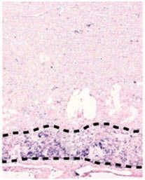
REVIEWS

                                regulator) and dynamin 2 (a vesicular regulator)62. Thus,           This association modifies the three-dimensional con-
                                when MET collaborates with Cd44, signal transduction                figuration of raf, which acquires signalling activity
                                is assisted by its structural and topographical regulation          and proceeds to phosphorylate and thereby activate
                                at the inner side of the plasma membrane rather than                MAPK/ErK kinase 1 (MEK1; also known as MAPKK1)
                                by its functional interaction with signalling amplifiers.           or MEK2 (also known as MAPKK2), the intermediate
                                The reasons why only some Cd44 variants contribute                  kinases of the system. in turn, MEK1 and MEK2 phos-
                                to MET signalling remain unclear.                                   phorylate ErK1 and ErK2, the final effectors of the
                                                                                                    cascade. Active ErKs translocate to the nucleus, where
                                Semaphorins activate MET in an HGF-independent                      they phosphorylate and stabilize several transcription
                                manner. The extracellular domain of MET exhibits                    factors that are involved in the early phases of the G1–S
                                structural homology with the semaphorins and plexins,               cell cycle transition. MET-dependent activation of the
                                a broad family of ligand–receptor pairs that are endowed            ras–ErK cascade can be decreased by molecules that
                                with either attractive or repulsive function in different           are involved in organogenesis and tissue repair, such
                                tissues (BOX 1). in particular, MET and class b plexins             as the membrane receptor Notch and the intracellu-
                                share a highly homologous Sema domain, which forms                  lar modulator Sprouty 68,69. interestingly, MET is able
                                a seven-bladed β-propeller that supports multiple inter-            to transcriptionally upregulate both Sprouty and the
                                actions between MET and some class b plexins. When                  Notch ligand delta, suggesting a mechanism of counter-
                                oligomerized with plexins, MET can be activated by                  feedback whereby MET induces negative regulators of
                                semaphorins in an HGF-independent manner, leading                   its own signalling, possibly to fine-tune the restricted
                                to stimulation of MET downstream effectors and execu-               execution of biological outputs in space and time. How
                                tion of MET-dependent biological responses63–65 (FIG. 1c).          the delta–Notch system and Sprouty impair MET-
                                This partnership expands the complement of soluble                  triggered stimulation of the ras–ErK pathway remains
                                factors that exert biological activity on MET-expressing            to be characterized.
                                cells and specifies the quality of cellular outcomes: cells             As well as the ErKs, MET is able to activate the JNK
                                that co-express MET and plexins react to either HGF or              and p38 MAPKs. For both families, the first MAPKKK
                                semaphorins by acquisition of invasive ability, which is            components of the cascade (MAPKKK1–MAPKKK4;
                                a typical MET-dependent effect; by contrast, cells that             also known as MEK kinase 1 (MEKK1)–MEKK4) are
                                express only plexins respond to semaphorins by arrest-              usually stimulated by rac, a small GTPase that is
                                ing migration and inducing cellular collapse, which is a            switched on by a ras–Pi3K-mediated pathway. MEKK-
                                plexin-specific repulsive function66.                               dependent phosphorylation of MEK4 and MEK7
                                                                                                    leads to activation of JNK1, JNK2 and JNK3, whereas
                                Major MET-regulated signalling pathways                             phosphorylation of MEK3 and MEK6 leads to activa-
                                MET-dependent signals are organized in pathways that                tion of p38α, p38β, p38γ and p38δ (FIG. 2a). Following
                                transmit biochemical information from the cell mem-                 MET-dependent stimulation, the JNKs and p38s
                                brane (where MET resides) to the nucleus (where modu-               control a range of cellular processes as diverse as cell
                                lation of gene expression occurs). As described above,              proliferation, differentiation, transformation and
                                the receptor-associated signalling apparatus, including             apoptosis70–74.
                                scaffolding adaptors and surface signal modifiers, is
                                somehow unique to MET. Conversely, the downstream                   The PI3K–Akt axis. Pi3K can be activated directly by
                                transducers are stereotypical signalling modules that are           MET and/or indirectly by ras, the activation of which
                                shared among different tyrosine kinase receptors. The               can also be mediated by MET (see above)37. Pi3K acti-
                                prominent examples of these biochemical pathways will               vation results in the formation of phosphatidylinositol-
                                now be discussed.                                                   3,4,5-triphosphate, which tethers pleckstrin homology
                                                                                                    domain (PH domain)-containing molecules, such as
                                The MAPK cascades. The subfamilies of MAPKs fea-                    the Ser/Thr kinase Akt, to the plasma membrane. Akt
                                ture a characteristic phospho-relay system in which a               is then able to suppress apoptosis through inactivation
                                series of three protein kinases (MAPK kinase kinase                 of the pro-apoptotic protein bCl-2 antagonist of cell
                                (MAPKKK), MAPK kinase (MAPKK) and MAPK)                             death (bAd) and activation of the E3 ubiquitin-protein
                                phosphorylate and activate one another 67. The ErKs are             ligase MdM2, which promotes the degradation of the
                                mainly triggered by tyrosine kinase-dependent stimula-              pro-apoptotic protein p53. Moreover, Akt inactivates
                                tion of ras. MET activates ras through the Grb2–SOS                 glycogen synthase kinase 3β (GSK3β), which antago-
                                complex, which can interact directly with the multi-                nizes the expression of positive cell cycle regulators
                                functional docking site in the C-terminal tail of MET               such as Myc and cyclin d1, and activates mammalian
                                or can be associated indirectly through the SHC adaptor             target of rapamycin (mTOr), which stimulates protein
                                protein37,45. Another route leading to ras–ErK activa-              synthesis and physical cell enlargement (FIG. 2b).
Pleckstrin homology             tion involves the tyrosine phosphatase SHP2, which
domain                          dephosphorylates the p120-ras-GAP binding site on                   The STAT pathway. Trans-phosphorylation of the dock-
(PH domain). A protein domain   GAb1; this impedes the recruitment of p120-ras-GAP,                 ing site of MET leads to the association of STATs —
that is characteristic of the
RNase PH family of bacterial
                                which usually deactivates ras, to promote ras activa-               namely, STAT3 — with its tail47,48. This docking event is
phosphate-dependent             tion44,53 (FIG. 2a). in the active, GTP-bound state, ras            followed by MET-dependent tyrosine phosphorylation
ribonucleases.                  attracts the Ser/Thr kinase raf through its effector loop.          of STAT3, which causes STATs to dissociate from the

838 | dECEMbEr 2010 | VOluME 11                                                                                          www.nature.com/reviews/molcellbio

                                                     © 2010 Macmillan Publishers Limited. All rights reserved
REVIEWS
a GRB2 SOS
SHC
MET GRB2 SOS Ras Raf MEK1,2 ERK1,2 Proliferation, transformation
GAB1
PI3K
SHP2 P p120 MEKK1,2,3,4 MEK4,7 JNK1,2,3
Proliferation, transformation,
Rac differentiation and apoptosis
MEKK1,2,3,4 MEK3,6 p38α,β,γ,δ
b
mTOR Protein translation and
MET BAD cell growth
PI3K Akt Protection from apoptosis
MDM2 p53
Ras GSK3β Proliferation
c P
STAT3 STAT3 P P
MET P P STAT3 STAT3 Proliferation, transformation
STAT3 STAT3 and tubulogenesis
P DNA
NF-κB
d IκB
Src
MET IKK
PI3K Akt P NF-κB
IκB
NF-κB Proliferation, survival and
tubulogenesis
Figure 2 | Major MeT-regulated signalling pathways. MET (also known as HGF receptor) triggers several downstream
pathways. a | The mitogen-activated protein kinase (MAPK) cascades consist of threeNature Reviews
subfamilies, | Molecular
each of whichCell Biology
comprises
three protein kinases that activate one another sequentially. The proximal elements are activated directly or indirectly
by the Ras small GTPase, which in turn is switched on by son of sevenless (SOS) and switched off by GTPase activating
proteins (GAPs), such as p120 Ras-GAP (p120). SOS can be activated and p120 can be inhibited in response to MET
signalling. The terminal effectors include extracellular signal-regulated kinases (ERKs), Jun amino-terminal kinases
(JNKs) and p38s. These translocate to the nucleus, where they influence the activity of various transcription factors.
b | Phosphoinositide 3-kinase (PI3K) is a lipid kinase that associates with the multifunctional docking site of MET and
catalyses the formation of phosphatidylinositol (3,4,5)-triphosphate (PtdIns(3,4,5)P3). Production of PtdIns(3,4,5)P3 creates
a docking site for Akt. Once compartmentalized at the inner side of the plasma membrane, Akt becomes activated and
phosphorylates several substrates involved in cell proliferation, survival and the regulation of cell size. c | Signal transducer
and activator of transcription 3 (STAT3) monomers bind to MET through their Src-homology-2 domain (SH2 domain) and
become trans-phosphorylated. Thereafter, each STAT3 moiety homodimerizes, using its SH2 domain to recognize the
phosphotyrosine of its partner, and translocates to the nucleus to operate as a transcription factor. d | In response to MET
stimulation and the ensuing activation of PI3K- and Src-dependent pathways, nuclear factor-κB inhibitor-α kinase (IKK) is
activated, and phosphorylates the nuclear factor-κB inhibitor-α (IκB) proteins (which are bound to nuclear factor-κB
(NF-κB)). This triggers the ubiquitin-dependent degradation of IκB, resulting in the nuclear translocation of NF-κB and
transcription. In all diagrams, dashed lines indicate that the signalling intermediates between the two nodes have not
been characterized in detail. BAD, BCL-2 antagonist of cell death; GAB1, GRB2-associated-binding protein 1; GRB2,
growth factor receptor-bound protein 2; GSK3β, glycogen synthase kinase 3β; MEK, MAPK/ERK kinase (also known as
MAPKK); MEKK, MEK kinase; mTOR, mammalian target of rapamycin; SHC, SH2 domain-containing transforming protein.
receptors and homodimerize through their SH2 domains. The IκBα–NF-κB complex. The NF-κb system comprises
Eventually, STAT dimers translocate to the nucleus, a family of transcription factors that are held inactive in
where they operate as transcription factors to regulate the cytoplasm by a family of inhibitory proteins known as
the expression of several genes that are implicated in cell iκbs75. These inhibitors are ubiquitylated and degraded by
proliferation or differentiation (FIG. 2c). a phosphorylation event that is triggered by the iκb kinase
NATurE rEViEWS | Molecular cell Biology VOluME 11 | dECEMbEr 2010 | 839
© 2010 Macmillan Publishers Limited. All rights reservedREVIEWS
(iKK). in response to MET, iKK activation is mediated degradation in the lysosomal compartment remains
by the Pi3K–Akt pathway and Src as signalling interme- the major determinant of receptor desensitization and
diates, but the direct distal effectors of iKK stimulation signal restraint 85. Prompt sequestration of the activated
are unknown76. iKK-induced destruction of iκbs leads to receptor into invaginating pits, and its endosomal sort-
the release of NF-κb, which translocates to the nucleus ing for degradation, precludes prolonged signal emis-
to stimulate the transcription of various genes, including sion and ensures faithful application of physiological
mitogenic and anti-apoptotic regulators76,77 (FIG. 2d). responses.
Trafficking to the lysosomes is triggered by ligand-
Regulation of MET signalling induced activation of MET, and requires the function
it is well established that signal transduction pathways that of casitas b-lineage lymphoma (Cbl), an E3 ubiquitin-
are downstream of tyrosine kinase receptors are modu- protein ligase that is recruited to a juxtamembrane phos-
lated not only by the identities of protein–protein inter- photyrosine residue in the active receptor (Tyr1003)86
actions but also by the colocalization of signal transducers (FIG. 3c). This leads to MET being monoubiquitylated at
in membrane domains or, more broadly, in organelles and multiple sites87, which enables its recognition by endo-
vesicles78. This notion has been recently applied to MET: cytic adaptors that contain ubiquitin-binding domains,
the prevailing view that the activated receptor recruits its sorting into clathrin-coated areas at the cell surface
signalling effectors at the plasma membrane, which in and its delivery to the endosomal network. From these
turn stimulate diffusible intermediates in the cytosol, has sorting endosomes, MET accumulates on the limiting
now been expanded by the finding that MET signals can and internal membranes of multivesicular bodies. This
also emanate from endosomal compartments. Moreover, terminates signalling by sequestering MET from the
the levels of MET expression at the cell surface are finely cytosol and preventing it being recycled by the plasma
tuned by other spatially restricted events, including extra- membrane. Finally, the multivesicular bodies fuse with
cellular shedding, intracellular cleavage and ubiquitin- the lysosomes and MET undergoes proteolytic demoli-
mediated degradation. All of these processes regulate the tion88,89. Seemingly active MET is not polyubiquitylated,
strength of MET activation and the ensuing robustness of and is therefore unlikely to be targeted for degradation
Endosomal MET-dependent signals. by the proteasome87; however, proteasomal destruction
Relating to a vesicle formed
of MET can occur in an ubiquitin-independent manner
by invagination of the plasma
membrane. MET internalization can promote signalling. Following (see below).
ligand binding, MET is rapidly internalized by clathrin- Cbl also attracts — through the scaffold molecule
Endocytosis mediated endocytosis and becomes recruited to periph- Cbl-interacting protein 85 (CiN85; also known as
Internalization and transport eral early endosomes79. Here, MET triggers the sustained SH3KbP1) — endophilins, a family of proteins that assist
of extracellular material and
plasma membrane proteins
activation of ErKs, which are then relocated to focal the negative curvature, invagination and scission of the
from the cell surface to complexes, where they mediate HGF-induced cell migra- planar plasma membrane during the early phases of
intracellular organelles tion (probably by phosphorylating paxillin and other focal endocytosis90. Therefore, Cbl promotes the physical
known as endosomes. adhesion targets80). A crucial regulator of this endosomal internalization of MET while tagging it for lysosomal
signal compartmentalization, and the consequent trans- degradation91 (FIG. 3c). Sustained association of Cbl
Focal complex
A small (1 μm diameter), fer of ErK to adhesion sites, is protein kinase Cε (PKCε) with active MET is favoured by decorin, an extracellu-
dot-like adhesion structure (FIG. 3a). rNA interference-mediated silencing of PKCε lar proteoglycan that binds directly to MET with high
that is present mainly at the negatively influences the recruitment of ErK1 and ErK2 affinity and acts as an antagonistic ligand by stimulating
edge of the lamellipodium. to focal adhesions81, possibly by reducing the delivery of receptor activation and, immediately thereafter, receptor
Focal adhesion
ErK–integrin complexes to the plasma membrane82. downmodulation92.
A cellular structure that links, From the peripheral endosomes, MET traffics along What skews the MET fate towards degradation rather
through integrin receptors, the the microtubule network and accumulates in a nondegra- than signalling remains unknown. in the case of the epi-
extracellular matrix outside dative perinuclear endomembrane compartment through dermal growth factor (EGF) receptor, it has been sug-
the cell to the actin
a process that is promoted by PKCα81,83 (FIG. 3b). This jux- gested that endosomal signalling predominates under
cytoskeleton inside the cell.
tanuclear delivery of MET is required for the optimal conditions of scarce ligand, whereas the degradative
E3 ubiquitin-protein ligase activation and nuclear translocation of STAT3 (REF. 84). pathway is favoured when abundant ligand is present 93.
An enzyme that is responsible MET-triggered phosphorylation of STAT3 at the plasma A similar scenario might also apply to MET but a for-
for the conjugation of ubiquitin membrane or early endosomes is likely to be below the mal demonstration of such a mechanism has not been
to substrate proteins.
threshold for nuclear uptake owing to the intense local provided so far.
Multivesicular body activity of phosphotyrosine phosphatases that dissi-
An endocytic intermediate pate STAT3 signal intensity during protein diffusion. Regulated proteolysis of MET. downregulation of MET
organelle in the lysosomal Conversely, post-endocytic trafficking to the perinuclear signalling also involves sequential proteolytic cleav-
degradative pathway that
compartment brings active MET into close proximity to ages. The first step is a disintegrin and metalloprotease
contains small vesicles and
is surrounded by a limiting the nucleus, which shields STAT3 against phosphatase (AdAM)-mediated release (known as shedding) of
membrane. activity and allows its nuclear accumulation (FIG. 3b). the extracellular domain, which generates a soluble
N-terminal fragment and a membrane-anchored cyto-
Proteasome MET internalization can inhibit signalling. Although plasmic tail. Then, the surface-associated cytoplasmic
A large multisubunit protein
complex that degrades
internalized receptors may remain able to signal in remnant undergoes regulated proteolysis by γ-secretase,
unnecessary or damaged peripheral and perinuclear compartments, the endo- which yields a labile intracellular portion that is rapidly
proteins by proteolysis. cytosis of ligand-activated receptors and their subsequent cleared by proteasome-mediated degradation94 (FIG. 3d).
840 | dECEMbEr 2010 | VOluME 11 www.nature.com/reviews/molcellbio
© 2010 Macmillan Publishers Limited. All rights reservedREVIEWS
a HGF b
Clathrin-
P Focal MET coated pit
ERK adhesion
Plasma
membrane
P P P P
P P P P
PKCε Phosphatase P
STAT3
STAT3
STAT3
ERK Early Early
ERK
endosome endosome
ERK PKCα
P
ERK P P P P
P P P P
Perinuclear
compartment
c
STAT3
P P
STAT3 P P STAT3 Nucleus
P
Endocytic STAT3
adaptor CBL P P P
CIN85 P P
Endophilins Ub
Early
d PMAs, suramin, LPA ‘Decoy’ MET
and anti-MET mAbs
endosome Multivesicular
body
ADAM
P P
P P γ-secretase
Lysosome
Proteasome
Figure 3 | compartmentalization of MeT signals. After being internalized from the cell surface, MET (also known as
HGF receptor) can maintain a signalling-competent modality or can be demolished. a |Nature The recruitment of MET into
Reviews | Molecular Cellearly
Biology
endosomes is mediated by protein kinase Cε (PKCε) and favours the delivery of active extracellular signal-regulated kinase
(ERK) to focal adhesions, where it can mediate hepatocyte growth factor (HGF; also known as scatter factor)-induced
cell migration. b | MET sorting into non-degradative perinuclear endomembrane compartments is mediated by PKCα.
In these compartments, the activity of phosphotyrosine phosphatases, which work on signal transducer and activator of
transcription 3 (STAT3) to dissipate signal intensity, is low. This favours efficient phosphorylation of STAT3 by MET and
translocation of STAT3 into the nucleus. c | Downregulation of active MET is initiated by MET association with casitas
B-lineage lymphoma (CBL), an E3 ubiquitin (Ub)-protein ligase that bridges MET and endocytic adaptors. CBL also
associates with endophilins through the scaffold molecule CBL-interacting protein 85 (CIN85), which promotes changes
in the plasma membrane that are necessary for cell surface curvature in the early phases of endocytosis. Following
endocytosis and intermediate accumulation in multivesicular bodies, MET is subjected to lysosomal degradation. d | MET
can also undergo sequential proteolytic cleavage at two juxtamembrane sites, one in the extracellular domain and the
other in the intracellular domain. The first cleavage (known as shedding), which is carried out by a disintegrin and
metalloprotease (ADAM)-like extracellular protease, generates a soluble extracellular ‘decoy’ fragment that interferes
with the activity of the full-length receptor and sequesters the ligand. This shedding is stimulated by various agents such
as phorbol esters (PMAs), suramin, lysophosphatidic acid (LPA) and monoclonal antibodies (mAbs) against the MET
extracellular domain. The second cleavage is performed by γ-secretase and yields an intracellular fragment that is
destroyed by the proteasome in an ubiquitin-independent manner. Dashed lines indicate that the signalling intermediates
between the two nodes have not been characterized in detail.
unlike Cbl-mediated endosomal degradation, MET signalling with chronic, low-grade attenuation
regulated proteolysis of MET is ligand- and ubiquitin- under steady-state conditions. The shedding of MET
independent and does not require the kinase activity of that is catalysed by AdAM metalloproteases can be
the receptor: the mechanism occurs basally and affords acutely enhanced by various agents such as phorbol
NATurE rEViEWS | Molecular cell Biology VOluME 11 | dECEMbEr 2010 | 841
© 2010 Macmillan Publishers Limited. All rights reservedREVIEWS
esters, suramin, lysophosphatidic acid and monoclonal using genetic knock-in approaches, several studies
antibodies (mAbs) against the MET ectodomain94–102. have addressed the physiological significance of the dif-
importantly, the extracellular shedding of MET not ferent signalling pathways that are regulated by MET
only decreases the number of receptor molecules on the in the whole organism. Specificity-switch mutants, in
cell surface but also generates a decoy moiety that inter- which the MET multifunctional docking site has been
acts with both HGF (by sequestering the ligand) and converted into an optimal binding motif for Pi3K,
full-length MET (by impairing dimerization and trans- Src or Grb2, exhibit a rescue of defined phenotypes
activation of the native receptor) to further inhibit MET (when compared with Met-null animals) in line with
signalling 103,104. Therefore, the use of targeted agents — the effectors that are recruited. Mice in which MET
such as mAbs, which increase MET shedding from the exclusively associates with Grb2 are viable, fertile and
cell surface — could have a great therapeutic effect on apparently normal — indicating that Grb2 is sufficient
cancers by reducing MET signalling. to recruit all of the necessary downstream effectors of
MET — whereas mutants with dedicated stimulation
MET signalling in development and disease of Src or Pi3K are embryonic lethal. However, although
To secure output production and to increase functional incompatible with survival, individual activation of Src
versatility in the face of recurring perturbations, tyrosine is sufficient to restore proliferation of placental tropho-
kinase receptors often expand and diversify individual blasts and myoblasts, and selective activation of Pi3K
components of their downstream signalling networks, is sufficient to promote axon outgrowth and branching
degenerate connections and duplicate entire modules of specific motor nerves113. Together, these findings
of protein–protein interactions. Signal redundancy, suggest that MET-mediated developmental events are
reciprocal signal reinforcement and feedback loops are accomplished by qualitatively different signals that are
stereotypical aspects of signal transduction105. because endowed with a tissue-specific activity.
of this, extracting functional information from the dis- The defects that are observed in Hg f and Met
section of individual signalling axes might be judged knockout embryos are phenocopied in Gab1-null mice
to be simplistic, if not futile. Signal compensation by — albeit in a slightly attenuated form — indicating
other transducers that have overlapping functions must that GAb1 has an essential role in MET-based signal
be considered. However, in the case of MET, different transduction pathways114 (FIG. 4). As mentioned above,
cell- and tissue-specific activities seem to be fulfilled by GAb1 associates with MET directly, through a specific
dedicated signalling cascades, with some transducers MbS, and indirectly, through the Grb2 adaptor 51,54. The
dominating over others depending on context, timing mechanism of recruitment of GAb1 to MET influences
and biological complexity (TABLE 1). Here, we outline biological outcomes in vivo in a tissue-specific manner:
a number of genetic and pharmacologic studies that both direct and indirect interactions with MET are nec-
have contributed to this knowledge. The coverage is essary for functional MET signalling during liver and
purposefully not exhaustive: we will limit our attention placenta formation, whereas either the Grb2 binding
to those settings in which a more documented connec- site or the MbS, but not both, are required for limb
tion between signals and phenotypes has been provided, muscle development 115. Similar to those observed for
as well as to those for which information has become MET, interactions of GAb1 with specific downstream
available only recently. effectors control distinct developmental processes. The
association between GAb1 and Pi3K seems to be essen-
Embryonic development. during development, HGF tial for EGF receptor-mediated embryonic eyelid closure
and MET convey essential signals for the growth and but this association is not involved in MET-regulated
survival of hepatocytes and placental trophoblast cells. organogenesis113. MET signalling in embryonic devel-
in Hgf- or Met-null embryos, the liver is considerably opment is mainly channelled by the GAb1–SHP2
reduced in size and the placental labyrinth is severely interaction; indeed, GAb1 mutants that are unable to
hypomorphic106–108 (FIG. 4). This hypomorphic phenotype bind SHP2 phenotypically copy Met-null mutants, with
eventually leads to death in utero owing to compromised defects such as a thin placental labyrinth layer (FIG. 4)
exchange of oxygen and nutrients between maternal and impaired migration of muscle precursors115. Again,
and fetal blood. The HGF–MET system also has a deci- we can conclude that the biological responses elicited
Placental labyrinth sive role in the proliferation and motility of long-range by GAb1, like those elicited by MET, have different sig-
The area of direct exchange
between the fetal and
migrating muscle progenitors. These myogenic precur- nal requirements in different tissues during embryonic
maternal blood supply in the sors delaminate from the dermomyotome, a structure development.
mammalian placenta. in the dorsolateral region of somites, and travel to the
limbs, tongue and diaphragm, where they differentiate Organ regeneration. The signalling mechanisms that
Hypaxial muscle
to form a subset of the hypaxial muscles109. Ablation of mediate MET-driven tissue patterning during organ
Skeletal muscle that is derived
from progenitor cell the Hgf or Met genes results in complete absence of this regeneration have been studied both in vitro, by per-
populations that are located in specific muscle type, leaving all other muscle groups forming morphogenetic assays under three-dimensional
the lateral myotome of each unaffected104–106. Finally, disruption of HGF–MET func- culture conditions, and in vivo, using Cre–loxP-mediated
somite. Examples of hypaxial tion affects the proper wiring of the nervous system, conditional deletion of MET in selected tissues.
muscles include, among others,
the diaphragm, the abdominal
leading to reduced survival of sensory and sympathetic When embedded in a collagen matrix, normal epi-
muscles and the muscles of the neurons as well as impaired outgrowth and fasciculation thelial cells proliferate and assemble into spheroids,
limb and girdles. (axon bundling) of certain motor nerves110–112. which feature a centrally localized, hollow lumen,
842 | dECEMbEr 2010 | VOluME 11 www.nature.com/reviews/molcellbio
© 2010 Macmillan Publishers Limited. All rights reservedREVIEWS
Table 1 | MET-dependent signals and biological responses
Transducer Position in the pathway Biochemical Biological activity in vitro Biological activity in vivo
activity
GAB1 Direct or indirect (through Adaptor Promotes epithelial tubulogenesis43,44,49,51 Promotes the proliferation of liver and
GRB2) interaction with MET placenta114, the migration of limb muscle
precursors114 and liver regeneration127
GRB2 Direct or indirect (through Adaptor Promotes cell proliferation, Promotes the proliferation of liver and
SHC) interaction with MET transformation and motility37,45,51,164 placenta and the migration of limb
muscle precursors113
Src Direct interaction with MET Tyrosine kinase Promotes anchorage-independent Promotes proliferation of placenta and
growth and motility37,165 myoblasts113
PI3K Direct or indirect (through Lipid kinase Promotes cell proliferation, survival and Promotes axon outgrowth and branching
GAB1) interaction with MET motility37,158,166,167 of some motor nerves113
SHP2 Direct or indirect (through Tyrosine Promotes epithelial tubulogenesis44,51 Promotes the proliferation of placenta113,
GAB1) interaction with MET phosphatase migration of limb muscle precursors113
and liver regeneration127
SHC Direct interaction with MET Adaptor Promotes proliferation and motility45 Not determined
STAT3 Direct interaction with MET Transcription Promotes epithelial tubulogenesis 47
Promotes tumorigenesis (specifically
factor and anchorage-independent growth48 leiomyosarcoma)48
CRK Indirect interaction Adaptor Promotes cell motility and anchorage- Not determined
(through GAB1) with MET independent growth71,168
Akt Downstream transducer Ser/Thr kinase Promotes cell survival169 Promotes liver regeneration126
ERK1 and Downstream transducer Ser/Thr kinase Promotes epithelial tubulogenesis and 118
Promotes liver regeneration126
ERK2 cell survival169; inhibits TGFβ fibrogenic
activity131,132
JNK Downstream transducer Ser/Thr kinase Promotes cell transformation70, Not determined
anchorage-independent growth71 and
apoptosis160
p38 MAPK Downstream transducer Ser/Thr kinase Promotes cell proliferation72 and Not determined
apoptosis73,74
NF-κB Downstream transducer Transcription Promotes cell proliferation77, epithelial Not determined
factor tubulogenesis77 and cell survival76,160
ERK, extracellular signal-regulated kinase; GAB1, GRB2-associated-binding protein 1; GRB2, growth factor receptor-bound protein 2; JNK, Jun amino-terminal
kinase; MAPK, mitogen-activated protein kinase; NF-κB, nuclear factor-κB; PI3K, phosphoinositide 3-kinase; SHC, Src-homology-2 domain-containing transforming
protein; STAT3, signal transducer and activator of transcription 3; TGFβ, transforming growth factor-β.
surrounded by polarized cells116. HGF causes sphe- the early formation of extensions and chains, seem to be
roids to form elongated tubules in a stepwise sequence necessary to produce cords and hollow tubules118. HGF
of biological events 117. First, cells undergo a partial has an important role in the regulation of the functions
epithelial–mesenchymal transition (EMT), which involves of MMPs by enhancing the transcriptional levels of a
the transformation of tightly packed elements into large number of MMPs as well as those of several pro-
invasive, spindle-shaped cells that emit long cytoplas- teases that convert the precursor forms of MMPs into
mic extensions in the surrounding matrix. Then, these active enzymes121–123.
extensions develop into single-file chains, which must In vivo, MET function is particularly important
resist anoikis to lengthen and thicken. ultimately, cells for the regeneration of the liver and the kidney in
redifferentiate: they restore polarity and turn into solid response to both acute and chronic insults. Production
cords that become mature tubules by the progressive of HGF increases steeply following liver damage, and
formation of a continuous lumen. At the signalling the ensuing activation of MET in hepatocytes provides
level, the series of events required for HGF-dependent strong mitogenic and anti-apoptotic stimuli for organ
tubule formation has been dissected. As a whole, this repair 14,15,124. Accordingly, conditional deletion of MET
process requires the integrity of the STAT3 and NF-κb in the liver results in impaired organ reconstitution
pathways47,77. The initial EMT relies on hypersustained after toxic insult and hepatectomy 125,126 (FIG. 4). in these
activation of the raf–MEK–ErK pathway, which, as mutant mice, phosphorylation of ErK1 and ErK2 is lost
Epithelial–mesenchymal discussed earlier, is triggered by the association of active and there is a delay in the timing of maximal activation
transition MET-bound GAb1 with the tyrosine phosphatase SHP2 of Akt that occurs after hepatic injury. The prolifera-
(EMT). The transformation of (REFS 44,53,118,119). Cell survival is promoted by trans- tion defect is reflected by low transcriptional induction
an epithelial cell into a
mesenchymal cell that has
cription factors of the Snail family 120. The later phases of cyclins and enhanced expression of the cyclin-
migratory and invasive of epithelial tubulogenesis entail the activity of matrix dependent kinase (CdK) inhibitor p21 (also known
properties. metalloproteases (MMPs), which, albeit dispensable for as CiP1 and WAF1)126. An analogous phenotype of
NATurE rEViEWS | Molecular cell Biology VOluME 11 | dECEMbEr 2010 | 843
© 2010 Macmillan Publishers Limited. All rights reservedREVIEWS
a HGF+/+ mice HGF–/– mice c GAB1+/+ mice GAB1∆SHP2/∆SHP2 mice
MET–/– mice
b GAB1+/+ mice GAB1–/– mice d Cre-ctrl (liver-specific knockout)
Figure 4 | The phenotypes of genetic abrogation of specific MeT signals. Mutant mice with targeted disruption of
hepatocyte growth factor (HGF; also known as scatter factor)- or MET (also known asNature Reviews | Molecular Cell
HGF receptor)-dependent Biology
signals
show various abnormalities that are related to proliferative impairment. a | The liver of HGF knockout (HGF–/–) mice
features severe loss of cellularity and enlarged sinusoids compared with that of wild-type embryos (HGF+/+ mice). Similarly,
the labyrinth layer (delimited by dotted lines) contains fewer trophoblast cells, embryonic vessels and maternal sinuses
and more mesenchymal cells. The junctional zone (denoted by an asterisk) lies above the labyrinth. b | An analogous
placental defect, with a large reduction in the size of the labyrinth layer, occurs in mice with genetic deletion of GRB2-
associated-binding protein 1 (GAB1). c | The same phenotype is also displayed in mice that harbour a knock-in point
mutation that abolishes GAB1 interaction with SHP2 (also known as PTPN11) (GAB1ΔSHP2/ΔSHP2 mice). d | In adult animals,
Cre–loxP-mediated conditional deletion of MET in the liver impairs hepatic regeneration following toxic insult, as shown
by the reduction of hepatocytes incorporating bromo-deoxyuridine (BrdU), which indicates a decrease in proliferating
hepatocytes. Larger necrotic areas are also evident in the liver of MET–/– mice compared with control liver (Cre-ctrl). A
similar decrease of proliferating hepatocytes can be observed during post-hepatectomy regeneration in mice with
liver-specific knockout of GAB1 and in mice with liver-targeted disruption of SHP2 (REF. 127). Images in part a modified,
with permission, from REFS 106,107 © (1995) Macmillan Publishers Ltd. All rights reserved. Images in part b modified, with
permission, from REF. 114 © (2000) The Rockefeller University Press. Images in part c modified, with permission, from
REF. 115 © (2007) National Academy of Sciences, USA. Images in part d modified, with permission, from REF. 125 © (2004)
National Academy of Sciences, USA.
compromised hepatic regeneration is also seen in mice agent by antagonizing TGFβ fibrogenic responses in
with liver-specific ablation of GAb1 or SHP2 (REF. 127), different ways, from transcriptional downmodulation of
further pointing to a crucial role for GAb1–SHP2 TFGβ130 to ErK-mediated inhibition of SMAds131,132, the
signalling in MET-driven outcomes (FIG. 4). cardinal transcriptional effectors of TGFβ signalling. At
MET also displays a similar protective activity in least in the liver, fibrosis is further reduced by an atypical
the kidney. HGF induces intense proliferative and anti- pro-apoptotic effect of HGF on myofibroblasts133 (BOX 2).
apoptotic responses in renal epithelial cells by activat- Epithelial cells also exploit the trophic properties
ing ras-dependent mitogenic signals, the Pi3K–Akt of the HGF–MET system during wound healing. At
pro-survival pathway and, subsequently, the transcrip- the wound edges, marginal keratinocytes form the so-
tional upregulation of the anti-apoptotic effectors b-cell called hyperproliferative epithelium, which undergoes
lymphoma-extra large (bCl-Xl) and bCl-2 (REF. 128). rounds of cell division to provide fresh elements that
This powerful reno-protective activity prevents acute migrate over the provisional matrix of the injured der-
renal failure due to tubular necrosis and accelerates mis and repopulate the wounded area134. Keratinocytes
kidney regeneration. from mutant mice with conditional deletion of MET
in the liver and the kidney, persistent damage in the epidermis are unable to contribute to the gen-
leads to a severe fibrotic evolution that is sustained by eration of the hyperproliferative epithelium in vivo135.
Myofibroblast
A modified fibroblast with
overproduction of TGFβ, which instructs interstitial In vitro, during the closure of scratch wounds, HGF
smooth muscle-like features myofibroblasts to deposit copious amounts of extra- is necessary to re-orient keratinocytes, so that focal
and contractile properties. cellular matrix 129. HGF acts as a powerful anti-fibrotic adhesion components, actin stress fibres and the plus
844 | dECEMbEr 2010 | VOluME 11 www.nature.com/reviews/molcellbio
© 2010 Macmillan Publishers Limited. All rights reservedYou can also read