Inhibition of HECT E3 ligases as potential therapy for COVID-19 - Biogem
←
→
Page content transcription
If your browser does not render page correctly, please read the page content below
Novelli et al. Cell Death and Disease (2021)12:310
                                    https://doi.org/10.1038/s41419-021-03513-1                                                                                                             Cell Death & Disease

                                     ARTICLE                                                                                                                                                    Open Access

                                    Inhibition of HECT E3 ligases as potential therapy
                                    for COVID-19
                                    Giuseppe Novelli 1,2,3, Jing Liu4, Michela Biancolella5, Tonino Alonzi6, Antonio Novelli7, J. J. Patten 8,
                                    Dario Cocciadiferro7, Emanuele Agolini 7, Vito Luigi Colona 1, Barbara Rizzacasa 1, Rosalinda Giannini1,
                                    Benedetta Bigio9, Delia Goletti6, Maria Rosaria Capobianchi10, Sandro Grelli11, Justin Mann12, Trevor D. McKee12,
                                    Ke Cheng12, Fatima Amanat13, Florian Krammer13, Andrea Guarracino 5, Gerardo Pepe 5, Carlo Tomino14,
                                    Yacine Tandjaoui-Lambiotte15,16, Yurdagul Uzunhan17, Sarah Tubiana18,19, Jade Ghosn20,21, COVID Human Genetic
                                    Effort, French COVID Cohort Study Group, CoV-Contact Cohort, Luigi D. Notarangelo22, Helen C. Su22,
                                    Laurent Abel9,23,24, Aurélie Cobat9,23,24, Gai Elhanan25,26, Joseph J. Grzymski25,26, Andrea Latini1, Sachdev S. Sidhu27,
                                    Suresh Jain28, Robert A. Davey29, Jean-Laurent Casanova9,23,24,30, Wenyi Wei4 and Pier Paolo Pandolfi4,26,31

                                      Abstract
                                      SARS-CoV-2 is responsible for the ongoing world-wide pandemic which has already taken more than two million lives.
                                      Effective treatments are urgently needed. The enzymatic activity of the HECT-E3 ligase family members has been
                                      implicated in the cell egression phase of deadly RNA viruses such as Ebola through direct interaction of its VP40
                                      Protein. Here we report that HECT-E3 ligase family members such as NEDD4 and WWP1 interact with and ubiquitylate
                                      the SARS-CoV-2 Spike protein. Furthermore, we find that HECT family members are overexpressed in primary samples
                  1234567890():,;
1234567890():,;
                  1234567890():,;
1234567890():,;

                                      derived from COVID-19 infected patients and COVID-19 mouse models. Importantly, rare germline activating variants
                                      in the NEDD4 and WWP1 genes are associated with severe COVID-19 cases. Critically, I3C, a natural NEDD4 and WWP1
                                      inhibitor from Brassicaceae, displays potent antiviral effects and inhibits viral egression. In conclusion, we identify the
                                      HECT family members of E3 ligases as likely novel biomarkers for COVID-19, as well as new potential targets of
                                      therapeutic strategy easily testable in clinical trials in view of the established well-tolerated nature of the Brassicaceae
                                      natural compounds.

                                                                                                                                        Introduction
                                                                                                                                          The Severe Acute Respiratory Syndrome Coronavirus 2
                                                                                                                                        (SARS-CoV-2) associated with the emerging disease
                                    Correspondence: Giuseppe Novelli (novelli@med.uniroma2.it) or
                                                                                                                                        (COVID-19) has resulted in an unprecedented global
                                    Pier Paolo Pandolfi (pierpaolo.pandolfiderinaldis@renown.org)
                                    1
                                     Department of Biomedicine and Prevention, Tor Vergata University of Rome,                          health and economic crisis1,2. To date (March, 9, 2021),
                                    00133 Rome, Italy
                                    2
                                                                                                                                        there are at least 24 putative drug treatments for the
                                     IRCCS Neuromed, Pozzilli, (IS), Italy
                                                                                                                                        disease. However, most are still at early stages of research.
                                    Full list of author information is available at the end of the article
                                    Lists of collaborators and their affiliations are listed at the end of the paper.                    The focus has been on the development of new and
                                    Leaders of the COVID Human Genetic Effort: Helen C. Su, Jean-Laurent                                repositioned vaccines, monoclonal antibodies, and
                                    Casanova
                                                                                                                                        drugs3–7. Another treatment option involves passive
                                    Edited by R.A. Knight
                                    COVID Human Genetic Effort consortium members and their affiliations                                 antibody administration via convalescent plasma trans-
                                    appears in Appendix I                                                                               fusion. Convalescent plasma has been successfully used in
                                    French COVID Cohort Study Group consortium members and their affiliations
                                                                                                                                        the past as post-exposure prophylaxis and in the ther-
                                    appears in Appendix II
                                    CoV-Contact Cohort consortium members and their affiliations appears in                              apeutic treatment of other coronavirus outbreaks (e.g.,
                                    Appendix III

                                    © The Author(s) 2021
                                                        Open Access This article is licensed under a Creative Commons Attribution 4.0 International License, which permits use, sharing, adaptation, distribution and reproduction
                                                        in any medium or format, as long as you give appropriate credit to the original author(s) and the source, provide a link to the Creative Commons license, and indicate if
                                    changes were made. The images or other third party material in this article are included in the article’s Creative Commons license, unless indicated otherwise in a credit line to the material. If
                                    material is not included in the article’s Creative Commons license and your intended use is not permitted by statutory regulation or exceeds the permitted use, you will need to obtain
                                    permission directly from the copyright holder. To view a copy of this license, visit http://creativecommons.org/licenses/by/4.0/.

                                    Official journal of the Cell Death Differentiation Association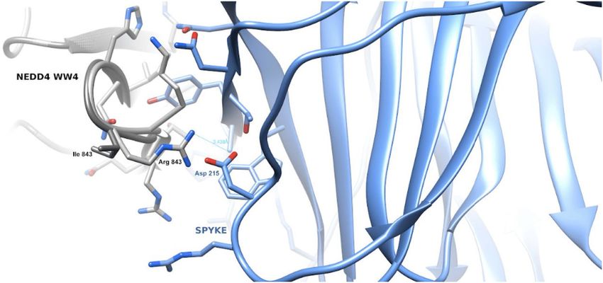
Novelli et al. Cell Death and Disease (2021)12:310                                                              Page 2 of 18

SARS-1 and Middle East respiratory syndrome [MERS])             overexpressed in COVID-19 vs. SARS-CoV-2 negative
and other autoimmune and chronic inflammatory dis-               patients in nasopharyngeal and oropharyngeal swab cells,
eases. Although some promising results have been initially      as well as in the lung of affected patients and in mouse
reported8, no significant clinical efficacy has been docu-        models of COVID-19. We also identified a subset of rare
mented in treated patients9. Several repurposed drugs           allelic variants in these genes and studied their distribu-
have been tested with disappointing results and many            tion in a large cohort of patients (COVID Human Genetic
other promising ones are undergoing clinical experi-            Effort (https://www.covidhge.com) severely affected by
mentation10–13. However, to date there is no effective          COVID-19 vs. asymptomatic or paucisymptomatic infec-
specific target drug against COVID-19. The unavailability        ted subjects. We showed that some of the identified var-
of selective and effective antiviral drugs is probably due to   iants display gain of function and aberrant activity. We
the poor knowledge of the pharmacological targets of the        finally evaluated whether selective inhibition of HECT
host cell necessary for the virus replication and/or for the    proteins by a natural NEDD4 and WWP1 inhibitor from
egress of new virions. Thus, a deeper knowledge of SARS-        Brassicaceae displayed anti-SARS-CoV-2 activity, thus
CoV-2 virus-host interaction for is fundamental to              providing preclinical support for the possible develop-
understand the molecular mechanisms that underly the            ment of clinical trials using this natural inhibitor in
life cycle of COVID-19 in order to develop treatments           COVID-19 patients.
worthy of a clinical trial assesement14.
   The enzymatic activity of the HECT-E3 ligases has been       Results
implicated in the cell egression phase of some RNA viruses      HECT family members interact and ubiquitinate the SARS-
possibly highjacking the endosomal sorting complexes            CoV-2 Spike protein
required for transport (ESCRT) machinery15–17, and spe-           To determine whether and how HECT type of E3 ligase
cifically members of a subgroup of HECT-E3 ligases,              family members are involved in SARS-CoV-2 pathology,
known as C2-WW-HECT (NEDD4-like) comprising at                  we at first focused on exploring how HECT E3 ligase
least nine members in humans (NEDD4, NEDD4L, ITCH,              might interact with the SARS-CoV-2 Spike protein, which
SMURF1, SMURF2, WWP1, WWP2, HECW1, and                          plays a critical role for the virus infection and egression
HECW2). This subgroup is characterized by a common              processes, and encode a PPxY motif (25-PPAY-28 in
modular architecture composed of a C2 domain related to         Spike protein)25. Notably, we found that the SARS-CoV-2
N-terminal C protein kinase, two to four domains with           S protein could interact with several HECT-E3 family
central tryptophan–tryptophan (WW), and a C-terminal            members, including NEDD4, WWP1, WWP2, SMURF1,
HECT domain18. The C2 domain is a Ca2+-dependent                and SMURF2 (Fig. 1A). Given that the PPxY motif is
binding domain and is mainly involved in targeting these        known to mediate the binding with NEDD4 family
enzymes to membrane compartments such as the plasma             members23, we next mutated the PPAY motif to Alanine
membrane, Golgi apparatus, endosomes, and lysosomes18.          (4A) or deleted this motif (delta-PPAY) altogether (Fig.
WW domains mediate protein– protein interactions                1B). We found that these mutants reduced the binding
through the recognition of Pro-rich motifs (PPxY, LPxY or       with NEDD4 while not abrogating it entirely (Fig. 1C).
related sequences) and phosphorylated Ser/Thr-Pro19,20.         Importantly and in support of a putative role of NEDD4 in
These domains provide a scaffold for recruiting protein         regulating SARS-CoV-2 viral life cycle, we found that
substrates and regulators. Several viral proteins have been     ectopic expression of NEDD4 could promote the
shown to recruit WW-domain host cell proteins of the            ubiquitination of the SARS-CoV-2 S protein in cells
NEDD4 family through PPxY motifs to facilitate their            (Fig. 1D).
egression and diffusion21,22. Among them, WWP1 was
found to interact with Ebola Virus VP40 to regulate             HECT genes and proteins expression in SARS-CoV-2
egression suggesting that viral PPxY-host WW domain-            patients and mouse models
mediated interaction could represent a potential new tar-         We first analyzed the gene expression levels of the nine
get for host-oriented inhibitors of EBOV and other virus        HECT family members by qRT-PCR using specific primer
egression23. Several studies have shown that the HECT           pairs (Table 2) on cDNA from residual unidentified
family members not only physically interact with specific        nasopharyngeal and oropharyngeal swabs of 37 COVID-
viral proteins to regulate the release of mature viral par-     19 patients with severe respiratory symptoms and 25
ticles through the ESCRT machine, but to regulate endo-         patients negative for the detection of SARS-CoV-2.
cytosis through ubiquitination24.                                 NEDD4 (FC = + 2.06, p ≤ 0.005), WWP1 (FC = + 1.85,
   Here, we investigated the involvement of HECT family         p ≤ 0.0005), WWP2 (FC = + 4.11; p < 0.005) and SMURF1
of E3 ligases in COVID-19 patients and their possible           (FC = + 1.7, p ≤ 0.05) showed a significant overexpression
involvement in SARS-CoV-2 infection. We found that              in nasopharyngeal and oropharyngeal swabs of COVID-19
WWP1, WWP2, SMURF1, and NEDD4 mRNA are                          positive patients compared to negative patients (Fig. 2A–D).

Official journal of the Cell Death Differentiation Association
Novelli et al. Cell Death and Disease (2021)12:310                                                                                Page 3 of 18

  Fig. 1 NEDD4 binds and ubiquitinates the SARS-CoV-2 S protein. A Immunoblotting (IB) of flag-immunoprecipates (IP) and whole cell lysis (WCL)
  derived from HEK293T cells that were transfected with HA-SARS-CoV-2 S and indicated NEDD4 family members. B A schematic diagram to show the
  PPxY motif in the SARS-CoV-2 S protein. C IB of flag-immunoprecipates and WCL derived from HEK293T cells that were transfected with NEDD4 and
  SARS-CoV-2 S WT or mutants. D IB of Ni-NTA pulldown and WCL derived from HEK293T cells that were transfected with SARS-CoV-2 S and NEDD4 WT
  (or EV as a negative control).

No significant differences were observed in the other ana-                 would affect their function could dictate the outcome and
lyzed genes (Fig. 2E–I).                                                  natural history of the disease. To test this hypothesis, we
   We then studied the expression of NEDD4 and WWP1                       initially collected a cohort of 130 unrelated Italian SARS-
at protein level taking advantage of a COVID-19 mouse                     CoV-2-positive patients, showing respiratory distress, Acute
model and of available human lung specimens from                          Respiratory Disease Syndrome (ARDS) or requiring invasive
infected patients necropsy. Overexpression at the protein                 ventilation and Intensive Care Unit (ICU) admission. We
level was observed for both WWP1 and NEDD4. How-                          identified a total of 408 HECT different pLOF, missense and
ever, WWP1 protein levels were more increased than                        in-frame monoallelic germline DNA variants. Data were
those of NEDD4 in the SARS-CoV-2-infected human lung                      extrapolated from previous studies performing WES analysis
tissue. In PCR negative COVID-19 human lungs, the basal                   described in previous publications26–28. Introducing a cut-
expression of NEDD4 and WWP1 was high. Interestingly,                     off at MAF < 0.01, we found 21 missense and 5 splice-region
however, in PCR positive COVID-19 human lungs,                            variants in NEDD4, NEDD4L, SMURF1, SMURF2, HECW1,
NEDD4 and WWP1 were downregulated everywhere                              HECW2, WWP1, and WWP2 genes, in a total of 24 patients
except in regions that expressed SARS-CoV-2 proteins                      (Table 1). No variant was detected in the ITCH gene. The
(Supplementary Fig. 1). In keeping with the human data,                   allelic frequencies of 12 genetic variants identified were
both NEDD4 (p < 0,0001) and WWP1 (p < 0,01) proteins                      significantly higher, when compared with those reported in
were significantly increased in mouse lungs over-                          GnomAD database for the EUR reference population (Table
expressing SARS-CoV-2 (Fig. 3). Thus, SARS-CoV-2                          1). One variant, M114I in WWP2 gene, was never detected
infection sustains and increases the expression levels of                 before (Table 1). Interestingly, five of the twelve variants
HECT Family members.                                                      observed with a higher frequency than that reported in
                                                                          GnomAD database are located in the NEDD4 gene.
HECT germline allelic variants in critical/life-threatening                  In a second step, we extended the genetic study to an
COVID-19 patients                                                         independent cohort of 710 unrelated COVID-19 critical
   We hypothesized that if high levels and sustained                      patients and 483 controls with asymptomatic or mild
expression of specific HECT-E3 ligase family members are                   SARS-CoV-2 infection belonging to the international
triggered by the SARS-CoV-2 infection, allelic variants that              CHGE Consortium data29,30, and we performed a

Official journal of the Cell Death Differentiation Association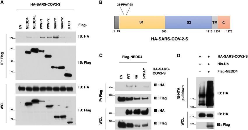
Novelli et al. Cell Death and Disease (2021)12:310                                                                                      Page 4 of 18

  Fig. 2 HECT E3 ubiquitin ligase gene expression level in SARS-CoV-2 positive and negative groups of subjects. A NEDD4 expression level in
  SARS-CoV-2 positive and negative groups of subjects, Mann–Whitney test, exact p value p = 0.0016, **; B WWP1 expression level in SARS-CoV-2
  positive and negative groups of subjects, Mann–Whitney test, exact p value p = 0.0005, ***; C WWP2 expression level in SARS-CoV-2 positive and
  negative groups of subjects, Mann–Whitney test, exact p value p = 0.0038, **; D SMURF1 expression level in SARS-CoV-2 positive and negative groups
  of subjects, Mann–Whitney test, exact p value p = 0.044, *; E SMURF2 expression level in SARS-CoV-2 positive and negative groups of subjects,
  Mann–Whitney test, non-significant p value; F NEDD4L expression level in SARS-CoV-2 positive and negative groups of subjects, Mann–Whitney test,
  non-significant p value; G ITCH expression level in SARS-CoV-2 positive and negative groups of subjects, Mann–Whitney test, non-significant p value;
  H HECW1 expression level in SARS-CoV-2 positive and negative groups of subjects, Mann–Whitney test, non-significant p value; I HECW2 expression
  level in SARS-CoV-2 positive and negative groups of subjects, Mann–Whitney test, non-significant p value.

PCA-adjusted burden test in order to evaluate a possible                     the six patients carrying any of these variants died
difference in the number of variants with MAF < 0.01. The                    (Table 1, Supplementary Table 1). Interestingly, WWP1
analysis did not reveal an enrichment of pLOF/missense/                      has a known binding activity with the protein S of the
inframe variants for any of the examined genes in severely                   virus, and the N745S missense variant previously char-
affected patients when compared to the asymptomatic                          acterized by Lee et al.31, leads to aberrant WWP1 enzy-
and paucisymptomatic infected controls (Supplementary                        matic activation with subsequent PTEN inactivation,
Table 1). As those tests involved a large number of var-                     thereby triggering hyperactive growth-promoting PI3K
iants, it is likely that most of them are neutral and strongly               signaling in cellular and murine models.
decreased the power of this analysis by diluting the signal.                   Next, we performed a more in depth in silico analysis of
Therefore, we performed a more detailed investigation of                     these three identified variants (I843R and R877G in NEDD4;
the variants that were present in at least two critical cases                N745S in WWP1). The I843R variant, mapping into the
and absent in infected controls. We identified 13 variants                    NEDD4 WW4 (in isoform 3), has a potential impact on
among which, three of them emerged as deleterious in all                     the protein ability to interact with its substrates. Specifically,
in silico prediction tools (Supplementary Table 2). Two of                   the 3D model of the variant WW4 domain in complex with
the three identified deleterious variants were in NEDD4                       the SARS-CoV-2 Spike (S) shows that the PPAY S residues
(I843R and R877G), and one in WWP1 (N745S). Each of                          interacting with the WW domain place the Asp215 S resi-
the three variants was present in two patients, and 3 out of                 due in close proximity with the Arg843 side-chain (Fig. 4),

Official journal of the Cell Death Differentiation Association
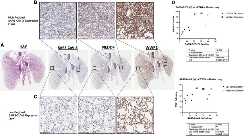
Novelli et al. Cell Death and Disease (2021)12:310                                                                                      Page 5 of 18

  Fig. 3 Mouse lungs (n = 3) expressing higher SARS-CoV-2 nucleocapsid protein express significantly higher NEDD4 (p < 0.0001*****) and
  WWP1 (p < 0.01**) in consecutive sections. Mouse lungs (n = 3) expressing higher SARS-CoV-2 nucleocapsid protein express significantly higher
  NEDD4 (p < 0.0001*****) and WWP1 (p < 0.01**) in consecutive sections (10X mag). Regions qualitatively defined as having high (>20% positive cells)
  Sars-CoV-2 expression (top row) and low (
Novelli et al. Cell Death and Disease (2021)12:310 Page 6 of 18
Table 1 HECT genes variants in a cohort of 130 SARS-CoV-2 positive patients (MAF < 0.01 in GnomAD v2.1.1; in bold p <
0.05).
Gene Genetic form Genotype dbSNP Consequence AF GnomAD Gender Age [years] p-value Outcome
HECW1 Known A1332T/WT rs200973212 missense 0.0000161 M 54 0.0114 Survived
Known E502Q/WT rs61756576 missense 0.0025630 M 83 0.5801 Deceased
Known N1265S/WT rs200912368 missense 0.0017820 F 50 0.4656 Survived
HECW2 Known S559G/WT rs779373864 missense 0.0000085 M 59 0.0072 Survived
Known N417S/WT rs138998510 missense 0.0005529 M 54 0.0791 Survived
Known A537P/WT rs750339715 missense 0.0000121 F 93 0.0092 Deceased
NEDD4 Known N888K/WT rs759199057 missense 0.0000119 M 59 0.01 Survived
Known G451A/WT rs60811367 missense 0.0017470 M 54 0.0013 Survived
M 39 Deceased
Known I1237T/WT rs373718024 missense 0.0003550 M 47 0.0159 Deceased
Known R877Q/WT rs201295772 missense 0.0000958 M 83 0.03176 Deceased
Known I843R/WT rs375088434 missense 0.0000199 F 77 0.0091 Deceased
Known T727I/WT rs61754989 missense 0.0036830 F 72 1 Deceased
Known D129N/WT rs150886795 missense 0.0002875 M 61 0.1233 Deceased
Known S29R/WT rs115484917 missense 0.0026250 M 39 0.1555 Deceased
NEDD4L Known c.698C>T/WT rs202231187 missense 0.0039100 M 73 1 Survived
Known c.1258-5A>C/WT rs768158353 splicing 0.0000853 M 64 0.0141 Deceased
Known c.698C>T/WT rs202231187 missense 0.0039100 M 54 1 Deceased
SMURF1 Known R564Q/WT rs182340234 missense 0.0000199 F 52 0.0068 Survived
Known T223M/WT rs371859465 missense 0.0000805 F 80 0.022 Survived
SMURF2 Known G10E/WT rs866321574 missense 0.0061730 F 36 0.1166 Survived
M 73 Deceased
Known I142V/WT rs145845053 missense 0.0005564 M 14 0.1281 Survived
WWP1 Known c.2395-4C>T/WT rs188228045 splicing 0.0000040 F 89 1 Deceased
Known c.540-5T>C/WT rs187132881 splicing 0.0023640 F 83 0.5073 Deceased
Known c.1836G>A/WT rs150841032 splicing 0.0002012 M 76 0.1019 Deceased
WWP2 Known R803C/WT rs747018644 missense 0.0000043 M 54 0.0049 Survived
New M114I/WT rs377573067 splicing / F 83 / Deceased
decrease for N745S-mediated ubiquitylation of the SARS- interaction with and ubiquitination of the SARS-CoV-2 S
COV2 S protein. These results argue that different WWP1 protein and associated proteins (Fig. 5G), but their
mutations might utilize different mechanisms to impact potential complementary roles, as well as their biological
COVID-19 biology, which requires additional in-depth and functional impacts in COVID-19 biology await further
studies in the future. On the other hand, in comparison investigation.
with the WT counterpart, the Smurf-1-T223M mutant
exhibited comparable binding or ubiquitination ability on The HECT inhibitor I3C is effective in mediating SARS-CoV-
SARS-CoV-2 S protein (Fig. 5E, F), at least in this 2 antiviral effect in in vitro cellular models
experimental setting. Given the close similarity between We next hypothesized that if HECT-E3 ligases are
their biochemical features and the reported functional indeed functionally relevant for the viral life cycle of
redundancies among NEDD4 family members, these data COVID-19, Indole-3-carbinol (I3C), a natural NEDD4
suggest that several NEDD4 family E3 ligases might par- and WWP1 inhibitor from Brassicaceae, might display a
ticipate in regulating COVID-19 egression via direct direct antiviral effect. We evaluated at first the impact of
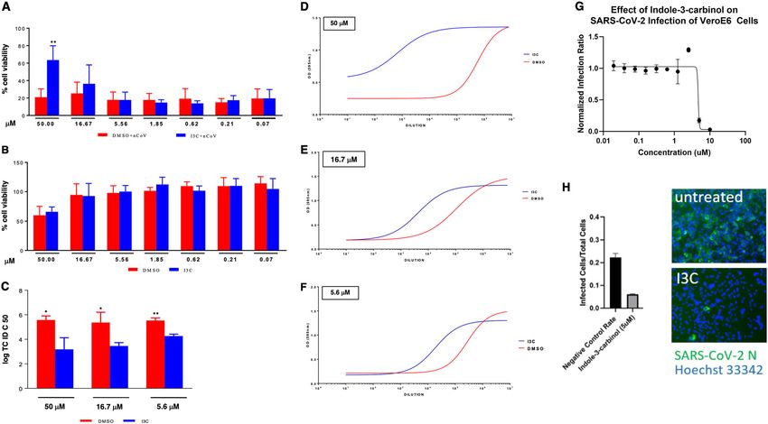
Official journal of the Cell Death Differentiation AssociationNovelli et al. Cell Death and Disease (2021)12:310 Page 7 of 18 Fig. 4 The Figure shows the 3D model of the WW domain of NEDD4 in complex with the SARS- CoV-2 Spike protein. The WW domain is displayed as a ribbon model, with the interface residue side-chains in gray. The Spike protein is displayed as a ribbon model, and the side-chains of its interface residues are shown in light blue. The Arg843–Asp215 residues are in close proximity and favor a stronger interaction between the variant WW domain and Spike with respect to the wt domain. Fig. 5 Gain-of-Function mutants in NEDD4, but not SMURF1 and WWP1, display elevated interaction with, but comparable ubiquitination of the SARS-CoV-2 S protein. A, C IB of flag-immunoprecipates and WCL derived from HEK293T cells that were transfected with HA-SARS-CoV-2 S and NEDD4/WWP1 WT and mutants. B, D IB of Ni-NTA pulldown and WCL derived from HEK293T cells that were transfected with SARS-CoV-2 S and NEDD4/WWP1 WT and mutants. E IB of flag-immunoprecipates and WCL derived from HEK293T cells that were transfected with HA-SARS-CoV-2 S and WT and mutants for Smurf1 or WWP1. F IB of Ni-NTA pulldown and WCL derived from HEK293T cells that were transfected with SARS-CoV-2 S and WT and mutants for Smurf1 or WWP1. G Binding and ubiquitination activity in WWP1/NEDD4/SMURF1 mutants vs. wild-type (WT). I3C on the cytopathic effect (CPE) induced by SARS- between 50 and 0.069 μM. The drug was added at dif- CoV-2 infection in Vero E6 cells. We treated the cells ferent time points, before (1 h) and after (1, 24, and 48 h) with I3C using a 3-fold concentration scale ranging SARS-Cov-2 multiplicity of infection (MOI = 0.001). CPE Official journal of the Cell Death Differentiation Association
Novelli et al. Cell Death and Disease (2021)12:310                                                                                             Page 8 of 18

  Fig. 6 I3C inhibited SARS-CoV-2-induced CPE and viral production in Vero E6 cells. Cells were treated with different doses of I3C (from 50 to
  0.069 μM; 1:3 serial dilutions) or DMSO (from 0.5 to 6.9 × 10⁻⁴ v/v percentage) 1 h before SARS-CoV-2 infection (MOI = 0.001) in four replicates.
  Absorption of the virus was allowed for 1 h at 37 °C in presence of I3C or DMSO treatments. The unabsorbed virus was removed and replaced by
  fresh medium with I3C or DMSO as above. Cells were then treated with either I3C or DMSO every 24 h and incubated at 37 °C with 5% CO2 for 72 h
  when the survival of infected (A) or not infected (B) cells was measured by crystal violet staining assay. The results were evaluated setting the
  uninfected control cells as 100% and the remaining values represented as a relative value. Experiments (n = 4) were performed in triplicate and data
  are expressed as mean +/−SD. Back-titration of virus progeny released by SARS-CoV-2-infected cells, treated as above, was performed on Vero E6
  cells. Survival of the cells was measured by crystal violet staining assay. Results were analyzed using Graph Pad (GraphPad Prism 8 XML ProjecT) with
  nonlinear regression curve fit (Inhibitor vs. response-Variable slope (four parameters)) ((D–F) and data expressed as log TCID50/100 μl (C)). Statistically
  significant differences between DMSO and I3C are represented as *P < 0.05 or **P < 0.002 determined using the paired t-test. Vero E6 cells were
  challenged with SARS-CoV-2. After 1.5 d, cells were fixed, stained with an N specific antibody and Hoechst 33342 for cell nuclei. Infected cells and
  total cell nuclei were counted by image analysis. The infection efficiency was normalized to infection seen in vehicle (DMSO) treated cells (G, H).

was evaluated 72 h post-infection, when culture media                            concentrations. The I3C-mediated decreased of viral
were collected for viral titer measurement. We found that                        production was also evident when cells were infected at
I3C reduced by about 60% the SARS-CoV-2-induced CPE                              higher MOI, although with lower efficacy (Supplementary
in Vero E6 cells at 50 μM, when compared to DMSO-                                Fig. 2). Since I3C reduced the viral production not only at
treated cells (Fig. 6A), while it was not effective at lower                     50 μM, when the CPE inhibition is clearly appreciated
concentrations. Similar results were obtained using a 10-                        (Fig. 6D), but also at 16.67 and 5.56 μM, when SARS-
fold increased MOI at 48 h post-infection. To note that                          CoV-2-induced CPE was not affected by I3C, it is likely
the concentration of 50 μM or 0.05% of I3C and DMSO,                             that I3C reduced the viral release rather than a viral entry
respectively, is partially toxic for the cells when treated for                  and/or replication leading to cell damage. Overall, these
72 h (Fig. 6B).                                                                  data demonstrated that I3C exerts a direct anti-SARS-
  Importantly, however, a much greater effect was                                CoV-2 replication activity.
observed when assessing the impact of I3C treatment on                             We further tested the potential efficacy of I3C utilizing a
the in vitro viral production. To this end, we measured the                      inhibition assay in Vero cells (see “Methods”: “Virus
amount of infectious SARS-CoV-2 released by the infec-                           infection inhibition assay”). Vero cells were grown to
ted cells treated with either I3C (50, 16.67, and 5.56 μM)                       70–90% confluency. After incubation with the virus cells
or DMSO (0.5, 0.167 and 0.056% (v/v)). Notably, I3C                              were fixed in formalin and then stained with SARS-CoV-2
significantly reduced the SARS-CoV-2 production at all                            antibody against the N protein and a fluorescently tagged
the concentrations tested (Fig. 6C–F), with a virus yield                        secondary antibody. Cell nuclei were stained with Hoechst
reduction ranging from 2 to 4 log at the various I3C                             33342 dye. The cells were imaged using a Cytation

Official journal of the Cell Death Differentiation Association
Novelli et al. Cell Death and Disease (2021)12:310                                                              Page 9 of 18

(Biotek) automated imaging system to visualize the blue         infection in lung tissue both in mice and humans. In
fluorescent nuclei and the green fluorescent infected cells       addition, we also demonstrated that NEDD4 and WWP1,
expressing virus N protein. Images were analyzed by             physically interact with and ubiquitylate the SARS-CoV-2
CellProfiler software using a customized analysis pipeline       S protein. This demonstrates a direct involvement of the
to count the nuclei and the infected cells. Infection effi-      HECT family proteins and, in particular, of NEDD4 and
ciency is expressed as a function of infected cells/cell        WWP1 in the virus life cycle. It is therefore conceivable
nuclei counted. Once again I3C was effective at inhibiting      that a greater production or an increased enzymatic
COVID-19 in this assay with an IC50 of 4.7 μm                   activity of members of these members of the HECT family
(Fig. 6G, H).                                                   could favor the exacerbation of the infection. The sole fact
                                                                that they are overexpressed in concomitance with the
Discussion                                                      SARS-CoV-2 infection suggests that the virus may take
   Several studies have shown that HECT proteins act as a       advantage from this pathway, as it has been shown for
functional interface between viral or cellular proteins         other RNA viruses.
containing PPxY motifs and the E-class vacuolar protein-           Additionally, and in line with this notion, we identified
sorting pathway (VPS)32. HECT domains can participate           three variants that bind more avidly to the SARS-CoV-2 S
in specific protein-protein interactions33 and hence ubi-        protein: two rare NEDD4 variants (I843R and R877G)
quitination of substrate proteins, which appears necessary      and the N745S germinal variant in WWP1, which was
for PPxY-dependent viral budding. Among the HECT                already characterized in cancer studies31. The increased
family members, WWP1 and NEDD4 have been the most               binding affinity could favor the ubiquitination of viral and
implicated in PPxY motif-dependent viral budding, and           cellular proteins thus implementing vesicular packaging
their HECT ubiquitin ligase activity is required for this       and virions release. These variants suggest the existence
activity34. Critically, these two HECT ubiquitin ligases can    of a particular genetic constraint against loss of function
physically and functionally interact forming heterodimeric      or gain of function given the multifunctionality of HECT
complexes, and are druggable by a well-tolerated natural        proteins31. It is worth noting that a recent paper reports
compound from Cruciferous vegetables35. Additionally,           that, in vitro, WWP1 K740 and 745S mutants displayed
gain of function germ line mutations of WWP1 have been          comparable ability as WT-WWP1 in largely mono-
identified in cancer susceptibility syndromes and in can-        ubiquitinating PTEN. These mutants, therefore, do not
cer patients31.                                                 appear to act as gain-of-function mutation in this in vitro
   We do not yet know the molecular mechanisms that             setting37. On one hand, this report is consistent with
govern several aspects of SARS-CoV-2 life cycle such as         what was previously reported in vivo where NEDD4 was
its entry, replication, assembly, budding, and particularly     found to mono-ubiquitinate PTEN and also cooperate
the egression of the virus. Recently, however, recently,        with WWP1 in promoting K27-polyubiquitination of
Ghosh et al.36, using virus-specific imaging and reporter        PTEN in cells, through heterodimeric interactions likely
methodologies, demonstrated that ß-Coronaviruses uti-           at plasma membrane38. On the other hand, the observed
lize lysosomal trafficking for exit, rather than biosynthetic    differences in WT-WWP1 vs. WWP1 mutants might
secretory pathway most commonly used by other envel-            stem from the monoubiquitination of PTEN observed
oped viruses. The biochemical and molecular character-          in vitro ub assay vs. K27-polyubiquitination of PTEN
ization of these steps and above all the identification of       detected in cells. It is possible that some key cellular
the proteins involved in these processes, is therefore          factors, likely WWP1 interacting proteins, such as
crucial to develop drugs that could interfere and block         NEDD4, might be required for polyubiquitination of
fundamental processes in the biology of the virus and pave      PTEN, both in cells and in vitro. Nonetheless, we also
the way for new therapeutic approaches.                         observed different ability for K740N and N745S WWP1
   Based on in silico analysis, it was recently proposed that   mutants in comparison with WT-WWP1 to promote
because SARS-CoV-2 encodes PPxY late domain motifs it           ubiquitination of the SARS-CoV-2-S protein in cells
might be capable of recruiting HECT family members              (Fig. 5D, F). These results are in keeping with the Cole
and, therefore, the ESCRT complex to improve virus              group37 to demonstrate that different WWP1 mutation
budding and release, favoring cellular reinfections. Inter-     might utilize different mechanism to control its down-
estingly, the PPxY motif is not present in SARS-CoV             stream pathways, including PTEN and SARS-CoV-2 S
proteins. The presence of the motif PPxY might con-             protein, which warrants additional in-depth investigation
tribute to explain why SARS-CoV-2 is more contagious            to reveal the underlying complicated mechanism that is
compared to SARS-CoV22.                                         likely to be context dependent or ever unique to each
   Here we demonstrated that WWP1, WWP2, and                    individual mutation of WWP1. Further studies are war-
NEDD4 are overexpressed during SARS-CoV-2 infection             ranted to analyze from a biochemical and functional
and that their expression co-localizes with areas of            point of view all the variants identified in these genes

Official journal of the Cell Death Differentiation Association
Novelli et al. Cell Death and Disease (2021)12:310 Page 10 of 18 (Supplementary Table 1) in order to access the genetic viral life cycle and the susceptibility and severity of enrichment found in a complete and unbiased way. It is COVID-19, our findings have immediate therapeutic also worth noting that several of the variants identified in implications for the treatment of infection and the pre- these genes have been observed in both asymptomatic vention of the most severe outcomes triggered by the and critical subjects. Interestingly, we extended the virus. The fact that I3C is effective in reducing SARS-CoV- genetic study to a second independent cohort of about 2 production in vitro prompts the immediate assessment 30,000 participants in the Healthy Nevada Project (HNP, of its efficacy in clinical trials (Supplementary Fig. 3). I3C Renown Health, Reno, Nevada, USA)39, to further cor- is, in fact, well-tolerated in both animal models and phase I roborate our results. This analysis led to results com- trials in humans at doses effective in the in vitro cell parable to the ones previously described. Moreover, we models35,44. It is therefore conceivable to rapidly reposi- identified 9 additional rare variants never detected before tion I3C in Phase II clinical trials in humans to test its in 9 COVID-19 patients, which may affect splicing ability to prevent the clinical severity of COVID-19. (Supplementary Table 3). HECT family members also play pivotal roles in the Materials and methods regulation of the innate immune response, and although International CHGE Consortium database the pathogenesis of COVID-19 is still under investigation, Between March and April 2020, 130 patients with it is clear that the innate immunity plays a crucial role in COVID-19 diagnosis were enrolled on Protocol no. 50/20 protective or destructive responses upon SARS-CoV-2 (Tor Vergata University Hospital). Informed consent was infection40. obtained from each patient. It is therefore conceivable that HEC family members To further improve our cohort, other 710 cases and 483 may affect the outcome and natural history of the controls were enrolled from the COVID Human Genetic COVID-19 infection also impacting non-cell autonomous Effort, International CHGE Consortium (Casanova J.L. anti-viral defense mechanisms. and Su H., https://www.covidhge.com), as described in For instance, ITCH controls the stability of critical Zhang et al.30. immune system proteins41 and acts upstream of B-cell The institutional review boards of each participating lymphoma 6 (Bcl-6), the main transcription factor Institution approved the protocol prior to patient enroll- involved in coordinating follicular helper T-cell differ- ment. The study was conducted in agreement with the entiation and immunoglobulin G (IgG) in response to principles of Declaration of Helsinki. acute viral infections42; WWP2 negatively regulates Toll-like receptor 3 (TLR3)-mediated innate immune Whole exome sequencing and data pre-processing response by targeting TIR-domain containing adapter- Genomic DNA was extracted from peripheral blood inducing interferon-β (TRIF) for ubiquitination and samples using standard procedures and Qiagen blood degradation43. The innate immune system acts as first DNA mini Kit (Qiagen, Hilden, Germany). Library pre- responder for the detection and clearance of viral paration and whole exome capture were performed by infections. Innate immune cells secrete proin- using the Twist Human Core Exome Kit (Twist flammatory cytokines that inhibit viral replication, sti- Bioscience, South San Francisco, CA, USA) according to mulate the adaptive immune response, and recruit other the manufacture’s protocol and sequenced on the Illu- immune cells to the site of infection40. In this respect, in mina NovaSeq 6000 platform. The BaseSpace pipeline an international cohort29, we recently showed that about (Illumina, Inc., San Diego, CA, USA) and the TGex soft- 3% of COVID-19 critical patients carried loss-of- ware (LifeMap Sciences, Inc., Alameda, CA, USA) were function variants in genes coding for proteins involved used for the variant calling and annotating variants, in type I IFN innate immunity, thus representing an respectively. Sequencing data were aligned to the hg19 important target for a deeper investigation of their role human reference genome. A minimum depth coverage of in the pathogenesis30. Collectively our results indicate 30X was considered suitable for analysis, based on the that the risk of susceptibility to severe COVID-19 is guidelines of the American College of Medical Genetics unlikely to be influenced predominantly by rare variants and Genomics. All variants were examined for coverage of HECT genes in the MAF range
Novelli et al. Cell Death and Disease (2021)12:310                                                                                              Page 11 of 18

62 subjects with acute respiratory symptoms or contacts                           (DeNovix) and 100 ng of total RNA was been reverse
with COVID-19 confirmed cases, arrived at the attention of                         transcribed into cDNA using the High Capacity cDNA
the Emergency Room (ER) of Policlinico Tor Vergata, PTV                           Reverse Transcription Kit (Applied Biosystems, USA). We
(Rome, Italy). As widely described by Amati et al.45, patients’                   analyzed the expression of the 9 members of the HECT3
swabs were referred to the Virology Unit of PTV for the                           ligase family: WWP1, WWP2, NEDD4, NEDD4L, ITCH,
molecular diagnostic test detecting the presence of SARS-                         SMURF1, SMURF2, HECW1, and HECW2 genes;
CoV-2 nucleic acids using used the Allplex™ 2019-nCoV                             GAPDH, ACTB, and RPLP0 genes were used for data
Assay (Seegene Inc, http://www.seegene.com/upload/                                normalization. Real-time PCRs (qRT-PCRs) have been
product/Allplex_2019_nCoV_performance_data.pdf).                                  performed using ABI7500 Fast Real-time PCR System
  SARS-CoV-2 positive (n = 37) and negative (n = 25)                              (Life Technologies) with Sybr Green Assay (Power Sybr
samples were used for RNA expression analysis.                                    Green PCR Master Mix, Life Technologies) and specific
                                                                                  primer pairs (Table 2).
Real-time PCR and statistical analysis                                               The qRT-PCR expression analyses were performed in
  The total RNA extracted from nasopharyngeal and                                 triplicate. Data analysis was performed using the com-
oropharyngeal swabs was evaluated by NanoDrop DS-11                               parative threshold cycle (Ct) method quantification

Table 2 Real-Time PCR primer sequences.

Gene                         Accession number                    Sequence (5’→3’)                                                            Product size (bp)

WWP1                         NM_007013.4                         Fw                   TGTAAATGTTACGCCACAGACT                                 105
                                                                 Rv                   GCTTGTTTCAAATCTATCGTTGC
WWP2                         NM_007014.5                         Fw                   GAAAGTGGTGTCCGCAAAGC                                   175
                                                                 Rv                   ATGACTCTGTGCCGTGACATT
NEDD4                        NM_006154.4                         Fw                   CTGCTACGGACAATTATACCCTA                                129
                                                                 Rv                   CATCCAACAGTTTGCCATGATA
NEDD4L                       NM_001144967.3                      Fw                   ACGTAGCGGATGAGAATAGAGAAC                               115
                                                                 Rv                   CTGTGATTAGATGGGTTTACCCTGA
ITCH                         NM_031483.7                         Fw                   GGTTCAGTATTTCCGGTTCTGGT                                118
                                                                 Rv                   GGGACTGAAGCTCATTATCTGTTG
SMURF1                       NM_020429.3                         Fw                   CCGCTCCAAGGCTTCAAGG                                    125
                                                                 Rv                   ATCCGGTTAAAGCAGGTATGGG
SMURF2                       NM_022739.4                         Fw                   GCAAATGGATCAGGAAGTCGGAAA                               100
                                                                 Rv                   CCGGAGGCCGGAGGA
HECW1                        NM_015052.5                         Fw                   CGAGCAACCACCCCCAGTGT                                   136
                                                                 Rv                   CCATGGCTTGGAAATCTGAGAGA
HECW2                        NM_001348768.2                      Fw                   CTACCAGCATAACCGCGACC                                   112
                                                                 Rv                   AAAGAATGCCTTGCCCTGGT
GAPDH                        NM_002046                           Fw                   AAGGTCGGAGTCAACGGATTT                                  100
                                                                 Rv                   TGAAGGGGTCATTGATGGCA
ACTB                         NM_001101                           Fw                   ATTGCCGACAGGATGCAGAA                                   150
                                                                 Rv                   GCTGATCCACATCTGCTGGAA
RPLP0                        NM_001002                           Fw                   ACCCAGCTCTGGAGAAACT                                    198
                                                                 Rv                   AAAAGGAGGTCTTCTCGGG

WWP1 WW domain containing E3 ubiquitin protein ligase 1, WWP2 WW domain containing E3 ubiquitin protein ligase 2, NEDD4 neural precursor cell expressed,
developmentally downregulated 4, E3 ubiquitin protein ligase, NEDD4L neural precursor cell expressed, developmentally downregulated 4-like, E3 ubiquitin protein
ligase, ITCH HECT-type E3 ubiquitin transferase itchy homolog, SMURF1 SMAD specific E3 ubiquitin protein ligase 1, SMURF2 SMAD specific E3 ubiquitin protein ligase
2, HECW1 HECT, C2 and WW domain containing E3 ubiquitin protein ligase 1, HECW2 HECT, C2 and WW domain containing E3 ubiquitin protein ligase 2, GAPDH
glyceraldehyde-3-phosphate dehydrogenase, ACTB β-actin, RPLP0 ribosomal protein, large, P0, Fw forward, Rev reverse. PCR polymerase chain reaction.

Official journal of the Cell Death Differentiation Association
Novelli et al. Cell Death and Disease (2021)12:310 Page 12 of 18 (2−ΔCt method) (as described by Rizzacasa et al. at between separate stains. Digital image analysis was per- https://www.protocols.io/view/comparative-ct-method- formed using Definiens TissueStudio 4.0 software (Astra- quantification-2-ct-method-zp7f5rn). Zeneca, Munich Germany), in which nucleus detection was Statistical analysis was performed using GraphPad performed on the hematoxylin counterstain, and positive Prism 7.0 (GraphPad Software, USA). D’Agostino & cells were identified using a threshold for DAB staining to Pearson, Shapiro–Wilk, and Kolmogorov–Smirnov nor- identify positive stained images for each biomarker mality tests were used to assess the distribution of gene (NEDD4, WWP1, and SARS-CoV2 Nucleocapsid protein). expression data derived from qRT-PCR assays. Since gene Following quantification, expression (as % positive cells) expression data did not pass the normality test (p ≤ 0.05), of NEDD4 and WWP1 was plotted against SARS-CoV2 Mann–Whitney test was used for data comparison expression using Prism software. Six random regions of between SARS-CoV-2 positive and negative groups. In high SARS-CoV2 expression (>20% positive cells) and 6 graphs, gene expression is represented as median with random regions low SARS-CoV-2 expressions (
Novelli et al. Cell Death and Disease (2021)12:310                                                                               Page 13 of 18

sp.; formerly called Cercopithecus aethiops). Cells were         passaged twice on Vero E6 cells by challenging at an MOI
maintained in Minimum Essential Medium (MEM), sup-               of less than 0.01 and incubating until cytopathology was
plemented with heat inactivated 10% fetal bovine serum           seen (after about 3 days). A sample of the culture super-
(FBS), 2 mM L-glutamine and 1% penicillin/streptomycin           natant was sequenced by NGS and was consistent with
solution (Sigma-Aldrich, Cat.No. R0883; F7524; G7513;            the original isolate without evidence of contaminants. The
P0781, respectively) and maintained at 5% CO2, 37 °C.            virus stock was stored at −80 °C until used.
                                                                   For evaluation of indole-3-carbinol against infection
I3C antiviral test                                               with wild type SARS-CoV-2, the compound was dissolved
  The antiviral activity of I3C has been tested by a cyto-       to 10 mM in DMSO and then diluted in culture medium
pathic effect (CPE) inhibition assay using Vero E6 cells         before addition to cells. The compound was added to
infected with the SARS-CoV-2 strain isolated at INMI L.          VeroE6 cells incubated for a minimum of 1 hour, then
Spallanzani IRCCS (2019-nCoV/Italy-INMI1; GenBank                challenged with virus at an MOI of less than 0.2. Dosing
MT06615656). The extent of in vitro inhibition of SARS-          ranged from a final concentration of 10 µM down to
CoV-2-driven cell damage (CPE) by I3C is expressed as            0.02 µM in a two-fold dilution series. As a positive con-
percentage of surviving cells.                                   trol, 5 µM E-64 was used as it was previously reported to
  Briefly, cell monolayers growing in 96-well plates (3 ×         inhibit SARS-CoV-2 infection57. Negative controls were
104 cells/well) were treated for 1 h with 1:3 serial dilutions
Novelli et al. Cell Death and Disease (2021)12:310 Page 14 of 18
Unit, Avicenne Hospital, APHP, Bobigny, France. 16INSERM U1272 Hypoxia & Conflict of interest
Lung, Bobigny, France. 17Pneumology Department, Reference Center for Rare The authors declare no competing interests.
Pulmonary Diseases, Hôpital Avicenne, APHP, Bobigny; INSERM UMR1272,
Université Paris 13, Bobigny, France. 18Hôpital Bichat Claude Bernard, APHP,
Paris, France. 19Centre d’investigation Clinique, Inserm CIC, 1425 Paris, France. Publisher’s note
20 Springer Nature remains neutral with regard to jurisdictional claims in
Infection, Antimicrobials, Modelling, Evolution (IAME), INSERM, UMRS1137,
University of Paris, Paris, France. 21AP-HP, Bichat Claude Bernard Hospital, published maps and institutional affiliations.
Infectious and Tropical Disease Department, Paris, France. 22Laboratory of
Clinical Immunology, NIAID, NIH, Bethesda, MD, USA. 23Laboratory of Human Supplementary information The online version contains supplementary
Genetics of Infectious Diseases, Necker Branch, INSERM, Necker Hospital for material available at https://doi.org/10.1038/s41419-021-03513-1.
Sick Children, Paris, France. 24University of Paris, Imagine Institute, Paris, France.
25
Center for Genomic Medicine, Desert Research Institute, Reno, NV 89502,
USA. 26Renown Institute for Cancer, Nevada System of Higher Education, Reno, Appendix I: COVID Human Genetic Effort
NV 89502, USA. 27The Donnelly Centre, University of Toronto, Toronto, Ontario, Laurent Abel1, Alessandro Aiuti2, Saleh Al-Muhsen3,
CanadaM5S 3E1 416-946-0863. 28Virna Therapeutics, West Roxbury, MA, USA.
29
Department of Microbiology Boston University, National Emerging Infectious Fahd Al-Mulla4, Mark S. Anderson5, Andrés Augusto
Diseases Laboratories, Boston, MA 02118, USA. 30Howard Hughes Medical Arias6, Hagit Baris Feldman7, Dusan Bogunovic8,
Institute, New York, NY, USA. 31MBC, Department of Molecular Biotechnology Alexandre Bolze9, Anastasiia Bondarenko10, Ahmed A.
and Health Sciences, University of Turin, Turin, TO 10126, Italy
Bousfiha11, Petter Brodin12, Yenan Bryceson13, Carlos D.
Bustamante14, Manish J. Butte15, Giorgio Casari16,
Author contributions
Samya Chakravorty17, John Christodoulou18, Antonio
G.N. and P.P.P. performed study concept and design, wrote the draft of the
paper, and supervised the project; J.L. and W.W. performed functional studies Condino-Neto19, Stefan M. Constantinescu20, Megan A.
using HECT proteins; M.B., and B.R. performed RNA expression analysis and Cooper21, Clifton L. Dalgard22, Murkesh Desai23, Beth
statistical tests; T.A., D.G., M.R.C., J.J.P., and R.A.D. provided in vivo SARS-CoV-2 A. Drolet24, Sara Espinosa-Padilla25, Jacques Fellay26,
assays and inhibition tests; A.N., D.C., and E.A. contributed to carry out the
molecular genetic data and performed the analysis; J.-L.C., A.C., L.A., B.B., V.L.C., Carlos Flores27, José Luis Franco6, Antoine Froidure28,
A.L., R.G., G.E., L.D.N., H.C.S., and J.J.G. provided acquisition, analysis and Peter K. Gregersen29, Filomeen Haerynck30, David
interpretation of genetic data, and wrote the paper; S.G. performed RNA
Hagin31, Rabih Halwani32, Lennart Hammarström33, Jim
expression experiments, contributed with reagents, materials and analysis
tools; K.C., T.M., F.A., F.K., and J.M. performed histological analysis in human Heath34, Sarah E. Henrickson35, Elena Hsieh36, Kohsuke
lung tissue and in mouse models; A.G. and G.P. provided in silico analysis; A.C. Imai37, Yuval Itan38, Timokratis Karamitros39, Kai
and A.L. evaluated and recruited patients to COVID and/or control cohorts; Y.T.
Kisand40, Cheng-Lung Ku41, Yu-Lung Lau42, Yun
L., Y.U., J.G., and V.L.C. contributed to provide clinical phenotype; J.M., C.T., L.D.
N., H.C.S., and S.S. performed review and revision of the paper. All authors read Ling43, Carrie L. Lucas44, Tom Maniatis45, Davoud
and approved the final version of the manuscript. Mansouri46, László Maródi47, Isabelle Meyts48, Joshua
D. Milner49, Kristina Mironska50, Trine H. Mogensen51,
Fundings Tomohiro Morio52, Lisa F.P. Ng53, Luigi D. Notar-
The Laboratory of Human Genetics of Infectious Diseases is supported by the angelo54, Antonio Novelli55, Giuseppe Novelli56, Satoshi
Howard Hughes Medical Institute, the Rockefeller University, the St. Giles
Foundation, the NIH (R01AI088364), the National Center for Advancing
Okada57, Tayfun Ozcelik58, Jana M. Pachlopnik59, Qiang
Translational Sciences (NCATS), the NIH Clinical and Translational Science Pan-Hammarström60, Rebeca Perez de Diaz61, Anna M.
Award (CTSA) program (UL1 TR001866), a Fast Grant from Emergent Ventures, Planas62, Carolina Prando63, Aurora Pujol64, Lluis
Mercatus Center at George Mason University, the Yale Center for Mendelian
Genomics and the GSP Coordinating Center funded by the National Human
Quintana-Murci65, Laurent Renia53, Carlos Rodríguez-
Genome Research Institute (NHGRI) (UM1HG006504 and U24HG008956), the Gallego66, Vanessa Sancho-Shimizu67, Vijay Sankaran68,
French National Research Agency (ANR) under the “Investments for the Future” Mohammed Shahrooei69, Andrew L. Snow22, Pere Soler-
program (ANR-10-IAHU-01), the Integrative Biology of Emerging Infectious
Diseases Laboratory of Excellence (ANR-10-LABX-62-IBEID), the French
Palacín70, András N. Spaan71, Stuart G. Tangye72, Stuart
Foundation for Medical Research (FRM) (EQU201903007798), the FRM and ANR Turvey73, Furkan Uddin74, Mohammed J. Uddin75,
GENCOVID project, ANRS-COV05, the Square Foundation, Grandir–Fonds de Diederik van de Beek76, Donald C. Vinh77, Horst von
Solidarité pour l’Enfance, the SCOR Corporate Foundation for Science, Institut
National de la Santé et de la Recherche Médicale (INSERM), the University of
Bernuth78, Pawel Zawadzki79, Helen C. Su54*, Jean-
Paris. The French COVID Cohort study group was sponsored by Inserm and Laurent Casanova80*
1
supported by the REACTing consortium and by a grant from the French INSERM U1163, University of Paris, Imagine Institute,
Ministry of Health (PHRC 20-0424). L.D.H. and H.C.S. are supported by the
Division of Intramural Research, National Institute of Allergy and Infectious
Paris, France. 2San Raffaele Telethon Institute for Gene
Disease, National Institutes of Health. This study was also supported in part by Therapy, IRCCS Ospedale San Raffaele, and Vita Salute San
a grant of Rome Foundation (Italy, Prot 317 A/I) to G.N. Raffaele University, Milan, Italy. 3Immunology Research
Laboratory, Department of Pediatrics, College of Medicine
Data availability and King Saud University Medical City, King Saud Uni-
The datasets used and/or analyzed during the current study are available from versity, Riyadh, Saudi Arabia. 4Dasman Diabetes Institute,
the corresponding author on reasonable request. Additional supporting
information may be found in the online version of this article at the publisher’s Department of Genetics and Bioinformatics, Dasman,
web site. Kuwait. 5Diabetes Center, University of California, San
Francisco, San Francisco, CA, USA. 6Universidad de Anti-
Ethics approval and consent to participate oquia, Group of Primary Immunodeficiencies, Antioquia,
See “Materials and methods” section. Colombia. 7The Genetics Institute, Tel Aviv Sourasky
Official journal of the Cell Death Differentiation AssociationNovelli et al. Cell Death and Disease (2021)12:310 Page 15 of 18
Medical Center and Sackler Faculty of Medicine, Tel Aviv Emirates. 33Department of Laboratory Medicine, Karolinska
University, Tel Aviv, Israel. 8Icahn School of Medicine at Institutet, Stockholm, Sweden. 34Institute for Systems
Mount Sinai, New York, NY, USA. 9Helix, San Mateo, CA, Biology, Seattle, WA, USA. 35Perelman School of Medicine,
USA. 10Shupyk National Medical Academy for Post- University of Pennsylvania Children’s Hospital of Philadel-
graduate Education, Kiev, Ukraine. 11Clinical Immunology phia, Division of Allergy-Immunology, Philadelphia, PA,
Unit, Department of Pediatric Infectious Disease, CHU Ibn USA. 36Department of Immunology and Microbiology,
Rushd and LICIA, Laboratoire d’Immunologie Clinique, Department of Pediatrics, Division of Allergy and Immu-
Inflammation et Allergie, Faculty of Medicine and Phar- nology, University of Colorado, School of Medicine Chil-
macy, Hassan II University, Casablanca, Morocco. 12SciLi- dren’s Hospital Colorado, Aurora, CO, USA. 37Riken,
feLab, Department of Women’s and Children’s Health, Tokyo, Japan. 38Institute for Personalized Medicine, Icahn
Karolinska Institutet, Stockholm, Sweden. 13Department of School of Medicine at Mount Sinai, New York, NY, USA;
Medicine, Center for Hematology and Regenerative Medi- Department of Genetics and Genomic Sciences, Icahn
cine, Karolinska Institutet, Stockholm, Sweden. 14Stanford School of Medicine at Mount Sinai, New York, NY, USA.
University, Stanford, CA, USA. 15Division of Immunology, 39
Bioinformatics and Applied Genomics Unit, Department
Allergy, and Rheumatology, Department of Pediatrics, of Microbiology, Hellenic Pasteur Institute, Athens, Greece.
40
UCLA, Los Angeles, CA, USA; Department of Micro- Molecular Pathology, Department of Biomedicine, Insti-
biology, Immunology, and Molecular Genetics, UCLA, Los tute of Biomedicine and Translational Medicine, University
Angeles, CA, USA. 16Medical Genetics, IRCCS Ospedale of Tartu, Tartu Estonia. 41Chang Gung University, Taoyuan
San Raffaele, Milan, Italy. 17Department of Pediatrics and County, Taiwan. 42Department of Paediatrics & Adolescent
Children’s Healthcare of Atlanta, Emory University, Atlanta, Medicine, The University of Hong Kong, Hong Kong,
GA, USA. 18Murdoch Children’s Research Institute, Vic- China. 43Shanghai Public Health Clinical Center, Fudan
toria, Australia. 19Department of Immunology - Institute of University, Shanghai, China. 44Department of Immuno-
Biomedical Sciences - University of São Paulo, São, Brazil. biology, Yale University School of Medicine, New Haven,
20
Endocrinology Department, Cliniques Universitaires Saint CT, USA. 45Zukerman Mind Brain Behavior Institute,
Luc, Brussels, Belgium. 21Washington University School of Columbia University, New York, NY, USA; New York
Medicine, St. Louis, MO, USA. 22Department of Anatomy, Genome Center, New York, NY, USA. 46Department of
Physiology & Genetics, Uniformed Services University of Clinical Immunology and Infectious Diseases, National
the Health Sciences, Bethesda, MD, USA. 23Bai Jerbai Research Institute of Tuberculosis and Lung Diseases, The
Wadia Hospital for Children, Mumbai, India. 24School of Clinical Tuberculosis and Epidemiology Research Center,
Medicine and Public Health, University of Wisconsin, National Research Institute of Tuberculosis and Lung Dis-
Madison, WI, USA. 25Instituto Nacional de Pediatria eases (NRITLD), Masih Daneshvari Hospital, Shahid
(National Institute of Pediatrics), Mexico City, Mexico. Beheshti University of Medical Sciences, Tehran, Iran.
26 47
School of Life Sciences, Ecole Polytechnique Fédérale de PID Clinical Unit and Laboratory, Department of Der-
Lausanne, Lausanne, Switzerland; Precision Medicine Unit, matology, Venereology and Dermato-oncology. 48Depart-
Lausanne University Hospital and University of Lausanne, ment of Pediatrics, University Hospitals Leuven, Leuven,
Lausanne, Switzerland. 27Genomics Division, Instituto Belgium; Laboratory for Inborn Errors of Immunity, KU
Tecnológico y de Energías Renovables (ITER), Santa Cruz Leuven, Leuven, Belgium. 49Department of Pediatrics,
de Tenerife, Spain; Research Unit, Hospital Universitario N. Columbia University Irving Medical Center. New York, NY,
S. de Candelaria, Santa Cruz de Tenerife, Spain; Instituto de USA. 50University Clinic for Children’s Diseases, Depart-
Tecnologías Biomédicas (ITB), Universidad de La Laguna, ment of Pediatric Immunology, Medical Faculty, University
San Cristóbal de La Laguna, Spain; CIBER de Enfermedades “St.Cyril and Methodij” Skopje, North Macedonia.
51
Respiratorias, Instituto de Salud Carlos III, Madrid, Spain. Department of Biomedicine, Aarhus University, Aarhus,
28
Institut de Recherche Expérimentale et Clinique, Pôle de Denmark; Department of Infectious Diseases, Aarhus Uni-
Pneumologie, Université catholique de Louvain, Belgium versity Hospital, Aarhus, Denmark. 52Tokyo Medical &
Service de pneumologie, Cliniques Universitaires Saint-Luc, Dental University Hospital, Tokyo, Japan. 53A*STAR ID
Brussels, Belgium. 29Feinstein Institute for Medical labs, Agency for Science, Technology and Research
Research, Northwell Health USA, Manhasset, NY, USA. (A*STAR), Singapore; Singapore Immunology Network,
30
Department of Paediatric Immunology and Pulmonology, Agency for Science, Technology and Research (A*STAR),
Centre for Primary Immunodeficiency Ghent (CPIG), PID Singapore. 54National Institute of Allergy and Infectious
Research Laboratory, Jeffrey Modell Diagnosis and Research Diseases, National Institutes of Health, Bethesda, MD, USA.
Centre, Ghent University Hospital, Edegem, Belgium. 31The 55
Laboratory of Medical Genetics, IRCCS Bambino Gesù
Genetics Institute Tel Aviv Sourasky Medical Center, Tel Children’s Hospital, Rome, Italy. 56Department of Biome-
Aviv, Israel. 32Sharjah Institute of Medical Research, College dicine and Prevention, Tor Vergata University of Rome,
of Medicine, University of Sharjah, Sharjah, United Arab Rome, Italy. 57Department of Pediatrics, Graduate School of
Official journal of the Cell Death Differentiation AssociationYou can also read