Alzheimer's disease brain-derived extracellular vesicles spread tau pathology in interneurons
←
→
Page content transcription
If your browser does not render page correctly, please read the page content below
doi:10.1093/brain/awaa376                                                                                                           BRAIN 2021: 144; 288–309              | 288

Alzheimer’s disease brain-derived extracellular
vesicles spread tau pathology in interneurons
  Zhi Ruan,1 Dhruba Pathak,1,2 Srinidhi Venkatesan Kalavai,1 Asuka Yoshii-Kitahara,1 Satoshi
Muraoka,1 Nemil Bhatt,3 Kayo Takamatsu-Yukawa,1 Jianqiao Hu,1 Yuzhi Wang,1 Samuel

                                                                                                                                                                                  Downloaded from https://academic.oup.com/brain/article/144/1/288/6007752 by guest on 23 April 2021
Hersh,1 Maria Ericsson,4 Santhi Gorantla,5 Howard E. Gendelman,5 Rakez Kayed,3 Seiko
Ikezu,1 Jennifer I. Luebke2,6 and Tsuneya Ikezu1,6,7

Extracellular vesicles are highly transmissible and play critical roles in the propagation of tau pathology, although the underlying
mechanism remains elusive. Here, for the first time, we comprehensively characterized the physicochemical structure and pathogen-
ic function of human brain-derived extracellular vesicles isolated from Alzheimer’s disease, prodromal Alzheimer’s disease, and
non-demented control cases. Alzheimer’s disease extracellular vesicles were significantly enriched in epitope-specific tau oligomers
in comparison to prodromal Alzheimer’s disease or control extracellular vesicles as determined by dot blot and atomic force mi-
croscopy. Alzheimer’s disease extracellular vesicles were more efficiently internalized by murine cortical neurons, as well as more
efficient in transferring and misfolding tau, than prodromal Alzheimer’s disease and control extracellular vesicles in vitro.
Strikingly, the inoculation of Alzheimer’s disease or prodromal Alzheimer’s disease extracellular vesicles containing only 300 pg of
tau into the outer molecular layer of the dentate gyrus of 18-month-old C57BL/6 mice resulted in the accumulation of abnormally
phosphorylated tau throughout the hippocampus by 4.5 months, whereas inoculation of an equal amount of tau from control
extracellular vesicles, isolated tau oligomers, or fibrils from the same Alzheimer’s disease donor showed little tau pathology.
Furthermore, Alzheimer’s disease extracellular vesicles induced misfolding of endogenous tau in both oligomeric and sarkosyl-insol-
uble forms in the hippocampal region. Unexpectedly, phosphorylated tau was primarily accumulated in glutamic acid decarboxyl-
ase 67 (GAD67) GABAergic interneurons and, to a lesser extent, glutamate receptor 2/3-positive excitatory mossy cells, showing
preferential extracellular vesicle-mediated GABAergic interneuronal tau propagation. Whole-cell patch clamp recordings of CA1
pyramidal cells showed significant reduction in the amplitude of spontaneous inhibitory post-synaptic currents. This was accompa-
nied by reductions in c-fos + GAD67 + neurons and GAD67 + neuronal puncta surrounding pyramidal neurons in the CA1 region,
confirming reduced GABAergic transmission in this region. Our study posits a novel mechanism for the spread of tau in hippocam-
pal GABAergic interneurons via brain-derived extracellular vesicles and their subsequent neuronal dysfunction.

 1    Department of Pharmacology and Experimental Therapeutics, Boston University School of Medicine, Boston, MA 02118, USA
 2    Department of Anatomy and Neurobiology, Boston University School of Medicine, Boston, MA 02118, USA
 3    Department of Neurology, University of Texas Medical Branch, Galveston, TX 77555, USA
 4    Department of Cell Biology, Harvard Medical School, Boston, MA 02115, USA
 5    Department of Pharmacology and Experimental Neurosciences, University of Nebraska Medical Center, Omaha, NE 68198, USA
 6    Center for Systems Neuroscience, Boston University, Boston, MA 02118, USA
 7    Department of Neurology and Alzheimer’s Disease Center, Boston University School of Medicine, Boston, MA 02118, USA

Received April 17, 2020. Revised July 29, 2020. Accepted August 17, 2020. Advance access publication November 27, 2020
C The Author(s) (2020). Published by Oxford University Press on behalf of the Guarantors of Brain.
V
This is an Open Access article distributed under the terms of the Creative Commons Attribution Non-Commercial License (http://creativecommons.org/licenses/by-nc/4.0/), which
permits non-commercial re-use, distribution, and reproduction in any medium, provided the original work is properly cited. For commercial re-use, please contact
journals.permissions@oup.com
Extracellular vesicles mediate tau spread                                                     BRAIN 2021: 144; 288–309    | 289

Correspondence to: Tsuneya Ikezu, MD, PhD
Professor of Departments of Pharmacology and Experimental Therapeutics and Neurology
Boston University School of Medicine, Boston, MA 02118, USA
E-mail: tikezu@bu.edu

Keywords: Alzheimer’s disease; extracellular vesicle; GABAergic interneuron; microtubule-associated protein tau; mouse model
Abbreviations: AP = action potential; dfPBS = double-filtered phosphate-buffered saline; EV = extracellular vesicle; KO = knock-
out; OML = outer molecular layer; pAD = prodromal Alzheimer’s disease; PK = proteinase K; sIPSC = spontaneous inhibitory
postsynaptic current; TEM = transmission electron microscopy

                                                                  to tau neuropathology (Guo et al., 2016; Narasimhan et al.,
Introduction                                                      2017). Notably, EVs isolated from transgenic tau mouse

                                                                                                                                    Downloaded from https://academic.oup.com/brain/article/144/1/288/6007752 by guest on 23 April 2021
Accumulation of misfolded microtubule-associated protein          brains, Alzheimer’s disease plasma, or human induced pluri-
tau is a neuropathological hallmark of Alzheimer’s disease.       potent stem cells expressing recombinant mutant tau also
The degree of Alzheimer’s disease cognitive decline is paral-     initiated propagation of tau in mouse brain tissues (Baker
leled by the progression of anatomical spread of misfolded        et al., 2016; Polanco et al., 2016; Winston et al., 2019).
tau (Arriagada et al., 1992). Abnormally aggregated and           Furthermore, pharmacological inhibition of exosome synthe-
phosphorylated tau (p-tau) first appears in the entorhinal        sis significantly reduced tau propagation (Asai et al., 2015;
cortex at the prodromal stage of Alzheimer’s disease, spread-     Bilousova et al., 2018). The molecular mechanisms of cell-
ing in a hierarchical pattern to hippocampal regions and          to-cell transmission of EVs and free tau aggregates via up-
then throughout neocortical areas (Braak and Braak, 1991).        take and secretion are subjects of intense investigation (Ruan
A growing body of evidence implicates a prion-like mechan-        and Ikezu, 2019; Brunello et al., 2020; Colin et al., 2020).
ism for this spread of tau. Healthy neurons internalize extra-    While the mode of neuronal uptake of free tau appears to
cellular tau, which serves as misfolding templates for            depend on conformational and post-translational modifica-
endogenous tau. The resulting misfolded tau is secreted, con-     tions (Mirbaha et al., 2015; Hu et al., 2016; Evans et al.,
tinuing this cycle of cell-to-cell transmission of tau.           2018), EV tau uptake is affected by its surface proteins. EVs
   Extracellular vesicles (EVs), composed of a cell-derived       can target specific cell types by the interaction of their cell
lipid bilayer, are classified as exosomes or microvesicles.       surface proteins (van Niel et al., 2018). To understand the
Exosomes, which are 30–150 nm in diameter, are secreted           molecular composition of human brain-derived EVs, we
after the fusion of endosomes with the cell surface.              have recently developed an isolation protocol for human
Microvesicles are 100–1000 nm in diameter and are secreted        and mouse brain-derived EVs by which we successfully
by the outward budding of plasma membranes (DeLeo and             enriched EVs with limited contamination from cytosolic
Ikezu, 2018; Ruan and Ikezu, 2019). EVs were originally           components, including the endoplasmic reticulum and Golgi
hypothesized to be part of the clearance system for unmeta-       (Muraoka et al., 2019, 2020a). Our proteomic profiling of
bolized cell composites. However, recent evidence suggests        Alzheimer’s disease and control brain-derived EVs identified
that EVs play critical roles in the spread of pathological pro-   glia-derived EV molecules enriched in Alzheimer’s disease
teins, motivating further investigation of their pathobiology     cases; linear discriminant analysis of the EV proteomes could
in neurodegenerative diseases (Fevrier et al., 2004; Danzer       distinguish Alzheimer’s disease from control cases with 88%
et al., 2012; Asai et al., 2015; Grad et al., 2015). Tau is       accuracy (Muraoka et al., 2020a). Furthermore, EVs isolated
secreted primarily in free form, while a minor fraction of tau    from interleukin (IL)-1b-stimulated human primary astro-
is found in EVs as observed in the CSF or blood of control        cytes showed increased expression of integrin-b3 (ITGB3),
subjects and Alzheimer’s disease patients (Arai et al., 1998;     which was critical for enhanced neuronal EV uptake (You
Saman et al., 2012; Zetterberg et al., 2013; Fiandaca et al.,     et al., 2020). These data demonstrate that disease-associated
2015; Ruan and Ikezu, 2019; Brunello et al., 2020). Levels        pathologies, such as glial inflammation, can alter the mo-
of free tau in the CSF of Alzheimer’s disease patients as well    lecular composition of EVs, affecting their neuronal uptake
as neuron-derived plasma EVs isolated from patients with ei-      and potency of tau spread.
ther mild cognitive impairment or Alzheimer’s disease corre-         There has been no comprehensive analysis of tau path-
lated with the progression of disease (Arai et al., 1998;         ology development after the injection of human brain-
Winston et al., 2016), suggesting potential pathogenic roles      derived EVs from control subjects or Alzheimer’s disease
of both forms of tau in disease development. It is a matter of    patients. Moreover, to fully understand the difference in tau
debate, however, whether EV-associated and free form tau          propagation potency between EV-associated and vesicle-free
may contribute differently to tau propagation. A disease-         tau, it is critical to compare neuropathology development
associated role for paired helical filament (PHF)-tau from        induced by different forms of tau isolated from the same
Alzheimer’s disease or other tauopathy brains was demon-          donor. Herein, we aimed to characterize brain-derived EVs
strated following its inoculation into mouse brains leading       separated from Alzheimer’s disease, prodromal Alzheimer’s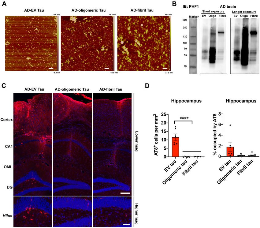
290   | BRAIN 2021: 144; 288–309                                                                                        Z. Ruan et al.

disease (pAD), and age/sex-matched control subjects for             for 70 min at 4 C using SW41Ti (Optima-XE SW41 Beckman
their biophysical, biochemical, and neurobiological proper-         Coulter). The pellet was resuspended in 2 ml of 0.475 M su-
ties as well as for tau pathology after their stereotaxic injec-    crose in double-filtered phosphate-buffered saline (dfPBS) with a
tion into the outer molecular layer (OML) of the dentate            0.22-lm pore-size filter and overlaid on five sucrose cushions (2
gyrus in aged C57BL/6 (B6) mice. The recipient mice were            ml each of 2.0 M, 1.5 M, 1 M, 0.825 M, and 0.65 M in
                                                                    dfPBS), then ultracentrifuged at 200 000g for 20 h at 4 C
tested by immunohistochemical and biochemical methods
                                                                    (Optima-XE SW41 Beckman Coulter). The gradient was col-
for characterization of tau accumulation in the hippocam-
                                                                    lected in 2 ml fractions, where fractions V and VI were enriched
pus. We also assessed the difference in tau pathology devel-        in EVs, except for the first and last fractions, which were 1 ml
opment after intrahippocampal injections of isolated EVs,           each. EV fractions V and VI were diluted to 12 ml in dfPBS and
tau oligomers, and tau fibrils in mice. Finally, hippocampal        ultracentrifuged at 100 000g for 70 min at 4 C using SW41Ti
pyramidal neurons of the recipient mice were assessed by            to pellet EVs, which were finally resuspended in 30 ll dfPBS.
whole-cell patch clamp recordings to determine whether tau          The bicinchoninic acid (BCA) assay (Pierce) was used to deter-
accumulation induces neurophysiological alterations.                mine the protein concentration for each sample.

                                                                                                                                         Downloaded from https://academic.oup.com/brain/article/144/1/288/6007752 by guest on 23 April 2021
                                                                    Nanoparticle tracking analysis
Materials and methods                                               The EVs in the enriched fractions were quantified as previously
                                                                    described (Muraoka et al., 2019, 2020b). Briefly, all samples
Animals                                                             were diluted in dfPBS by 1:1000 or greater to be read by the
                                                                    Nanosight 300 (Malvern Panalytical Inc), which can read 10–
Aged C57BL/6 (18–19 months old), tau knockout (KO)
                                                                    100 particles per frame. Using a syringe pump infusion system
(B6.129X1-Mapttm1Hnd/J, # 007251), and pregnant CD-1 mice
                                                                    (Harvard Laboratories/Malvern), five 30-s videos of each sam-
were purchased from National Institute of Aging (NIA), Jackson
                                                                    ple were taken at 21 C. Analysis of particle counts was carried
laboratory, and Charles River Laboratory, respectively. B6 mice
                                                                    out with Nanosight NTA 3.3 software (Malvern Panalytical
were housed as groups in regular light/dark cycles with free ac-
                                                                    Inc), using a detection threshold of five. Particle counts were
cess to food and water, and welfare-related assessments were
                                                                    normalized by the dilution factors for particle reading, the final
carried out before and after the surgery. Animals were randomly
                                                                    pellet, and starting material for exosome extraction. Finally, the
allocated to experimental groups and variability was assessed
                                                                    counts were averaged for fractions V and VI.
based on the body weight. Animals were used for intracerebral
inoculation of human brain-derived materials. Adult tau-KO
mice were used for isolation of brain-derived EVs. E16 (embry-      Atomic force microscopy
onic Day 16) CD-1 mice were used for primary culture of cor-
                                                                    Ten micrograms of EVs (1 lg/ll) were incubated with 100 ll
tical neurons. All animal procedures followed the guidelines of
                                                                    0.5% sarkosyl (Sigma-Aldrich) for 30 min on ice in ultracentri-
the National Institutes of Health Guide for the Care and Use of
                                                                    fuge-compatible Beckman microcentrifuge tubes for solubiliza-
Laboratory Animals and were approved by the Boston
                                                                    tion of vesicles, followed by dilution in dfPBS to 1.2 ml. The
University Institutional Animal Care and Use Committee
                                                                    sample was ultracentrifuged at 100 000g for 70 min at 4 C. The
(IACUC).
                                                                    supernatant was removed until 50 ll sample remained, to which
                                                                    dfPBS was added to a volume of 1.2 ml for the second ultracen-
Isolation of extracellular vesicles                                 trifugation at 100 000g for 70 min at 4 C. The pellet was disso-
from Alzheimer’s disease brains                                     ciated in 10 ll dfPBS and imaged via atomic force microscopy
                                                                    using the Multimode 8 AFM machine (Bruker) under ScanAsyst
Human and mouse brain-derived EVs were isolated according           mode, as previously described (Sengupta et al., 2018).
to our recently published methods (Muraoka et al., 2020b),
which were adapted from a previous publication (Perez-
Gonzalez et al., 2012). Briefly, fresh frozen human frontal cor-
                                                                    Tau purification from extracellular
tex was sliced with a razor blade on ice while frozen to generate   vesicles with proteinase K
2–3 mm3 sections. The cut sections were dissociated
(LK003178, Worthington Biochemical Corporation) in 3 ml
                                                                    treatment
HibernateTM-E medium (Thermo Fisher Scientific) containing          EVs (90 mg) were diluted to a concentration of 860 lg/ml pro-
20 units of papain at 37 C for 15 min. After incubation, the       tein and incubated with 20 lg/ml proteinase K (PK, QIAGEN)
samples were immediately added with 6 ml of ice-cold                and 5 mM CaCl2 in PBS for 1 h at 37 C with gentle vortexing
HibernateTM-E solution (A1247601 Gibco) and filtered using a        every 15 min. The PK activity was then inhibited by adding
40 lm mesh filter (Fisher scientific). The tissue sample was se-    5 mM phenylmethylsulphonyl fluoride (PMSF) for 10 min at
quentially centrifuged at 300g for 10 min at 4 C. The super-       room temperature. A quarter volume of each sample was col-
natant was transferred to a new 15 ml tube and then                 lected for western blot analysis with CD63 and actin antibodies.
centrifuged at 2000g for 10 min at 4 C (Eppendorf). This super-    The rest was lysed with TENT buffer (50 mM Tris HCl pH 7.5,
natant was then transferred to a 30-ml conical tube and then        2 mM EDTA, 150 mM NaCl, 1% TritonTM X-100) (1:1) for
centrifuged at 10 000g for 10 min at 4 C (Avanti J-E JA25-50       30 min on ice, followed by centrifugation at 48 300g for 20 min
Beckman Coulter). The supernatant was filtered through a 0.22       at 4 C. The supernatant and pellet were designated as S1 and
lm pore-size filter (Millipore) and ultracentrifuged at 100 000g    P1 fractions, respectively. The S1 fraction was ultracentrifuged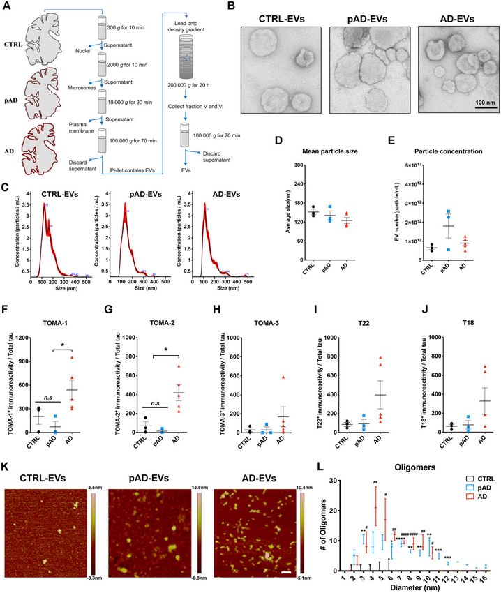
Extracellular vesicles mediate tau spread                                                            BRAIN 2021: 144; 288–309     | 291

at 186 000g at 4 C for 40 min to collect the pellet fraction          ELISA of brain tissue extraction
(S1p) as the tau oligomer-enriched fraction. The P1 fraction was
resuspended in 1 ml of buffer (1% sarkosyl, 10 mM Tris, pH
                                                                       and extracellular vesicle samples
7.4, 800 mM NaCl, 10% sucrose, 1 mM EGTA, 1 mM PMSF),                  Brain tissue homogenate and EV samples were diluted 1:10 in
and incubated by rotating with the benchtop thermomixer at             8 M guanidine buffer for solubilization for 3 h at room tempera-
room temperature for 1 h. The sample was ultracentrifuged at           ture with agitation, followed by dilution in TENT buffer supple-
186 000g for 1 h at 4 C. After completely removing the super-         mented with protease and phosphatase inhibitors (Pierce HALT
natant and rinsing the pellet in sterile PBS, the sarkosyl-insoluble   inhibitor), and subjected to human total tau ELISA (Thermo
pellet (P2), as the tau fibril-enriched fraction, was removed.         Fisher Scientific) according to the manufacturer’s instructions.

                                                                       Extracellular vesicle labelling with
Transmission electron and                                              PKH26
immunoelectron microscopy                                              EVs were labelled with lipophilic red fluorescent dye (PKH26,

                                                                                                                                             Downloaded from https://academic.oup.com/brain/article/144/1/288/6007752 by guest on 23 April 2021
                                                                       Sigma-Aldrich), according to the manufacturer’s protocol.
Transmission electron microscopy (TEM) of EVs and tau con-             Briefly, 0.32 ll PKH26 was mixed with 5 lg EV samples in
tents purified from human brain-derived EV samples was con-            40 ll diluent C, and incubated for 5 min at room temperature.
ducted as previously described (Asai et al., 2015; Muraoka             dfPBS was used as a negative control. The labelling reaction
et al., 2020b). Briefly, 5 ll of the EV sample was adsorbed for        was stopped by adding 50 ll chilled dfPBS, and subjected to
1 min on a carbon-coated grid (Electron Microscopy Sciences)           Exosome Spin Columns (MW 3000, ThermoFisher) at 750g for
that had been made hydrophilic by a 20 s exposure to a glow            2 min to remove the free dye and enrich the labelled EVs, which
discharge (25 mA). Excess liquid was removed with filter paper         were adjusted to 5 lg/100 ll for the neuronal EV uptake assay.
(#1 Whatman), then the grid was floated briefly on a drop of
water (to wash away phosphate or salt), blotted on filter paper,
and then stained with 0.75% uranyl formate (EMS) for 15 s.
                                                                       Primary tissue culture of murine
After removing the excess uranyl formate with a filter paper, the      cortical neurons
grids were examined in a JEOL 1200EX transmission electron             Primary murine cortical neurons were isolated from E16 em-
microscope and images were recorded with an AMT 2k CCD                 bryos from pregnant CD-1 mice (Charles River Laboratory).
camera. For immunogold labelling on tau fibrils, samples were          Dissociated cortical tissues were digested with trypsin-EDTA
adsorbed to the grid for 5 min, blocked on 1% bovine serum al-         (diluted to 0.125%, Invitrogen), triturated with polished pip-
bumin (BSA) for 10 min, transferred to 5 ll drops of primary           ettes, strained into single neurons using a 40-lm pore-size
antibody (PHF1, mouse mAb, 1:10; kindly provided by Dr P.              Falcon cell strainer (Thermo Fisher Scientific), and finally plated
Davies) and incubated for 30 min, then washed in four drops of         onto sterilized 12-mm high precision thickness coverslips
PBS (total 10 min) before incubation in rabbit anti-mouse bridg-       (Bioscience Tools) at 375 000 cells per coverslip in 24-well
ing antibody (1:50, #ab6709, Abcam) for 30 min followed by             plates, as previously described (You et al., 2020). Coverslips
5-nm protein A–gold (University Medical Center, Utrecht, The           were precoated with 100 lg/ml poly-D-lysine (Sigma-Aldrich)
Netherlands) for 20 min. Grids were washed in two drops of             diluted in borate buffer (0.05 M boric acid, pH 8.5) and washed
PBS followed by four drops of water (total 15 min) and subse-          with sterile water prior to plating. Neurons at day in vitro 7
quent staining in 0.75% uranyl formate and imaged as                   were treated with PKH26-labelled EVs for neuronal uptake or
described above. For immunogold labelling on EVs, pelleted             tau transfer study.
exosomes were fixed with 4% paraformaldehyde in 0.1 M
phosphate buffer (pH 7.4) and processed for ultrathin cryosec-         Tau seeding assay
tioning. Pellets were infiltrated with 2.3 M sucrose in PBS for
                                                                       HEK-TauRD P301S Förster resonance energy transfer (FRET)
15 min, frozen in liquid nitrogen and sectioned at –120 C.
                                                                       biosensor cells (ATCC) were plated on a poly-D-lysine-coated
Sections 60–80-nm thick were picked up and transferred to for-
                                                                       96-well plate (# 354461, Corning) in growth media (Dulbecco’s
mvar carbon-coated copper grids, and immunogold labelling
                                                                       modified Eagle medium, 10% foetal bovine serum, 1 penicil-
was carried out at room temperature on a piece of Parafilm.
                                                                       lin/streptomycin, all from Invitrogen). The next day, human
The antibody was diluted in 1% BSA in PBS. Grids were floated
                                                                       brain-derived EVs were mixed with 80 ll Opti-MEM and 20 ll
on drops of 1% BSA for 10 min to block non-specific labelling,         LipofectamineTM 2000, and incubated at room temperature for
transferred to 5-ll drops of primary antibodies (PHF1, mouse           10 min. Subsequently, growth media was removed from the
mAb, 1:10) and incubated for 30 min, then washed in four               cells, replaced with samples containing Lipofectamine, and incu-
drops of PBS (total 10 min) before incubation in rabbit anti-          bated at 37 C, 5% CO2. After 1 h, Lipofectamine-containing
mouse bridging antibody (1:50, Abcam ab6709) for 30 min fol-           media was removed from the cells and replaced with growth
lowed by 5 nm protein A–gold for 20 min. Grids were washed             media. Cells were maintained in culture at 37 C, 5% CO2 for
in two drops of PBS followed by four drops of water (total 15          72 h afterward. The day of the analysis, cells were washed in
min) and contrasted in a mixture of 0.3% uranyl acetate in 2%          PBS, detached with 0.25% Trypsin-EDTA (Invitrogen), and
methylcellulose for 5 min. Excess liquid was blotted off with fil-     washed with the fluorescence-activated cell sorting (FACS) buf-
ter paper and the grids were examined at 80 kV with a JEOL             fer (PBS + 0.5% BSA). Subsequently, cells were fixed in 2%
1200EX transmission electron microscope and images were                paraformaldehyde, 2% sucrose solution in PBS for 15 min at
recorded with an AMT 2k CCD camera.                                    4 C, spun at 13 500g for 15 min at 4 C, then resuspended in
292   | BRAIN 2021: 144; 288–309                                                                                        Z. Ruan et al.

FACS buffer. FRET assays were performed with LSRII flow            Confocal image processing and
cytometer (BD Bioscience) using pacific-orange and pacific-blue
dye filter settings for yellow and cyan fluorescent protein, re-
                                                                   quantification by Imaris
spectively. Data were analysed using FlowJo and quantified as      All confocal imaging was performed on a LSM710 using Zen
integrated FRET density.                                           2010 software (Zeiss) or a Leica TCS SP8 lightning microscope
                                                                   on an inverted Leica DMi8 microscope stand, using the confocal
                                                                   mode for 63 oil immersion/1.4N.A objective, and 1.1 optical
                                                                   zoom at a pinhole of 1.0 airy unit. Confocal stacks of images
Stereotaxic surgery                                                with 2048  2048 pixel resolution were collected while using a
C57BL/6 mice at 18–19 months old were deeply anaesthetized         system-optimized z-interval of 0.28 lm. GAD67 + puncta were
with isoflurane and immobilized in a mouse stereotaxic frame       imaged with a 552 nm laser, and emission was collected at
(David Kopf Instruments) installed with robotic stereotaxic in-    565–650nm; for imaging c-fos, a 488 nm laser line was used
                                                                   and emission was collected at 490–600 nm. All co-localization
jection system (Drill and Injection Robot, Neurostar). Animals
                                                                   images were scanned frame-by-frame in the sequential scanning
were unilaterally inoculated with human or tau-KO mouse

                                                                                                                                         Downloaded from https://academic.oup.com/brain/article/144/1/288/6007752 by guest on 23 April 2021
                                                                   mode, which showed no cross-talk among multiple channels.
brain-derived EVs, or tau aggregates purified from Alzheimer’s
                                                                   Gain and offset were set at values which prevented saturated
disease human brain in the dorsal hippocampal OML (bregma:
                                                                   and empty pixels. All images were processed via adaptive
–2.18 mm; lateral: 1.13 mm; depth: –1.9 mm from the skull)
                                                                   LIGHTNING deconvolution (Leica) after image acquisition.
using a 10-ll Hamilton syringe. Each injection site received
                                                                   The quantification of GAD67 + puncta was counted using the
1.0 ll of inoculum, containing 300 pg tau/ll for EV samples, sa-
                                                                   ‘Spot’ module of Imaris 9.5, 64-bit version (Bitplane AG). CA1
line, 300 pg or 2 lg of tau per microlitre oligomers or fibrils,   pyramidal cells were manually cut with GAD67 field in 3D.
and euthanized 4.5 months after the injection. We noted that       This program analyses stacks of confocal sections acquired in
the majority of injected materials were deposited at the OML of    two channels (red for GAD67, blue for DAPI represents the nu-
the hippocampus (Fig. 4A).                                         clei). Final data analysis was performed with Excel (Microsoft)
                                                                   and Prism 8 (GraphPad).

Immunochemistry and                                                Biochemical sequential extraction
immunofluorescence                                                 from mouse brains
Brains were removed after transcardial perfusion fixation with     Brain tissues were removed from control EV, pAD EV and
ice-cold 4% paraformaldehyde/PBS followed by post-fixation         Alzheimer’s disease EV-injected mice at the designated time
for 16 h and cryoprotection with 15%, then 30% sucrose/PBS         points after transcardial perfusion of animals by ice-cold PBS to
over 3–5 days, and embedded in O.C.T. compound (Thermo             minimize contamination of blood-derived mouse immunoglobu-
Fisher). They were cryosectioned coronally in 20-mm thickness      lins. Hippocampal and cortical regions were dissected separate-
using a cryostat, and three hippocampal sections separated by      ly, snap-frozen in dry ice, and stored at –80 C before protein
at least 200 mm per mouse were used for immunohistochemistry       extraction. For enrichment of tau oligomers and fibrils, sequen-
per primary antibody. The sections were processed by antigen       tial extractions were performed as follows: each hippocampal
retrieval with Tris-EDTA (pH 8.0) at 80 C, permeabilized in       tissue was homogenized in nine volumes of TBS buffer (50 mM
0.5% TritonTM X-100/PBS, and blocked in 10% normal goat            Tris-Cl, pH 8.0 in saline) and supplemented with protease and
serum, 1% BSA, and 0.1% Tween-20 in PBS, as previously             phosphatase inhibitor cocktails (Thermo Fisher Scientific). The
described (Asai et al., 2015). Sections were incubated with pri-   homogenate was centrifuged at 48 300g for 20 min at 4 C. The
mary antibodies against glutamic acid decarboxylase 67             supernatant and pellet were designated as S1 (TBS-supernatant)
(GAD67, PA5-36054, ThermoFisher Scientific), GAD67-biotin-         and P1 (TBS-pellet) fractions, respectively. The S1 fraction was
                                                                   ultracentrifuged at 186 000g at 4 C for 40 min. The pellet frac-
conjugated (MAB5406B, Millipore), parvalbumin (PV,
                                                                   tion (S1p) was resuspended in 10 ml PBS and frozen at –80 C as
ab11427, Abcam), neurogranin (Ab5620, Millipore Sigma),
                                                                   the tau oligomer-enriched fraction. The P1 fraction was resus-
pSer202/pSer205 tau (AT8, MN1020, ThermoFisher Scientific),
                                                                   pended in five volumes of wet weight of the original tissue of
GluR2/3 (AB1506, Millipore Sigma), MAP-2 (mab3418,
                                                                   buffer B (1% sarkosyl, 10 mM Tris, pH 7.4, 800 mM NaCl,
Millipore Sigma), c-fos (226003, Synaptic Systems), pSer422 tau
                                                                   10% sucrose, 1 mM EGTA, 1 mM PMSF, all from Sigma-
(PS422, 44-764G, ThermoFisher Scientific), along with mis-
                                                                   Aldrich), and incubated by rotating with the benchtop thermo-
folded tau (Alz50 and MC1), pSer202 tau (CP13) and pSer396/        mixer at room temperature for 1 h. The sample was ultracentri-
pSer404 tau (PHF-1, all kind gifts provided by Dr Peter Davies),   fuged at 186 000g for 1 h at 4 C. After completely removing
diluted with 1% BSA, 0.025% Tween-20 in PBS at 4 C over-          the supernatant and rinsing the pellet in sterile PBS, sarkosyl-in-
night (see Supplementary Table 1 for antibody dilution informa-    soluble pellet (P2) was resuspended with 10 ll PBS, and frozen
tion). Sections were then washed and incubated in secondary        at –80 C as tau fibril-enriched fraction.
antibodies (Alexa Fluor 647 goat anti-mouse; 1:1000, Alexa
Fluor 488 goat anti-rabbit; 1:1000, Alexa Fluor 568 streptavi-
din 1:1000) for 1 h at room temperature. All images were cap-
                                                                   Western and dot blotting
tured on Nikon deconvolution wide-field epifluorescence system     For EV samples, after being lysed with TENT buffer, the equiva-
(Nikon Instruments) or confocal microscopic imaging, as            lent amount of protein was loaded on 4–20% SDS-PAGE gels
described below.                                                   (Bio-Rad). For mouse brain tissue samples, homogenates of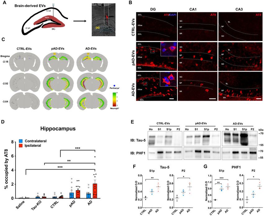
Extracellular vesicles mediate tau spread                                                                   BRAIN 2021: 144; 288–309        | 293

                                                                                                                                                     Downloaded from https://academic.oup.com/brain/article/144/1/288/6007752 by guest on 23 April 2021

 Figure 1 Characterization of EVs by TEM, nanoparticle tracking analysis, tau oligomer dot-blotting and atomic force micros-
 copy. (A) A schema of EV separation from human frozen brain tissue. (B) TEM image of human brain-derived EVs. (C–E) Nanoparticle tracking
 analysis of isolated EVs (C), quantification of EV size (D) and EV density (E). (F–J) Semi-quantification of tau oligomers in EVs by multiple tau
 oligomer antibodies. Dot blot images are provided in Supplementary Fig. 1A. *P 5 0.05, as determined by one-way ANOVA (alpha = 0.05) and
 Tukey’s post hoc. Graphs indicate mean ± SEM. Each dot represents an individual donor, three replicates per subject, three donors per group for
 control (CTRL) and pAD, five donors for the Alzheimer’s disease (AD) group (Supplementary Table 2). (K and L) Atomic force microscopy
 images showing brain-derived EV-tau oligomers isolated from CTRL, pAD, and Alzheimer’s disease brains (K). Scale bars = 200 nm. Size distribu-
 tion histogram of EV-tau oligomers (L). *P 5 0.05, **P 5 0.01, ***P 5 0.005 and ****P 5 0.0001 for pAD EVs versus CTRL EVs; #P 5 0.05,
 ##
    P 5 0.01, and ####P 5 0.0001 for Alzheimer’s disease EVs versus CTRL EVs as determined by one-way ANOVA (alpha = 0.05) and Tukey’s post
 hoc. Graphs indicate mean ± SEM. n = 3 images per sample. AD = Alzheimer’s disease; CTRL = control.
294   | BRAIN 2021: 144; 288–309                                                                                        Z. Ruan et al.

hippocampus from each experimental group and an equal pro-          12.5 kHz sampling frequency) to assess repetitive AP firing.
portion of corresponding homogenates, S1, S1p and P2, were          Those neurons that did not fire repetitively during the depolariz-
loaded on 10% SDS-PAGE gels (Bio-Rad) and electrotrans-             ing step were discarded. Firing rates in response to current steps
ferred to 0.45 lm nitrocellulose membranes (Bio-Rad). For dot       were determined by fitting to a generalized linear model, using
blotting, an equal volume of EV samples were dotted onto 0.45       the genotype, CA1 pyramidal cells types, rheobase, input resist-
lm nitrocellulose membranes (Bio-Rad) and washed twice with         ance, injected current level, and their respective interactions as
TBS buffer. The membranes were then blocked in freshly pre-         independent variables. Whole-cell voltage clamp was used to
pared 5% BSA diluted in TBS before being immunoblotted with         measure AMPA receptor-mediated spontaneous excitatory cur-
specific primary antibodies (Supplementary Table 1). The mem-       rents (sEPSCs) response for 2 min at a holding potential of –80
brane was further incubated with HRP-labelled secondary anti-       mV (6.67 kHz sampling frequency). The same neuron was held
bodies (Santa Cruz Biotech) and scanned using the C300 digital      at –40 mV (6.67 kHz sampling frequency) for 2 min to obtain
chemiluminescent imager (Azure Biosystems). The band den-           enough sample size to measure GABA receptor-mediated spon-
sities were digitally measured using ImageJ (NIH).                  taneous inhibitory currents (sIPSCs). All recorded traces were
                                                                    run through Mini Analysis Program (Synaptosoft), which

                                                                                                                                         Downloaded from https://academic.oup.com/brain/article/144/1/288/6007752 by guest on 23 April 2021
Whole-cell patch clamp recording                                    allowed for quantification of synaptic current properties such as
                                                                    frequency, amplitude, area, time to rise, and time to decay. To
Preparation of brain slices for recording and filling               determine the kinetics of EPSCs and IPSCs, the rise and decay
Immediately after decapitation, mouse brains were rapidly           of averaged traces were each fit to a single-exponential function.
removed and placed in oxygenated (95% O2 and 5% CO2) ice-           In all of the synaptic current measurements, the event detection
cold Ringer’s solution containing the following ingredients (in     threshold was set to the maximum root mean squared noise
mM): 25 NaHCO3, 124 NaCl, 1 KCl, 2 KH2PO4, 10 glucose,              level (5 pA). All neurons had resting membrane potentials be-
2.5 CaCl2, 1.3 MgCl2 (pH 7.4; Sigma-Aldrich). A total of four       tween –55 and –75 mV (somatic recordings) and were con-
to five 300-mm thick acute coronal sections containing the          firmed to have intact somas and apical tufts by fluorescence
hippocampus were obtained from each subject. Over an 8–10-h         microscopic imaging of biocytin-filled cells.
period, slices were individually transferred from the incubation
chamber to submersion-type recording chambers (Harvard
Apparatus) affixed to the stages of Nikon E600 infrared-differ-
                                                                    Statistical analyses
ential interference contrast (IR-DIC) microscopes (Micro Video      All data are presented as means ± standard error of the mean
Instruments) with a water-immersion lens (40, 0.9 NA;              (SEM). Comparisons between two groups were done by two-
Olympus) for recording. During recordings, slices were super-       tailed paired or unpaired Student’s t-tests. Multiple comparisons
fused in room-temperature Ringer’s solution bubbled with carb-      were performed by either one- or two-way ANOVA, followed
ogen (95% O2, 5% CO2) at a rate of 2.5 ml/min. Whole-cell           by Tukey’s or Bonferroni’s post hoc test. Statistical analyses
patch clamp recordings were obtained from the soma of visually      were performed using Prism 8.0 (GraphPad). A statistically sig-
identified CA1 pyramidal cells in both the dorsal and ventral       nificant difference was assumed at P 5 0.05.
hippocampus of the ipsilateral side of the brain. Electrodes were
created from borosilicate glass with a Flaming/Brown micropip-      Data availability
ette puller (Model P-87, Sutter Instruments). These pulled patch
pipettes were filled with potassium methanesulphonate (KMS)-        The authors confirm that the data supporting the findings of
based intracellular solution, with concentrations in mM as fol-     this study are available within the article and its Supplementary
lows: (KCH3SO3 122, MgCl2 2, EGTA 5, Na-HEPES 10,                   material.
Na2ATP 5). Each had a resistance of 5.5–6.5 MX in external
Ringer’s solution.

Physiological inclusion criteria
                                                                    Results
Single action potential (AP) properties [including threshold,
amplitude, action potential half-width (APHW), rise and fall]
                                                                    Detection of tau oligomers in
were measured on the second evoked AP in a 200 ms current-          Alzheimer’s disease brain-derived
clamp series that preferentially evoked three or more APs after
depolarizing step-current. We proceeded to high input resistance
                                                                    extracellular vesicles
or low input resistance only if neurons were unable to elicit AP    While tau has been found in both exosomes and microvesicles
at 200 ms. APHW was computed at half-max of AP amplitude,           in tauopathy mouse brains, neuroblastoma cells, and the CSF
where the amplitude was measured from the threshold to the          and plasma in Alzheimer’s disease patients (Arai et al., 1998;
absolute peak of the spike. All quantification of AP properties     Saman et al., 2012; Dujardin et al., 2014; Fiandaca et al.,
was carried out in an expanded timescale, using the linear meas-
                                                                    2015), there has been no study to date which reports a
ure tool from FitMaster analysis software (HEKA Elektronik).
                                                                    detailed analysis of control, pAD, and Alzheimer’s disease
An algorithm designed in MATLAB was used to automatically
detect these parameters. In the few cases where it failed to do     brain-derived EVs. To this end, we isolated exosome-enriched
so, a manual detection method was used. The final paradigm in       EV fractions from Alzheimer’s disease, pAD, and control sub-
the current-clamp configuration was to inject 2 s hyperpolariz-     ject brain samples (three cases per group, see Supplementary
ing and depolarizing steps (–100 to + 120 pA with increments        Table 2 for demographics) and tau-KO mouse brain tissues
of 20 pA or –220 pA to + 330 pA with increments of 50 pA,           (Supplementary Table 2) using a combination of high-speed
Extracellular vesicles mediate tau spread BRAIN 2021: 144; 288–309 | 295
centrifugation, filtration, discontinuous sucrose gradient ultra- addition, atomic force microscopy analysis showed that the
centrifugation, and additional ultracentrifugation, as recently detergent-insoluble fraction of Alzheimer’s disease and pAD,
published (Muraoka et al., 2020a, b) (Fig. 1A). Analysis of but not of control EVs, contained significantly more globular
the isolated fractions by TEM (Fig. 1B) and nanoparticle particles at a mode height of 4–6 nm, consistent with tau
tracking analysis (NTA, Fig. 1C) demonstrated enrichment of oligomers (Fig. 1K and L). Taken together, these data suggest
brain EVs in the exosome size range (Budnik et al., 2016). that Alzheimer’s disease and pAD EVs are enriched in tau
There was no difference in terms of EV particle concentration oligomers compared to control subjects, suggesting their EV
or size among groups (Fig. 1D and E). We detected total tau tau seeding potency and pathogenic activities.
and amyloid-b42 but not amyloid-b40 in EVs by ELISA.
Amyloid-b42 was significantly enriched in Alzheimer’s disease
EVs compared to pAD or control EVs (Supplementary Table Detection of sarkosyl-insoluble tau
3). Considering that the tau oligomer is roughly a nanometer aggregates within Alzheimer’s
in size (Combs et al., 2017), we postulated that brain-derived
disease extracellular vesicles
Downloaded from https://academic.oup.com/brain/article/144/1/288/6007752 by guest on 23 April 2021
EVs contain tau in oligomeric form. Indeed, there was a sig-
nificantly higher amount of oligomeric tau in EVs derived To determine whether the tau is inside or associated with
from Alzheimer’s disease compared to controls as determined EVs, we first treated EV samples with PK to digest
by dot blotting of tau oligomer-specific monoclonal antibod- EV-associated proteins, which has been used in EV-related
ies TOMA-1 and TOMA-2, but not by TOMA-3 (Fig. 1F–H studies. PK can digest proteins on the outer leaflet of EVs,
and Supplementary Fig. 1). There was no difference in such as CD9 and CD63, but does not digest the luminal pro-
immunoreactivity to tau oligomer polyclonal antibodies T22 teins, such as Tsg101 and actin (Cvjetkovic et al., 2016). In
and T18 among three patient groups (Fig. 1I and J). In our study, we also determined which form of tau in EVs is
Figure 2 PK treatment of human brain derived EVs for biochemical characterization. (A) Workflow of the tau purification by se-
quential centrifugation after with or without PK treatment. (B) Western blot analysis of non-treated and PK-treated EVs from three groups
(CTRL, pAD and Alzheimer’s disease) with CD63 and actin antibodies. (C) Immunoelectron microscopy images of ultrathin-sectioned
Alzheimer’s disease EVs for PHF1+ tau with or without PK-treatment. Images were captured at direct magnification 30 000, with the 10 nm
immunogold labelling. (D) Western blot analysis of oligomer-enriched (S1p) and sarkosyl-insoluble tau-enriched (P2) fractions from EVs for PHF1
with or without PK-treatment. (E) Semi-quantification of PHF1 immunoreactivity. Two donors per group for CTRL and pAD and four donors for
the Alzheimer’s disease group. (F) Immunoelectron microscopy of isolated tau fibrils, oligomers or sarkosyl-insoluble fraction of EVs from human
Alzheimer’s disease brain tissue. Images were captured by TEM at direct magnification 40 000, with the 5-nm immunogold labelling for PHF1.
(A–F) Donors 1 and 2 (control, CTRL), 4 and 5 (pAD), and 7–10 (Alzheimer’s disease, AD) were used (Supplementary Table 2).296 | BRAIN 2021: 144; 288–309 Z. Ruan et al.
enriched by sarkosyl solubilization and sequential centrifuga- excitatory amino acid transporter 1 (EAAT1), glial fibrillar
tion of lysed EVs to separate the oligomer-enriched fraction acidic protein (GFAP), and aquaporin-4 (AQP4). We
(S1p) and sarkosyl-insoluble fraction (P2) using control, observed a trend of enrichment of EAAT1, but reduction of
pAD, and Alzheimer’s disease EVs containing the same GFAP in pAD or Alzheimer’s disease EVs compared to con-
amount of protein per sample (Fig. 2A). PK treatment suc- trol EVs, whereas AQP4 showed no difference
cessfully reduced the amount of CD63 in all the EV samples (Supplementary Fig. 2A and C). For neuron-specific
(control, pAD, and Alzheimer’s disease) as a positive control, markers, synaptophysin (SYP) showed a significant decrease
but did not affect the amount of actin within EVs as a nega- in the Alzheimer’s disease EV samples compared to the
tive control (Fig. 2B). Immunoelectron microscopic analysis control EV samples, along with a similar trend in L1 cell ad-
shows that PK treatment did not significantly change detec- hesion molecule (L1CAM) band intensity (Supplementary
tion of EVs with PHF1 monoclonal antibody, which detects Fig. 2A and D), consistent with the proteomics data.
pS396/pS404 tau (Fig. 2C). Furthermore, PHF1 + tau is mostly The discrepancy observed in astrocytic markers could be
enriched in the P2 fraction in Alzheimer’s disease EVs, while due to the limitation of the quantification by label-free
Downloaded from https://academic.oup.com/brain/article/144/1/288/6007752 by guest on 23 April 2021
PHF1 + tau was also detected in the S1p fraction (Fig. 2D proteomic mass spectrometry, which depends on the quanti-
and E). PK treatment did not affect the P2 tau in tative comparisons using the label-free intensity-based abso-
Alzheimer’s disease EV samples while it partially reduced the lute quantification (iBAQ) method with all samples
S1p tau. PHF1 + tau was almost undetectable in control and normalized by total ion current for the run. Taken
pAD EV samples (Fig. 2D and E). together, these findings suggested that more glia- and less
To observe the structure of tau in the human brain-derived neuron-derived molecules are enriched in the Alzheimer’s
EV samples, we lysed Alzheimer’s disease and pAD EVs disease EVs.
with 2% sarkosyl buffer and ultracentrifuged to isolate the
sarkosyl-insoluble fraction of tau aggregates. As controls, Increased uptake of Alzheimer’s
tau oligomers and fibrils were isolated by sarkosyl solubil-
ization and sequential centrifugation/ultracentrifugation
disease extracellular vesicles by
from the same Alzheimer’s disease brain tissues. The isolated neurons and subsequent tau
samples were subjected to immunoelectron microscopy with transfer
PHF1 and immunogold. As shown in Fig. 2F, we detected a
Protein-protein interactions between EVs and cell surface
PHF1 + paired helical structure in isolated tau fibril and
molecules of recipient cells can facilitate the binding of EVs
globular structures of tau oligomers as the control.
and subsequent endocytosis (Mulcahy et al., 2014; van Niel
Interestingly, we detected mostly PHF1 + globular structures
et al., 2018). We hypothesized that Alzheimer’s disease-
in both Alzheimer’s disease and pAD EVs (Fig. 2F), likely
induced inflammatory brain conditions affect the efficiency
representing oligomer or protofibril tau. Taken together,
of EV uptake by altering molecular complexes of the EV’s
these data show that PHF1 + tau detected in Alzheimer’s dis-
surface. Therefore, we investigated the efficiency of the brain-
ease EVs are mostly inside the EVs in a sarkosyl-insoluble
derived EV uptake by primary cultured murine neurons
globular form, whereas the tau oligomer form can also be
in vitro. After 7 days of neuronal differentiation, the cells
associated with the outer-leaflet of EVs. PHF1 + tau was de-
were incubated with PKH26-labelled EVs isolated from the
tectable at a much lower level in the S1p and P2 fractions in
brain tissue of Alzheimer’s disease, pAD, and control samples
a PK-insensitive manner, while it was undetectable in either
over 24 h, as previously described (You et al., 2020)
fraction of the control EVs.
(Fig. 3A). Alzheimer’s disease EVs showed significantly
higher neuronal uptake compared to control EVs, but there
Detection of neuronal and glial was no significant change between pAD and control EV
molecules in brain-derived groups (Fig. 3B and C). The transfer efficiency of tau from
EV to neurons was significantly higher in Alzheimer’s disease
extracellular vesicles EVs compared to that of control EVs (Fig. 3D). We labelled
We have previously reported enrichment of glial molecules the supernatant with PKH26 as a negative control at the last
and reduction in neuronal molecules in Alzheimer’s disease ultracentrifugation wash step of the EV isolation and applied
EVs (Muraoka et al., 2020a). To validate these findings it to neuronal cells. There was no PKH26 positivity found in
from proteomics analysis, we performed western blot with supernatant applied neurons (data not shown). Finally, to
several cell type-specific proteins identified from the prote- understand if EV-tau has different tau seeding activity de-
omic profiling of the brain-derived EV samples (Muraoka pendent on the disease conditions, we used a FRET-based
et al., 2020a). Interestingly, we detected myelin oligodendro- tau seeding assay, as previously described (Holmes et al.,
cyte glycoprotein (MOG), a mature oligodendrocyte marker, 2014). Astonishingly, the Alzheimer’s disease EVs showed
as being significantly more enriched in the Alzheimer’s dis- significantly higher seeding activity compared to the pAD
ease EV samples than in pAD or control EV samples and control EV groups (Fig. 3E), suggesting higher potency
(Supplementary Fig. 2A and B), consistent with the proteo- of Alzheimer’s disease EVs to induce tau pathology. In sum-
mics data. For astrocyte-specific markers, we tested mary, the data demonstrate the pathogenic functions ofExtracellular vesicles mediate tau spread BRAIN 2021: 144; 288–309 | 297
Downloaded from https://academic.oup.com/brain/article/144/1/288/6007752 by guest on 23 April 2021
Figure 3 Neuronal uptake, tau transfer efficiency and tau seeding activities of human brain-derived EVs. (A) A diagram illustrat-
ing the primary culture model with primary neurons used to measure the transfer of EVs containing tau and a biosensor cell assay system for
seeding activity. (B) Cellular uptake of PKH26-labelled EVs (red) by primary culture murine cortical neurons (MAP-2, green; DAPI, blue). Original
magnification: 20 (left and middle columns); 40 (right column, taken by Zeiss LSM710 confocal microscopy). Scale bars = 40, 20, 10 mm (left
to right). (C) Quantification of PKH26 fluorescent intensity in neurons. **P 5 0.0001 and ****P 5 0.0001 compared with PBS or dye only group;
##
P 5 0.01 compared with the CTRL-EV group; determined by one-way ANOVA (alpha = 0.05) and Tukey’s post hoc. Each dot represents aver-
age data per image (10–20 cells per image), nine images per group (for PBS and dye only), 10 images per donor and three donors per group (for
control EV, pAD EV, and Alzheimer’s disease EVs), total n = 30 per group. (D) Total human tau ELISA of neuronal cell lysates. #P 5 0.05 compared
with pAD EV and ##P 5 0.01 compared with the control EV group; n.s denotes no significance as determined by one-way ANOVA (alpha = 0.05)
and Tukey’s post hoc. Three donors per group, three independent experiments. Graphs indicate mean ± SEM. (E) EVs were tested in the tau-
FRET assay for tau seeding activity. Results are plotted as integrated FRET density values for each sample. ###P 5 0.001 compared with control
EV and pAD EV groups; as determined by one-way ANOVA (alpha = 0.05) and Tukey’s post hoc. Three donors per group, and each dot represents
one well. Graphs indicate mean ± SEM. (B–E) Donors 1–3 (control), 4–6 (pAD), and 7–9 (Alzheimer’s disease) were used (Supplementary Table
2). AD = Alzheimer’s disease; BF = bright-field; CTRL = control; FRET = Förster resonance energy transfer; ICC = immunocytochemistry.298 | BRAIN 2021: 144; 288–309 Z. Ruan et al.
Alzheimer’s disease EVs with efficient neuronal uptake, tau monoclonal HT7 (data not shown), indicating that endogenous
transfer, and tau seeding potency. mouse tau was recruited and aggregated by the inoculation of
human brain-derived EV tau. A growing body of evidence sug-
Inoculation of Alzheimer’s disease gests that misfolded tau tends to be truncated and frequently
consists of different conformers or structural polymorphisms,
extracellular vesicles propagates tau dependent on the stages and disease of tauopathy (Fitzpatrick
pathology in aged mice et al., 2017; Falcon et al., 2018, 2019; Sigurdsson, 2018;
We further investigated whether brain-derived EVs can initiate Zhang et al., 2020). Therefore, we performed neuropathologic-
tauopathy in vivo using 2-month-old B6 mice. Brain-derived al analysis of tau by immunohistochemistry using conform-
EVs isolated from the brain tissue of Alzheimer’s disease, pAD, ation-specific (Alz50 and MC1) and p-tau epitope-specific
and control cases were unilaterally injected in the OML of the monoclonal antibodies (CP13 for pSer202 tau, PS422 for
dentate gyrus (Fig. 4A). The amount of injected tau (300 pg/ll, pSer422 tau, and PHF1 for pSer396/pSer404 tau). All five anti-
bodies detected misfolded or phosphorylated tau mainly in the
1 ll per injection) was much lower than that used in previous
Downloaded from https://academic.oup.com/brain/article/144/1/288/6007752 by guest on 23 April 2021
hilus of hippocampal region of Alzheimer’s disease and pAD
tau propagation studies (1–8 lg) (Guo et al., 2016;
EV groups (Supplementary Fig. 4B–F).
Narasimhan et al., 2017). The concentration was in the range
We next examined whether EV-tau could induce templated
of the extracellular tau concentration in mouse interstitial fluid
misfolding of original tau aggregates in the endogenous tau
of the CNS (Yamada et al., 2014). Immunofluorescence against
of the recipient mice. Aggregated tau was extracted from the
pSer202/pSer205 tau (AT8) yielded a considerable, yet not abun-
recipient mouse brains via sarkosyl solubilization and sequen-
dant amount of AT8 + cells in the hippocampal region of
tial centrifugation, and immunoblotted using Tau-5 and
Alzheimer’s disease and pAD EV-injected female mice
PHF1 monoclonal antibodies, as previously described
(Supplementary Fig. 3), while Alzheimer’s disease and pAD
(Fig. 4E) (Apicco et al., 2018; Jiang et al., 2019). We
EV-injected male mice show no AT8 positivity (data not
observed a significant increase in oligomeric tau in the S1p
shown). A previous study reported enhancement of tau propa-
fraction of both Alzheimer’s disease and pAD EV injected
gation induced by tau fibril injection in aged B6 mice in com-
mouse hippocampi in comparison to the control EV group,
parison to young mice (Guo et al., 2016). Therefore, we tested
determined by both Tau-5 (total tau) and PHF1 immunoblot-
aged female mice as recipients to determine whether tau path-
ting (Fig. 4F and G). The amount of sarkosyl-insoluble tau in
ology induced by brain-derived EVs reflects the donor’s disease
the P2 fraction was also significantly elevated in Alzheimer’s
conditions. Brain-derived EVs were isolated from three donors
disease EV injected mouse hippocampi when compared to
each of Alzheimer’s disease, pAD, and control cases, with tau-
the control EV group (Fig. 4F and G). These data indicate
KO mouse brain-derived EVs serving as a negative EV control.
that Alzheimer’s disease EV inoculation induced accumula-
Each EV sample (containing 300 pg tau/injectate for human
tion of oligomeric and fibrillar tau, while pAD EV inocula-
brain-derived EVs or saline control), was unilaterally injected
tion induced accumulation of oligomeric tau. Taken together,
into the OML of the dentate gyrus of 18–19-month-old B6 fe-
these data show the efficient induction of tau propagation in
male mice (Fig. 4A). The spread of tau pathology was evaluated
the hippocampus of the aged B6 female mouse brain after the
by immunofluorescence against AT8 in the hippocampal region
OML injection of Alzheimer’s disease EVs containing a
at 4.5 months post-injection (Fig. 4B and Supplementary Figs 3
physiological concentration of tau. Conformational changes
and 4A). Interestingly, abundant perikaryal AT8 + inclusions
of tau in the recipient mice appear to reflect the original tau
were detected in both ipsilateral and contralateral sides of the
conformation of Alzheimer’s disease EVs and pAD EVs,
hippocampal region including the CA1, CA3, dentate granule
which was also reported in mice injected with Alzheimer’s
cells, subgranular zone, and hilus, in the Alzheimer’s disease
disease brain-derived tau fibrils (Guo et al., 2016).
and pAD EV groups, suggesting that tau transfers between ana-
tomically connected pathways (Fig. 4B). Semiquantitative
brain-wide mapping of tau pathologies revealed that AT8 + Extracellular vesicles propagate
pathogenic tau was accumulated throughout the hippocampus, more tau than tau oligomers or
and was predominantly distributed in the caudal hilus region in fibrils when injected into mouse
the mouse brains injected with Alzheimer’s disease or pAD
EVs, while control EV injected mouse brains showed very little brains
AT8 positivity (Fig. 4C). Notably, the percentage of the area To determine how propagation of tau pathology may differ
occupied by AT8 + cells in the hippocampal region was signifi- between the injection of EV-associated or free form tau, we
cantly higher in Alzheimer’s disease EVs compared to the con- compared EV tau with oligomeric and fibrillar tau derived
trol EV, saline, or tau-KO EV groups (Fig. 4D). There was no from the same donor for tau pathology development.
significant difference between pAD and control EV injected Oligomeric and fibrillar tau was isolated from the same
groups, and no AT8 + staining was observed in the saline or Alzheimer’s disease EV donor as the S1p and P2 fractions
tau-KO injected groups (Fig. 4D and Supplementary Fig. 4A). according to the previous publications (Guo et al., 2016;
All AT8 + neurons were negative for human tau, as determined Apicco et al., 2018; Jiang et al., 2019). The p-tau immunor-
by immunofluorescent staining against human tau-specific eactivity and structure of the isolated tau aggregates wereExtracellular vesicles mediate tau spread BRAIN 2021: 144; 288–309 | 299
Downloaded from https://academic.oup.com/brain/article/144/1/288/6007752 by guest on 23 April 2021
Figure 4 Alzheimer’s disease EV but not control EV injection causes progressive tauopathy in aged B6 mouse brains. (A) A
schema illustrating 300 pg of tau containing EVs from human brain unilaterally injected to the hippocampus of B6 mice at 18–19 months of age.
DiI (red) indicated the injection site of the OML of the hippocampus. (B) Representative image of AT8 staining (red) 4.5 months after intrahippo-
campal injection of Alzheimer’s disease EV and pAD EV into aged B6 mouse brain. Original magnification: 20. Scale bar = 50 lm. (C) Semi-
quantitative analysis of Alzheimer’s disease-like tau pathologies based on AT8 immunostaining of brains from control, pAD, and Alzheimer’s dis-
ease (AD) EV-injected mice at 4.5 months post-injection. Blue dots represent AT8 + perikaryal inclusions. AT8 + density from green (0, low) to
red (3, high). (D) Quantification of AT8 + occupied area in the contralateral (blue) and ipsilateral (red) in entire hippocampal regions of recipient
mice. **P 5 0.01 and ***P 5 0.001 compared with the control EV group determined by one-way ANOVA (alpha = 0.05) and Tukey’s post hoc.
Total mice in each group for the quantification were 4, 6, 12, 12, and 11 for saline, tau-KO, control, pAD, and Alzheimer’s disease. Two donors
for EVs per group for control (Donors 1 and 2), pAD (Donors 4 and 5), and Alzheimer’s disease (Donors 7 and 9), (n = 5–6 mice per donor).
Bregma –1.34 to –3.64, four sections per mouse were analysed. Each dot represents mean value from one animal. Graphs indicate mean ± SEM.
(E) Immunoblotting of biochemically fractionated brain tissue samples for homogenate (Ho), TBS supernatant (S1), tau oligomer enriched (S1p)
and tau fibril enriched fractions (P2) by Tau-5 (total tau) and PHF1 (pSer396/pSer404 tau) (top) and their quantification (bottom). Equal propor-
tions of homogenate (Ho), S1, S1p, and P2 fractions were analysed (n = 3 mice/group). Optical density (OD) was normalized to that for the hom-
ogenate fraction from each corresponding mouse. *P 5 0.05, **P 5 0.01 and ***P 5 0.001 compared with the control group as determined by
one-way ANOVA (alpha = 0.05) and Tukey’s post hoc. Graphs indicate mean ± SEM. CTRL = control; DG = dentate gyrus; IB = immunoblot.
examined by western blot using a PHF1 antibody and atom- sonicated tau fibril preparations (Fig. 5A), which is consist-
ic force microscopy (Fig. 5A and B). The atomic force mi- ent with the description of the fibril structure as previously
croscopy images showed mostly small, oligomeric, globular reported (Ghag et al., 2018). We observed a mainly mono-
particles (6–8 nm in height) in EV and tau oligomer prepara- meric PHF1 + band in p-tau in EV and tau oligomer
tions, and large, globular structures (30–70 nm in height) in enriched samples, and a trimeric PHF1 + band in fibrilYou can also read