Asymptomatic Infection of Marburg Virus Reservoir Bats Is Explained by a Strategy of Immunoprotective Disease Tolerance
←
→
Page content transcription
If your browser does not render page correctly, please read the page content below
Article

Asymptomatic Infection of Marburg Virus Reservoir
Bats Is Explained by a Strategy of Immunoprotective
Disease Tolerance
Highlights                                                        Authors
         +
d   CD14 monocytes show Marburg virus replication after           Jonathan C. Guito, Joseph B. Prescott,
    infection of reservoir bats                                   Catherine E. Arnold, ...,
                                                                  Gustavo F. Palacios,
d   Infected bat monocytes and tissues show canonical antiviral   Mariano Sanchez-Lockhart,
    response gene induction                                       Jonathan S. Towner
d   Infected bat host response lacks significant induction of
    immunopathogenic genes
                                                                  Correspondence
                                                                  jit8@cdc.gov (J.S.T.),
d   Immunoprotective bat response contrasts starkly to that       mariano.sanchez-lockhart.ctr@mail.mil
    reported for sick primates                                    (M.S.-L.)

                                                                  In Brief
                                                                  A long-standing question for zoonotic
                                                                  diseases is how animal reservoirs such as
                                                                  bats immunologically control infection by
                                                                  pathogens that often kill spillover hosts
                                                                  such as humans. Guito et al. present clear
                                                                  evidence that Marburg virus-infected
                                                                  Egyptian rousette bats avert pathology by
                                                                  limiting induction of proinflammatory
                                                                  genes linked to severe disease.

             Guito et al., 2021, Current Biology 31, 257–270
             January 25, 2021 Published by Elsevier Inc.
             https://doi.org/10.1016/j.cub.2020.10.015                                            ll
ll
                                                                                                                       OPEN ACCESS

Article
Asymptomatic Infection of Marburg Virus
Reservoir Bats Is Explained by a Strategy of
Immunoprotective Disease Tolerance
Jonathan C. Guito,1 Joseph B. Prescott,2 Catherine E. Arnold,3 Brian R. Amman,1 Amy J. Schuh,1 Jessica R. Spengler,1
Tara K. Sealy,1 Jessica R. Harmon,1 JoAnn D. Coleman-McCray,1 Kirsten A. Kulcsar,4 Elyse R. Nagle,5 Raina Kumar,5
Gustavo F. Palacios,5 Mariano Sanchez-Lockhart,5,6,* and Jonathan S. Towner1,7,8,*
1ViralSpecial Pathogens Branch, Centers for Disease Control and Prevention, Atlanta, GA 30329, USA
2Center  for Biological Threats and Special Pathogens, Robert Koch Institute, Berlin, Germany
3Diagnostic Systems Division, U.S. Army Medical Research Institute of Infectious Diseases (USAMRIID), Fort Detrick, MD 21702, USA
4Department of Microbiology and Immunology, University of Maryland School of Medicine, Baltimore, MD 21201, USA
5Center for Genome Sciences, USAMRIID, Fort Detrick, MD 21702, USA
6Department of Pathology and Microbiology, University of Nebraska Medical Center, Omaha, NE 68198, USA
7Department of Pathology, College of Veterinary Medicine, University of Georgia, Athens, GA 30602, USA
8Lead Contact

*Correspondence: mariano.sanchez-lockhart.ctr@mail.mil (M.S.-L.), jit8@cdc.gov (J.S.T.)
https://doi.org/10.1016/j.cub.2020.10.015

SUMMARY

Marburg virus (MARV) is among the most virulent pathogens of primates, including humans. Contributors to
severe MARV disease include immune response suppression and inflammatory gene dysregulation (‘‘cytokine
storm’’), leading to systemic damage and often death. Conversely, MARV causes little to no clinical disease in
its reservoir host, the Egyptian rousette bat (ERB). Previous genomic and in vitro data suggest that a tolerant
ERB immune response may underlie MARV avirulence, but no significant examination of this response in vivo
yet exists. Here, using colony-bred ERBs inoculated with a bat isolate of MARV, we use species-specific an-
tibodies and an immune gene probe array (NanoString) to temporally characterize the transcriptional host
response at sites of MARV replication relevant to primate pathogenesis and immunity, including CD14+ mono-
cytes/macrophages, critical immune response mediators, primary MARV targets, and skin at the inoculation
site, where highest viral loads and initial engagement of antiviral defenses are expected. Our analysis shows
that ERBs upregulate canonical antiviral genes typical of mammalian systems, such as ISG15, IFIT1, and
OAS3, yet demonstrate a remarkable lack of significant induction of proinflammatory genes classically impli-
cated in primate filoviral pathogenesis, including CCL8, FAS, and IL6. Together, these findings offer the first
in vivo functional evidence for disease tolerance as an immunological mechanism by which the bat reservoir
asymptomatically hosts MARV. More broadly, these data highlight factors determining disparate outcomes
between reservoir and spillover hosts and defensive strategies likely utilized by bat hosts of other emerging
pathogens, knowledge that may guide development of effective antiviral therapies.

INTRODUCTION                                                           clearance within 14 days.3,5,6 In contrast, MARV loads in blood
                                                                       of infected NHPs are comparatively high, with death usually
Egyptian rousette bats (ERBs, Rousettus aegyptiacus), cave-            occurring within 6–9 days.4,7,8 Much remains unknown about
dwelling fruit bats common across sub-Saharan Africa and parts         how ERBs, along with bat reservoir species harboring other
of the Middle East, have been identified as the Marburg virus          zoonotic pathogens such as Nipah, Hendra, and SARS-related
(MARV) natural reservoir.1,2 MARV, a member of the family Filo-        coronaviruses, are able to control viral replication and mitigate
viridae that includes Ebola virus (EBOV), is the only human-path-      the severe disease these agents cause in primates.9–11 Eluci-
ogenic filovirus for which a natural reservoir has been identified.1   dating the immunological mechanisms likely facilitating this con-
Filoviruses often cause fatal illness in spillover hosts such as hu-   trol in these bats, including cell, pathway, and molecular factors
mans and non-human primates (NHPs), yet distinctive of a reser-        contributing to disease protection, which in turn could inform
voir species, MARV-infected ERBs remain asymptomatic.3–6               spillover dynamics and antiviral drug design, has increasingly
Experimentally infected ERBs develop low-level viremia that            earned recognition as a core component of global initiatives to
peaks between days 5–7 post-infection, and typically shed infec-       prevent and prepare for emerging zoonotic disease outbreaks.
tious virus in oral secretions for up to 3 weeks.3,5,6 Virus dissem-      To date, at least two prominent immunoprotective mecha-
inates widely, with highest levels found in liver and spleen, before   nisms in bat reservoirs have been posited. One mechanism,

                                                           Current Biology 31, 257–270, January 25, 2021 Published by Elsevier Inc. 257
                     This is an open access article under the CC BY-NC-ND license (http://creativecommons.org/licenses/by-nc-nd/4.0/).
ll
     OPEN ACCESS                                                                                                                Article

resistance, suggests that bats are better equipped to control            from tissues/CD14+ splenocytes. In this report, we demonstrate
viral replication through noncanonical means, such as quickly            in vivo that disease tolerance is a major strategy by which the
mounting innate and adaptive immune responses from a pre-ex-             ERB reservoir host controls MARV infection, as opposed to a
isting high basal state.12–14 This strategy has been postulated for      model of initial resistance conferred by more potent antiviral de-
large Pteropus bats that carry Nipah and Hendra viruses.12,13            fenses.14 Specifically, we show significant induction of a cluster
Another mechanism, disease tolerance, contends that bat hosts            of canonical antiviral genes (including DDX58 [RIG-I], IFIT1,
have co-evolved with their respective resident pathogens to              IRF7, ISG15, and OAS3) typical of viral infection in mammals,
tolerate infection, allowing viral replication only to levels suffi-     but strikingly, almost no significant changes in expression of tradi-
cient enough for transmission while concurrently mounting a              tional markers of adaptive immunity or inflammation, including the
subdued antiviral immune response that controls clinical disease         cytokines and chemokines IFNg, CCL8, FAS, and IL6, normally
(and tissue damage) by actively minimizing proinflammatory re-           associated with MARV pathogenesis in primates.8,25,27–31 Our
sponses.11,14–17 This concept of tolerance is supported for              findings identify putative virulence-determining differences be-
ERBs, as recent efforts to map the bat’s genome and transcrip-           tween reservoir and spillover host, as well as immunoprotective
tome has shown that this MARV reservoir (1) has diversified im-          commonalities likely shared between ERBs and bat reservoir
mune-inhibitory domain-containing natural killer (NK) receptor           hosts of other emerging zoonotic pathogens (e.g., SARS, Ebola),
loci and accompanying expansion of major histocompatibility              which could be exploitable for human therapeutic development.
(MHC) loci; (2) has expanded type I interferon (IFN) gene loci,
particularly of the IFNu family, that are less potently induced          RESULTS
in vitro, suggesting more subdued effector signaling; and (3) fails
to induce IFN genes even after ERB cell infection with a mutant          CD14+ Splenocytes from MARV-Infected ERBs Are
MARV that is incapable of antagonizing IFN-stimulated gene               Efficiently and Specifically MACS-Selected and Capable
(ISG) signaling.18–20 Further, infecting ERB bone marrow-derived         of Robust Direct Infection
dendritic cells (BMDCs) failed to increase inflammatory gene             To validate that our ERB-specific anti-CD14 antibody could be
expression, while experimental MARV infections of ERBs pro-              used within our time course MARV infection study to isolate
duced minimal, if any, gross histological signs of inflammation,         CD14+ monocytes/macrophages from ERB spleen with high
even in tissues with highest viral loads.3,5,6,21,22 The culmination     specificity and efficacy (along with separately using an anti-
of these data led us to hypothesize that ERBs have indeed devel-         CD19 antibody to target B lymphocytes), we quantified repre-
oped a system of disease tolerance to MARV. However, signifi-            sentative monocyte and lymphocyte populations pre- and
cant testing of this hypothesis has so far been limited to immor-        post-MACS selection by flow cytometry using total splenocyte
talized cell lines, ex vivo tissue culture infections, or genomic        suspensions harvested from MARV-infected ERBs during the
approaches that cannot reproduce or examine the complexity               acute phase of infection at D5. Approximately 12% of spleno-
and context of an immune response in a whole animal, which               cytes were CD14+, while 44% stained CD19+ (Figures 1A and
to our knowledge remains uncharacterized at a broad transcrip-           1B). These percentages were moderately higher than ranges
tional level for any bat reservoir of a human-pathogenic virus,          previously identified for splenic monocytes/macrophages and
including ERBs infected with MARV. A particularly important              B cells of other mammals, including NHPs and P. alecto.32–35
focus of any in vivo host response study in MARV-infected                This could be due to infiltration of activated macrophages into
ERBs would be the role of monocytes and macrophages, as                  the tissue (as the ERB spleens used for cytometry were collected
these CD14+ immune cells, along with dendritic cells (DCs), are          at the peak of viral infection whereas the prior mammalian data
primary sites of MARV infection in primates and are critical for         were obtained from naive animals), naturally higher basal pro-
initiating innate and adaptive responses.4,7,23–25                       portions of resident CD14+ splenocytes in ERBs, or some degree
   To address these critical knowledge gaps, we conduct a 40-an-         of both. Rat immunoglobulin G (IgG) isotype control showed min-
imal in vivo time course experiment in which ERBs are inoculated         imal events due to nonspecific protein binding (Figure 1C). These
with a bat isolate of MARV (or sham control) and euthanized in           stains were exclusive and based on gating of live, singlet cells
groups of five on days (D)1, 2, 3, 5, 8, 14, and 28 (or D13 for unin-    (Figure 1C). CD19+ cells had a forward-/side-scatter (FSC/
fected control bats).26 All bats come from our captive breeding          SSC) profile indicative of lymphocytes (Figure 1D, red), while
colony originally founded from ERBs caught in Uganda where               CD14+ cells were larger and had a profile indicative of mono-
ERB-linked MARV outbreaks had occurred. Focusing on early                cytes/macrophages (Figure 1D, blue). Following MACS, CD14+
and acute phases of infection, we measure the ERB transcrip-             and CD19+ splenocytes retained both their fluorescence proper-
tional immune response at various tissue sites that are both             ties (Figures 1E and 1F), with 95% CD14+ enrichment and
supportive of MARV replication in ERBs and important in primate          97% CD19+ enrichment compared to pre-cell isolation staining
filoviral pathogenesis/immunity. These sites include skin at the         or isotype control. Separately, ex vivo microscopy of MACS-
inoculation site (where viral loads are highest and frontline host de-   selected splenocytes from naive colony ERBs also showed mor-
fenses are likely) and spleen-derived CD14+ monocytes/macro-             phologies consistent with monocytes/macrophages (CD14+) or
phages, as well as whole spleen, liver, and colon.3,6,7,22,24,25 Our     lymphocytes (CD19+), and both populations were homoge-
study incorporated custom ERB-specific reagents, including an            neous, in contrast to the heterogeneity of total splenocytes (Fig-
anti-CD14 antibody used in tandem with positive magnetic bead            ure S1A). To further validate that in vivo CD14+ splenocyte re-
selection (MACS) of CD14+ splenocytes and a panel of 380 im-             sponses to MARV reflected direct infection of and replication in
mune-related gene probes (nCounter ERB CodeSet) used to                  monocytes/macrophages, which are primary MARV targets in
quantify differential gene expression (DGE) of total RNA extracted       primates, we infected ex vivo CD14+ splenocytes from naive

258 Current Biology 31, 257–270, January 25, 2021
ll
Article                                                                                                                        OPEN ACCESS

                                                                                                 Figure 1. ERB-Specific CD14 and CD19 An-
                                                                                                 tibodies Detect and Magnetically Select
                                                                                                 CD14+ or CD19+ Splenocyte Populations
                                                                                                 with High Efficiency
                                                                                                 (A and B) Proportions of live, singlet events for
                                                                                                 CD14+ (A) (consistent with monocytes/macro-
                                                                                                 phages) or CD19+ (B) (consistent with B lympho-
                                                                                                 cytes), respectively, as measured by representative
                                                                                                 PE fluorescence in total splenocyte suspensions
                                                                                                 harvested from MARV-infected ERBs at D5.
                                                                                                 (C) Gating strategy for flow cytometry on singlet,
                                                                                                 live splenocyte sample events using rat IgG as a
                                                                                                 negative isotype control stain to differentiate
                                                                                                 nonspecific antibody binding.
                                                                                                 (D) Overlapping forward- and side-scatter (FSC/
                                                                                                 SSC) profiles of CD14+ (blue)/CD19+ (red) events
                                                                                                 as determined in (A and B).
                                                                                                 (E and F) Histogram profiles showing post-mag-
                                                                                                 netic bead selection (MACS) enrichment of CD14+
                                                                                                 or CD19+ populations visualized by PE fluores-
                                                                                                 cence (orange), as compared to pre-selected
                                                                                                 events (determined in A and B, shown in gray) and
                                                                                                 IgG control events (blue).
                                                                                                 See also Figure S1.

colony ERBs with a recombinant version of the same MARV bat               NHPs, we observed that all infected bats were asymptomatic
isolate used for our ERB inoculations.7,21,25,26,36,37 This rMARV         and tissues were without gross signs of inflammation.3,7,22
encodes a ZsGreen (ZsG) fluorescent protein able to visualize
successful filovirus replication.36,37 As expected, CD14+ popula-         Early/Acute Phase Host Gene Responses in MARV-
tions showed robust ex vivo infection, with ZsG fluorescence              Infected ERB Tissues Indicate Declining Lymphocyte
increasing across the cell monolayer in both breadth and inten-           and Increasing Macrophage Proportions
sity out to D3 (Figure S1B). Thus, these two species-specific an-         Using total RNA probed with the ERB CodeSet, we analyzed ERB
tibodies efficiently and selectively target and magnetically isolate      immune response gene expression focusing on CD14+ spleno-
ERB cells characteristic of monocytes/macrophages and B                   cytes and skin (inoculation site). This targeted transcriptomics
cells, agreeing with their cytometric analysis in our previously          data allowed us to utilize the CIBERSORT gene expression
published BMDC ex vivo study, and ERB CD14+ monocytic cells               profiling software to estimate proportionate trends in immune
are a direct cellular target of MARV infection, reflecting the es-        cell populations in skin (Figures 3A and S2A) and spleen (Figures
tablished MARV tropism of their primate counterparts.7,21,25              3B and S2B) over the course of MARV infection. Both tissues con-
                                                                          sisted of DC, monocyte, macrophage, NK cell, and T and B
MARV-Infected ERBs Become Temporarily Viremic with                        lymphocyte populations. As expected for mammals, higher esti-
Virus Dissemination to Tissues                                            mated proportions of monocytes/macrophages were seen in
MARV RNA levels in blood were first detected on D1, peaked at             skin and higher DC and lymphocyte proportions in spleen. NK
D5, and were undetectable by D14 (Figure 2A). Consistent with             cells were a small, mostly stable population in both tissues, rising
previous studies, similar viral RNA (vRNA) kinetics were observed         transiently at D5 in spleen during peak viremia but significantly
in skin at the inoculation site (Figure 2B) and in whole liver and        decreasing by D5 and D8 in skin. In skin, mast cells and neutro-
spleen (Figure 2C), persisting in the spleen for longer (D14) than        phils proportionately increased (D1–D5) and decreased (D1–
in liver or skin (D8).3,6,22 Bat #861 (D5 cohort) had the highest over-   D14) compared to control bats, respectively, with significant pro-
all vRNA levels detected for any animal and among the highest             portional neutrophil decline on D8. Consistent with primate
blood vRNA levels at D4–D5 along with bats #319 (D8), #371                studies, B cells showed marked proportional declines by D1
(D28), and #857 (D28), as well as #206 (D14) at D10 (Figures 2A           (skin, significantly so from D2–D14) and D5 (spleen) before
and 2C; Data S1). Characteristic of a negative-sense RNA virus,           partially or fully recovering (or even significantly increasing
viral gene counts in skin, as measured by nCounter (Figure 2B,            compared to uninfected bats as with splenic B cells from D8–
left), showed a mild 3ʹ to 5ʹ transcriptional gradient, while three       D28).4,7 T cell proportions modestly declined in skin (to a signifi-
genomic vRNA targets measuring replication (vRNA 1–3) did not             cant degree on D8) without fully recovering by D28. Conversely,
(Figure 2B, right).37 This gradient is also observable in isolated        macrophage proportions increased (D2–D5) in skin and spleen,
CD14+ splenocytes of bat #861 (Figure 2D, left, compared to               the latter of which show significant increases on D2 and D5, coin-
vRNA targets, right), further indicative of transcription/replication     cidently with a proportional reduction of monocytes between
in monocytic cells as previously observed in primate CD14+ pop-           those time points. However, monocyte proportions in skin overall
ulations.24,25,37 Consistent with data from prior studies of ERBs         showed expansion across the time course (significantly on D8
naturally or experimentally infected with MARV (including histol-         and D14) compared to uninfected bats; based on population
ogy), and in contrast to reports on severely ill MARV-infected            trends for individual bats (Figure S2A), this expansion appears

                                                                                        Current Biology 31, 257–270, January 25, 2021 259
ll
      OPEN ACCESS                                                                                                                                 Article

Figure 2. Viral RNA Is Detected in Blood, Skin, and Whole Tissues from MARV-Infected ERBs with Kinetics like Previous Studies and Indi-
cates Viral Gene Transcription and Replication in CD14+ Splenocytes
(A) Daily viral RNA levels of all remaining individual bats were quantitated by MARV TaqMan assay of total RNA extracted from whole blood. Each time point
cohort was color-coded as indicated in figure legend at top, with each bat denoted by a different symbol. Pre, pre-inoculation.
(B) RNA level in skin for each nCounter-based MARV gene (left side) or each of three vRNA targets (right side) following nSolver analysis was calculated using the
average normalized counts for each infected or control bat cohort. Error bars indicate standard deviation of the averaged counts. Dotted red lines indicate
background count threshold value.
(C) Viral RNA levels for each individual bat were quantitated by MARV TaqMan assay of total RNA extracted from liver and spleen.
(D) CD14+ splenocyte nCounter-based normalized viral gene counts (left) or vRNA counts (right) were calculated for bat #861, the only bat in the CD14+ dataset
with MARV targets above background.

to only lack statistical significance prior to D8 due to an unusually             and whole tissues (spleen, liver, and colon/rectum) of MARV-in-
high monocyte percentage identified for bat #557 among unin-                      fected ERBs using the nCounter Advanced Analysis (nCAA) soft-
fected cohort members. Finally, bat #861 (D5), which had the                      ware module. Both CD14+ splenocytes and skin showed early
highest splenic viral load, showed the greatest reduction in B                    gene induction (D1) and had highest DEG numbers (Figure S2C).
cell percentage and greatest increase of NK/macrophage propor-                    Skin and CD14+ splenocytes had 36 and 63 unique DEGs,
tions of all bats for that tissue type (Figure S2B).                              respectively, and shared an additional 42 targets (Figure S2D;
                                                                                  Table S1). Many of these unique and shared DEGs were from
MARV-Infected ERBs Show Broad Gene Induction in                                   functionally related gene classes. Complement and apoptosis
CD14+ Splenocytes and Skin but an Overall Moderate,                               factors, IFN type I/II-related genes, and lymphocyte/DC/macro-
Transient Immune Response that Includes Genes from                                phage markers and regulatory genes were notable DEG classes
Functionally Related Classes                                                      unique to skin (Table S1). CD14+ splenocyte-specific DEGs
We measured fold-change (FC) of significantly differentially ex-                  included viral restriction, cell growth/stress response, and anti-
pressed genes (DEGs) in RNA from CD14+ splenocytes, skin,                         inflammatory factors, as well as TNF and kinase family members

260 Current Biology 31, 257–270, January 25, 2021
ll
Article                                                                                                                              OPEN ACCESS

                                                                                    beta 2-microglobulin (B2M) and HLAs with liver (Table S1), which
                                                                                    may be linked to the recently identified expansion of MHC class I
                                                                                    genes within the ERB genome.18 Lastly, limited sets of DEGs
                                                                                    were unique to and shared among other tissue/cell groupings,
                                                                                    including one DEG each specific to liver (TIGIT), colon
                                                                                    (HSH2D), and spleen (CD163) (Figure S2D; Table S1).
                                                                                      Globally averaged FC levels across datasets were modest:
                                                                                    less than 16-fold in skin (where viral load was highest) and barely
                                                                                    extending beyond 7-fold in CD14+ splenocytes (Figure S2C).
                                                                                    Primary responses were also temporally limited, usually peaking
                                                                                    by D2 (CD14+ splenocytes and whole tissues) or D3 (skin) and
                                                                                    quickly returning to baseline by D5–D8 (CD14+ splenocytes
                                                                                    and tissues) or by D8–D14 (skin) (Figure S2C). Peak responses
                                                                                    occurred within the early exponential phase of viral replication,
                                                                                    just prior to maximal viral loads (compare Figure S2C to Figure 2).
                                                                                    Significant downregulation, an otherwise rare occurrence seen
                                                                                    at select time points for few DEGs, was observed in spleen at
                                                                                    D8 (Figure S2C).

                                                                                    The Transcriptional Immune Response to MARV in ERBs
                                                                                    Is Characterized by Early and Acute-Phase Induction of
                                                                                    Canonical Antiviral Genes and Pathways Typical of
                                                                                    Mammalian Systems
                                                                                    To better visualize immune response progression in MARV-in-
                                                                                    fected ERBs, we generated heatmaps of nCAA-derived, time
                                                                                    point-averaged DEG datasets (Figures 4 and 5). In addition to
                                                                                    our core analysis of CD14+ splenocytes, skin, and whole tis-
                                                                                    sues (spleen, liver, and colon) in which viral replication has
                                                                                    been previously observed, we separately analyzed antiviral
                                                                                    gene expression in CD19+ splenocytes (Figure S3).3,6 As this
                                                                                    expression profile was largely similar to that found for CD14+
                                                                                    splenocytes, and was presumably indirect as B lymphocytes
                                                                                    are not a known MARV replication target in any species, we
                                                                                    excluded this dataset from comparative analyses in this study.4
                                                                                    Among the five core tissue/cell types, we further discriminated
                                                                                    potential trends by categorizing DEGs into various generalized
                                                                                    response networks with functional roles annotated by
Figure 3. Skin and Spleen Gene Expression Patterns Suggest
                                                                                    PANTHER and DAVID programs (denoted by color-coded
Lymphocyte Depletion and Monocyte Expansion following MARV
Infection of ERBs
                                                                                    squares in Figures 4 and 5). The clearest response trend was
Normalized nCounter gene counts from tissue datasets were analyzed by the           a common cluster of 26 DEGs (D2–D5) that accounted for the
web-based CIBERSORT program to match time point-averaged ERB gene                   most strongly upregulated genes (denoted by asterisks in Fig-
expression from (A) skin or (B) spleen, to known signatures identified for human    ures 4 and 5); 14 DEGs within the cluster were shared by four
marker genes of various immune cell types. Immune cell types identified in each     of these tissue/cell types, while the other 12 were upregulated
tissue are represented by color, and bar length of each indicates the relative      in all five types. The cluster was dominated by canonical anti-
estimated percentage of that cell type present within the total mixed-cell pop-
                                                                                    viral factors usually upregulated in mammalian immune re-
ulation. Black and white asterisks denote significant p values % 0.05 (*) or 0.01
(**) obtained by two-tailed t test for individual cell types in each infected bat   sponses and involved in IFN induction (IRF7, IFI6, XAF1) and/or
tissue in comparison to their estimated proportions in uninfected control bats;     antiviral defenses such as pattern recognition (DDX58 [RIG-I],
colored asterisks correspond to the identically colored cell type below them.       EIF2AK2 [PKR]), ISGylation (ISG15, HERC5, USP18), the
See also Figure S2.                                                                 RNase L pathway (OAS3), and ISG effector functioning (MX1,
                                                                                    IFIT1/2, STAT1).25,28,29,31,38 Some were significantly but tran-
(Table S1). Skin and CD14+ splenocytes shared interleukin (IL)-                     siently downregulated in spleen on D8 (CD163, RIG-I, ISG15,
related genes and IFN regulatory factors (IRFs), which were                         USP18), most after strong initial induction (Figure 5C). Indeed,
mostly induced in CD14+ splenocytes, whereas almost all co-                         all but one of the three highest-induced genes across tissue/
stimulator upregulation occurred in skin. Colon and liver shared                    cell types were cluster DEGs, except late (D14) upregulation
some viral entry, replication/DNA damage response, and                              of IL8 in skin, which was the study’s strongest induced gene.
egress-related antiviral DEGs with skin, including SAMD9L,                          A hierarchy of induction was also noted within the cluster,
poly (ADP-ribose) polymerases (PARPs) and PML, and BST2                             particularly ISG15, the leading DEG in liver and colon, among
(Tetherin), respectively (Table S1). Further, skin and/or CD14+                     the top three in spleen and CD14+ splenocytes, and followed
splenocytes shared induction of MHC-related factors, including                      by COMP, OAS1, IFI6, and DHX58 (Figures 4 and 5). These

                                                                                                  Current Biology 31, 257–270, January 25, 2021 261
ll
OPEN ACCESS Article
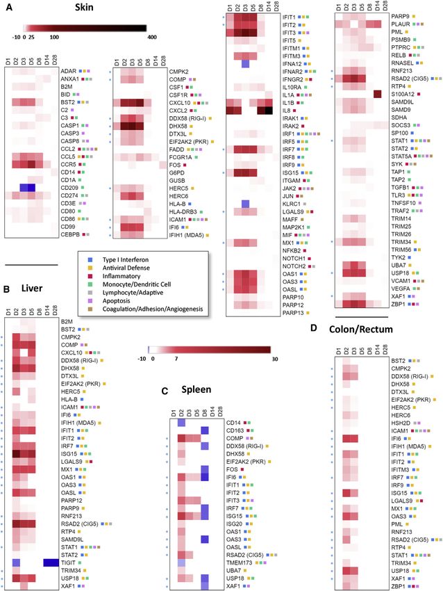
Figure 4. The ERB Transcriptional Immune
Response to MARV Infection in CD14+ Sple-
nocytes
nCAA analyzed differential gene expression (DGE)
using gene counts of infected versus uninfected
bats for CD14+ splenocytes. Selection as a differ-
entially expressed gene (DEG) at each time point
was based on meeting ± 2 fold-change (FC),
p value % 0.05, and above-background count
criteria (described in Methods). At a minimum,
DEGs met these criteria at one or more time points
in each dataset. A heatmap was produced using
Morpheus. Linear FC intensity for each DEG was
denoted in red (upregulation), blue (down-
regulation), or white (no change/failed criteria).
Only datapoints meeting above inclusion criteria
were visualized. Each column denotes a different
time point, as indicated on top of each graph.
Gradient bars represent FC intensity scales, set to
a maximum of 30-fold. Blue asterisks denote the
cluster of 26 canonical antiviral genes common to
at least four of the five major datasets. Color-
coded squares next to most DEGs denote their
functional annotation by PANTHER and DAVID
programs within generalized response networks
as indicated in the bottom legend.
See also Figures S2, S3, and S4 and Tables S1, S2,
S3, and S4.
bat #861 (D5), which generally had the
highest viral loads and showed markedly
greater global induction of DEGs than other
bats within its time point cohort in CD14+
splenocytes or whole tissues. Collectively,
DEG profiles between nCounter analysis
methods (nCAA-derived versus individu-
ally quantitated) were similar.
Finally, to discern in which main
signaling pathways the DEGs sets may
be involved, we performed Ingenuity
Pathway Analysis (IPA). Two pathways
were unique to CD14+ splenocytes: ‘‘Toll-
like Receptor (TLR) Signaling’’ and ‘‘[DC]
Maturation,’’ the latter of which is ex-
pected for a monocyte-enriched popula-
tion (Figure 6A). IPA of skin featured
five genes alone accounted for 11 of the 15 top three DEGs entirely unique positively regulated pathways: ‘‘JAK/STAT
across the five tissue/cell types. Despite strong upregulation Signaling,’’ ‘‘IL6 Signaling,’’ ‘‘IL10 Signaling,’’ ‘‘Th2 Pathway,’’
relative to other ERB DEGs, cluster gene induction showed and ‘‘Role of Macrophages, Fibroblasts and Endothelial Cells in
markedly reduced potency when compared qualitatively to pre- Rheumatoid Arthritis,’’ potentially selected due to infiltration of
viously reported gene induction for NHP homologs in whole immune cells into the inoculation site (Figure 6B). Strong positive
blood, total peripheral blood mononuclear cells (PBMCs), or regulation of IL6, JAK/STAT, and Th2 pathways extended to D28,
PBMC-derived immune cells (including CD14+ monocytes) perhaps because of virus lingering within the site. CD14+ spleno-
following in vivo infection with various MARV or EBOV cytes shared two innate immune response pathways with whole
isolates.25,28,29,31,39 tissues (Figures 6C–6E): ‘‘Activation of IRF by Cytosolic Pattern
Complementary heatmaps based on the DEGs identified by Recognition Receptors [PRRs]’’ and ‘‘Role of [PRRs] in Recogni-
nCAA, inclusive of each infected bat, were also produced (Figures tion of Bacteria and Viruses.’’ Two other immune response
S4 and S5). Immune response profiles of individual bats within a pathways were unique to all whole tissues: IFN signaling and
cohort were generally very consistent. Further, this analysis re- ‘‘Role of RIG-I Receptors in Antiviral Innate Immunity.’’ Most
vealed broad response intensity differences for individual bats immune response-related pathways showed strong positive
that correlated with their vRNA levels. The clearest example was regulation, particularly during early/acute phases (D2–D5).
262 Current Biology 31, 257–270, January 25, 2021ll
Article OPEN ACCESS
Figure 5. The ERB Transcriptional Immune Response to MARV Infection in Skin and Whole Tissues
nCAA analyzed DGE as described in Figure 4 for each tissue dataset: (A) skin/inoculation site (across top), (B) liver, (C) spleen,( and D) colon. Heatmaps were
produced as described in Figure 4. Gradient bars represent FC intensity scales, set to a maximum of 30-fold and identical for all datasets except skin (bar at top),
which instead has a maximum of 400-fold. Blue asterisks denote the cluster of 26 canonical antiviral genes common to at least four of the five major datasets.
Color-coded squares next to most DEGs denote their functional annotation by PANTHER and DAVID programs within generalized response networks as
indicated in the center legend.
See also Figures S2 and S5 and Tables S1, S2, S3, and S4.
Current Biology 31, 257–270, January 25, 2021 263ll
OPEN ACCESS Article
Figure 6. A Handful of Canonical Signaling
Pathways Regulated in CD14+ Splenocytes,
Skin, and Whole Tissues Define ERB Tran-
scriptional Immune Responses
Ingenuity pathway analysis (IPA) was used to
determine the top five canonical pathways for each
major dataset: (A) CD14+ splenocytes, (B) skin/
inoculation site, (C) liver, (D) colon, and (E) spleen.
Top five pathways are ranked by p value. Z score
specifies an activation score calculated by IPA
where a positive value indicates that the gene
expression in the dataset is changing in a way that
suggests positive regulation. Negative values
indicate that the gene expression in the dataset is
changing in a way that suggests negative regula-
tion. Pathways without a calculated Z score are
indicated with an ‘‘x.’’
adaptive immune response-related gene
markers or receptors, including CD19,
CD8A/B, FOXP3, HLA-A, HAVCR1/2,
and KLRs, were also unaltered (Tables
S2 and S3).
Notably, we observed that many pro-
and anti-inflammatory and receptor
genes, including cytokines, chemokines,
and adaptive immunity-related genes,
were not significantly induced in MARV-in-
fected ERBs (Table S4). Among them were
most IFN genes, CXCL11, CCL8, CSF2
(GM-CSF); various IL genes such as IL6,
IL33, GZMA, and NOS2/3; and receptors
including CD40, FAS, IFNAR1, and IL6R.
To further clarify the impact of the mini-
mally regulated cytokine and chemokine
signaling/receptor pathway in CD14+
splenocytes and skin of infected ERBs,
we used Pathview Web to visualize regula-
tion of over 80 DEG and non-DEG
pathway members present in the ERB
CodeSet. The lack of significant cytokine,
chemokine, or receptor induction identi-
ERB Response to MARV Shows Minimal Regulation of fied by Pathview was remarkable. In CD14+ splenocytes, cyto-
Cytokine, Chemokine, and Adaptive Immunity-Related kine induction was mostly limited to the overall modest D1
Genes response, with strongest but transient induction of IL8, IL1A/B,
While ERB immune-related gene induction reflected typical and TNF (Figure 7). In skin at the inoculation site, where presence
mammalian responses (albeit with reduced potency than of these inflammatory response factors is expected due to the
observed in filovirus-infected NHPs), we wanted to determine if high viral load at this location following MARV inoculation, only
any DEGs previously identified in MARV-infected spillover hosts a handful were identified as significantly upregulated, particularly
(like NHPs) were not significantly regulated in the natural bat IL8, IL1B, CXCL10 (IP10), CCL5 (RANTES), and CCR5 (Fig-
reservoir.25,38,39 Thus, we compiled all genes within our ERB Co- ure S6A). Few additional genes with any role in inflammation as
deSet not defined by nCAA as significant DEGs (Tables S2, S3, annotated by PANTHER and DAVID programs were induced
and S4); these were nearly half the CodeSet’s genes. Many beyond low, often transient levels, including TNFAIP3 (CD14+),
non-DEGs were signaling intermediates or transcription factors ICAM1, and LGALS9 (CD14+ and skin) and CXCL2, S100A12,
(Table S2), including those not usually upregulated such as and ZBP1 (skin) (Figures 4 and 5). Overall, inflammatory re-
MAVS, JAKs, STATs, CTNNB1, MAP kinase genes, and MYC. sponses contrasted with those for other antiviral pathways such
Other genes unchanged following MARV infection of ERBs as RIG-I, TLR, and TNF signaling, which showed significant
have known involvement in antiviral responses, including DEG involvement in skin and CD14+ splenocyte datasets during
DDX50, IFI35, MX2, OAS2, and UBE2L6. Several innate and/or early/acute phases of MARV infection (Figures S6 and S7).
264 Current Biology 31, 257–270, January 25, 2021ll
Article OPEN ACCESS
Figure 7. ERB Transcriptional Immune Responses Occur in the Absence of Significant Expression Changes for All but a Few Cytokine and
Chemokine Signaling/Receptor Genes
Pathview Web analysis shows representative DGE of the cytokine/chemokine receptor interaction pathway for CD14+ splenocytes. Genes in white boxes are
those without ERB CodeSet probes; genes in greyed boxes are those represented in the CodeSet. Red bands indicate upregulation of a given gene, blue bands
indicate downregulation, and gray bands indicate lack of expression change/failed inclusion criteria; each band indicates a different post-infection time point
from D1 to D28, arranged left to right across a gene box. Legend at top right defines color intensity scale set at indicated log2FC values.
See also Figures S6 and S7 and Table S4.
DISCUSSION filovirus reservoir.11,12,14–16 In this scenario, the antiviral state in
MARV-infected ERBs would restrict replication while avoiding
Soon after the discovery of MARV, key questions quickly immune-mediated pathology caused by cytokine and chemo-
emerged, including what the natural reservoir is, how the virus kine dysregulation. Thus, the absence of DEGs thought to cause
spills over into humans and NHPs, and importantly, how does such pathology is remarkable and holds at least three major im-
the reservoir host immunologically control the pathogen while al- plications regarding the differences in pathogenesis between in-
lowing sufficient viral replication to enable transmission.1,2,5,6,11,15 fected reservoirs and accidental spillover hosts and the exact
In this report, we characterize the in vivo transcriptional immune physiological avenues by which reservoirs and viruses co-
response in MARV natural reservoir, focusing on CD14+ spleno- evolved a tolerant host environment. First, the paucity of signifi-
cytes and skin at the inoculation site, both pathogenesis-relevant cant cytokine/chemokine responses in ERBs is clearly distinct
sites of virus replication. from those observed in filovirus-infected primates and relates
DGE analysis across tissue datasets revealed a broad, distinc- to disease burden.7,8,27,28,30,31 Only a handful of such genes
tive host response in which inflammatory genes and their recep- (including CCL5, IP10, IL1B, IL8, and TNF) were significantly up-
tors were not significantly changed in MARV-infected ERBs, in regulated either at the infection site or transiently in CD14+ sple-
stark contrast to the extensive dysregulation seen in pri- nocytes and almost none in whole tissues, consistent with previ-
mates.7,27–29,31,39 While we cannot completely rule out nonca- ous clinical observations of minimal tissue inflammation in
nonical strategies by which some non-DEGs could contribute ERBs.3,6 Non-DEG cytokines/chemokines within the ERB Code-
to MARV resistance (e.g., enhanced basal expression that Set included most IFNs, ILs such as IL12B and IL33, NOS genes,
‘‘primes’’ ERBs to better engage antiviral defenses, as demon- and a few CCLs, CCRs, and CXCRs. Indeed, recently published
strated in Pteropus bats), this transcriptional phenotype offers in vitro data from our lab using MARV-infected ERB BMDCs also
the first in vivo functional evidence for disease tolerance in a showed limited inflammatory gene regulation, expanding
Current Biology 31, 257–270, January 25, 2021 265ll
OPEN ACCESS Article
support for ERB disease tolerance to MARV.21 One notable lymphocytes from these tissues at around or just prior to when
distinction between our current in vivo-based work and the MARV-infected ERBs exhibit initial IgG production, which could
BMDC study was that IL33 was strongly suppressed in BMDCs, possibly further contribute to the IgG response phenotype and/
which might simply suggest DC-specific gene regulation. Mean- or reduced antibody-mediated inflammation.4–6,44 Given these
while, the lack of significant induction of IFNs agrees with previ- open questions, a better understanding of antibody loci, devel-
ous data from an ERB kidney cell line even after being infected opment, and response in these animals is required. Third, this
with a recombinant MARV ablated in its usual ability to antago- finding offers insight into the broader spectrum of antiviral
nize IFN-mediated antiviral gene induction via VP35, and despite response strategies used by viral reservoir hosts in vivo, and
those cells showing marked induction of multiple IFN genes by into how disease tolerance in ERBs compares to other host-virus
an unrelated control virus (Sendai virus).19 Type I IFN genes relationships. This relationship has been well characterized for
also showed minimal, if any, detectable basal gene counts in un- hantavirus rodent hosts. Like ERBs, bank voles (Myodes glareo-
infected bats, suggesting that ERBs do not rely on constitutively lus), male Norway rats (Rattus norvegicus), and deer mice (Pero-
activated IFN as posited for P. alecto bats by Zhou et al.12 It is myscus maniculatus) infected with Puumala virus, Seoul virus, or
tempting to speculate that IFN-independent responses therefore Sin Nombre virus, respectively, show no clinical signs of disease,
occur in these animals, allowing induction of specific antiviral lack tissue-specific proinflammatory cytokine induction, and
genes even in the presence of viral antagonist proteins. For generate low or non-neutralizing antibody responses.45–48
instance, recent work by the Mühlberger lab showed that Disease tolerance has also been suggested in studies with rac-
MARV VP35 inhibits PKR in human but not ERB cells, and there coons infected with raccoon rabies virus and mallards infected
is precedent for IFN-independent response induction by IRF3 or with low-pathogenic H1N1.49,50 However, to our knowledge,
STAT1 of subsets of canonical antiviral genes during in vitro hu- no other significant in vivo study of broad transcriptional immune
man cytomegalovirus (HCMV) infection.40–42 This does not pre- responses in any bona fide bat reservoir has been performed.
clude, however, the alternative possibilities that induction of Coronavirus inoculation of a suspected but unconfirmed ances-
IFN gene transcription simply occurs at levels too low for tral reservoir, the Jamaican fruit bat (Artibeus jamaicensis), re-
nCounter to detect, certain IFN proteins are activated on a sulted in subclinical disease and transient, low-level induction
post-transcriptional level, and/or the ability of MARV proteins of MX1, IFIT1, and RANTES, supporting tolerance.51 More
to fully suppress IFN is cell type-dependent, any of which would recently, a second, proteomics-based study compared the
suggest that IFNs still act as master antiviral response regulators lungs of Hendra virus-infected P. alecto bats and ferrets 60 h
in MARV-infected ERBs, perhaps in a paracrine fashion, without post-infection, in conjunction with limited IFN and cytokine
a need for noncanonical explanations. Of most relevance to TaqMan analysis.52 This report found downregulation of consti-
pathogenesis among the cytokine/chemokine non-DEGs in tutively expressed IFN in these bats, with a significantly more
ERBs is that they appear to be largely pro- or ambi-inflammatory; likely enhancement of IFN signaling and neutrophil-, T cell-,
several are highly induced in primate filoviral infections, including and antibody-mediated immunity pathways than ferrets, sug-
IFNg, IL2, IL4, MIP-1b (CCL4), and CSF2.8,24,25,27,30,31,39 Indeed, gesting inflammatory response suppression amidst concomitant
CCL8, FAS, and particularly IL6 are commonly reported hall- cell-mediated response activation. These two studies, however,
marks of the primate inflammatory response and subsequent se- remain rare examples of in vivo immune response data for bats.
vere disease.24,27,39 While basally expressed in naive ERBs, Intra-order comparisons beyond gross clinical observations thus
none of these genes displayed any significant activation upon remain challenging, highlighting the necessity for expanded
MARV infection, suggesting no role in the ERB response to in vivo reservoir host research to capture a more comprehensive
MARV, again consistent with a disease tolerance model.11,14–16 and contextual picture of the complex biological processes that
Second, tolerance may offer a molecular explanation for why follow infection by pertinent pathogenic threats like filoviruses.
these bats have previously exhibited non-neutralizing antibody The overarching immunological principles and precise host fac-
activity when tested in vitro and rapidly waning IgG in serum tors dictating reservoir protection against disease that such
despite protective secondary responses upon MARV rechal- studies uncover have become essential in a globalizing world
lenge.43 Our data show insufficient adaptive immune gene in- where human contact with wild hosts, and emerging disease
duction (such as AGER, CD40, HLA-A, IL13, and TNFSF13) outbreaks from the viruses they harbor, becomes an ever more
that could be associated with unique mechanisms dictating frequent event, as the likely recent spillover of SARS-CoV-2
lymphocyte maturation, Ig structure/function and/or memory from an unidentified bat reservoir has illustrated to a profoundly
cell generation/activity. Prior genomic studies have posited devastating effect.
that in some bat species, there is a diminished emphasis on DGE analysis also revealed commonalities and differences
the role of somatic hypermutation, which could subsequently between significant ERB and primate responses, as well as the
lessen a role for IgG during primary acute infection and poten- potential importance of several specific DEGs. MARV infection
tially promote dampened antibody-mediated inflammation.10 of ERBs stimulated canonical immune response genes, such
Our results extend the possibility of quantitatively reduced adap- as ISG15, IFIT2, RIG-I, and OAS1.28,38 Similar upregulation
tive immunity to ERBs on a transcriptional level. We also identi- was not seen previously using MARV-infected ERB cell lines;
fied (likely indirect) antiviral gene expression in CD19+ spleno- rather, antiviral genes were largely suppressed.19 The apparent
cytes following MARV infection, possibly affecting the role of B discrepancy in immune gene signaling between infected cells
cells in adaptive immune processes, as well as a conspicuous and whole animals has also been reported in primate studies,
proportional reduction of skin and spleen B cells in our again emphasizing the value of investigating in vivo host re-
CIBERSORT analysis. This latter data suggest depletion of sponses.24 This phenotypic disagreement between in vitro and
266 Current Biology 31, 257–270, January 25, 2021ll
Article OPEN ACCESS
in vivo responses may be due in part to conditional roles of afore- D3 in skin and spleen play roles in transitional innate-adaptive
mentioned viral antagonists like VP35, which at high multiplicity processes, including T cell activation by DCs (DC-SIGN, also a
of infection common for in vitro assays could accentuate sup- filovirus receptor), NK cell recognition of MHC class I molecules
pressive antagonist abilities (another potential explanation for (KLRC1 [NKG2]), T cell activation via MHC class II-presenting B
the lack of significant IL33 induction in vivo compared to cells (TMEM173 [STING]), and monocyte-macrophage differen-
BMDCs).19,21,53 Further, their functions could be restricted tiation (CD14). Suppressing these factors could impact estab-
temporally or by cell type and/or unresolvable in mixed cell pop- lished downstream features of MARV pathogenesis, such as
ulations. Indeed, primate macrophages, a primary filoviral target, lymphopenia due to lack of DC maturation and T cell activation
show robust antiviral gene responses both in vitro and in vivo (which itself modulates antibody production).4,7,8
following EBOV infection, as do the ERB-derived BMDCs after Another notable aspect of our study was an ability to correlate
ex vivo MARV infection, both reflecting those we observed in vivo MARV replication levels with specific ERB immune response
in MARV-infected ERB CD14+ splenocytes.21,24,25 Comparing gene kinetics across tissue/cell types, including at early and
in vivo data from our work using the ERB-derived MARV isolate acute time points post-infection, thereby greatly expanding
371Bat2007 to data from filoviral primate studies is vital to under- upon previous work that was able to detect only MARV RNA in
standing how the natural reservoir has evolved to better control these tissues.3,6 Indeed, virus disseminated quickly from the
infection compared to the maladaptive immunopathology eli- inoculation site, evidenced by virus positivity in spleen and liver
cited in primate spillover hosts. For instance, most MARV pri- by D1 and corresponding broad early host gene induction in
mate experiments use the Musoke or Angola virus isolates skin and CD14+ splenocytes. This D1 response in CD14+ spleno-
that, at the whole-genome level, are 93% identical to each cytes occurred prior to responses in the surrounding spleen tis-
other and 92% identical to 371Bat2007. Importantly, each of sue. These kinetics leave open the likely possibility that infiltrating
these viruses is linked to, or isolated from, fatal human infections monocytes may be initially infected at the inoculation site and
and causes near-uniformly fatal disease in NHPs. Indeed, the then transport the virus into the spleen and other tissues. Thus,
MARV bat isolate used in our study is greater than 99% identical CD14+ splenocyte response induction following infection, repli-
to 01Uga07, a virus isolated from a fatally infected miner working cation, and viral protein expression is likely critical to MARV path-
at Kitaka mine in Uganda where the ERB harboring 371Bat2007 ogenesis, irrespective of infectious virion formation from this cell
was captured.26,54 Therefore, the high degree of sequence type (not specifically measured in the context of this study), which
identity between 371Bat2007 and 01Uga07, combined with the might very well guide toward a non-productive infection given a
known diversity among fatal human filoviruses, strongly sug- fully functional, co-evolved ERB immune system capable of miti-
gests that this bat isolate would also cause severe, if not gating viral disease burden. Further, since B lymphocytes are not
lethal, disease in primates. To this end, DEGs in MARV-infected known to be MARV replication targets, any observed antiviral
ERBs consisted of canonical antiviral genes typical of those seen gene transcription in CD19+ splenocytes is likely due to paracrine
in infected primates and other mammalian systems.25,28,29,31,38 induction by host factors secreted from these early infected
Most, if not all, of the 26 cluster DEGs common across ERB tis- spleen monocytes.4 Indeed, as the majority of total splenocytes
sues/cells, such as ISG15, OAS1, and MX1, have also been iden- (56%) consisted of CD14+ and CD19+ cells and expression pro-
tified as elevated in MARV-infected primates.25,29,31 Moreover, files were largely similar between them, upregulation of their
cluster DEGs acted within a limited spectrum of traditional path- DEGs probably formed the basis of the overall response seen
ways shared by primates and other bat species, such as OAS/ for whole spleen. Despite the early kinetics in skin and CD14+
RNase L signaling, ISGylation, and JAK/STAT signaling.13,25,31,53 splenocytes, onset of the bulk of the host response was delayed
Assuming these ERB genes have antiviral capabilities akin to until D2. Responses correlated with viral load through D5. As
their primate orthologs, this implies that cluster gene induction identified by CIBERSORT in the skin and spleen, kinetics of viral
alone does not confer MARV protection to ERBs, as we hypoth- loads and/or immune responses also corresponded to propor-
esized in our ERB kidney cell line-based study.19 However, these tional, in some cases significant, estimated changes in lympho-
data suggest that antiviral responses are better regulated in cyte and macrophage populations indicative of cell depletion/
ERBs than in primates, with a subset of genes dictating a large apoptosis and activation/infiltration, respectively (although we
proportion of the global response but confined to a short-lived in- must caveat that this analysis is based solely on gene expression
duction window in which these presumably normal responses data from our large, though finite, ERB CodeSet and will need
are actively controlled. Further, ERB response genes were validation in future studies in order to rule out possible biases
substantially less induced than those previously measured in pri- due to reliance on nCounter values or human-specific marker
mate whole blood, monocytes, or tissues.25,29,31,39 For a prob- gene signatures). Nevertheless, together these observations sug-
able ERB infection target like CD14+ splenocytes to show mostly gest that innate immunity in ERBs, capable of robust upregulation
as limited, tightly regulated, and canonical a response profile as even during peak infection, swiftly controls viral replication,
ERB whole tissues, whereas in primates these immune cells possibly without substantial lymphocytic support. Simulta-
show extensive, strong ISG, and adaptive immune and neously, negative feedback pressures (e.g., USP18, a known
inflammatory gene responses, is perhaps also indicative of dis- IFN signaling modulator) coordinate equally rapid cessation of
ease tolerance.25 Among non-cluster DEGs, some showed primary gene responses once virus control is secured. The corre-
notable changes. The intense mid/late upregulation of IL8, lation of viral replication to host response also implies proportion-
IL1B, and S100A12 at the skin inoculation site was possibly ality in individual bats. For example, as seen in bat #861, high viral
due to an uncleared bolus of virus and/or prolonged immune load may be linked to transcriptional ‘‘superinduction’’ and
cell infiltration. Conversely, several DEGs downregulated D2– immune cell population phenotypes. This putative relationship
Current Biology 31, 257–270, January 25, 2021 267ll
OPEN ACCESS Article
raises intriguing questions regarding host susceptibility (whether B Data visualization
bats under stressful or immunomodulating conditions permit
greater viral replication), immune adaptability (whether bat SUPPLEMENTAL INFORMATION
response gene expression equalizes with viral yield as needed),
Supplemental Information can be found online at https://doi.org/10.1016/j.
and virus maintenance (how these higher yields and stronger re-
cub.2020.10.015.
sponses affect viral shedding and transmission potential) and
therefore warrants further exploration. Additionally, as previous ACKNOWLEDGMENTS
data suggest that bat-to-bat transmission could occur through
biting (i.e., subcutaneous inoculations in skin), future studies The authors wish to thank Tatyana Klimova for her assistance with critical
focused on the subcutaneous skin inoculation site are of particu- reading of the manuscript. Opinions, interpretations, conclusions, and recom-
larly high biological relevance for understanding MARV replica- mendations are those of the authors and are not necessarily endorsed by the
U.S. Army or the Centers for Disease Control and Prevention. This work was
tion and control in nature.5,6
partially funded by the Defense Threat Reduction Agency, Department of De-
In conclusion, we hypothesize a refined model for disease fense (HDTRA1-14-1-0016).
tolerance to MARV infection in its bat reservoir. This model is
defined by tissue-wide, moderate stimulation of canonical anti- AUTHOR CONTRIBUTIONS
viral genes early in MARV infection, possibly relying on putative,
noncanonical mechanisms (like IFN-independent induction) to Conceptualization, J.S.T., G.F.P., M.S.-L., J.C.G., and J.B.P.; Experimenta-
tion, J.C.G., J.B.P., B.R.A., A.J.S., J.R.S., T.K.S., J.R.H., J.D.C.-M., and
finely calibrate a cluster of the innate immune response with
J.S.T.; Data Analysis, J.C.G., C.E.A., and J.B.P.; Funding Acquisition, G.F.P.
rheostat-like precision. ERBs may thus have evolved to suc-
and J.S.T.; Resources, K.A.K. and E.R.N.; Writing – Original Draft, J.C.G.; Re-
cessfully disrupt viral replication without an evolutionary view and Editing, J.C.G., J.S.T., M.S.-L., J.B.P., J.R.S., and R.K.
emphasis on adaptive responses for long-lived immunity or
without triggering uncontrolled inflammatory gene expression DECLARATION OF INTERESTS
responsible for the classical ‘‘cytokine storm’’ and subsequent
severe immunopathology characterizing MARV disease in The authors declare no competing interests.
primates, including humans. Moving forward, it will be of para-
Received: May 18, 2020
mount interest to elucidate the gene functions, molecular
Revised: July 28, 2020
mechanisms, and immune cell types that mediate this disease Accepted: October 7, 2020
tolerant phenotype following MARV infection, which may in Published: November 5, 2020
turn be applicable to bat reservoir hosts of other emerging path-
ogens and translate into novel spillover control or antiviral ther- REFERENCES
apeutic strategies that better protect humans from zoonotic
1. Amman, B.R., Swanepoel, R., Nichol, S.T., and Towner, J.S. (2017).
viral infection and pathogenicity.
Ecology of Filoviruses. Curr. Top. Microbiol. Immunol. 411, 23–61.
2. Towner, J.S., Pourrut, X., Albariño, C.G., Nkogue, C.N., Bird, B.H., Grard,
STAR+METHODS G., Ksiazek, T.G., Gonzalez, J.P., Nichol, S.T., and Leroy, E.M. (2007).
Marburg virus infection detected in a common African bat. PLoS ONE 2,
Detailed methods are provided in the online version of this paper e764.
and include the following: 3. Jones, M.E., Schuh, A.J., Amman, B.R., Sealy, T.K., Zaki, S.R., Nichol,
S.T., and Towner, J.S. (2015). Experimental Inoculation of Egyptian
d KEY RESOURCES TABLE Rousette Bats (Rousettus aegyptiacus) with Viruses of the Ebolavirus
d RESOURCE AVAILABILITY and Marburgvirus Genera. Viruses 7, 3420–3442.
B Lead Contact 4. Messaoudi, I., Amarasinghe, G.K., and Basler, C.F. (2015). Filovirus path-
ogenesis and immune evasion: insights from Ebola virus and Marburg vi-
B Materials Availability
rus. Nat. Rev. Microbiol. 13, 663–676.
B Data and Code Availability
5. Schuh, A.J., Amman, B.R., Jones, M.E., Sealy, T.K., Uebelhoer, L.S.,
d EXPERIMENTAL MODEL AND SUBJECT DETAILS
Spengler, J.R., Martin, B.E., Coleman-McCray, J.A., Nichol, S.T., and
d METHOD DETAILS Towner, J.S. (2017). Modelling filovirus maintenance in nature by experi-
B Biosafety mental transmission of Marburg virus between Egyptian rousette bats.
B Viruses and antibodies Nat. Commun. 8, 14446.
B Inoculations and sampling 6. Amman, B.R., Jones, M.E., Sealy, T.K., Uebelhoer, L.S., Schuh, A.J., Bird,
B Euthanasia and necropsy B.H., Coleman-McCray, J.D., Martin, B.E., Nichol, S.T., and Towner, J.S.
B Cell preparation (2015). Oral shedding of Marburg virus in experimentally infected Egyptian
fruit bats (Rousettus aegyptiacus). J. Wildl. Dis. 51, 113–124.
B Cell labeling and magnetic selection
+
B Ex vivo CD14 splenocyte infectivity assay 7. Hensley, L.E., Alves, D.A., Geisbert, J.B., Fritz, E.A., Reed, C., Larsen, T.,
and Geisbert, T.W. (2011). Pathogenesis of Marburg hemorrhagic fever in
B RNA isolation and TaqMan assay
cynomolgus macaques. J. Infect. Dis. 204 (Suppl 3 ), S1021–S1031.
B nCounter hybridization and count detection
8. Fernando, L., Qiu, X., Melito, P.L., Williams, K.J., Feldmann, F., Feldmann,
d QUANTIFICATION AND STATISTICAL ANALYSIS H., Jones, S.M., and Alimonti, J.B. (2015). Immune Response to Marburg
B ERB probe-based nCounter CodeSet Virus Angola Infection in Nonhuman Primates. J. Infect. Dis. 212 (Suppl 2 ),
B nCounter transcriptomic analysis S234–S241.
B CIBERSORT analysis 9. Baker, M.L., Schountz, T., and Wang, L.F. (2013). Antiviral immune re-
B Gene Ontology (GO) and pathway analysis sponses of bats: a review. Zoonoses Public Health 60, 104–116.
268 Current Biology 31, 257–270, January 25, 2021You can also read