SPATIALLY RESTRICTED IMMUNE RESPONSES ARE REQUIRED FOR MAINTAINING ROOT MERISTEMATIC ACTIVITY UPON DETECTION OF BACTERIA - MPG.PURE
←
→
Page content transcription
If your browser does not render page correctly, please read the page content below
Article

Spatially Restricted Immune Responses Are
Required for Maintaining Root Meristematic Activity
upon Detection of Bacteria
Graphical Abstract                                                        Authors
                                                                             lia Emonet, Feng Zhou,
                                                                          Aure
                                                                          Jordan Vacheron, ...,
                                                                          Paul Schulze-Lefert, Christoph Keel,
                                                                          Niko Geldner

                                                                          Correspondence
                                                                          niko.geldner@unil.ch

                                                                          In Brief
                                                                          By installing immune responsiveness
                                                                          specifically in peripheral root meristem
                                                                          cells, Emonet et al. generate
                                                                          hyperresponsive plants, showing
                                                                          flagellin-dependent root collapse and
                                                                          growth inhibition by commensal bacteria.
                                                                          This highlights the relevance of spatially
                                                                          resolved immune responses and their
                                                                          importance for bacterial accommodation
                                                                          by roots.

Highlights
d   Cell-specific expression of an immune receptor reveals
    general and specific responses

d   Immune signaling appears to separate into autonomous and
    non-autonomous branches

d   Immune response in some meristem cells causes meristem
    collapse and growth arrest

d   Mis-localized immune perception interferes with
    accommodation of commensal bacteria

           Emonet et al., 2021, Current Biology 31, 1–17
           March 8, 2021 ª 2021 The Authors. Published by Elsevier Inc.
           https://doi.org/10.1016/j.cub.2020.12.048                                                      ll
Please cite this article in press as: Emonet et al., Spatially Restricted Immune Responses Are Required for Maintaining Root Meristematic Activity upon
 Detection of Bacteria, Current Biology (2021), https://doi.org/10.1016/j.cub.2020.12.048

                                                                                                                                            ll
                                                                                                                                      OPEN ACCESS

Article
Spatially Restricted Immune Responses
Are Required for Maintaining Root
Meristematic Activity upon Detection of Bacteria
   lia Emonet,1 Feng Zhou,1,4 Jordan Vacheron,2 Clara Margot Heiman,2 Vale
Aure                                                                      rie De
                                                                                nervaud Tendon,1 Ka-Wai Ma,3
Paul Schulze-Lefert,3 Christoph Keel,2 and Niko Geldner1,5,6,*
1Department   of Plant Molecular Biology, University of Lausanne, Biophore, UNIL-Sorge, 1015 Lausanne, Switzerland
2Department   of Fundamental Microbiology, University of Lausanne, Biophore, UNIL-Sorge, 1015 Lausanne, Switzerland
                                                                                                          -Weg 10, 50829 Cologne,
3Department of Plant Microbe Interactions, Max Planck Institute for Plant Breeding Research, Carl-von-Linne

Germany
4Present address: National Key Laboratory of Plant Molecular Genetics, CAS Center for Excellence in Molecular Plant Sciences, Chinese

Academy of Sciences, 300 Feng Lin Road, Shanghai 200032, China
5Twitter: @NikoGeldner
6Lead Contact

*Correspondence: niko.geldner@unil.ch
https://doi.org/10.1016/j.cub.2020.12.048

SUMMARY

Plants restrict immune responses to vulnerable root parts. Spatially restricted responses are thought to be
necessary to avoid constitutive responses to rhizosphere microbiota. To directly demonstrate the impor-
tance of spatially restricted responses, we expressed the plant flagellin receptor (FLS2) in different tissues,
combined with fluorescent defense markers for immune readouts at cellular resolution. Our analysis distin-
guishes responses appearing cell autonomous from apparently non-cell-autonomous responses. It reveals
lignification as a general immune response, contrasting suberization. Importantly, our analysis divides the
root meristem into a central zone refractory to FLS2 expression and a cortex that is sensitized by FLS2
expression, causing meristem collapse upon stimulation. Meristematic epidermal expression generates su-
per-competent lines that detect native bacterial flagellin and bypass the weak or absent response to com-
mensals, providing a powerful tool for studying root immunity. Our manipulations and readouts demonstrate
incompatibility of meristematic activity and defense and the importance of cell-resolved studies of plant im-
mune responses.

INTRODUCTION                                                                  with growth inhibition,5,10 a plethora of publications have estab-
                                                                              lished a growth-promoting action of the soil microbiome.11 It is
Similar to the intestinal microbiome of animals, plant roots host a           therefore of particular interest to understand how roots accom-
vast range of micro-organisms in their rhizosphere. Among those,              modate a rhizosphere community, while avoiding a constant
some can act as pathogens, negatively impacting growth and                    activation of PRRs and the growth-defense trade-off that
reproduction. In both animals and plants, a sophisticated immune              comes with it. Many researchers have argued that growth inhi-
system keeps the vast majority of pathogens at bay, while allowing            bition can be overcome by the ability of commensal micro-or-
colonization with commensal and beneficial microbes.1,2 In plants,            ganisms to suppress plant immunity.12 In addition, it was
this immunity rests on the recognition of highly conserved                    recently shown that the root has an inherently dampened
microbe-associated molecular patterns (MAMPs), recognized by                  MAMP response until it encounters damage, which locally
an extended set of plasma-membrane-localized pattern-recogni-                 boosts immune responsiveness.13
tion receptors (PRRs).3 One of the most investigated MAMPs is a                  Indeed, root immune responses are often lower than in the
22-amino-acid fragment of the bacterial flagellin protein (flg22). It         shoot, in part because of an absence or low abundance of
is detected by the FLAGELLIN SENSING 2 (FLS2) receptor4–7                     PRRs.14–16 Moreover, plants restrict their defense to regions
and induces a signaling cascade, including reactive oxygen spe-               considered vulnerable, such as regions with absent or broken
cies (ROS) production, calcium signaling, MAPKs (mitogen-acti-                endodermal barriers, such as the elongation zone or lateral
vated protein kinase) phosphorylation, and gene transcription,                root emergence sites. It is also there where bacteria are found
eventually leading to defense responses, such as callose and lignin           to preferentially accumulate.13–15,17–20
deposition or phytoalexin production.8,9                                         Here, we set out to address the relevance of spatially limited
   Yet plant PRRs equally perceive MAMPs from commensal or                    immune responses. Wyrsch et al.21 ectopically expressed
beneficial microbes, which are part of the normal plant rhizo-                FLS2 under tissue-specific promoters, and their data suggested
sphere. Whereas MAMP-triggered immunity (MTI) is associated                   that all root tissues were competent to mount an immune

                                                Current Biology 31, 1–17, March 8, 2021 ª 2021 The Authors. Published by Elsevier Inc. 1
                       This is an open access article under the CC BY-NC-ND license (http://creativecommons.org/licenses/by-nc-nd/4.0/).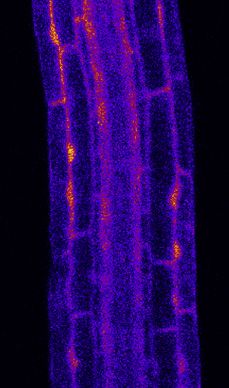
Please cite this article in press as: Emonet et al., Spatially Restricted Immune Responses Are Required for Maintaining Root Meristematic Activity upon
 Detection of Bacteria, Current Biology (2021), https://doi.org/10.1016/j.cub.2020.12.048

           ll
      OPEN ACCESS                                                                                                                          Article

response provided that FLS2 is expressed, although the nature                 the differentiated stele14 (Figure 1Ai) but also weakly in all tissues
of the tissue had a large influence on the strength of the innate             from the elongation to the differentiation zone, as well as in root
immune responses. Yet the immune readouts used were at                        cap cells (Figure 1Bi).13 WER::FLS2, by contrast, was strongly ex-
whole-plant or organ-level resolution and did not allow the                   pressed in the epidermis of the meristematic zone (Figures 1Aii and
authors to conclude from which cell type responses were origi-                1Bii), as predicted,30 with some weak signal in the elongating cor-
nating and whether they were cell autonomous, regional, or sys-               tex (Figure S1A). CASP1::FLS2 had the predicted exclusive
temic. MAMPs induce ROS production, as well as cytosolic                      expression in differentiated endodermis (Figures 1Aiii, 1Biii, and
calcium increases, both of which are known to act in paracrine,               S1B). In agreement with its established expression,31,32 we de-
even systemic signaling.22–25 Indeed, calcium waves were re-                  tected SHR::FLS2 in the stele close to the meristem (Figures
ported to initiate in the root elongation zone and to spread across           1Aiv and 1Biv) but also faintly in the neighboring endodermis, sug-
tissues after flg22 treatment,26,27 opening the possibility that              gesting that either FLS2 proteins or mRNAs move through plasmo-
MAMP responses could be induced in cell layers far away from                  desmata or that the SHR promoter has a low, overlooked activity in
the site of MAMP perception.                                                  the endodermis (Figure S1D). UBQ10::FLS2 was detected in all tis-
   By adding cellular resolution reporters to these cell-type-spe-            sues throughout the root, from meristem to differentiation zone
cific FLS2 expression lines, we were able to both manipulate and              (Figures 1Av, 1Bv, and S1C).
quantitatively map defense responses at cellular resolution in the
root. Our approach reveals the presence of regions refractory to              Quantitative Analysis of MAMP Response Patterns at
FLS2 presence, as well as others which are super-competent.                   Cellular Resolution
We show that restricted FLS2 expression in meristematic                       We then crossed our selection of promoter::FLS2 lines with two
epidermis has drastic impact on root development, affecting                   lines expressing transcriptional markers of defense, PER5::NLS-
growth, cell wall composition, and cell viability. To assess the              3xmVenus (PEROXIDASE 5) and MYB51::NLS-3xmVenus (MYB
impact of FLS2 misexpression in response to natural microbiota,               DOMAIN PROTEIN 51), and generated homozygous lines at all
we use our super-competent lines in the presence of commensal                 three loci (marker, prom::FLS2, and fls2 [SAIL691_C04]). As a con-
bacteria that are not or only weakly inducing responses in wild-              trol, we used the same two markers in WT Col-0 background.
type (WT) plants. We demonstrate stimulation of FLS2 directly                 Markers were chosen for their strong response to flg22 and their
by native, bacteria-derived flagellin and reveal the importance               distinct response patterns.13,20,21 In addition, we developed a
of spatial restriction of immune responses in order to adequately             pipeline using tissue-specific quantitative analysis for measuring
balance growth and defense.                                                   and comparing MAMP responses in an unbiased fashion (Fig-
                                                                              ure S2). For this, we additionally introduced ubiquitous nuclear
RESULTS                                                                       markers (UBQ10::NLS-mTurquoise2 or UBQ10::NLS-tdTomato)
                                                                              in all our genotypes, which allows to call all nuclei as separate,
In order to obtain cellular resolution readouts of immune re-                 individual 3D regions of interests (ROIs), even those with weak or
sponses in the different FLS2-expressing lines, we combined                   undetectable MAMP response. After mock or flg22 treatment
them with a set of fluorescent transcriptional response markers.              and fixation, cell-wall-stained roots were imaged at three different
These marker lines use a triple mVenus fluorochrome coupled to                zones of the root: meristem (MZ); elongation (EZ); and differentia-
a nuclear localization signal (prom::NLS-3xmVenus). Combining                 tion (DZ). Each nucleus was automatically detected as a 3D object,
concatemerization with nuclear concentration generates high                   and the obtained nuclei object maps were then combined to the
sensitivity and allows for a clear cellular assignment, not achiev-           cell wall marker channels to manually curate and assign each nu-
able with cytosolic, endoplasmic reticulum (ER), or plasma-                   cleus to a tissue. Once the selected nuclei were assigned, mean in-
membrane-localized markers. These lines now enable us to                      tensity for each cell type per zone per treatment per genotype were
observe damage and defense responses with cellular resolution,                calculated and color coded for the generation of a quantitative
adding a crucial layer of complexity to our analyses of damage                MAMP-response atlas for each prom::FLS2 line (Figure S2; values
and immune responses.13,20,25,28 In addition to these transcrip-              in Figure S3).
tional reporter lines, we also employed fluorescence-based
markers for cytosolic calcium changes triggered by flg22.29                   Ectopic FLS2 Expression Alters MAMP Response
                                                                              Patterns
Tissue-Restricted Expression of the FLS2 Receptor in                          Our cell-specific quantification and microscopic analysis in PER5-
fls2 Mutants                                                                  expressing WT plants confirmed that PER5 is not expressed in
In order to analyze the ability of the different root tissues to respond      absence of flg22 treatment (Figures 2A–2C) but that MYB51 pre-
to flg22, we used lines expressing FLS2 under cell-type-specific              sents a constitutive, flg22-independent expression in the
promoters in an fls2 (SAIL691_C04) mutant background.21 We                    epidermis and root cap cells of the undifferentiated tissues (MZ
selected lines expressing FLS2-GFP driven by three different tis-             and EZ) and in the stele and the cortex of the DZ (Figures 3A and
sue-specific promoters: WEREWOLF for epidermis (WER::FLS2);                   3C). Both MAMP markers are strongly induced by flg22 in the
CASPARIAN STRIP DOMAIN PROTEIN 1 for endodermis                               EZ, recapitulating previous observations (Figures 2A and
(CASP1::FLS2); and SHORT-ROOT for inner cell layers                           3A).13,19,20 Specifically, PER5 is induced almost exclusively in
(SHR::FLS2). As controls, we monitored FLS2-GFP driven by the                 the elongating epidermis and root cap cells (Figures 2C and 2D).
constitutive promoter UBIQUITIN 10 (UBQ10::FLS2) and by the                   Strong response in these tissues might be enhanced by feedfor-
native FLS2 promoter (FLS2::FLS2 in Ws-0). As described previ-                ward regulation of flg22 on the endogenous FLS2 promoter.13
ously, endogenous FLS2 expression was observed principally in                 MYB51 induction is restricted to these same tissues close to the

2 Current Biology 31, 1–17, March 8, 2021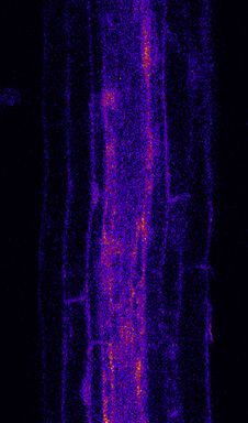
Please cite this article in press as: Emonet et al., Spatially Restricted Immune Responses Are Required for Maintaining Root Meristematic Activity upon
Detection of Bacteria, Current Biology (2021), https://doi.org/10.1016/j.cub.2020.12.048
ll
Article OPEN ACCESS
A B
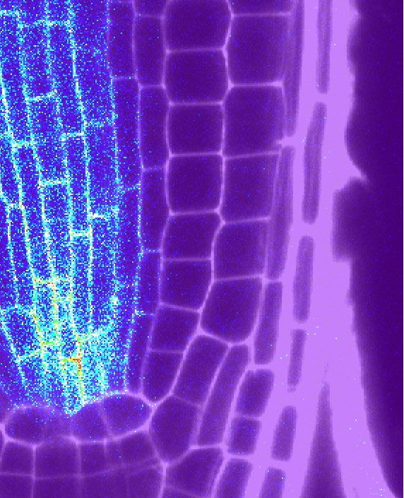
Figure 1. Tissue-Specific Promoters Drive FLS2 Receptor Expression Ectopically
(A) Tile scan of fls2 (SAIL691_C04) roots Col-0 background complemented with GFP-tagged FLS2 receptor under epidermal (WER::; ii), endodermal (CASP1::; iii),
central cylinder (SHR::; iv), and ubiquitous (UBQ10::) promoters (v). For comparison, endogenous FLS2 expression is shown in FLS2::FLS2-GFP Ws-0 lines
(natural fls2 mutant; i). Root shape is highlighted with propidium iodide (PI) staining cell wall (PI, red). Scale bar, 100 mm. Developmental regions of the roots are
labeled: differentiated zone (DZ); elongation zone (EZ); and meristematic zone (MZ). White arrows represent start of CASP1::FLS2 signal.
(B) Close-up view of FLS2-GFP expression at selected regions of the complemented lines. FLS2 driven by its endogenous promoter is expressed in all tissues in
the DZ (i). Note that, in contrast to previous report, low FLS2 expression is observed in epidermis and cortex (white arrow). In the meristem (MZ), WER promoter
expresses FLS2 specifically in epidermis (ep) and root cap (rc) (ii) and SHR promoter in the stele (st) and endodermis (en) (iv). In the DZ, FLS2 is expressed in all
tissues under UBQ10 promoter (v) but is restricted to endodermis with CASP1 promoter (iii). FLS2-GFP (BlueGreen) is co-visualized with PI-stained cell wall (red).
Separated and overlaid channels (right column) are presented. Scale bar, 25 mm. c, cortex; en, endodermis; ep, epidermis; rc, root cap cells; st, stele. Repre-
sentative pictures are presented. Nine to twelve seedlings over three to four independent biological replicates were analyzed for each genotype.
See also Figure S1.
meristem, but induction expands to cortex and pericycle cells in pattern. The defense markers extend to the whole root in
the later root regions (Figures 3C and 3D). Therefore, induction of UBQ10::FLS2, although they are restricted to the DZ or the MZ
transcriptional readouts matches largely, but not completely, the in CASP1::FLS2 and WER::FLS2, respectively. As expected, the
FLS2 expression observed in WT plants. Some mismatches, fls2 (SAIL691_C04) mutant does not respond to flg22 in any tissue.
such as the absence of induction in the differentiated stele—
despite FLS2 expression there—can be partially explained by PER5 and MYB51 Appear to Separate Cell-Autonomous
the Casparian strip blocking the entry of flg22 into these tissues.13 and Non-Cell-Autonomous Branches of the MAMP
For both markers, changing expression of FLS2 had an obvious Response
impact on the pattern of responses (Figures 2A and 3A). Rather PER5 responds only in the differentiated endodermis in the
than remaining restricted to the elongation zone, PER5 and CASP1::FLS2 recombinant line, which matches the very spe-
MYB51 induction largely follows the ectopic FLS2 expression cific expression pattern of CASP1 promoter (Figure 2C).
Current Biology 31, 1–17, March 8, 2021 3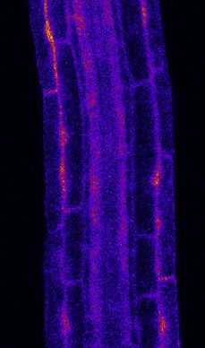
Please cite this article in press as: Emonet et al., Spatially Restricted Immune Responses Are Required for Maintaining Root Meristematic Activity upon
 Detection of Bacteria, Current Biology (2021), https://doi.org/10.1016/j.cub.2020.12.048

                                  ll
                   OPEN ACCESS                                                                                                                                                                                                                Article

   A                                                                                                                                prom::FLS2-GFP fls2

                                               WT                                 UBQ10::                              WER::                        CASP1::                             SHR::                                          fls2
                    mock                        flg22              mock                  flg22               mock          flg22             mock        flg22           mock              flg22                    mock                      flg22
   PER5::NLS-3mV

                   100 um

   B                                                                           fls2                                    C                                                  fls2
                                  WT                UBQ10::FLS2-GFP                   WER::FLS2-GFP                                WT                 CASP1::FLS2-GFP            SHR::FLS2-GFP                   fls2
                     mock              flg22             mock          flg22          mock           flg22                 mock            flg22        mock     flg22           mock           flg22   mock            flg22
      DZ
      EZ
      MZ

                   PER5::NLS-3mV
                   Cell wall

   D                                       WT                           UBQ10::FLS2-GFP fls2                   WER::FLS2-GFP fls2                   CASP1::FLS2-GFP fls2                SHR::FLS2-GFP fls2                             fls2
                                  mock                   flg22            mock               flg22                  mock           flg22               mock         flg22                mock           flg22                   mock                  flg22
                    30c
                    12c
                    EZ
                    MZ

                          log10 (Mean Intensity)
                                                                                PER5::NLS-3mVenus
                            2.5          3.0            3.5      4.0

Figure 2. PER5 Marker Gene Is Induced Cell Autonomously by flg22 Treatment
(A) Overview of PER5::NLS-3mVenus marker response to flg22 in different FLS2 recombinant lines. All lines contain PER5::NLS-3mVenus in the background. Wild
type (WT) is in Col-0 background; fls2 (SAIL691_C04) is an insertion line in Col-0 background. Tile scan images of 1 mM flg22-treated plants versus mock are
shown. Pictures were taken with similar settings. Settings were always identical between mock and corresponding flg22 treatment. Region of responsiveness is

                                                                                                                                                                                                                (legend continued on next page)
4 Current Biology 31, 1–17, March 8, 2021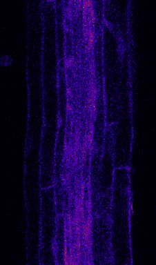
Please cite this article in press as: Emonet et al., Spatially Restricted Immune Responses Are Required for Maintaining Root Meristematic Activity upon
  Detection of Bacteria, Current Biology (2021), https://doi.org/10.1016/j.cub.2020.12.048

                                                                                                                                                       ll
Article                                                                                                                                          OPEN ACCESS

Sequential captures before and after flg22 treatment allow for                       Regional Ca2+ Waves Are Initiated from FLS2-
visualization of FLS2 receptors and PER5 marker in the same                          Expressing Cells
cells, suggesting a cell-autonomous response (Figure S4A).                           Because MYB51 induction by flg22 appeared to have a non-cell-
For WER::FLS2 line, the PER5 response also follows detectable                        autonomous component, we wondered whether cytosolic cal-
FLS2 expression. We could quantify a strong response in root                         cium increases play a part in non-cell-autonomous flg22
cap cells and the meristematic epidermis, extending until the                        responses. Indeed, cytosolic Ca2+ increases are among the
early DZ, as well as in cortex cells (Figures 2B–2D), where we                       earliest responses upon MAMP perception, preceding transcrip-
could also detect FLS2 protein (Figure S1A). In contrast to                          tional changes.33,34 In roots, Ca2+ influx after flg22 perception
PER5, we detected MYB51 response to flg22 not only in cells                          was shown to spread across tissues and was proposed to be
expressing FLS2 but also some degree of induction in neigh-                          part of Ca2+/ROS waves, spreading immune signaling over
boring cells (Figures 3B–3D). Intensity ratio between flg22 and                      long distances.23,24,26,27,35 However, because many cells ex-
mock-treated plants were calculated and represented graphi-                          press some degree of FLS2 in WT plants, it is impossible to
cally in Figure S4B. Non-cell-autonomous responses seem                              dissect to what extent such waves represent a non-cell-autono-
obvious for MYB51 in the DZ of CASP1::FLS2. Although FLS2                            mous propagation of the Ca2+ signaling or are due to flg22 diffu-
is specifically expressed in the endodermis (Figures 1Biii and                       sion and direct flg22-dependent activation of the receptor in
S1B), we could barely detect any MYB51 responses in this tis-                        different tissue layers and regions.
sue, although the neighboring stele and cortex cells strongly                           Using the intensity-based Ca2+ reporter R-GECO1 in our
upregulated MYB51 (Figures 3C, 3D, and S4B). Similarly,                              transgenic lines,26 we observed that, in WER::FLS2, calcium
flg22 treatment led to strong MYB51 expression not only in                           signals initiate in the epidermis and spread to inner tissues
the epidermis and cortex but also in central tissues in                              (Videos S1 and S2; Figures 4A and 4B). Because the receptor
WER::FLS2 (Figures 3C and 3D). Thus, we suggest that                                 has no detectable expression in central tissues, we are taking
MYB51 induction by MAMPs is controlled by non-cell-autono-                           this as evidence that FLS2 stimulation in epidermis/root cap
mous mechanisms, contrasting with the relatively cell-autono-                        can cause cytosolic calcium increases in the stele and thus
mous induction of other markers, such as PER5 and FRK1                               causes a propagating Ca2+ wave. This propagation of Ca2+
responses (this work and Zhou et al.13).                                             transients could be observed in all recombinant lines tested,
                                                                                     with the intriguing property that the wave direction could be
FLS2 Expression Is Insufficient to Cause flg22                                       manipulated—i.e., in both CASP1::FLS2 and SHR::FLS2 lines,
Responses in the Vascular Meristem                                                   the wave started first in the endodermis then spread to outer
Intriguingly, some tissues were also completely refractory to                        and inner tissues (Figures 4C and 4D; Video S2). Moreover, in
flg22-triggered responses. Despite a clear presence of FLS2 in                       these two lines, the wave starts in the differentiated rather
the vascular meristem (Figures 1Biv, S1C, and S1D), flg22 treat-                     than in the elongation zone (Video S1). When FLS2 was ex-
ment did not trigger PER5 or MYB51 expression in this tissue in                      pressed in all tissues under the UBQ10 promoter, all tissues re-
SHR and UBQ10::FLS2 lines (Figures 2 and 3), except for some                         sponded almost simultaneously (Figure 4E; Videos S1 and S2).
weak MYB51 induction in meristematic pericycle cells in                              As expected, flg22 does not induce any calcium waves in fls2
UBQ10::FLS2 (Figures 3D and S4B). Because SHR::FLS2 is                               (SAIL691_C04) background (Figure 4F; Video S2). Taken
not sufficient to induce MYB51 in the pericycle cells, despite                       together, although transcriptional readouts are largely cell
its expression there, we conclude that flg22 induction of                            autonomous, cytosolic calcium transients represent a non-
MYB51 in the pericycle in the UBQ10::FLS2 line is due to a                           cell-autonomous response to MAMP stimulation. This implies
perception of flg22 in outer cell layers. Thus, central meriste-                     that even cells that are neither exposed to flg22 nor able to
matic tissues differ from outer tissue layers in their competence                    perceive this peptide nevertheless rapidly receive a kind of
to respond to flg22 in the presence of receptor.                                     stress signal in the form of a calcium wave.

modified by the different expression patterns of FLS2. Brackets indicate responsive regions. For SHR, close-up view was generated with increased brightness to
highlight stelar signal (white arrow). Scale bar, 100 mm.
(B) Maximal projection of transverse sections views of PER5 expression pattern in UBQ10:: and WER::FLS2-GFP fls2 compared to WT (PER5::NLS-3mVenus in
Col-0) shown for MZ, EZ, and DZ (30 cells after start of elongation). Seedlings were treated for 24 h with 1 mM flg22. Note the refractory region in the central
cylinder in UBQ10::FLS2-GFP fls2 (white arrows). Nuclear localized mVenus signal (yellow) was co-displayed with PI cell wall marker (purple). Images were taken
with similar settings, but corresponding mock and flg22 treatment pictures for each zone separately always have identical parameters. Note that epidermal signal
in flg22-treated WT seedlings is faint (EZ, black arrow), due to settings chosen to avoid saturation of signal in the transgenic lines. Compare to (C), WT. Scale bar,
25 mm.
(C) Maximal projection of transverse section views of PER5::NLS-3mVenus expression pattern in CASP1:: and SHR::FLS2-GFP fls2 as well as WT (PER5::NLS-
3mVenus in Col-0) and fls2 (SAIL691_C04) control. White arrows point at ectopic response in the endodermis. Images were acquired as in (B), with similar settings
between genotypes, but with identical parameter for corresponding mock and flg22 treatment. Pictures were acquired with increased gain compared to (B) due to
lower average signal intensity. Scale bar, 25 mm.
(A–C) Representative pictures are presented. 10–20 seedlings over 3–5 independent biological replicates were analyzed for each genotype and treatment.
(D) Quantitative map of PER5::NLS-3mVenus responses inferred from tissue-specific quantification after 24-h treatment with 1 mM flg22. Nuclear signals were
quantified in ROI delimited with UBQ10::NLS-mTurquoises2 for all tissue-specific promoter lines, and WT signal was quantified with UBQ10::NLS-tdTomato
marker. Mean intensity is therefore comparable between prom::FLS2-GFP fls2 lines, but not to WT. Four to six seedlings per genotype per treatment were
analyzed (n = 51; 16,680 different nuclei analyzed).
See also Figures S2–S4.

                                                                                                                 Current Biology 31, 1–17, March 8, 2021 5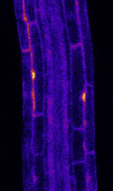
Please cite this article in press as: Emonet et al., Spatially Restricted Immune Responses Are Required for Maintaining Root Meristematic Activity upon
 Detection of Bacteria, Current Biology (2021), https://doi.org/10.1016/j.cub.2020.12.048

           ll
       OPEN ACCESS                                                                                                                         Article

   A

   B                                                               C

   D

                                                                                                                                    (legend on next page)

6 Current Biology 31, 1–17, March 8, 2021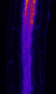
Please cite this article in press as: Emonet et al., Spatially Restricted Immune Responses Are Required for Maintaining Root Meristematic Activity upon
  Detection of Bacteria, Current Biology (2021), https://doi.org/10.1016/j.cub.2020.12.048

                                                                                                                                                      ll
Article                                                                                                                                         OPEN ACCESS

Epidermal Meristematic Expression of FLS2 Leads to                                  3-OH-C10:0, nor the endogenous danger-associated molecular
flg22 Hypersensitivity and Meristem Collapse                                        pattern (DAMP) Atpep1 enhanced PER5 expression in
Having demonstrated that we can profoundly alter the pattern of                     WER::FLS2 (Figure 5D). This demonstrates that ectopic FLS2
immune responses in the root by ectopic FLS2 expression, we                         expression does not cause a global upregulation of responsive-
wanted to study the consequences of such altered spatial pat-                       ness to MAMPs or DAMPs but specifically affects flg22-depen-
terns for plant growth and defense. Sustained stimulation with                      dent signaling.
MAMPs causes root growth inhibition (RGI) often explained by                           Interestingly, treatment of the WER::FLS2 super-competent
an antagonism between growth and defense responses.10 We                            line with flg22 induces profound morphological changes in the
therefore assessed root length of seedlings transferred to                          root not observed in WT. After 2 days of treatment, cells reaching
flg22-containing medium (Figures 5A and 5B). As expected,                           the transition zone start to swell and division patterns become
treated WT plants showed only a mild reduction in root length.                      disorganized, giving rise to bulky meristem shapes (Figures 5E,
By contrast, the root length of the constitutive, overexpressing                    upper panel, and 5G). Virtual cross-sections revealed that cortex
UBQ10::FLS2 line was strongly reduced with additionally                             cells expand tremendously, dislocating epidermal cells (Fig-
stunted shoot growth. More surprisingly, a strong root length in-                   ure 5F). Thus, precise spatial regulation of FLS2 expression
hibition was also observed in the WER::FLS2 line, although this                     levels is necessary to avoid severe root growth inhibition and
line expresses FLS2 only in young epidermal and root cap cells.                     flg22-induced disorganized cell expansion in the meristem.
SHR::FLS2 and CASP1::FLS2, by contrast, showed root growth
similar to WT.                                                                      FLS2 Ectopic Expression Leads to Cell-Autonomous,
   In order to more precisely identify the tissue responsible for                   flg22-Triggered Lignin Deposition
root growth inhibition, we generated two additional prom::FLS2                      Considering the strong impact of flg22 on meristem morphology
lines using the RCH1 (RECOGNITION OF C.HIGGINSIANUM)                                in WER::FLS2, we decided to assess whether flg22 causes lignin
and PRP3 (PROLINE-RICH PROTEIN 3) promoters.36 RCH1 is                              deposition in our lines. Indeed, MAMP responses are known to
expressed in the whole meristem, although PRP3 is expressed                         modify cell wall composition, such as callose deposition or ligni-
strongly in differentiating root hair cells (Figure S1E). Although                  fication.8,19,37 Lignin and suberin depositions are long-known
PER5 induction followed the expression of FLS2 in both lines                        damage- and immunity-associated responses38–43 but have
(Figures S1G and S1H), only RCH1::FLS2 presents an increased                        not been widely adopted in recent studies on MTI.44,45
root growth inhibition, whereas PRP3::FLS2 responds as WT                              We found that flg22 treatment induced strong lignification from
(Figure S1F). Therefore, we conclude that it is the expression                      transition to differentiated zone in WER::FLS2 (Figures 5E, lower
of FLS2 only in the meristematic epidermal cell layers that                         panel, and S5A). Lignin was deposited between epidermis and
causes strongly enhanced root growth inhibition in response to                      cortex cells, mainly at the corners (Figure 5F). In younger regions,
flg22, implying this tissue is extremely sensitive to MAMP re-                      lignin was also found between epidermis and root cap cells.
sponses. Indeed, when comparing the pattern of PER5 expres-                         Interestingly, all other recombinant lines also showed lignin
sion between WT and WER::FLS2 at cellular resolution, it is                         deposition that coincided with the respective FLS2 expression
evident that root cap cells responded strongly to flg22 in both                     patterns, except in the stele (Figure S5). The latter observation
genotypes but that meristematic epidermal cells only show re-                       corresponded to the absence of flg22-mediated PER5 induction
sponses in WER::FLS2 (Figure 5C). This indicates that MAMP re-                      in these tissues, as can be observed in SHR::FLS2, as well as the
sponses in meristematic epidermal cells are the cause of the                        GRP:FLS2 line (Figures 2C and S1I) that expresses FLS2 specif-
growth inhibition in WER::FLS2 plants. Importantly, neither the                     ically in the pericycle cells (Figure S1E). Interestingly, no lignin
treatment with MAMPs, such as elf18, chitin, or the LPS fragment                    deposition could be observed in flg22-treated WT roots (Figures

Figure 3. MYB51 Marker Is Induced Non-Cell-Autonomously by flg22 Treatment
(A) Overview of MYB51::NLS-3mVenus response to 1 mM flg22 after 1 day in different prom::FLS2-GFP fls2 (SAIL691_C04) lines. MYB51 zone of responsiveness
follows FLS2 expression pattern. Tile scan images were taken with similar settings. Settings are always identical between mock and corresponding flg22
treatment. Brackets indicate zone of responsiveness. Scale bar, 100 mM.
(B) Maximal projection of transverse sections views of MYB51 expression pattern in UBQ10:: and WER::FLS2-GFP fls2 compared to WT shown for MZ, EZ, and
DZ (30 cells after start of elongation). Seedlings were treated for 24 h with 1 mM flg22. Nuclear localized mVenus signal (yellow) was co-displayed with PI cell wall
marker (purple). Images were taken with similar settings, and corresponding mock and flg22 treatment pictures for each zone separately have identical pa-
rameters. Pictures were acquired with low gain compare to (C) due to strong average intensity of UBQ10:: and WER::FLS2-GFP fls2 responses, explaining the
faint signal in WT (white arrowheads). Scale bar, 25 mm.
(C) Maximal projection of transverse sections views of MYB51::NLS-3mVenus expression in CASP1:: and SHR::FLS2 as well as WT and fls2 (SAIL691_C04).
MYB51 expression pattern stay conserved (epidermis-cortex-stele), but intensity is increased in neighborhood of cells expressing FLS2, such as in cortex in
CASP1::FLS2-GFP fls2 or stele in SHR::FLS2-GFP fls2 (white arrowheads). Imaged were acquired as in (B), with similar settings between genotypes, and
corresponding mock and flg22 treatment pictures have identical parameters. Due to lower average signal intensity, pictures were acquired with increased gain
compare to (B). Scale bar, 25 mm.
(A–C) 10–20 seedlings over 3–5 independent biological replicates were analyzed for each genotype and treatment.
(D) Quantitative map of MYB51::NLS-3mVenus responses inferred from tissue-specific quantification after 24-h treatment with 1 mM flg22. Nuclear signals were
quantified in ROI delimited with UBQ10::NLS-mTurquoises2 for all tissue-specific promoter lines, and WT signal was quantified with UBQ10::NLS-tdTomato
marker. Mean intensity is comparable between prom::FLS2-GFP fls2 (SAIL691_C04) lines, but not to WT. Note the constitutive signal present in untreated
seedlings. WT represents Col-0 background throughout the whole figure. Three to six seedlings per genotype per treatment were analyzed (n = 59; 15,180
different nuclei analyzed).
See also Figures S2–S4.

                                                                                                                Current Biology 31, 1–17, March 8, 2021 7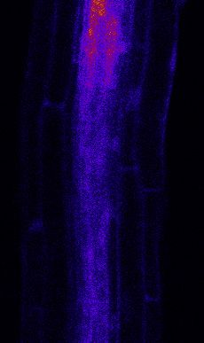
Please cite this article in press as: Emonet et al., Spatially Restricted Immune Responses Are Required for Maintaining Root Meristematic Activity upon
 Detection of Bacteria, Current Biology (2021), https://doi.org/10.1016/j.cub.2020.12.048

            ll
      OPEN ACCESS                                                                                                                             Article

                 A

                 B

                 C

                 D

                 E

                 F

Figure 4. Ca2+ Waves Are Non-Cell-Autonomous Responses
Ca2+-dependent signal in (A) WT (Col-0 background) and (B) WER::FLS2-GFP fls2, (C) CASP1::FLS2-GFP fls2, (D) SHR::FLS2-GFP fls2, (E) UBQ10::FLS2-GFP
fls2, and (F) fls2 (SAIL691_C04) lines in response to 1.25 mM flg22. Time series of UBQ10::R-GECO1 fluorescence: pictures are longitudinal middle sections of
                                                                                                                            (legend continued on next page)
8 Current Biology 31, 1–17, March 8, 2021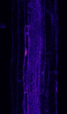
Please cite this article in press as: Emonet et al., Spatially Restricted Immune Responses Are Required for Maintaining Root Meristematic Activity upon
 Detection of Bacteria, Current Biology (2021), https://doi.org/10.1016/j.cub.2020.12.048

                                                                                                                                                  ll
Article                                                                                                                                     OPEN ACCESS

5E, 5F, and S5), fitting with previous reports.37 It is intriguing to             ectopic defenses in sensitive tissues might suffer from the pres-
speculate that PER5, ROS production, and other flg22-respon-                      ence of normally harmless bacteria and tip the balance between
sive genes, categorized as ‘‘oxidative stress’’ response genes,46                 growth and defense. The model commensal/beneficial Pseudo-
are actually part of a lignification response that stays below a                  monas protegens CHA0 does not induce MAMP responses in
productive threshold in WT but pivots into a full lignification                   WT plants, except at high concentration or if the root is
upon flg22 stimulation of FLS2 overexpression lines.                              wounded.13 However, when CHA0 was inoculated on roots of
                                                                                  WER::FLS2 seedlings in the absence of synthetic flg22 peptide,
Lignin Deposition Is Not Sufficient to Explain the Strong,                        a strong PER5 induction was observed (Figure 7A).
flg22-Induced Root Growth Inhibition                                                 To confirm that the induction of PER5 was caused by native,
The stronger root growth inhibition observed in the super-                        bacterial flg22, we inoculated seedlings with a CHA0 deletion
competent WER::FLS2 line could be due to the impact of lignin                     mutant defective for fleQ, which is required for the induction of
deposition in the transition zone. To test whether cell wall rein-                flagellum development,50,51 as well as fliC, coding for the flagellin
forcement by lignin prevents cell division and elongation, we in-                 protein itself.52 In contrast to the WT strain, DfleQ and DfliC
hibited lignin formation with the monolignol synthesis inhibitor                  mutants could not trigger any response in WER::FLS2, demon-
piperonylic acid (PA), expecting to restore root growth. However,                 strating that MAMP responses are induced by the direct FLS2-
even when lignin was no longer detectable, WER::FLS2 still                        mediated detection of bacteria-derived flagellin molecules
showed root meristem collapse (Figure 5I) and root growth inhi-                   (Figure 7A). In order to exclude that the absence of PER5 induc-
bition (Figure 5H). The rather weak and slow root growth inhibi-                  tion might be due to the impaired motility of the mutants (Fig-
tion observed in WT plants is not associated with meristem                        ure S6A), we inoculated both plant genotypes with the CHA0
collapse or lignification, suggesting that this growth inhibition is              insertion mutant fliC::pEMG, which produces non-functional
more indirectly related to immune activation in other tissues                     flagellin, including the flg22 sequence, due to the insertion of
and organs.                                                                       the pEMG plasmid (Figure S6B). Despite being equally impaired
                                                                                  in motility (Figure S6A), we found that fliC::pEMG mutant induces
Suberin Lamellae Deposition after flg22 Treatment Is an                           PER5 as efficiently as WT CHA0 strains (Figure 7A), excluding
Endodermis-Specific Response                                                      that impaired motility underlies the absence of PER5 induction
In the root endodermis, ectopic lignin deposition occurs as a                     in DfleQ and DfliC mutants. We also ascertained that all bacterial
compensatory mechanism for impaired Casparian strip forma-                        mutants were in direct contact with root epidermal cells (Fig-
tion in a manner similar to lignification in CASP1::FLS2 lines                    ure S6C) and that both mutant and WT bacteria were alive, as
treated with flg22. In the endodermis, compensatory lignification                 shown by colony-forming unit (CFU) counting from colonized
is often followed by suberin lamellae deposition.47 We therefore                  roots (Figure S6D). Although DfleQ mutant colonization was
investigated whether flg22 treatment would induce suberin                         slightly impaired, the DfliC mutant showed similar CFU counts.
deposition in the endodermis-expressing FLS2 lines. In WT                         Finally, heat-killed CHA0 WT bacteria and fliC::pEMG mutants,
plants, suberin deposition is usually restricted to the endo-                     but not DfleQ and DfliC mutants, induce PER5 (Figure S6E).
dermis, starting in the late differentiation zone by patches                      Together, this clearly indicates that the MAMP responses in the
(‘‘patchy zone’’) and then progressing to a fully suberized                       super-competent WER::FLS2 line are caused exclusively by
zone.48,49 Although no suberin induction by flg22 was found in                    native flg22 released from the bacteria.
WT (Figures 6A and 6B), lines expressing FLS2 in the endo-
dermis, such as CASP1::, SHR::, and UBQ10::FLS2, showed                           Root Colonization with P. protegens Does Not Cause
increased endodermal suberization, leading to a complete                          Enhanced Root Growth Inhibition in WER::FLS2
disappearance of the patchy zone (Figure 6B). WER::FLS2 roots,                    We then assessed the impact of CHA0 bacteria on root growth.
by contrast, displayed a normal proportion of patchy to fully su-                 Surprisingly, despite its induction of PER5, CHA0 did not signif-
berized zone despite a shorter root length (Figure 6A).                           icantly cause significant root growth inhibition in WER::FLS2
Conversely, root growth was not affected in CASP1:: and                           compared to WT in most replicates (Figure 7B). It has been re-
SHR::FLS2 lines, but suberin formed nevertheless much earlier                     ported that some commensal bacteria are able to attenuate
in response to flg22. Therefore, flg22 can induce suberization                    MAMP-triggered immune responses, either broadly or only the
only when FLS2 is expressed in the endodermis and suberization                    branches of the response that causes root growth inhibition,53,54
can be seen as a cell-type-specific flg22 response.                               which could explain this observation. Indeed, Ma et al.55 were
                                                                                  unable to observe any growth phenotype of WER::FLS2 plants
Super-competent WER::FLS2 Plants Can Detect Native                                grown in non-sterile soil, which they explain by their finding
Bacterial Flagellin                                                               that 41% of root commensals can suppress MAMP-triggered
The strong impact of flg22 on WER::FLS2 root growth and cell                      root growth inhibition in mono-associations.55 Interestingly,
wall modification prompted us to evaluate whether commensal                       they found that only a part of flg22-transcriptional responses
bacteria stimulate similar MAMP responses. Plants that mount                      were inhibited by a suppressive synthetic bacterial community,

roots at the EZ or DZ. Time 0:00 corresponds to the start of flg22 treatment. White arrows point at tissues showing a strong increase in Ca2+ content. Scale bar,
50 mm. Right panel shows normalized R-GECO1 fluorescence intensity (DF/F) measured in tissue-specific ROIs. Values present the dynamics of Ca2+ cytosolic
concentration in response to flg22 in a single root shown in left panel for each tissue type. Black arrows point at the maximum intensity of the trace. Gray
background corresponds to flg22 treatment. Representative pictures are presented. Eight to fifteen seedlings over two to six independent biological replicates
were analyzed for each genotype. Quantification was performed on at least three seedlings with similar results. See also Videos S1 and S2.

                                                                                                              Current Biology 31, 1–17, March 8, 2021 9
Please cite this article in press as: Emonet et al., Spatially Restricted Immune Responses Are Required for Maintaining Root Meristematic Activity upon
Detection of Bacteria, Current Biology (2021), https://doi.org/10.1016/j.cub.2020.12.048
ll
OPEN ACCESS Article
A B
C
D
E F
G H I
(legend on next page)
10 Current Biology 31, 1–17, March 8, 2021Please cite this article in press as: Emonet et al., Spatially Restricted Immune Responses Are Required for Maintaining Root Meristematic Activity upon
Detection of Bacteria, Current Biology (2021), https://doi.org/10.1016/j.cub.2020.12.048
ll
Article OPEN ACCESS
suggesting that suppression of root growth inhibition can be though often with great variation. However, one Pseudomonas
achieved without full suppression of all transcriptional isolate, R569, caused strongly enhanced PER5 induction (Fig-
responses. ure 7D) and root growth inhibition compared to WT (Figure 7E).
For this isolate, the effect was very robust and was repeatedly
WER::FLS2 Shows Enhanced Root Growth Inhibition to a observed both in Lausanne and Cologne laboratory growth con-
Restricted Subset of Commensal Bacteria ditions (Figure S6F). We demonstrated that synthetized flg22
In order to obtain a more comprehensive picture of how from isolate R569 (flg22R569) induced PER5 marker expression
WER::FLS2 affects responses to bacteria, we screened a set and root growth inhibition similarly to the ‘‘standard’’ flg22
of 34 bacterial strains from the At-SPHERE culture collection of from Pseudomonas aeruginosa (Figures S6G and S6H). These
root-derived commensals56 (Table S1) for both induction of effects were absent in the fls2 (SAIL691_C04) mutant back-
PER5 marker and enhanced root growth inhibition in WER::FLS2 ground. In addition, R569, but also the CHA0, Pseudomonas
compared to WT lines. We selected isolates to represent strains induce lignin deposition in WER::FLS2, but not in WT
different phyla, with a bias toward bacteria predicted by Gar- plants (Figure 7F). We conclude that mis-localized defense acti-
rido-Oter et al.53 to possess a flg22 peptide sequence recog- vation in the WER::FLS2 line can lead to strong root growth inhi-
nized by FLS2, based on sequence homology with the canonical bition in response to a root commensal isolate that is innocuous
flg22 sequence (Table S2). We also included some bacteria not when growing on WT roots.
predicted to be detected by FLS2. Among the 17 strains pre-
dicted to be recognized, only ten triggered an enhanced PER5 DISCUSSION
marker induction in WER::FLS2 (Figure 7C). Moreover, five
strains, with flg22 sequences predicted not to be detectable, It is not understood why only a restricted subset of root tissues
did induce the PER5 marker. This underlines the limitations in directly respond to MAMPs in the absence of other stimuli.13,19,20
computationally predicting flg22 activity from sequence alone The combination of tissue-specific receptor expression and
and the potential of the WER::FLS2 line to rapidly test experi- cellular resolution readouts presented here demonstrates the
mentally, using living and heat-killed bacteria, whether a native consequences of altering the spatial patterns of MAMP re-
bacterial flg22 can be detected by the plant. sponses in roots and reveals four important features.
Although half of the bacterial isolates could induce PER5 First, different MAMP responses are highly tissue specific and
marker specifically in the WER::FLS2 line in our conditions, varying in cell autonomy. Suberin, for example, is only induced in
most of them did not cause enhanced root growth inhibition (Fig- the endodermis, although lignification can be induced in a wider
ure 7C; Table S2). However, five of these isolates did show an variety of tissues by FLS2 expression. Although PER5 induction
enhanced effect on WER::FLS2 root growth compared to WT, appears cell autonomous, MYB51 and calcium signals are also
Figure 5. Epidermal Meristematic Expression of FLS2 Leads to flg22 Hypersensitivity and Meristem Collapse
(A) Flg22 treatment increases root growth inhibition in WER::FLS2 hypersensitive line. Root length quantification of prom::FLS2-GFP fls2 lines, fls2 (SAIL691_C04)
mutant, and WT (Col-0 background) transferred on 1 mM flg22 for 6 days post-inoculation (dpi). Boxplot center represents the median (n = 23–28 roots). Different
letters indicate statistically significant difference between means by Kruskal-Wallis test and Dunn’s multiple comparison (p < 0.05). Similar results were obtained
in two biological replicates.
(B) Representative pictures of quantification in (A), seedlings transferred for 6 days on 1 mM flg22. Scale bar, 1 cm.
(C) Flg22 induces strongly PER5::NLS-3mVenus in the epidermis of WER::FLS2-GFP fls2 hypersensitive line compared to WT (Col-0 background). On the right,
maximum projection of z stacks taken in root tips of plants treated for 24 h with 1 mM flg22 or mock is shown. Schematic represents the depth of the z stack.
Pictures were taken with identical settings. Scale bar, 25 mm. Representative pictures are presented (n = 15 on four replicates).
(D) WER::FLS2-GFP fls2 hypersensitivity is specific to flg22. WER::FLS2-GFP fls2 and WT plants (PER5::NLS-3mVenus in Col-0 background) were treated for
24 h with either 1 mM elf18, 2 mg/mL chitin, 1 mM 3-OH-C10:0, or 1 mM AtPep1. Maximum projection of z stacks taken in root tips is shown. PER5 induction is
highlighted with mVenus (Fire Lookup Table [LUT]). Parameters were identical for mock and treatment. Scale bar, 25 mm. Representative pictures are presented
(n = 12 on three replicates).
(E) Treatment of WER::FLS2-GFP fls2 for 2 days with 1 mM flg22 induces meristem swelling and lignin deposition. Upper panel shows median projection of
calcofluor-white-stained cell wall in the transition zone of the root tip (cyan). Note bulky cells of the epidermis (white arrowhead). Lower panel presents maximum
projection of lignin deposition stained with basic fuchsin (red). Lignin accumulates between cells only in WER::FLS2-GFP fls2 after flg22 treatment. WT represents
Col-0 background. Scale bar, 25 mm.
(F) Cross-section of pictures in (E). Cell wall stained with calcofluor white (cyan) is co-visualized with lignin stained with basic fuchsin (red). Flg22 treatment
induces massive swelling of cortex cells (white arrowheads) only in WER::FLS2-GFP fls2. Lignin is principally deposited between epidermal and cortex cells.
Epidermal cells are pushed apart by the swelling cortex and are sometimes missing. Scale bar, 25 mm.
(E and F) Representative pictures are presented (n = 15–20 on five replicates).
(G) Epidermal view of plasma membrane visualized by the construct UBQ10::mScarlet-SYP122 in WER::FLS2-GFP fls2. Cell division is disorganized after 1 mM
flg22 treatment. Scale bar, 25 mm.
(H) Inhibition of monolignol synthesis does not rescue meristem flg22-driven increased root growth inhibition of WER::FLS2-GFP fls2 compared to WT (Col-
0 background). Root growth measured after overnight pre-treatment with 10 mM PA inhibitor followed by 36 h 1 mM flg22 combined to 10 mM PA treatment is
shown. Boxplot center represents the median (16 % n % 27). Different letters indicate statistically significant difference (p < 0.05) between means by Kruskal-
Wallis test and Dunn’s multiple comparison. Similar results were obtained in two biological replicates.
(I) Flg22 induces meristem swelling despite inhibition of monolignol by PA treatment. Pictures were taken from samples quantified in (H). Upper panel shows
median projection of calcofluor-white-stained cell wall in the transition zone of root tip (cyan). Lower panel presents maximum projection of lignin deposition
stained with basic fuchsin (red). White arrowheads point at examples of bulky cells. Representative pictures are presented. Scale bar, 25 mm.
See also Figure S5.
Current Biology 31, 1–17, March 8, 2021 11Please cite this article in press as: Emonet et al., Spatially Restricted Immune Responses Are Required for Maintaining Root Meristematic Activity upon
Detection of Bacteria, Current Biology (2021), https://doi.org/10.1016/j.cub.2020.12.048
ll
OPEN ACCESS Article
A Figure 6. Suberin Deposition Is Triggered
by flg22 when Endodermal Cells Expressed
FLS2
(A) Quantification of suberized zone length in
seedlings treated for 1 day with 1 mM flg22 (18 % n
% 27). Data of two replicates were pooled. Roots
regions were classified as suberized, patchy, and
unsuberized zones. Error bars represent standard
error (SE). Different letters indicate statistically
significant difference among lines for the specified
zone (p < 0.05). Multiple comparison was per-
formed using ANOVA and Tukey’s tests for the
suberized zone, whereas Kruskal-Wallis and
Dunn’s tests were used for patchy and non-su-
berized zones.
(B) Whole root views of suberin lamellae deposi-
tion in CASP1:: and UBQ10::FLS2-GFP fls2 lines
compared to WT (Col-0 background) after 1 mM
flg22 treatment versus mock. Suberin was stained
B with Fluorol Yellow. Representative pictures from
quantification in (A) are presented. White arrow-
heads, start of patchy zone; yellow arrowheads,
start of fully suberized zones. Scale bar, 1 mm.
metabolites influencing root micro-
biota.17,59 However, epidermal cells
might need to have a competency for
strong responses if root cap damages
by pathogens or other stresses induce
FLS2 expression. Yet a constitutive
expression of FLS2 in the meristematic
epidermis leads to drastic changes in
induced in cells lacking detectable FLS2 receptor expression. It the root structure upon exposure to flg22, strongly affecting mer-
will be important to describe larger numbers of response genes istem activity and growth. This inhibition of root growth is faster,
for a comprehensive view of the cell-type-specific, cell-autono- much more severe, and possibly of a different nature than the
mous, and non-cell-autonomous branches of the flg22 rather weak and slow root growth inhibition observed upon
response. Cell-type-specific transcriptomic approaches, for flg22 treatment of WT roots.
example, can contribute to our understanding of tissue-specific Finally, despite lignification upon actual bacterial infection
immune pathways.57 The prom::FLS2 lines analyzed here can be being well documented and shown to restrict bacterial inva-
valuable tools to distinguish cell-autonomous responses from sion,8,60,61 treatment with single MAMPs was rarely seen to stim-
non-cell-autonomous MAMP responses. ulate lignin deposition, particularly in roots.37,62–64 Here, we
Second, we found that the vascular meristem is refractory to show that strong FLS2 expression enables a single MAMP to
flg22 even when expressing FLS2 receptor. The seemingly contra- induce lignification, probably by overriding endogenous negative
dictory finding to Wyrsch et al.21 can be explained by the whole-or- feedbacks that prevent this from happening at WT receptor
gan readouts used in the earlier work, as well as use of levels. This provides an opportunity to study MAMP-induced
LBD16::FLS2, thought to be a stele-specific line but that we found lignification in a simplified and reproducible setting. Interestingly,
to also express in other tissues (Figure S1J). Lack of downstream overstimulation of the developmental SCHENGEN pathway also
signaling components or increased activity of negative regulators leads to ectopic lignification, as well as defense gene induction,
could both be responsible for the stele’s inability to respond to suggesting unexpected commonalities between developmental
flg22. The vascular meristem might be particularly vulnerable to and immune receptor pathways.65–68 Nevertheless, lignification
an activation of defense as it contains early-differentiating phloem, only partly explains the severe root growth inhibition we observe.
providing nutrition and hormones to the entire growing meristem. Other factors produced in response to flg22 might also interfere
Third, we observed root regions that can be rendered super- with meristem function, such as basic coumarins,69 which inhibit
competent by FLS2 expression. We speculate that epidermal cellulose biosynthesis, resulting in meristem swelling similar to
meristematic cells are kept non-responsive in WT,13,19 because the one observed on WER::FLS2.70
the outer root cap cells can mount MAMP responses that are not Importantly, our work also reveals that overexpression of a sin-
detrimental to meristem function. This might be linked to the gle PRR in a competent but otherwise non-responsive cell type
particular fate of root cap cells that rapidly enter apoptosis bypasses the weak or absent immune responses to commensal
once they reach the transition zone.58,59 They do not contribute bacteria.13,19,53,71 Though bacteria can inhibit MTI,12,72 MAMPs
to the body of the root but excrete mucilage and secondary produced by rhizosphere bacteria might often be too low in
12 Current Biology 31, 1–17, March 8, 2021Please cite this article in press as: Emonet et al., Spatially Restricted Immune Responses Are Required for Maintaining Root Meristematic Activity upon
Detection of Bacteria, Current Biology (2021), https://doi.org/10.1016/j.cub.2020.12.048
ll
Article OPEN ACCESS
A
PER5::NLS-3mV
B C
PER5 induction
D E
PER5::NLS-3mV
F
Figure 7. WER::FLS2 Line Detects Endogenous Bacterial flg22
(A) CHA0 bacteria trigger a strong induction of PER5::NLS-3mVenus marker (Fire LUT) on WER::FLS2-GFP fls2 compared to WT (Col-0 background). Mutants
DfliC and DfleQ defective for flagellum lose their ability to induce detectable MAMP responses. The fliC::pEMG mutant presenting non-functional flagellin still
(legend continued on next page)
Current Biology 31, 1–17, March 8, 2021 13Please cite this article in press as: Emonet et al., Spatially Restricted Immune Responses Are Required for Maintaining Root Meristematic Activity upon
Detection of Bacteria, Current Biology (2021), https://doi.org/10.1016/j.cub.2020.12.048
ll
OPEN ACCESS Article
concentration to activate MAMP responses in the first place. B Plant growth conditions
Therefore, roots can appear largely unresponsive to bacterial B Bacterial strains and growth conditions
presence without additional stresses.13 The obvious root growth d METHOD DETAILS
phenotype triggered by MAMPs in WER::FLS2 lines proves to be B Plant plasmid construction
a powerful tool to investigate the effect of commensals on root B Elicitors and inhibitors treatments
immune responses. Our super-competent lines have allowed B Microscopy settings and image processing
for the first time to demonstrate stimulation of FLS2 by a native B Fixation and staining
2+
flagellin peptide from a commensal bacterium. Generally, the B Ca imaging on roots
cocktail of elicitors that bacteria is thought to release prevents B Suberin staining
assignment of a MAMP response to an individual MAMP.73 B Bacterial root inoculation assays
The WER::FLS2 line now generates a cell type that responds B CFU counting
only to a single MAMP and can test predictions about flg22 pep- B Swimming assay
tide detectability, release, and processing. Extending our d QUANTIFICATION AND STATISTICAL ANALYSIS
approach, the ectopic overexpression of potential PRR recep- B Cell-specific quantification
tors in the epidermal meristem cells could be used to functionally B Calcium signaling quantification
pair novel receptors and ligands. B Statistical analysis
It has become evident that immune responses cannot be un-
derstood without taking into consideration the specificities of
SUPPLEMENTAL INFORMATION
different cell types and developmental stages. Our data establish
the necessity for the plant to spatially restrict its immune re- Supplemental Information can be found online at https://doi.org/10.1016/j.
sponses. This spatial allocation of defense capacities might in cub.2020.12.048.
turn influence the microbial colonization pattern of the rhizo-
sphere. The new tools presented will pave the way for a better
understanding of bacterial community structures in roots. ACKNOWLEDGMENTS
We thank the Central Imaging Facility (CIF) of the University of Lausanne for
STAR+METHODS expert technical support. We particularly thank Thomas Boller (Basel), Jean-
Pierre Me traux (Fribourg), Silke Lehman (Fribourg), and Ines Wyrsch (Basel)
Detailed methods are provided in the online version of this paper for initiating this project with us. We thank Youssef Belkhadir (Vienna), Thors-
and include the following: ten Nürnberger (Tübingen), Corne Pieterse (Utrecht), Cyril Zipfel (Zürich), and
Peter Kupferschmied (Bern) as well as all members from the Geldner lab for
d KEY RESOURCES TABLE sharing material and/or helpful discussions and input. Finally, we are grateful
d RESOURCE AVAILABILITY to Artan Graf and Yasmine Genolet for their patience and assiduity for the
B Lead Contact manual curation of cell-type-specific quantification. This work was supported
by funds to N.G. from an ERC Consolidator Grant (GA-N: 616228–ENDOFUN)
B Materials Availability
and two consecutive Swiss National Science Fondation (SNSF) grants
B Data and Code Availability (CRSII3_136278 and 31003A_156261). F.Z. was supported by a European Mo-
d EXPERIMENTAL MODEL AND SUBJECT DETAILS lecular Biology Organization (EMBO) Long-Term Fellowship (ALTF 1139-2014)
B Plant material and A.E. by an EMBO Short-Term Fellowship (STF8066).
induces PER5 transcriptional readout. Maximum projection of z stacks imaging MZs and EZs treated with drop inoculation of bacterial solution of a concentration
of optical density 600 (OD600) = 0.01 or mock, respectively, is shown. Representative images were acquired at 1 dpi (n = 15 on five replicates). Acquisition was
done with identical settings. Scale bar, 25 mm.
(B) CHA0 does not induce consistently increased root growth inhibition in WER::FLS2-GFP fls2 compared to WT (Col-0 background). Root growth was quantified
at 6 dpi on plate inoculated with bacteria at OD600 = 10 3. Different letters indicate statistically significant difference (p < 0.05). Multiple comparison was per-
formed using ANOVA and Tukey’s test (n = 12–20). Similar results were obtained in half our replicates (5/10). In the other half, CHA0 induced a small, often non-
significant RGI.
(C) Proportion of natural isolates from At-SPHERE culture collection triggering stronger PER5::NLS-3mVenus induction and increased RGI on WER::FLS2-GFP
fls2 compared to WT seedlings (PER5::NLS-3mVenus in Col-0 background; yes) or not (no). Bacteria classified in ‘‘variable’’ presented contradictory results
between replicates. Bacteria flg22 sequence was predicted to be recognized by FLS2 (flg22 predicted) or not (flg22 not predicted). Numbers of bacterial isolates
in each category are indicated in color. Gray surfaces indicate identical bacteria strains.
(D) Pseudomonas isolate R569 from At-SPHERE culture collection triggers strong PER5::NLS-3mVenus (Fire LUT) induction on WER::FLS2-GFP fls2, but not in
WT (Col-0 background). Seedlings were imaged after 1-day treatment with OD600 = 0.01. Maximum projection of z stacks at MZ and EZ is shown. Scale bar,
50 mm. Representative pictures are presented. Nine to twelve seedlings over three independent biological replicates were analyzed for each genotype and
treatment.
(E) Isolate R569 induces a robust increased root growth inhibition on WER::FLS2-GFP fls2 compared to WT plants (Col-0 background). High concentration of
bacteria (OD600 = 0.1) is deleterious to both genotypes. Root growth was quantified at 6 dpi on plates inoculated with bacteria at OD600 = 10 1 to 10 4. Different
letters indicate statistically significant difference (p < 0.05). Multiple comparison was performed using Kruskal-Wallis and Dunn’s test (n = 20–27 by genotype by
treatment). Similar results were obtained in at least three replicates.
(F) Treatment of WER::FLS2-GFP fls2 for 3 days with 2 mL of CHA0 or R569 bacterial solution (OD600 = 0.01) induces lignin deposition (RedHot LUT) between the
epidermis and cortex of the differentiated zone. Cell wall is highlighted with calcofluor white (cyan). Representative images are presented (n = 10 on two rep-
licates). White arrowhead, lignin deposition. Scale bar, 25 mm.
See also Figure S6 and Tables S1 and S2.
14 Current Biology 31, 1–17, March 8, 2021You can also read