Protective Functions of Reactive Astrocytes Following Central Nervous System Insult
←
→
Page content transcription
If your browser does not render page correctly, please read the page content below
REVIEW
                                                                                                                                     published: 30 September 2020
                                                                                                                                   doi: 10.3389/fimmu.2020.573256

                                          Protective Functions of Reactive
                                          Astrocytes Following Central
                                          Nervous System Insult
                                          Mathias Linnerbauer 1,2 and Veit Rothhammer 1,2*
                                          1 Department of Neurology, Klinikum rechts der Isar, Technical University of Munich, Munich, Germany, 2 Department of

                                          Neurology, University Hospital Erlangen, Friedrich–Alexander University Erlangen–Nürnberg, Erlangen, Germany

                                          Astrocytes play important roles in numerous central nervous system disorders including
                                          autoimmune inflammatory, hypoxic, and degenerative diseases such as Multiple
                                          Sclerosis, ischemic stroke, and Alzheimer’s disease. Depending on the spatial and
                                          temporal context, activated astrocytes may contribute to the pathogenesis,
                                          progression, and recovery of disease. Recent progress in the dissection of
                                          transcriptional responses to varying forms of central nervous system insult has shed
                                          light on the mechanisms that govern the complexity of reactive astrocyte functions. While
                         Edited by:
                                          a large body of research focuses on the pathogenic effects of reactive astrocytes, little is
                        Edgar Meinl,
      Ludwig Maximilian University of     known about how they limit inflammation and contribute to tissue regeneration. However,
                 Munich, Germany          these protective astrocyte pathways might be of relevance for the understanding of the
                      Reviewed by:        underlying pathology in disease and may lead to novel targeted approaches to treat
                       Cinthia Farina,
       San Raffaele Scientific Institute
                                          autoimmune inflammatory and degenerative disorders of the central nervous system. In
                         (IRCCS), Italy   this review article, we have revisited the emerging concept of protective astrocyte
                  Khalil Sherali Rawji,
                                          functions and discuss their role in the recovery from inflammatory and ischemic disease
            University of Cambridge,
                     United Kingdom       as well as their role in degenerative disorders. Focusing on soluble astrocyte derived
                *Correspondence:          mediators, we aggregate the existing knowledge on astrocyte functions in the
                  Veit Rothhammer         maintenance of homeostasis as well as their reparative and tissue-protective function
    veit.rothhammer@uk-erlangen.de
                                          after acute lesions and in neurodegenerative disorders. Finally, we give an outlook of how
                  Specialty section:      these mediators may guide future therapeutic strategies to tackle yet untreatable
        This article was submitted to     disorders of the central nervous system.
               Multiple Sclerosis and
                   Neuroimmunology,       Keywords: protective, astrocytes, neuroinflammation, astrogliosis, neurodegeneration, multiple sclerosis,
              a section of the journal    ischemic stroke, Alzheimer’s disease
             Frontiers in Immunology

            Received: 01 July 2020
     Accepted: 14 September 2020
     Published: 30 September 2020
                                          INTRODUCTION
                            Citation:     Astrocytes are the most abundant cell type in the mammalian central nervous system (CNS) and
  Linnerbauer M and Rothhammer V
                                          responsible for a multitude of functions. During development, astrocytes arise from neural stem
       (2020) Protective Functions of
Reactive Astrocytes Following Central
                                          cells (NSCs) in the subventricular zone (SVZ) and migrate along radial glia processes to populate the
              Nervous System Insult.      CNS (1). Once their migration is complete, astrocytes differentiate into subgroups with a high
         Front. Immunol. 11:573256.       degree of functional and regional specialization (1–6). During postnatal development, astrocytes
   doi: 10.3389/fimmu.2020.573256          instruct the formation of excitatory and inhibitory synapses, support developmental myelination,

Frontiers in Immunology | www.frontiersin.org                                    1                                   September 2020 | Volume 11 | Article 573256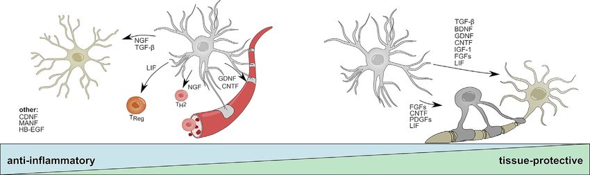
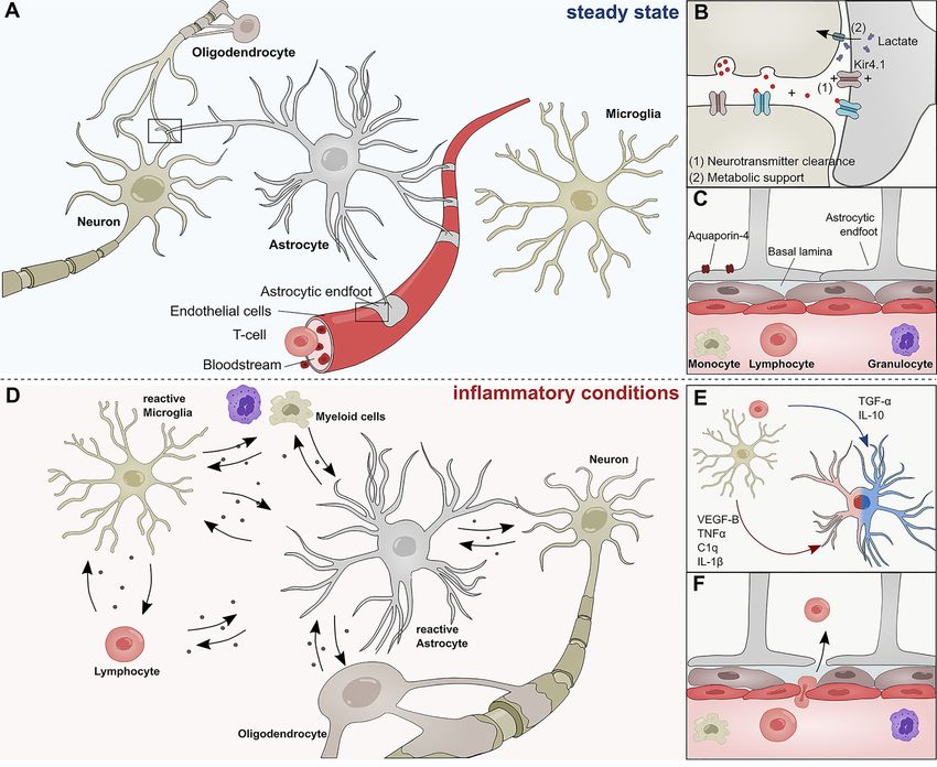
Linnerbauer and Rothhammer                                                                                                    Protective Functions of Reactive Astrocytes

and aid the establishment of complex neural circuitry through                           and other extracellular factors (16–20) (Figure 1B). In this
the secretion of soluble factors (7–10) and physical cell contact                       context, the inward rectifying K+ channel Kir4.1 has gained
(11–13). Throughout adulthood, astrocytes form close                                    attention as part of a K+ spatial buffering system that is
interactions with neurons to provide structural support and                             required for neuronal transmission and functioning (Figure
engage in metabolic coupling, serving as nutrient source and                            1B). Kir4.1 is highly expressed in astrocytic endfeet, and its
storage for neurons (Figures 1A, B) (14). Particularly lactate                          misregulation has been linked to numerous neurological
produced by astrocytes has been demonstrated to play an                                 disorders (21–23).
important role in the modulation of neuronal excitability and                              Besides their versatile role during neurogenesis and their
plasticity (15). Furthermore, astrocytes actively take part in                          contribution to the maintenance of neuronal circuitry, astrocytes
synaptic transmission and have been shown to modulate                                   are key participants in the formation and maintenance of the
cognitive functions through the clearance of neurotransmitter                           blood brain barrier (BBB) (24, 25) (Figure 1C). During CNS

 FIGURE 1 | Role of astrocytes in the steady state and inflammatory conditions. (A) Astrocytes interact with neurons, oligodendrocytes, microglia, and cells of the
 BBB during steady state conditions. (B) Astrocytes form tripartite synapses with neurons and regulate their synaptic transmission through metabolic support and the
 clearance of neurotransmitters. (C) Astrocytic endfeet line the cerebral vasculature and are a constituent of the blood brain barrier, thus limiting the infiltration of
 pathogens and peripheral immune cells into the central nervous system. Their endfeet express high levels of Aqp-4 and form a close interaction with pericytes and
 the basal lamina of the brain parenchyma. (D) During inflammatory conditions, reactive astrocyte secrete a plethora of inflammatory mediators that regulate functions
 of myeloid cells, lymphocytes, oligodendrocytes, neurons, and microglia. (E) Soluble inflammatory mediators derived from mircoglia and other immune cells
 differentially induce pathogenic (red) or protective (blue) astrocyte functions. (F) Peripheral immune cells pervade the BBB during inflammatory conditions and
 transgress into the CNS. C1q, Complement component 1q; IL-1b, Interleukin-1 b; IL-10, Interleukin 10; TNF-a, Tumor necrosis factor a; TGF-a, Transforming
 growth factor a; VEGF-B, Vascular endothelial growth factor B.

Frontiers in Immunology | www.frontiersin.org                                       2                                    September 2020 | Volume 11 | Article 573256
Linnerbauer and Rothhammer                                                                                   Protective Functions of Reactive Astrocytes

angiogenesis, astrocytes extend their polarized endfeet around               to factors secreted by reactive astrocytes under inflammatory
the abluminal side of cerebral blood vessels and aid early sprout            conditions (49). For instance, reactive astrocytes use contact- and
guidance and maturation by the expression of transporters, anti-             diffusion-mediated mechanisms to modulate trafficking of
permeability proteins and the secretion of growth factors (24–               peripheral immune cells into the CNS, a topic that has been
26). As key constituent of the glial limitans, astrocytic endfeet            extensively reviewed by Sofroniew and others (25, 28) (Figure
line the basement membrane surrounding the cerebral                          1F). Once the peripheral cells have extravasated, they accumulate
vasculature and provide a physical barrier between CNS and                   in perivascular spaces where they are in close contact to astrocytic
the peripheral blood system, thus limiting the influx of pathogens            endfeet (50). It is possible that during this stage, MHCII+ astrocytes
and large hydrophilic molecules (Figure 1C) (24, 25, 27–29).                 function as antigen-presenting cells to reactivate infiltrating
Furthermore, astrocytes control water homeostasis in the CNS                 lymphocytes and promote inflammation (51–53). Furthermore,
via Aquaporin-4 (Aqp4) and other channel proteins involved in                there is increasing evidence that astrocytes control the survival
bidirectional fluid exchange across the BBB (30) (Figure 1B).                 of T-cells and B-cells via co-regulatory and secreted factors.
The importance of Aqp4 in the CNS is demonstrated in a                       Indeed, while FasL expression by astrocytes induces cell death in
series of publications that link the (mal-)function of Aqp4 to               infiltrating lymphocytes, B cell–activating factor of the tumor
multiple neurological disorders (31–34). Aqp4 has also been                  necrosis factor (TNF) family (BAFF) produced by astrocytes
identified as a major target of autoantibodies in patients suffering          promotes B-cell survival in inflammatory conditions and
from neuromyelitis optica (NMO), a rare CNS inflammatory                      primary B cell lymphoma (54–56). Interestingly, astrocytes
disorder that has historically been closely associated to MS (35).           themselves respond to myeloid-derived APRIL, another member
    In addition to their versatile functions in the steady-state,            of the TNF superfamily with an increase in IL-10 production,
astrocytes sense and react to danger signals in a multistep process          consequently suppressing pro-inflammatory T-cell functions (57).
referred to as astrogliosis (36, 37). Combinatorial exposure to a            These interactions between reactive astrocytes and cells of the
broad spectrum of extracellular cues, including cytokines, growth            adaptive immune system are complemented by their functions as
factors, and hormones induces transcriptional remodeling,                    part of the cerebral innate immune system (58).
resulting in cellular hypertrophy, proliferation and secretion of                Another degree of complexity is added when analyzing the
inflammatory mediators (Figure 1D) (36). The severity and                     temporal dynamics of astrogliosis in the context of disease. In
permanence of these transcriptional changes is dependent on                  vivo ablation experiments of astrocytes in experimental
the type and strength of the stimuli and can range from reversible           autoimmune encephalomyelitis (EAE), an animal model of
alterations to severe astrogliosis with compact scar formation               Multiple Sclerosis (MS), demonstrated that astrocytes are
(36, 38). Most forms of astrogliosis share the upregulation of glial         required for disease suppression in early EAE stages, as loss of
fibrillary acidic protein (GFAP), a phenomenon that has been                  astrocytes worsened disease, characterized by increased BBB
observed in multiple CNS disorders (1, 39–41).                               permeability, leukocyte infiltration, and neuronal death
    For many decades, it was believed that severe astrogliosis and the       (59–62). Conversely, selective ablation of reactive astrocytes
formation of a glial scar inhibits axonal re-growth and is detrimental       during the chronic phase of EAE ameliorated disease, marked
for neurological outcome. However, an increasing amount of                   by decreased microglial activation and monocyte infiltration
evidence suggests that astrocytes also play beneficial roles in               (60). This and other studies underline the dire need to further
disease (42, 43). Methodological advances in the genomic analysis            dissect the contribution of astrocytes to the pathogenesis and
of reactive astrocytes have begun to shed light on the molecular             progression of numerous CNS disorders.
mechanisms that define the fine line between pathogenic and                        While many studies focus on the pathogenic potential of reactive
protective astrocyte functions. For instance, a landmark study by            astrocytes, molecular mechanisms underlying their protective effects
Zamanian and colleagues (44) demonstrated that astrocytes                    remain elusive at large. Here, we will discuss astrocyte-derived
respond differentially to varying forms of CNS insult. While                 mediators with anti-inflammatory or tissue-protective properties,
exposure to lipopolysaccharide (LPS) resulted in the upregulation            and examine how these factors may guide future therapeutic
of pro-inflammatory genes and skewed astrocytes toward a                      strategies. In this context we will not focus on protective astrocyte
cytotoxic profile, ischemia induced transcriptional programs that             functions mediated by inflammatory cytokines or cell-cell contact,
are associated with neuroprotective functions (44–47). In this               which have been reviewed extensively elsewhere (49, 63, 64), but
context, particularly intercellular crosstalk with microglia has been        rather concentrate on soluble factors often overlooked in the field
identified as key regulator of astrocyte functions. Work by several           of neuroinflammation.
groups including ours has unraveled molecular mechanisms
through which microglia-derived molecules such as interleukin
(IL)-1b, IL-10, tumor necrosis factor (TNF)-a, vascular
endothelial growth factor (VEGF)-B, or transforming growth                   PROTECTIVE EFFECTS OF REACTIVE
factor (TGF)- a, among others, modulate transcriptional                      ASTROCYTES FOLLOWING CNS INSULT
programs in astrocytes that are associated to degenerative or
protective functions (Figure 1E) (45, 48). In addition to                    A widely recognized protective function of astrogliosis is the
microglia, numerous other CNS-resident and non-CNS-resident                  formation of a physical barrier, which limits the influx of
cell types modulate astroglial properties and are themselves subject         peripheral immune cells and thus restricts lesion size

Frontiers in Immunology | www.frontiersin.org                            3                               September 2020 | Volume 11 | Article 573256
Linnerbauer and Rothhammer Protective Functions of Reactive Astrocytes
(28, 65–67). This function has been discussed in depth in a series BDNF
of excellent reviews (24, 25, 28, 37, 63). Here, we will focus on Brain-derived neurotrophic factor (BDNF) and nerve growth
astrocyte secreted mediators relevant for astrocyte protective factor (NGF) are members of the neurotrophin family and
functions. Advances in single cell sequencing, spatial highly expressed by astrocytes during development (109, 111–
transcriptomics, and conditional knock-down approaches 113). Throughout adulthood, astrocytes express low levels of
demonstrate that reactive astrocytes secrete a plethora of anti- BDNF but significantly upregulate its production in response to
inflammatory and tissue-protective mediators that act spinal cord injury (SCI) (114, 115), ischemia (115), and
on numerous cells to control their inflammatory state (Table 1). neuroinflammation (68, 69). BDNF signals through two
This review will focus on three major domains to summarize the receptors, the high-affinity TrkB receptor and the low-affinity
existing knowledge on astrocyte protective function: neurotrophic p75NTR receptor, both of which are expressed throughout the
factors, neuropoetic cytokines, and growth factors. CNS by neurons, astrocytes, and oligodendrocytes (116). While
BDNF/TrkB signaling on neurons has been shown to promote
Neurotrophic Factors survival and neurite outgrowth (117), p75NTR signaling induces
Neurotrophic factors (NTFs) play an essential role in the growth, apoptosis in cultured neurons (118). This dualistic signaling
differentiation, and survival of neurons in health and disease. system corresponds to the dichotomic effector functions of
They can broadly be divided into neurotrophins, members of the BDNF. Early studies in the context of axotomy and SCI
ciliary neurotrophic factor (CNTF) family, and members of the demonstrated beneficial effects of BDNF on the regeneration
glia derived neurotrophic factor (GDNF) family. While their role and long-term survival of neurons (119, 120) (Figure 2). In
in the survival of neurons is relatively well defined, little is EAE, reports suggest that BDNF depletion in CNS resident cells
known about inflammatory functions and how astrocytes during the initial phase worsens disease, while deletion during
contribute to their production. Generally, glial cells are known later stages does not lead to significant differences (121). Although
to express low levels of NTFs under homeostatic conditions, but this protective effect might depend on multiple cell types,
significantly upregulate their production following CNS damage astrocytes have been suggested to be a key participant in BDNF-
(109, 110). dependent remyelination in the cuprizone model of de- and
remyelination (68). This is supported by observations of
increased progenitor cell proliferation and maturation of
TABLE 1 | Tissue-protective mediators secreted by astrocytes.
neurons following lentiviral overexpression of Bdnf in
hippocampal astrocytes (70) (Figure 2). Furthermore, a study by
Mediator Disease Protective effect References Linker and colleagues (69) demonstrated that conditional depletion
model of BDNF in astrocytes worsens EAE severity. Interestingly, the
BDNF Ischemia; Promotes neuronal survival; increases (68–70) authors did not observe changes in infiltrating immune cells,
SCI; remyelination but demonstrated a significant increase in axonal loss
EAE and demyelination.
NGF SCI; Pro-NGF induces neuronal death; (71–74) Overall, these findings suggest that BDNF regulates axonal
TBI; mature-NGF promotes TH2
EAE differentiation, neuronal survival and
myelination and neuronal function through Trk/p75NTR signaling
increases phagocytosis of microglia on neurons and potentially oligodendrocytes, making it a key
GDNF PD; Promotes neuronal survival; increases (75–80) constituent of neuronal health. This also becomes clear in the
EAE tight junction function; regulates context of multiple neurodegenerative disorders, where a single
microglial activation
nucleotide polymorphism (SNP) in the BDNF gene is associated to
CNTF EAE; Increases neuronal survival, promotes (81–83)
SCI; tight junction functions; increases
increased susceptibility, incidence and severity of MS (122–124) and
remyelination Alzheimer’s disease (AD) (125), correlating with cognitive
MANF/ Ischemia; Reduces pro-inflammatory cytokine (84–86) dysfunction (126, 127). Although the relative contribution of
CDNF AD; production; promotes neuronal survival BDNF producing cells to the protective effects of BDNF remains
ER stress
under debate, a substantial body of evidence points to astrocytes as
PDGF Acute and Increases OPC population density; (87, 88)
family chronic regulates oligodendrocyte key drivers of BDNF mediated effects in disease. Furthermore,
members demyelination differentiation and proliferation astrocyte-derived BDNF has been identified as mediator of the
FGF Ischemia; Promotes neuronal survival; (89–91) therapeutic functions of glatiramer acetate (GA), a FDA-approved
family SCI; regulates oligodendrocyte drug for the treatment of relapse-remitting MS (RRMS) in a
members viral induced differentiation and proliferation;
demyelination reduces glial reactivity
mouse model of neurodegeneration, demonstrating the potential
HB-EGF in vitro Increases neuronal survival (92, 93) of astrocyte-derived BDNF for future therapeutic strategies (128).
IGF TBI Promotes neuronal survival (94–96) Nonetheless, recent evidence suggest that there is a fine line
TGF-b Ischemia; Reduces myeloid cell activation and (97, 98) between protective and pathogenic astrocyte-mediated functions
Toxoplasma pro-inflammatory cytokine production; of BDNF, as astrocytes themselves respond to increased levels of
infection promotes neuronal survival
LIF EAE; Increases stem cell renewal, (99–108)
BDNF with the secretion of neurotoxic amounts of nitric oxide
SCI; promotes oligodendrocyte (NO), demonstrating a sophisticated feedback loop that prevents
TBI differentiation and myelination excessive BDNF signaling (129).
Frontiers in Immunology | www.frontiersin.org 4 September 2020 | Volume 11 | Article 573256Linnerbauer and Rothhammer Protective Functions of Reactive Astrocytes FIGURE 2 | Anti-inflammatory and tissue-protective functions of reactive astrocytes. Activated astrocytes secrete soluble mediators with anti-inflammatory functions that help to resolve acute inflammation following CNS insult. NGF and TGF-b promote beneficial functions in microglia; LIF skews CD4 T-cells towards a regulatory phenotype; NGF promotes the differentiation into TH2 cells; GDNF and CNDF have beneficial effects on blood brain barrier permeability. CNDF, MANF, and HB-EGF have been associated to anti-inflammatory functions on multiple cell types or cells that are not displayed. During later stages, astrocyte-derived mediators promote the survival of neurons and oligodendrocytes and aid the long-term regeneration following CNS insult. TGF-b, BDNF, FGF family members, DNF, CNTF, IGF-1, and LIF increase neuronal survival; CNTF, LIF, and PDGF family members promote oligodendrocyte differentiation and myelination. BDNF, Brain derived neurotrophic factor; CNTF, Ciliary neurotrophic factor; FGF, Fibroblast growth factor; GDNF, Glial cell line-derived neurotrophic factor; HB-EGF, Heparin-binding epidermal growth factor; IGF-1, Insulin-like growth factor 1; LIF, Leukemia inhibitory factor; MANF, Mesencephalic astrocyte-derived neurotrophic factor; NGF, Nerve growth factor; PDGF, Platelet-derived growth factor; TH2, T helper type 2 cell; TReg, T regulatory cell; TGF-b, Transforming growth factor b. NGF and the presence of TrkA vs. p75NTR receptors on adjacent cells In contrast to BDNF, little is known about the immunomodulatory (72, 139). Of note, in addition to effects mediated by astrocyte and tissue-protective functions of astrocyte-derived NGF. Early derived NGF, a suppressive function of exogenously administered studies suggest upregulation of Ngf mRNA in astrocytes in NGF in reactive astrocytes has been proposed, implicating a models of traumatic injury, Parkinson’s disease (PD), and potential autocrine feedback loop that limits excessive astroglial neuroinflammation (130–132). Similar to BDNF, mature NGF is activation (140). cleaved from its precursor pro-NGF and signals via a dual receptor system consisting of TrkA and p75NTR (133). While mature NGF GDNF preferentially binds to TrkA and promotes neuronal survival, pro- The GDNF family of neurotrophic factors consists of GDNF, NGF has a higher affinity to p75NTR and has been shown to induce neurturin (NTRN), artemin (ARTN), and persepin (PSPN) apoptotic signaling in oligodendrocytes and neurons (134, 135). (141). All four members belong to the TGF-b superfamily and Under homeostatic conditions, pro-NGF and its mature form signal through the RET Tyrosine kinase to regulate the signal synergistically through TrkA/p75NTR to promote the differentiation and survival of multiple distinct sets of neurons survival and differentiation of neuronal cells; however, imbalances (141, 142). Interestingly, alternative signaling receptors, such as in the relative abundances of TrkA and p75NTR have been described the neural cell adhesion molecule (NCAM) have been proposed in multiple CNS disorders (133, 136–138). Interestingly, several and numerous studies suggest synergistic signaling with NGF, reports demonstrate that activated astrocytes secrete increased BDNF, and TGF-b (143–147). Reactive astrocytes rapidly amounts of neurotoxic pro-NGF in vitro and following SCI, upregulate the production of GDNF in response to LPS, IL-1b, suggesting a tissue-destructive role of endogenous, astrocyte- IFN-g and microglia-derived TNF-a, and have been shown to derived pro-NGF (71, 72). In contrast, treatment with exogenous promote the survival of dopaminergic neurons in vitro (75, 76). NGF has been shown to be beneficial in models of traumatic injury This is in line with in vivo studies that demonstrate beneficial and neuroinflammation (73, 74). For instance, administration of effects of astrocyte-specific overexpression of Gdnf in models of human NGF into the ventricle of marmoset monkeys prevented the Parkinson’s disease (PD) (77, 78). Furthermore, transplantation of development of lesions in an EAE model by skewing infiltrating T- human NPCs committed to a glial fate that have been genetically cells towards an anti-inflammatory TH2 phenotype (73) (Figure 2). engineered to overexpress GDNF promoted neuronal survival and In line with this observation, a recent study reports that NGF regeneration in primate models of amyotrophic lateral sclerosis instructs TrkA-mediated phagocytosis of neurotoxic Amyloid-b (ALS) (148, 149) (Figure 2). Besides the supportive effects of plaques by microglia in a mouse model of AD (74) (Figure 2). astrocyte-derived GDNF on neurons, GDNF/GDNFRa signaling While it remains to be seen, which impact astrocyte-derived NGF has been shown to promote the trans-endothelial resistance in an has on the net effect of NGF, the activation of astrocytes and in vitro BBB model, suggesting a positive effect of astrocyte- subsequent induction of Ngf expression by inflammation or stress- derived GDNF on tight-junction function and BBB permeability related events may contribute to both beneficial and harmful effects during neuroinflammation (79) (Figure 2). Collectively, further of NGF, depending on the availability of pro-NGF vs. mature NGF investigation into the anti-inflammatory and tissue-protective Frontiers in Immunology | www.frontiersin.org 5 September 2020 | Volume 11 | Article 573256
Linnerbauer and Rothhammer                                                                                  Protective Functions of Reactive Astrocytes

effects of astrocyte-derived GDNF is needed, but given the                  sense and respond to ER stress by negatively regulating NF-kB
synergistic signaling of GDNF in combination with TGF-b and                 dependent inflammatory programs (84, 165, 169–174). In
other NTFs, astrocytic GDNF may contribute to the reduction of              astrocytes, upregulation of both MANF and CDNF has been
inflammation and regenerative capacities following CNS insult.               observed in response to ER stress and experimental stroke, where
                                                                            they alleviate the secretion of pro-inflammatory cytokines IL-1b,
CNTF                                                                        TNF-a, and IL-6 (84–86). This is supported by a study using
CNTF composes a separate family of neurotrophic factors and has             astrocyte-specific overexpression of Manf, which resulted in a
been extensively studied as inducer of neuronal differentiation,            downregulation of pro-inflammatory cytokines (84). Taken
survival and neurite outgrowth (150). Besides its effect on                 together, this indicates that astrocytic MANF and CDNF
neurons, CNTF has been shown to support the maturation of                   function as cell-autonomous safety switch that prevents ER
oligodendrocytes and astrocytes (151–154) (Figure 2). It signals            stress induced overactivation and provides neurotrophic
through a heterotrimeric receptor complex consisting of the non-            support for neurons (Figure 2). Evidence from a Drosophila
signaling subunit ciliary neurotrophic factor receptor alpha                model of retinal tissue damage further suggests that MANF
(CNTFRa), and the two signaling chains glycoprotein-130                     counteracts the pro-inflammatory functions of VEGF–related
(gp130) and leukemia inhibitor factor receptor (LIFRb), which are           factor 1 (Pvf-1) homologue and is required for successful tissue
shared with the distantly related leukemia inhibitory factor (LIF)          repair (169). This is of particular interest in the context of glial
and interleukin-6 (IL-6) (155). Upon CNTF binding,                          communication, as VEGF secreted by microglia has been
heterodimerization of gp130 and LIFRb induces JAK/STAT                      demonstrated to induce pro-inflammatory signaling in
dependent transcriptional programs that are associated with the             astrocytes, and the successive MANF secretion by astrocytes
differentiation and survival of neurons (156). Under homeostatic            may present an anti-inflammatory mechanism that counteracts
conditions, the expression of low levels of Cntf in astrocytes is           pathogenic VEGF signaling (48).
limited to the white matter, indicating region-specific effects on
distinct neuronal subpopulations (157). Interestingly, this finding is
concordant with observations of increased Cntf expression in                Growth Factors and Neuropoietic
astrocytes and the upregulation of CNTFRa on neurons located                Cytokines
in white matter lesions of MS patients (158). A study investigating         PDGF Family Members
the spatial and temporal dynamics of multiple NTFs in a cuprizone           Platelet-derived growth factors (PDGFs) and their cognate
model of demyelination suggests that astrocytes express CNTF in a           receptors compose a signaling network that consists of five
biphasic manner during initial demyelination and remyelination              ligand-dimers (PDGF-AA, PDGF-BB, PDGF-AB, PDGF-CC,
(159). Mechanistically, it has been proposed that loss of physical          PDGF-DD) and three receptors (PDGFR-aa, PDGFR-bb,
interaction between astrocytes and neurons following injury induces         PDGFR-ab) (175). While PDGFs have originally been
STAT3-mediated Cntf expression in astrocytes, which promotes                identified as growth factor for smooth muscle cells (176), they
survival of neurons and oligodendrocytes and may counteract TNF-            are nowadays viewed as potent inducer of oligodendrocyte
a induced myelin disintegration during EAE (81–83) (Figure 2).              proliferation and differentiation (177, 178). Interestingly, the
Similar to GDNF, beneficial effects on BBB permeability and a                PDGF family of cysteine-knot growth factors also includes
reduction of immune cell infiltrates have been observed following            members of the VEGF subfamily, of which VEGF-B has been
administration of exogenous CNTF in a mouse model of                        shown to induce pro-inflammatory gene expression in astrocytes
neuroinflammation (160) (Figure 2). Collectively, the current data           (48). Similarly, astrocytes can also respond to PDGF-A and
indicates that astrocyte derived CNTF might contribute to the               PDGF-C by expression of PDGFR-a, which serves as mitogen
reduction of acute inflammation and increases the survival of                and inducer of astrocytic branching (179, 180). In addition, a
neurons and oligodendrocytes in the context of CNS insult. In               series of studies demonstrated that astrocytes express PDGF-A
addition, CNTF may promote the activation of surrounding                    and PDGF-B monomers, but not PDGF-C or PDGF-D
astrocytes in an autocrine/paracrine manner.                                (181–185). In the developing brain, these astrocyte-derived
                                                                            PDGF variants modulate the proliferation and differentiation
MANF/CNDF                                                                   of oligodendrocyte precursor cells (OPCs) (184, 186) and
Mesencephalic Astrocyte-Derived Neurotrophic Factor (MANF)                  potentially regulate the proliferation and survival of neurons
and Cerebral Dopamine Neurotrophic Factor (CDNF) constitute                 (187, 188). In the adult CNS, it remains unclear to what extent
a novel, evolutionary conserved family of NTFs with regenerative            astrocytes contribute to the PDGF signaling network, as neurons
capacities in health and disease. Although MANF and CDNF                    have also been proposed as source of PDGF-A and PDGF-B
have been originally identified to provide neurotrophic support              (189–191). Nevertheless, early work by Silberstein et al. (182)
for dopaminergic neurons, it has become clear that their                    indicates that cultured astrocytes upregulate the expression of
functions extend beyond those of classical NTFs (161–168).                  PDGFs in response to TNF-a and TGF-b, suggesting a role of
Both NTFs have been associated to numerous tissue-protective                PDGF signaling in inflammatory conditions. In this context, two
and anti-inflammatory functions in models of PD, ischemia and                independent studies investigated the therapeutic effects of
nerve injury (162–164, 168, 169). In addition, a series of recent           astrocyte-derived PDGF-A by conditional overexpression in
studies demonstrated that MANF and CDNF are partially                       mouse models of chronic and acute CNS demyelination and
retained within the endoplasmic reticulum (ER), where they                  revealed that elevated expression of PDGF-A by astrocytes

Frontiers in Immunology | www.frontiersin.org                           6                               September 2020 | Volume 11 | Article 573256
Linnerbauer and Rothhammer Protective Functions of Reactive Astrocytes
significantly increased OPC survival and population density (87, induces the expression of inflammatory genes in oligodendrocytes.
88) (Figure 2). While these findings may prove useful to address Overall, the existing data fails to produce a coherent picture on
the progressing demyelination in primary and secondary under which conditions FGF family members exhibit beneficial or
progressive MS and other degenerative CNS pathologies, harmful functions during CNS insult (206, 211) and extensive
important questions remain outstanding. Which programs research is needed to illuminate the effects of astrocyte-derived
control the expression of PDGFs in astrocytes? To what extent FGFs. Nevertheless, accumulating evidence strongly suggests that
do astrocyte-derived PDGFs modulate the functions of FGFs play an important role in the pathophysiology of MS and
oligodendrocytes and neurons? And what is their role in the (209, 212, 213) and new insights may guide the development of
remyelinating brain? Further research into the basic mechanisms FGF-based therapeutic strategies.
of CNS intrinsic signaling of PDGFs is needed to warrant success
in their use as future therapeutic target. HB-EGF
HB-EGF has originally been identified in macrophage-like cells
FGF Family Members with mitogenic functions for numerous cell types (214). Similar to
Fibroblast growth factors (FGFs) constitute a family of at least 20 NGF and other neurotrophins, HB-EGF is synthesized in a pre-
secreted ligands with pleiotropic roles in the developing and mature transmembrane form (pro-HB-EGF) before it is cleaved by
mature CNS (192–203). Most FGF receptors (FGFRs) can numerous metalloproteinases (MMP3, MMP9, ADM9, ADAM10,
respond to multiple FGF ligands (e.g. FGFR2 binds FGF1 to ADAM12, ADAM17) into its mature, soluble form (215). While
FGF10, whereas FGFR3 binds FGF1/2/4/8/9/17/18), creating a the membrane anchored pro-HB-EGF functions as juxtacrine
complex signaling network where a single FGF can induce growth factor and receptor for diphtheria toxin in some species,
distinct cellular responses. This notion is highlighted in a soluble HB-EGF has recently been described to modulate cell
recent article by Duong et al. (196), in which the authors migration, differentiation, and inflammatory functions in multiple
report FGF8 to function as cell fate switch that controls the cell types (216–222). In addition, HB-EGF enhances neurogenesis
differentiation of radial glial cells in the SVZ into neurons or in models of ischemic injury and promotes the survival of
astrocytes. Additional studies have demonstrated that FGFs dopaminergic neurons (223, 224) (Figure 2). Mature HB-EGF
regulate astrocyte morphogenesis, maturation, and function in signals through EGFR, ErbB4 and a newly defined N-arginine
both health and disease (197–199). For instance, in dibasic convertase, but may also be able to induce ErbB2 through
remyelinating lesions of MS, FGF-1 may act as a promoter of heterodimerization (214, 225–227). In astrocytes, upregulation of
remyelination by an indirect mechanism that involves the HBEGF mRNA has been observed in response to sphingosine-1-
induction of CXCL8 and LIF expression in astrocytes (204). phosphat (S1P)-receptor activation by S1P or S1P receptor
Furthermore, astrocytes have been recognized as important modulator fingolimod (92, 93). This may be dependent on
source of FGFs (192). Indeed, reactive astrocytes have been combined S1P1R and S1P2R signaling and the activation of the
found to upregulate FGF2 expression following CNS insult in immediate early transcription factors ERG1 and AP1, indicating
multiple species in vivo (89, 90) and in vitro (205). In particular, a that astrocyte-derived HB-EGF is part of a rapid response
study by Messersmith et al. (90) found significantly increased mechanism that counteracts pro-inflammatory astrocyte
FGF2 mRNA transcripts and protein levels associated to white functions (93). Indeed, it has been suggested that HB-EGF
matter astrocytes in the initial phase of remyelination, indicating suppresses the nuclear translocation of NF-kB by inhibition of
that astrocyte-derived FGF2 may modulate the differentiation of IkB kinase (IKK) mediated inhibitor of kB (IkB) degradation
oligodendrocytes (206) (Figure 2). Other potential effects of (222). Collectively, astrocyte-derived HB-EGF may not only serve
astrocyte-derived FGF2 include the attenuation of neuronal as neurotrophic factor but also dampen pro-inflammatory gene
death via signaling through FGFR3 (207) and autocrine/ transcription in Egfr-expressing microglia and infiltrating immune
paracrine regulation of glia reactivity (199). The therapeutic cells (220).
potential of FGF2 is recapitulated in a comprehensive study by
Ruffini et al. (208), in which the authors demonstrate that viral IGF
delivery of FGF2 to the CNS of mice 1 week after EAE induction Insulin-like growth factor 1 (IGF-1) is a polypeptide hormone
significantly ameliorated the clinicopathological outcome, and functions as primary mediator of growth hormone (GH)
marked by reduced infiltration of peripheral immune cells, and dependent growth effects in most peripheral tissues (228). In the
an increase of myelin-forming oligodendrocytes. It is unclear, brain, IGF-1 regulates the proliferation and differentiation of
however, to what extent astrocytes contribute to these beneficial multiple CNS resident cells and has been implicated in several
effects of FGF2. Indeed, several reports suggest that FGF2 in neurological disorders (229–231). IGF-1 signals through its
general, and astrocyte-derived FGF2 in particular can also inhibit cognate receptor IGFR-1R, but can also form functional
oligodendrocyte repopulation and their remyelinating capacities hybrids with the insulin receptor (229). Besides IGF-1, IGF-2
in multiple models of CNS insult (206, 209–211). Besides FGF2, and its receptor IGF-2R share a similar expression pattern in the
astrocyte-derived FGF9 has been implicated to play a role during developing and mature CNS (229). Both IGF/IGFR pairs signal
remyelination and CNS inflammation (212). Lindner et al. (212) through phosphoinositide 3-kinase (PI3K)–AKT–forkhead box
demonstrated in a series of in vitro experiments and post- protein O (FOXO) and RAS–mitogen-activated protein kinase
mortem tissue analyses of MS patients that FGF9, upregulated (MAPK) pathways to induce downstream expression of growth
by astrocytes following CNS insult, inhibits remyelination and promoting genes. Interestingly, IGF-1R can furthermore
Frontiers in Immunology | www.frontiersin.org 7 September 2020 | Volume 11 | Article 573256Linnerbauer and Rothhammer Protective Functions of Reactive Astrocytes
modulate transcription directly by acting as transcriptional its neurotrophic functions support axonal regeneration
regulator in the nucleus (232). Numerous studies indicate roles during recovery.
for IGF-1/IGF-1R signaling in the pathogenesis and progression
of neurological disorders and show that their expression is LIF
differentially modulated by CNS insult (159, 229, 231, 233, Leukemia inhibitory factor (LIF) is another member of the IL-6
234). While microglia have been implicated as main source of class cytokine family. Analogous to CNTF, LIF signals through
IGFs under pathological conditions, the neuroprotective LIFRa and gp130 to induce JAK/STAT dependent gene
potential of astrocyte-derived IGFs has recently gained transcription. It was first described as a suppressor of
attention (94–96). For example, conditional overexpression of proliferation in a myeloid leukemia cell line, but has since
IGF-1 in astrocytes promoted neuronal survival and reduced been associated to functions in multiple peripheral organs
hippocampal neurodegeneration in a controlled cortical injury (252–257). In addition, LIF has been recognized as
(CCI) model, highlighting the therapeutic efficacy of astrocyte- neuropoetic cytokine, regulating the differentiation and
derived IGFs (96) (Figure 2). Although the role of endogenous, activation of multiple cell types in the CNS (258–262). Under
astrocyte-derived IGFs in the context of neuroinflammation homeostatic conditions, expression of Lif remains low in the
must be further investigated, its broad spectrum of growth CNS, but is heavily ramped up in response to various types of
promoting effects on CNS-resident cells may provide beneficial insult (99, 100, 263–265). Astrocytes are thought to play an
for neuronal and non-neuronal regeneration. important role in the upregulation of LIF, and have been
identified as major source of Lif mRNA in the injured brain
TGF-b (100, 263). Consequently, astrocyte-derived LIF may potentiate
Transforming growth factor b (TGF-b) belongs to a family stem cell renewal in the adult SVZ and increase the regenerative
of pleiotrophic cytokines with potent regulatory and capacities following CNS insult (101, 262) (Figure 2). Although
inflammatory functions in numerous cell types (235–237). In it is not entirely clear what mechanisms modulate the
mammals, TGF-b exists in three isoforms (TGF-b1. TGF-b2, upregulation of Lif expression in astrocytes, S1PR signaling
TGF-b3), with TGF-b1 being the most prevalent one. The has been shown to be a potent inducer (92, 93). Apart from
immunoregulatory cytokine elicits its function through its beneficial functions on stem cell regeneration and
binding to TGF-b type I (TGF-bRI) and type II (TGF-bRII) neurogenesis, accumulating evidence shows that LIF plays
receptors, which induce Smad protein phosphorylation and essential roles during oligodendrocyte maturation and
downstream transcriptional regulation of their target genes function in the context of autoimmune inflammation and
(236, 238). Generally, TGF-b has been identified as master remyelination (102–107). This becomes important both in
regulator of immune tolerance, T cell differentiation and health and disease. A study by Ishibashi (100) demonstrated
mediator of inflammatory responses in multiple cell types that astrocytes secrete LIF in response to ATP stimulation and
(235, 236, 239, 240). In addition, work by Krieglstein and promote the oligodendrocyte-mediated myelination of axons,
others suggests that TGF-b also exerts neurotrophic functions defining a mechanism that mediates myelination in an activity-
through direct or indirect regulation of neuronal development dependent manner. During EAE, increased levels of LIF have
and survival (143, 241–248) (Figure 2). While members of the been associated to protective functions and increased survival of
TGF-b superfamily are widely expressed among numerous cell oligodendrocytes (102, 107) (Figure 2). In line with this notion,
types in the CNS, astrocytes have been implicated as key blockage of LIF worsened oligodendrocyte loss while
contributor of endogenous TGF-b in the CNS (249). Indeed, conditional deletion of a LIFR/gp130 suppressor protected
astrocyte-derived TGF-b has been linked to anti-inflammatory against cuprizone-induced demyelination (102, 266). Aside
and neuroprotective functions in models of experimental from oligodendrocytes, LIF has been implicated in the
stroke, Toxoplasma infection, and AD (97, 250, 251). regulation of T-cell responses by altering their pathogenic
Although the molecular mechanisms underlying the anti- potential. Indeed, several studies show that LIF suppresses
inflammatory and neuroprotective functions of astrocyte- pro-inflammatory gene expression in CD4 T-cells and skews
derived TGF-b in the context of neuroinflammation remain their polarization in an anti-inflammatory manner (267–269)
to be defined, TGF-b may exert its beneficial role through the (Figure 2). Collectively, these data suggest that astrocyte-
suppression of glial NF-kB signaling and the associated pro- derived LIF may contribute to the resolution of acute tissue
inflammatory functions of CNS resident macrophages and inflammation, promote the remyelinating capacities of
microglia (97) (Figure 2). This is in line with a study oligodendrocytes, and induce stem-cell renewal to prevent
defining an IL-10/TGF-b signaling loop between activated long-term neurodegeneration.
astrocytes and microglia that limits CNS inflammation (98).
Microglia-derived IL-10, an anti-inflammatory cytokine, Non-Secreted Factors
redirected astrocyte pathogenic functions and stimulated the In addition to neurotrophic factors, neuropoetic cytokines and
production of TGF-b, which in turn reduced microglial growth factors, astrocytes secrete a plethora of other protective
activation and the secretion of pro-inflammatory IL-1b factors, including cytokines, metabolites, extracellular matrix
(98). Taken together, astrocyte-derived TGF-b may serve as (ECM) proteins, and metalloproteinases (MMPs) (23, 50, 266,
immunosuppressive cytokine during initial inflammation while 267, 270). In the healthy brain, tight metabolic coupling
Frontiers in Immunology | www.frontiersin.org 8 September 2020 | Volume 11 | Article 573256Linnerbauer and Rothhammer Protective Functions of Reactive Astrocytes
between neurons and astrocytes is key to sustain high firing temporal expression (18, 289–293). Consequently, exogenous
rates and neuronal wellbeing (267). Recently, it has been activation of astrocytes at an improper time-point and in the
suggested that the metabolic crosstalk between astrocytes and wrong microenvironment might result in harmful, rather than
neurons also plays important roles during neuroinflammation beneficial effects, ultimately worsening clinical outcome. Further
and neurodegeneration (271–273). In this context, it remains to research is needed to determine (1) how many functionally
be defined which metabolites with protective functions during distinct astrocyte-subsets exist, (2) which factors induce their
homeostasis have similar effects in the inflamed or injured differentiation, (3) how the underlying transcriptional programs
brain. Similarly, astrocyte-derived ECM proteins and MMPs relate to the differential secretion of astrocyte-derived mediators,
have been associated to numerous protective functions in the and (4) whether these transcriptional subsets also correlate with
healthy brain. However, it has been well documented that different secretional and functional astrocyte subpopulations.
astrocyte-derived chondroitin sulfate proteoglycans (GSPGs), Most protective effects mediated by astrocytes are the result of
which are a key component of the ECM, restrict remyelination, a transient response to environmental cues present in the
neurite outgrowth and limit functional recovery following CNS disease-specific micro-environment. It is conceivable that the
injury (274). Among multiple other strategies to overcome diversity and strength of the intercellular crosstalk, specific to a
CSPG mediated inhibition of neuronal regeneration, MMPs given lesion type, also strongly influences the outcome of specific
have been proposed to exhibit protective effects in the post- therapeutic strategies. Indeed, while a transient and highly
acute phase of CNS injury (275, 276). Indeed, astrocyte-derived disease-specific astrocyte response allows for an adapted
MMPs may promote neuronal plasticity in the healthy brain reaction to the respective insult and prevents extensive
and enhance functional recovery through ECM dependent and overactivation/-suppression, exogenous induction of specific
independent mechanisms (277, 278). Future research will need tissue-protective pathways may only provide short-term
to determine which parameters dictate the protective effects of solutions to long-term problems, and eventually wear off when
astrocyte-derived ECM components and MMPs, and how they the local microenvironment changes over the course of
can be harnessed for therapeutic strategies to enhance recovery the disease.
following CNS insult. In these lines, genetic modifications of astrocytes to foster their
tissue-protective and anti-inflammatory functions have been
proposed (70, 73, 84, 96, 148, 149, 288). These approaches
might be particularly useful for the treatment of chronic
THERAPEUTIC OUTLOOK AND conditions and allow for the targeted activation of protective
DISCUSSION subpopulations. In this context, adeno-associated viruses
(AAVs) have been proven to be efficient vectors for viral gene
Currently, only few effective therapies exist to tackle the vast delivery. Interestingly, a landmark study by Foust and colleagues
complexity of neurological disorders and the development of (294) demonstrated that AAV serotype 9 successfully bypasses the
novel strategies is hampered by their limited access to the CNS. BBB and predominantly transduces astrocytes in the adult mouse
Exogenously administered agents require a high permeability brain (295). Further modifications such as the conditional
through the BBB and a persisting bioavailability to ensure long- expression of target genes under the astrocyte-specific GFAP
lasting therapeutic effects (279). Only a limited number of small promoter may enhance viral delivery and present a robust
molecules has shown beneficial “protective” effects on glial cells delivery system (296, 297). However, AAVs are limited to a
following acute CNS insult so far, which is best documented during relatively small insert size (4.7 kb) and viral delivery may have
neuroinflammation (92, 280–286). Thus, there is a dire need for unpredictable off-target effects. To overcome this complication,
novel strategies that mediate recovery after acute CNS insult and cell replacement strategies (using genetically modified or
lead to long-term regeneration in chronic inflammatory and unmodified cells) may present a useful alternative to harness the
degenerative diseases. Based on their strategic location and anti-inflammatory and tissue-protective functions of astrocytes.
versatile roles in the pathogenesis and progression of CNS Several transplantation trials of human fetal mesencephalic stem
disorders, astrocytes have been proposed as therapeutic targets cells into striatal regions of PD patients have demonstrated
(230, 231). Generally, most existing approaches targeting successful functional integration and long-term benefits (298–
astrocytes in the context of neurological disorders are based on 302). This is different for astrocytes, as they require differentiation
gene therapy, cell replacement, or the exogenous administration of into their mature form before being grafted. Although we have
compounds that induce neuroprotective functions in astrocytes little information about whether human astrocytes can be
(287, 288). As discussed above, multiple exogenously generated from embryonic stem cells (ESCs) or NSCs, several
administered molecules mimic the protective functions of studies report successful and stable differentiation of human ESCs
astrocyte-derived mediators or induce their endogenous into dopaminergic neurons, and transplantation of glial-restricted
production (70, 73, 84, 92, 93, 96, 148, 149, 160). Although these pluripotent stem cells in mouse models of ALS suggest that this
strategies represent promising approaches, several issues will need to might represent a feasible approach (303–307). One significant
be addressed. advantage over existing cell replacement therapies using neurons
First, the multi-faceted functions of astrocyte derived is that one astrocyte has the capacity to induce differentiation and
mediators are determined by their differential spatial and survival in numerous neurons (through the secretion of soluble
Frontiers in Immunology | www.frontiersin.org 9 September 2020 | Volume 11 | Article 573256Linnerbauer and Rothhammer Protective Functions of Reactive Astrocytes
factors), thus making it an efficient approach to tackle subsets beyond a dualistic concept will be inevitable to
neurodegeneration. Indeed, two ongoing Phase I/IIa trials understand their roles in the context of CNS inflammation.
(NCT03482050, with GDNF overexpression NCT02943850) Novel high-throughput technologies will pave the way for a
currently examine the therapeutic potential of grafted human better understanding of what signals drive the secretion of protective
stem cell derived astrocytes for the treatment of ALS. factors by astrocytes, to what extent these transcriptional profiles are
Overall, numerous studies presented in this review suggest influenced by intercellular communication, and how we can harness
that exogenous application or the genetic overexpression of the protective potential of reactive astrocytes in a clinical setting.
astrocyte derived factors limit inflammation and aid central
nervous regeneration. These findings need to be strengthened
and extensively recapitulated in other clinically relevant model AUTHOR CONTRIBUTIONS
species and CNS injuries before taking the next step towards
clinical application. Such models will allow to find common Both ML and VR researched data and reviewed and edited the
mechanisms underlying tissue-protective functions of astrocytes manuscript. ML wrote the manuscript.
and assess their translatability in a defined setting. Furthermore,
it will become essential to investigate the combinatorial effects of
astrocyte-derived factors as multiple studies have demonstrated FUNDING
synergistic effects and cross-regulatory mechanisms between
several of the discussed mediators (308–315). Lastly, we are This work was supported by grants RO4866/2-1 and RO4866/4-1
just beginning to grasp the versatile roles glial cells play in the from the German Research foundation (DFG) and an ERC Starting
diseased CNS and the extensive characterization of astrocytic Grant (851693 – HICI) by the European Research Council.
REFERENCES 13. Murai KK, Nguyen LN, Irie F, Yamaguchi Y, Pasquale EB. Control of
hippocampal dendritic spine morphology through ephrin-A3/EphA4
1. Molofsky AV, Deneen B. Astrocyte development: A Guide for the Perplexed. signaling. Nat Neurosci (2003) 6(2):153–60. doi: 10.1038/nn994
Glia (2015) 63(8):1320–9. doi: 10.1002/glia.22836 14. Magistretti PJ. Neuron–glia metabolic coupling and plasticity. J Exp Biol
2. Hochstim C, Deneen B, Lukaszewicz A, Zhou Q, Anderson DJ. (2006) 209(12):2304–11. doi: 10.1242/jeb.02208
Identification of positionally distinct astrocyte subtypes whose identities 15. Magistretti PJ, Allaman I. Lactate in the brain: from metabolic end-product
are specified by a homeodomain code. Cell (2008) 133(3):510–22. doi: to signalling molecule. Nat Rev Neurosci (2018) 19(4):235–49. doi: 10.1038/
10.1016/j.cell.2008.02.046 nrn.2018.19
3. Molofsky AV, Kelley KW, Tsai H-H, Redmond SA, Chang SM, Madireddy 16. Santello M, Toni N, Volterra A. Astrocyte function from information
L, et al. Astrocyte-encoded positional cues maintain sensorimotor circuit processing to cognition and cognitive impairment. Nat Neurosci (2019) 22
integrity. Nature (2014) 509(7499):189–94. doi: 10.1038/nature13161 (2):154–66. doi: 10.1038/s41593-018-0325-8
4. Ge W-P, Miyawaki A, Gage FH, Jan YN, Jan LY. Local generation of glia is a 17. Pascual O, Casper KB, Kubera C, Zhang J, Revilla-Sanchez R, Sul J-Y, et al.
major astrocyte source in postnatal cortex. Nature (2012) 484(7394):376–80. Astrocytic Purinergic Signaling Coordinates Synaptic Networks. Science
doi: 10.1038/nature10959 (2005) 310(5745):113–6. doi: 10.1126/science.1116916
5. Deneen B, Ho R, Lukaszewicz A, Hochstim CJ, Gronostajski RM, Anderson 18. Haim LB, Rowitch DH. Functional diversity of astrocytes in neural circuit
DJ. The transcription factor NFIA controls the onset of gliogenesis in the regulation. Nat Rev Neurosci (2017) 18(1):31–41. doi: 10.1038/nrn.2016.159
developing spinal cord. Neuron (2006) 52(6):953–68. doi: 10.1016/ 19. Santello M, Volterra A. Synaptic modulation by astrocytes via Ca2
j.neuron.2006.11.019 +-dependent glutamate release. Neuroscience (2009) 158(1):253–9. doi:
6. Rowitch DH, Kriegstein AR. Developmental genetics of vertebrate glial–cell 10.1016/j.neuroscience.2008.03.039
specification. Nature (2010) 468(7321):214–22. doi: 10.1038/nature09611 20. Walz W. Role of astrocytes in the clearance of excess extracellular potassium.
7. Christopherson KS, Ullian EM, Stokes CCA, Mullowney CE, Hell JW, Agah Neurochem Int (2000) 36(4):291–300. doi: 10.1016/S0197-0186(99)00137-0
A, et al. Thrombospondins are astrocyte-secreted proteins that promote 21. Cui Y, Yang Y, Ni Z, Dong Y, Cai G, Foncelle A, et al. Astroglial Kir4.1 in the
CNS synaptogenesis. Cell (2005) 120(3):421–33. doi: 10.1016/j.cell.2004. lateral habenula drives neuronal bursts in depression. Nature (2018) 554
12.020 (7692):323–7. doi: 10.1038/nature25752
8. Mauch DH, Nägler K, Schumacher S, Göritz C, Müller E-C, Otto A, et al. 22. Tong X, Ao Y, Faas GC, Nwaobi SE, Xu J, Haustein MD, et al. Astrocyte
CNS Synaptogenesis Promoted by Glia-Derived Cholesterol. Science (2001) Kir4.1 ion channel deficits contribute to neuronal dysfunction in
294(5545):1354–7. doi: 10.1126/science.294.5545.1354 Huntington’s disease model mice. Nat Neurosci (2014) 17(5):694–703. doi:
9. Hughes EG, Elmariah SB, Balice-Gordon RJ. Astrocyte secreted proteins 10.1038/nn.3691
selectively increase hippocampal GABAergic axon length, branching, and 23. Song F, Hong X, Cao J, Ma G, Han Y, Cepeda C, et al. Kir4.1 channels in
synaptogenesis. Mol Cell Neurosci (2010) 43(1):136–45. doi: 10.1016/ NG2-glia play a role in development, potassium signaling, and ischemia-
j.mcn.2009.10.004 related myelin loss. Commun Biol (2018) 1(1):1–9. doi: 10.1038/s42003-018-
10. Traiffort E, Kassoussi A, Zahaf A, Laouarem Y. Astrocytes and Microglia as 0083-x
Major Players of Myelin Production in Normal and Pathological Conditions. 24. Obermeier B, Daneman R, Ransohoff RM. Development, maintenance and
Front Cell Neurosci (2020) 14:79. doi: 10.3389/fncel.2020.00079 disruption of the blood-brain barrier. Nat Med (2013) 19(12):1584–96. doi:
11. Hama H, Hara C, Yamaguchi K, Miyawaki A. PKC signaling mediates global 10.1038/nm.3407
enhancement of excitatory synaptogenesis in neurons triggered by local 25. Abbott NJ, Rönnbäck L, Hansson E. Astrocyte–endothelial interactions at
contact with astrocytes. Neuron (2004) 41(3):405–15. doi: 10.1016/S0896- the blood–brain barrier. Nat Rev Neurosci (2006) 7(1):41–53. doi: 10.1038/
6273(04)00007-8 nrn1824
12. Barker AJ, Koch SM, Reed J, Barres BA, Ullian EM. Developmental control 26. Lee S-W, Kim WJ, Choi YK, Song HS, Son MJ, Gelman IH, et al. SSeCKS
of synaptic receptivity. J Neurosci Off J Soc Neurosci (2008) 28(33):8150–60. regulates angiogenesis and tight junction formation in blood-brain barrier.
doi: 10.1523/JNEUROSCI.1744-08.2008 Nat Med (2003) 9(7):900–6. doi: 10.1038/nm889
Frontiers in Immunology | www.frontiersin.org 10 September 2020 | Volume 11 | Article 573256You can also read