The Genomic Response to TGF-β1 Dictates Failed Repair and Progression of Fibrotic Disease in the Obstructed Kidney - Frontiers
←
→
Page content transcription
If your browser does not render page correctly, please read the page content below
REVIEW
                                                                                                                                                published: 02 July 2021
                                                                                                                                        doi: 10.3389/fcell.2021.678524

                                            The Genomic Response to TGF-β1
                                            Dictates Failed Repair and
                                            Progression of Fibrotic Disease in
                                            the Obstructed Kidney
                                            Craig E. Higgins 1 , Jiaqi Tang 1 , Stephen P. Higgins 1 , Cody C. Gifford 1 , Badar M. Mian 2,3 ,
                                            David M. Jones 4 , Wenzheng Zhang 1 , Angelica Costello 1 , David J. Conti 5 ,
                                            Rohan Samarakoon 1 and Paul J. Higgins 1,2,3*
                                            1
                                              Department of Regenerative and Cancer Cell Biology, Albany Medical College, Albany, NY, United States, 2 The Urological
                          Edited by:        Institute of Northeastern New York, Albany, NY, United States, 3 Division of Urology, Department of Surgery, Albany Medical
                        Meilang Xue,        College, Albany, NY, United States, 4 Department of Pathology and Laboratory Medicine, Albany Medical College, Albany,
   The University of Sydney, Australia      NY, United States, 5 Division of Transplantation Surgery, Department of Surgery, Albany Medical College, Albany, NY,
                       Reviewed by:         United States
                  Halesha Dhurvigere
                      Basavarajappa,
  Beckman Research Institute of City
                                            Tubulointerstitial fibrosis is a common and diagnostic hallmark of a spectrum of
              of Hope, United States        chronic renal disorders. While the etiology varies as to the causative nature of the
        Padmanabhan Pattabiraman,           underlying pathology, persistent TGF-β1 signaling drives the relentless progression
 Indiana University, Purdue University
          Indianapolis, United States       of renal fibrotic disease. TGF-β1 orchestrates the multifaceted program of kidney
                  *Correspondence:          fibrogenesis involving proximal tubular dysfunction, failed epithelial recovery or re-
                       Paul J. Higgins      differentiation, capillary collapse and subsequent interstitial fibrosis eventually leading to
                    higginp@amc.edu
                                            chronic and ultimately end-stage disease. An increasing complement of non-canonical
                    Specialty section:      elements function as co-factors in TGF-β1 signaling. p53 is a particularly prominent
         This article was submitted to      transcriptional co-regulator of several TGF-β1 fibrotic-response genes by complexing
                   Molecular Medicine,
                a section of the journal
                                            with TGF-β1 receptor-activated SMADs. This cooperative p53/TGF-β1 genomic cluster
  Frontiers in Cell and Developmental       includes genes involved in cellular proliferative control, survival, apoptosis, senescence,
                                Biology
                                            and ECM remodeling. While the molecular basis for this co-dependency remains to
           Received: 09 March 2021
                                            be determined, a subset of TGF-β1-regulated genes possess both p53- and SMAD-
            Accepted: 07 June 2021
            Published: 02 July 2021         binding motifs. Increases in p53 expression and phosphorylation, moreover, are evident
                              Citation:     in various forms of renal injury as well as kidney allograft rejection. Targeted reduction
     Higgins CE, Tang J, Higgins SP,        of p53 levels by pharmacologic and genetic approaches attenuates expression of the
    Gifford CC, Mian BM, Jones DM,
      Zhang W, Costello A, Conti DJ,
                                            involved genes and mitigates the fibrotic response confirming a key role for p53 in
Samarakoon R and Higgins PJ (2021)          renal disorders. This review focuses on mechanisms underlying TGF-β1-induced renal
  The Genomic Response to TGF-β1
                                            fibrosis largely in the context of ureteral obstruction, which mimics the pathophysiology
           Dictates Failed Repair and
Progression of Fibrotic Disease in the      of pediatric unilateral ureteropelvic junction obstruction, and the role of p53 as a
                   Obstructed Kidney.       transcriptional regulator within the TGF-β1 repertoire of fibrosis-promoting genes.
      Front. Cell Dev. Biol. 9:678524.
     doi: 10.3389/fcell.2021.678524         Keywords: fibrosis, PAI-1, transcription, TGF-β, p53

Frontiers in Cell and Developmental Biology | www.frontiersin.org                  1                                               July 2021 | Volume 9 | Article 678524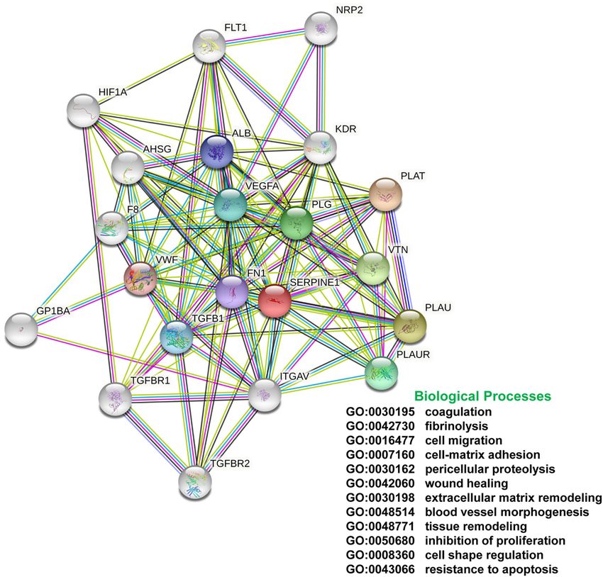
Higgins et al. TGF-β1 in Renal Fibrosis
THE CLINICAL REALITIES OF CHRONIC Indeed, the extent of tubulointerstitial pathology (i.e., degree of
RENAL DISEASE inflammation, tubular dysmorphism and atrophy, progressive
fibrosis) has important functional and prognostic implications
Acute kidney injury (AKI) and chronic kidney disease (CKD) (Grande et al., 2010; Truong et al., 2011; Eddy, 2014). While
comprise a rapidly growing medical and economic burden within older individuals constitute the majority of the at-risk cohort
the US as well as globally. Renal tubular epithelial trauma and (60% of the population >80 years have CKD), with collateral
subsequent cell death correlates with patient morbidity and age-dependent increases in cardiovascular complications (Ruiz-
mortality and, when severe or episodic, often progresses to CKD Ortega et al., 2020), children are also susceptible. In 2016,
and eventual end-stage renal disease (ESRD) (Bonventre and approximately 5,700 pediatric patients developed ESRD due to
Yang, 2011; Kaissling et al., 2013; Ferenbach and Bonventre, several causative factors with a mortality incidence 30-times that
2015; Kumar, 2018; Liu et al., 2018). Epidemiologic data suggest of their healthy counterparts (McDonald et al., 2004; Kramer
that CKD may be the most under-recognized public health issue et al., 2009; Centers for Disease Control and Prevention, 2016; US
impacting 1 in 7 (35 million) adults in the US with 90% of Renal Data System, 2018). Indeed, the primary causes of pediatric
affected individuals unaware of their underlying condition (Tuot CKD and ESRD are congenital anomalies of the kidney and
et al., 2011; Centers for Disease Control and Prevention, 20161 ; urinary tract (Ingraham and McHugh, 2011; Chevalier, 2016).
US Renal Data System, 2018). The Global Burden of Disease Unilateral ureteropelvic junction (UPJ) obstruction, with an
Study2 (Bowe et al., 2018; O’Brien, 2019) estimated that over incidence of 1:500–1,500 live births, is the most common form of
the period from 2002 to 2016, deaths due to CKD rose 58%. obstructive uropathy associated with end-stage disease although
Moreover, disability adjusted life years lost to CKD climbed other contributors include ureterovesical junction blockage,
41% while years living with disability and years of life lost posterior urethral valve disease, urethral atresia or stricture and
to CKD increased by 48 and 56%, respectively. Diabetes and neuropathic bladder (Ucero et al., 2010; Weitz et al., 2017).
hypertension are the primary and secondary drivers, respectively,
of CDK and ESRD (NIDDK Health Information Website)3 ; other
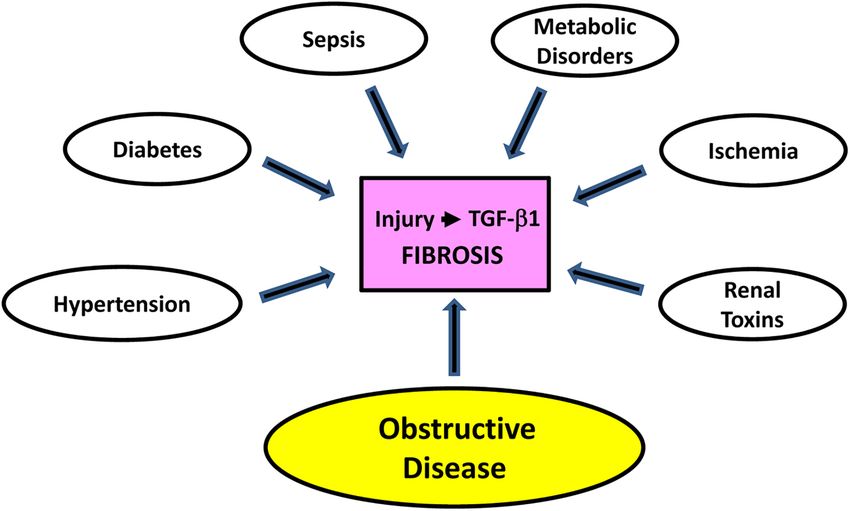
prominent contributors include sepsis, ischemia/reperfusion TUBULOINTERSTITIAL INJURY: THE
injury, obstructive nephropathy, metabolic disorders, and dietary BASICS
exposure to nephrotoxins (Uchino et al., 2005; Bagshaw et al.,
2008; Emlet et al., 2015; Figure 1). Medicare costs for patients Extensive or recurring sublethal epithelial trauma, usually in
with all stages of CKD approximated $114 billion in 2016 alone the context of persistent transforming growth factor-β1 (TGF-
($35 billion for ESRD and $79 billion for the treatment of β1) pathway activation, initiates and sustains a program of
individuals with CKD without end-stage organ failure). Race, age maladaptive repair that facilitates the progression of AKI to
and economic disparities are prevalent in the renal disease patient CKD (Friedman et al., 2013; Emlet et al., 2015; Ferenbach
population (Luyckx et al., 2018) and the overall incidence as well and Bonventre, 2015; Venkatachalam et al., 2015; Basile et al.,
as expenditures continue to rise with limited effective therapies 2016; Takaori et al., 2016; Chang-Panesso and Humphreys,
on the horizon (Ruiz-Ortega et al., 2020). 2017; Schnaper, 2017; Chung et al., 2018; Qi and Yang, 2018;
Regardless of etiology, progressive tubulointerstitial fibrosis Gewin, 2019; Tang et al., 2020; Figure 2). The major source
is the final common pathway to CKD and a hallmark of ESRD of TGF-β1, as well as other proinflammatory cytokines, in
(Eddy, 2005, 2014; Bonventre, 2010; Zeisberg and Neilson, 2010). the kidney is the injured epithelium although both resident
and infiltrative macrophages are also major contributors
(Bonventre and Yang, 2011; Liu et al., 2018; Black et al., 2019;
Abbreviations: AQP2, aquaporin 2; ALK, activin-like kinase; AKI, acute kidney
injury; ATM, ataxia telangiectasia mutated; ATR, ataxia telangiectasia and Rad3-
Zhang et al., 2020). Repetitive tubular damage triggers renal
related serine/threonine-protein kinase; CASP, chronic kidney disease-associated inflammation, pericyte loss, subsequent capillary rarefaction and
secretory phenotype; CHK1, checkpoint kinase 1; CHK2, checkpoint kinase 2; tissue hypoxia, epithelial dedifferentiation, G2 /M growth arrest,
CKD, chronic kidney disease; CTGF, connective tissue growth factor; DEG, tubule dysfunction and nephron dropout (Basile, 2004; Fine and
differentially expressed genes; Dot1l, disruptor of telomeric silencing-1-like; ECM,
extracellular matrix; EDA, extra domain A; ESRD, end stage renal disease; Norman, 2008; Yang et al., 2010; Moonen et al., 2018; Kumar,
FnEDA, extra domain A splice variant of fibronectin; GARP, glycoprotein A 2018; Zhang D. et al., 2018; Zhang S. et al., 2018; Liu et al.,
repitious predominant; KIM-1, kidney injury molecule-1; LAP, latency-associated 2019). Necrotic or apoptotic renal epithelial cells also release
peptide; LTBP, latent transforming growth factor-β1 binding protein; MRTF,
various damage-associated molecular pattern (DAMP) factors
myocardin-related transcription factors; PAI-1, plasminogen activator inhibitor-1;
PIF-alpha, pifithrin-α; RGD, arginine-glycine-aspartic acid; SASP, senescence- that activate toll-like receptors and stimulate the innate immune
associated secretory phenotype; SERPIN, serine protease inhibitor; SERPINE1, system prolonging the inflammatory response (Liu et al., 2018).
serine protease inhibitor, clade E, member 1; siRNA, small interfering RNA; SRF, Non-resolving inflammation precedes, and likely promotes,
serum response factor; TASCC, target or rapamycin-autophagy spatial coupling
components; TCF, ternary complex factors; TGF-α1, transforming growth
renal interstitial fibrosis (Bascands and Schanstra, 2005;
factor-β1; TGF-βR, transforming growth factor-β receptor; TAZ, transcriptional Chevalier et al., 2010; Meng et al., 2015, 2016; Li et al., 2017).
coactivator with PDZ-binding motif; UPJ, ureteropelvic junction; UUO, unilateral The extent of tubulointerstitial pathology (i.e., degree of
ureteral obstruction; YAP, yes-associated protein. inflammation, tubular dysmorphism and atrophy, progressive
1
nccd.cdc.gov/ckd fibrosis) has critical functional and prognostic implications
2
www.healthdata.org/gbd (Grande et al., 2010; Truong et al., 2011; Eddy, 2014). Increased
3
www.nikkd.nih.gov/health-information/kidneydisease angiotensin II and TGF-β1 levels in the injured kidney stimulates
Frontiers in Cell and Developmental Biology | www.frontiersin.org 2 July 2021 | Volume 9 | Article 678524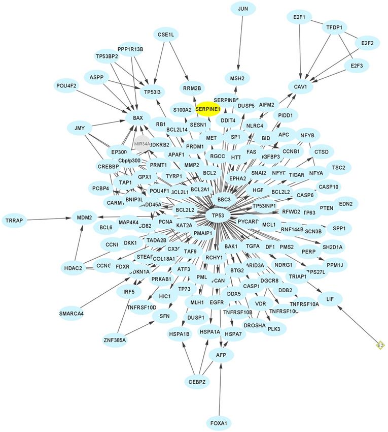
Higgins et al.                                                                                                                                       TGF-β1 in Renal Fibrosis

  FIGURE 1 | Contributors to renal damage. Extensive trauma or episodic epithelial injury, regardless of etiology and usually in the context of persistent transforming
  growth factor-β1 (TGF-β1) pathway activation, initiates and sustains a program of maladaptive repair that facilitates the progression of AKI to CKD. While diabetes
  and hypertension are preeminent initiators of CKD, sepsis, metabolic disorders, ischemia/reperfusion injury, exposure to nephrotoxins, and obstructive nephropathy
  are other significant causative factors. Several animal models of renal injury lend themselves to the discovery of genes and pathways that contribute to the onset and
  progression of kidney fibrosis. Unilateral ureteral obstruction (UUO) in rodents (either complete or partial/reversible), for example, is one of the most widely used as it
  approximates the pathophysiology of human obstructive nephropathy in children and adults. Ureteral ligation is a relatively simple procedure and produces a highly
  reproducible pathological response over a short time course with minimal inter-animal variability. UUO provides a translationally-relevant in vivo platform to probe the
  genomic complexity of kidney injury, mechanisms underlying maladaptive repair and the efficacy of new therapeutic approaches to the management of fibrotic
  disease (Martínez-Klimova et al., 2019).

the conversion of activated Gli1+ /FOXD1+ vascular pericytes                              an accelerated context) human obstructive nephropathy while
and interstitial fibroblasts to matrix-producing myofibroblasts                           bridging the pathologic features of AKI and CKD (Moller et al.,
driving the pathophysiology of tissue fibrosis (Qi et al., 2006;                          1984; Hruska, 2002; Ucero et al., 2014). Ureteral ligation provides
Picard et al., 2008; Ricardo et al., 2008; Grande and Lopez-                              an accessible, translationally-relevant, in vivo opportunity to
Novoa, 2009; Cook, 2010; Humphreys et al., 2010; LeBleu et al.,                           clarify the genomic complexity of renal fibrotic disease, dissect
2013; Duffield, 2014; Gomez and Duffield, 2014; Kramann and                               critical pathophysiologic events underlying the kidney response
Humphreys, 2014; Richter et al., 2015; Kramann et al., 2015;                              to injury and identify mechanisms involved in maladaptive repair
Mack and Yanagita, 2015). Pericyte mobilization in response                               (Klahr and Morrissey, 2002; Truong et al., 2011; Eddy et al.,
to injury, moreover, results in their interstitial translocation,                         2012; Samarakoon et al., 2012; Arvaniti et al., 2016; Sun et al.,
effectively promoting peritubular capillary collapse and creation                         2016; Jackson L. et al., 2018; Jackson A. R. et al., 2018; Martínez-
of a hypoxic environment (Kramann et al., 2013; Kramann and                               Klimova et al., 2019; Pavkovic et al., 2019).
Humphreys, 2014).                                                                             Surgical interference with the flow of urine increases
                                                                                          hydrostatic pressure initially in the collecting ducts expanding
                                                                                          rapidly to the distal and proximal tubules (Martínez-Klimova
EXPERIMENTAL OBSTRUCTIVE                                                                  et al., 2019). Long-term obstruction results in outer medullar
NEPHROPATHY: A TOOL TO PROBE                                                              ablation and tubular atrophy; a 65% decrease in proximal
MECHANISMS AND PATHWAYS                                                                   tubule mass becomes evident within 14 days of ureteral
                                                                                          ligation. Tubule dilation, epithelial necrosis/apoptosis, basement
Several animal models of acute and chronic renal disease are                              membrane denudation, rapid influx of inflammatory cells,
amenable to the discovery of causative factors underlying the                             interstitial expansion with increased cellular proliferation and
onset and progression of kidney fibrosis while affording a                                eventual fibrosis are prominent in the cortex of the ligated
platform to assess the efficacy of therapeutic interventions (Ortiz                       kidney (Cochrane et al., 2005; Manucha, 2007; Forbes et al., 2011,
et al., 2015; Nogueira et al., 2017; Bao et al., 2018). Unilateral                        2012; Ucero et al., 2014). The proximal tubule appears to be the
ureteral obstruction (UUO) in rodents (e.g., Chevalier, 2015;                             predominant sensor and immediate effector of renal damage and
Martínez-Klimova et al., 2019), for example, closely mirrors (in                          may well orchestrate disease progression via injury-associated

Frontiers in Cell and Developmental Biology | www.frontiersin.org                     3                                                 July 2021 | Volume 9 | Article 678524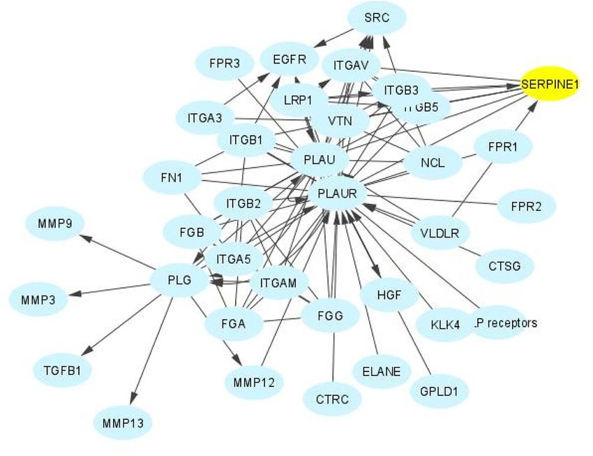
Higgins et al.                                                                                                                                          TGF-β1 in Renal Fibrosis

  FIGURE 2 | Repair outcomes in the injured kidney. Mild or sublethal AKI initiates a process of adaptive repair that involves resolution of inflammation and restoration
  of tubular architecture with a regain of renal function. Recurring AKI or severe tubular trauma, in contrast, results in maladaptive repair which is characterized by a
  sustained inflammatory response, stalling of injured proximal tubular epithelial cells in the G2 /M stage of the cell cycle, interstitial translocation of vascular pericytes
  and their differentiation into ECM-producing myofibroblasts, tubular atrophy, capillary loss and failure to regenerate a functional epithelium. G2 /M-stalled proximal
  tubular cells express significant levels of the potent pro-fibrotic effectors TGF-β1 and CCN2 which contribute to the initiation and progression of renal fibrosis.

tubular shortening and/or paracrine mechanisms that impact                                  phosphorylation, susceptibility to ischemic injury and relative
several resident renal cell types (Endo et al., 2015; Tan et al., 2016;                     deficiency of anti-oxidant/anti-apoptotic factors (Chevalier,
Gewin et al., 2017). A significant fraction (46%) of glomeruli,                             2016). The distal nephron including the collecting duct, however,
moreover, exhibit atrophic proximal tubules and 39% eventually                              also contributes significantly to the overall response of the kidney
become atubular indicating that the glomerulotubular junction                               to ureteral ligation-induced injury (Hiatt et al., 2013). Tubular
tubular epithelium is particularly sensitive to UUO-induced                                 dilation and myofibroblast accumulation in the distal nephron
necrosis and/or apoptosis (Chevalier et al., 2011). One suggestion                          increases by 2—3- and 6-fold, respectively, in the obstructed
is that glomerulotubular junction cell death may be a key driver                            kidney and coupled to a change in cellular composition of
of nephron loss and that the subsequent fibrotic response reflects                          the collecting duct. Aquaporin 2 (Aqp2)-expressing principal
an attempt at self-limiting tissue repair (Chevalier, 2016). Such                           cells decline by 65% and intercalated cell abundance decreases
congenital reduction in nephron density impairs recovery from                               by 75%. E-cadherin- and β-catenin-mediated collecting duct
obstructive injury and exacerbates the fibrotic process (Sergio                             epithelial adhesion is also disrupted. Notably, these features are
et al., 2015).                                                                              replicated in the distal and connecting tubules (Hiatt et al.,
    Partial and complete UUO in neonatal rodents are similar                                2013) confirming that the distal nephron is a major target of
except for a temporal offset in acquisition of pathologic features                          UUO-initiated renal disease, highlighting the utility of UUO
(Jackson L. et al., 2018; Jackson A. R. et al., 2018). UUO                                  as a model to dissect the involvement of collecting duct and
modeling largely focuses on the proximal tubular compartment                                distal tubule injury to kidney repair and fibrosis. Principal cells
due to its high mitochondrial load, dependency on oxidative                                 in the collecting duct are fundamental to the development of

Frontiers in Cell and Developmental Biology | www.frontiersin.org                       4                                                 July 2021 | Volume 9 | Article 678524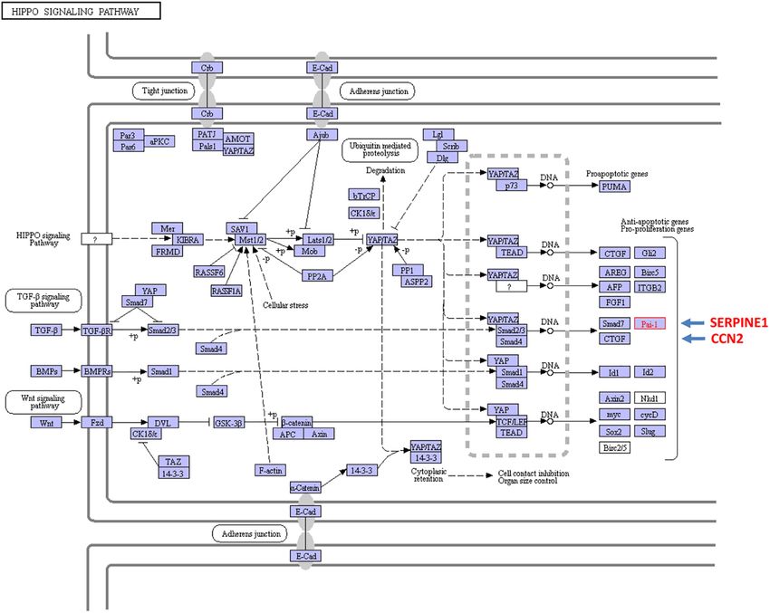
Higgins et al.                                                                                                                TGF-β1 in Renal Fibrosis

tubulointerstitial fibrosis (Butt et al., 2007; Ivanova et al., 2008;        Following tubular cell necrosis or apoptosis, the remaining viable
Fujiu et al., 2011), at least in part through Notch signaling, and are       epithelium undergoes morphologic dedifferentiation (i.e., loss of
subject to epigenetic regulation (Zhang et al., 2020). Mib1, an E3           polarity with cell spreading and migration to cover the exposed
ligase produced by ligand-expressing cells, is required for efficient        areas of the basement membrane) and subsequent proliferation
Notch mobilization while inactivation of Mib1 in the collecting              as an attempt to restore the functional integrity of the nephron
duct results in increased tubulointerstitial fibrosis and apoptosis          (Bonventre, 2003). Fate mapping studies indicate, moreover, that
of principal cells in response to UUO. Furthermore, CKD can be               tubular regeneration is orchestrated by surviving epithelial cells
induced by connecting tubule/collecting duct-specific disruption             (Humphreys et al., 2008; Berger et al., 2014; Lombardi et al.,
of the β1 integrin (Mamuya et al., 2017), integrin-linked kinase             2016). Although it is apparent that upon injury a subpopulation
(Huang et al., 2019), and histone H3 K79 methyltransferase Dot1l             of renal cells exhibits significant regenerative potential, these are
(Zhang et al., 2020) or ameliorated by collecting duct-specific              not likely a fixed pre-existing progenitor population but rather
ablation of Krüppel-like factor 5 (Fujiu et al., 2011).                      derive from viable dedifferentiated proximal tubular cells that
   Recent genetic studies, moreover, implicate connecting                    acquire a specific phenotype in response to injury (Kusaba and
tubule/connecting duct endothelin-1, a potent vasoconstrictor                Humphreys, 2014; Kusaba et al., 2014; Humphreys et al., 2016;
with proinflammatory and profibrotic properties, in not only                 Andrianova et al., 2019). Early successful repair, nevertheless,
UUO-mediated injury but also in streptozotocin-induced as                    involves activation of a Sox+ /KIM1+ cohort which regresses
well as age-related kidney disease (Zhang et al., 2020). Four                after regeneration of a functional epithelium (Kumar, 2018).
groups of engineered mice including (1) those with floxed alleles            Retention of the Sox+ /KIM1+ phenotype, however, signals
of histone H3 lysine79 (H3 K79) methyltransferase disruptor                  tubules with unresolved injury while Snai1 and Twist1 induction
of telomeric silencing-1 (Dot1lf /f ) and endothelin-1 (Edn1f /f );          predispose to a more plastic phenotype, failed differentiation
(2) Dot1lf /f Aqp2Cre (Dot1lAC ); (3) Dot1lf /f Edn1f /f Aqp2Cre             and accumulation of cells in G2 /M with engagement of a
(DEAC ); and (4) Edn1f /f Aqp2Cre (Edn1AC ) were subjected to                proinflammatory/profibrotic genomic program (Kumar, 2018).
UUO. An Aqp2 promoter-driven Cre construct provided for                          G1 phase arrest in the injured kidney allows for repair of DNA
Cre expression specifically in the epithelial cells of the collecting        damage prior to replication in S phase. G2 /M-stalling provides
duct. Dot1lAC vs. WT or Edn1AC mice developed severe fibrosis                an additional opportunity to assess DNA integrity but also
and renal dysfunction. Dot1lAC phenotypes were mitigated in                  mobilizes the c-JUN N-terminal kinase stress pathway resulting
the double-knockout DEAC mice with similar results evident in                in the transcription of several major pro-fibrotic senescence-
streptozotocin-induced diabetes and normal aging (Zhang et al.,              associated secretory phenotype (SASP)-type effectors. These
2020). This is the first demonstration that loss of histone H3               include connective tissue growth factor (CTGF, CCN2), TGF-β1
K79 methyltransferase Dot1l promotes renal fibrosis due, in large            and the clade E member 1 serine protease inhibitor SERPINE1,
measure, to endothelin-1 up-regulation in the collecting duct                also known as plasminogen activator inhibitor-1 (PAI-1), a
epithelium consistent with the implication that Dot1l exerts an              potent negative regulator of the pericellular proteolytic cascade
antifibrotic function by repressing endothelin-1 transcription.              (Yang et al., 2010; Sturmlechner et al., 2017; Liu et al., 2019;
Kidney fibrosis in response to UUO, moreover, is epigenetically              Figure 3). Cytoscape profiling, moreover, implicates SERPINE1
regulated through Dot1l action in the connecting tubule and                  as a major hub gene in the genomic program of tissue
collecting duct. It appears, therefore, that the pathophysiology             fibrosis where it functions as a key interacting modulator of
of obstructive uropathy is both complex and likely involves                  focalized uPA/uPAR-dependent pericellular proteolysis as well as
the entire nephron. The growing appreciation for the extensive               a binding partner and activator of the signaling competent low-
cross-talk and mutual inducibility between the TGF-β1 and                    density lipoprotein receptor-related protein-1 (LRP1) (Figure 4).
endothelin-1 signaling systems in the kidney, their shared potent            String Protein-Protein Interaction Network and Gene Ontology
fibrogenic activities and ability to impact virtually all renal cell         analyses confirmed the cooperative role of SERPINE1, TGF-
types (e.g., Eddy, 2000; Castañares et al., 2007; Dhaun et al., 2012;        β1 and the extracellular matrix (ECM) protein fibronectin in
Wermuth et al., 2016) suggests that nephron segment-specific                 the more global process of normal and maladaptive wound
fibrotic factors may need to be considered in the formulation of             repair (Figure 5).
targeted therapies.                                                              Events underlying the coupling of G2 /M and expression of
                                                                             a fibrotic program, however, are complex. TGF-β1-induced G2
                                                                             phase prolongation in proximal tubular cells appears mediated,
TUBULAR REPAIR AND CELL CYCLE                                                at least in part, by Twist1 and Snai1 since overexpression
ARREST IN THE INJURED KIDNEY                                                 of either is sufficient for induction of the p53 target gene
                                                                             p21 and protracted residence in G2 (Lovisa et al., 2015; Qi
Depending on the severity and duration of injury to the proximal             and Yang, 2018). p21, moreover, is likely involved in the
tubular epithelium (a critical initiator of the tubulointerstitial           increase in G2 cells in the very initial stages of renal injury
fibrotic process), the response of the kidney can be adaptive                (Koyano et al., 2019). While the p53→p21 pathway contributes
(i.e., regenerative; restoration of function) or maladaptive                 to G2 /M arrest and acquisition of a fibrotic program, an
(i.e., fibrotic; compromised function) (Grgic et al., 2012;                  additional highly up-regulated p53-dependent gene (at least in
Lee et al., 2012; Kumar et al., 2014; Ferenbach and Bonventre,               aristolochic acid [AA]-induced kidney injury) is cyclin G1 which
2015; Kumar, 2018; Liu et al., 2018; Qi and Yang, 2018; Figure 2).           promotes the extended duration of G2 /M and also increases

Frontiers in Cell and Developmental Biology | www.frontiersin.org        5                                        July 2021 | Volume 9 | Article 678524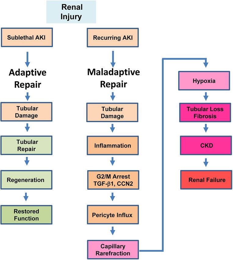
Higgins et al.                                                                                                                                     TGF-β1 in Renal Fibrosis

  FIGURE 3 | PAI-1 (SERPINE1) is a critical factor in the regulation of the pericellular proteolytic microenvironment and fibrotic response to tissue injury. Plasminogen
  activators (urokinase, uPA; tissue-type, tPA) are the physiologically and pathophysiologically-relevant plasmin-generating proteinases that impact extracellular matrix
  (ECM) accumulation/degradation through a complex and highly interdependent proteolytic cascade. Pro-uPA is cleaved to the active enzyme uPA by
  membrane-anchored serine proteases (e.g., Matriptase, Hepsin, Serase-1B) or catalytically-active levels of plasmin. uPA-induced conversion of plasminogen to
  plasmin results in the significant downstream mobilization of several matrix metalloproteinases (MMPs). Collectively, both the plasmin-dependent and MMP
  proteolytic systems dictate the extent and locale of ECM remodeling. Elevated expression or bioactivity of PAI-1, generally in response to tissue injury-induced
  TGF-β1, facilitates ECM accumulation and inhibits ECM degradation which, if prolonged or chronic, leads to the initiation and progression of fibrotic disease.

formation of target of rapamycin (TOR)-autophagy spatial                                 number of senescent cells induces inflammation and fibrosis
coupling components (TASCCs) stimulating, thereby, expression                            (Kim et al., 2020).
of SASP genes (Canaud et al., 2019). The p53 inhibitor pifithrin-                           The maladaptive tubular repair and the cellular
α (PIF) attenuates the fraction of G2 /M-arrested epithelial cells                       senescence programs (e.g., G2 /M stalling, expression of
while deletion of cyclin G1 , mTOR, LC3, or lysosomal associated                         proinflammatory/profibrotic factors) both involve p53 and
membrane protein 2 (LAMP2) reduces the onset and progression                             transcription of the p53 target genes p21 and PAI-1. There is, in
of renal disease (Canaud et al., 2019).                                                  fact, considerable overlap among the SASP, the chronic kidney
                                                                                         disease-associated secretory phenotype (CASP) and the SASP
                                                                                         aging and disease biomarker gene sets that includes increases in
INJURY-ASSOCIATED ACQUISITION OF                                                         the scar-promoting proteins TGF-β1, PAI-1 (SERPINE1) and
A SENESCENCE-LIKE PHENOTYPE                                                              CNN2 (Wang et al., 2017; Basisty et al., 2020). A percentage
                                                                                         of tubular epithelial cells gradually acquire a senescence-like
Multiple sublethal injuries to the kidney leads to the emergence                         phenotype with advancing age and express elevated levels of
of a senescence-like state in some surviving tubular cells                               TGF-β1, p16, and p21 (Ding et al., 2001; Braun et al., 2012;
resulting in a failure to respond with adaptive proliferation                            Ferenbach and Bonventre, 2015). Indeed, senescence promotes
(Ferenbach and Bonventre, 2015). Senescent epithelial cells are                          interstitial fibrosis, tubular atrophy and renal graft deterioration
evident in the kidney in the pathologic context of hypertension,                         limiting tubular regeneration and transplant survival (Braun
diabetes, IgA nephropathy and ischemia/reperfusion injury                                et al., 2012). The elevated levels of reactive oxygen species
particularly in aged mice, where progressive immune system                               (ROS) that accompany the DNA damage response, moreover,
dysfunction may drive the development of CKD (Verzola et al.,                            are likely major contributors to the initiation of the senescent
2008; Satriano et al., 2010; Qi and Yang, 2018; Xiong and                                phenotype (Moonen et al., 2018; Beck et al., 2020a,b). Indeed,
Zhou, 2019; Schroth et al., 2020). Indeed, aging in rodents is                           in some cell types, TGF-β1 functions as a senescence driver
associated with enhanced tubular cell senescence, elevated TGF-                          via ROS-stimulated NF-κB signaling and induction of SASP
β1, p16, and p21 expression and increasing tubulointerstitial                            factors, including PAI-1 (Kwon et al., 2017; You et al., 2019;
fibrosis (Ding et al., 2001; Knoppert et al., 2019). While                               Figure 6). This appears critically important in the establishment
reparative CD24+ /CD133+ epithelial cells contribute to healing                          of the growth arrest state as PAI-1 is not merely a biomarker
and functional recovery, exogenous delivery of even a small                              of the senescent phenotype but is necessary and sufficient for

Frontiers in Cell and Developmental Biology | www.frontiersin.org                    6                                               July 2021 | Volume 9 | Article 678524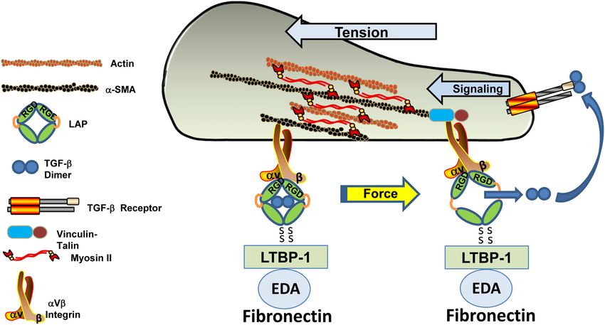
Higgins et al.                                                                                                                                   TGF-β1 in Renal Fibrosis

  FIGURE 4 | The SERPINE1 interactome. SERPINE1 (PAI-1) is a major hub factor in the regulation of the immediate pericellular proteolytic cascade. PAI-1 titrates the
  conversion of plasminogen to plasmin by binding to and inhibiting the catalytic activity of urokinase plasminogen activator (PLAU), effectively attenuating stromal
  proteolysis while promoting matrix accumulation and the onset and progression of fibrotic disease regardless of etiology. PAI-1 also regulates cellular attachment and
  migration, key aspects of the injury repair program, largely by altering interaction of the PLAU-PLAU receptor (PLAUR) complex with its associated integrins and by
  functioning as a ligand for LRP1 to initiate post-receptor downstream signaling.

the induction of replicative senescence downstream of p53                               Liu et al., 2018). Ataxia telangiectasia mutated (ATM) and ATM
(Kortlever et al., 2006; Hiebert et al., 2018).                                         and RAD3-related (ATR), which function as sensors of DNA
    Once renal repair becomes dysfunctional (i.e., elevated                             damage in the maintenance of genomic stability, are involved and
expression of the cell cycle arrest protein p21, down-regulation                        alterations in their expression has consequences. ATM and ATR
of the anti-aging factor Klotho, telomere shortening, increased                         have several DNA repair targets in common including p53 and
oxidative stress), continued activation of the SASP and CASP                            the cell cycle checkpoint kinases CHK1 (ATR) and CHK2 (ATM)
programs accelerate cellular aging leading to the development of                        (Bradbury and Jackson, 2003; Awasthi et al., 2015). ATR deletion
age-related pathologies (Wang et al., 2017; Andrade et al., 2018;                       in renal proximal tubular epithelial cells exacerbates maladaptive
Dai et al., 2019). Cellular senescence is evident in many forms                         repair, increases the number of senescent cells and promotes
of kidney injury (Li and Lerman, 2020) and older mice have                              expression of a profibrotic secretory phenotype (Kishi et al.,
increased senescence-associated β-galactosidase, p53, and p21                           2019). These findings suggest that ATR provides a protective role
expression in response to ischemia/reperfusion injury compared                          in the injured proximal tubular epithelium to restrict or attenuate
to young mice (Clements et al., 2013; Valentijn et al., 2018).                          exuberant (i.e., fibrotic) repair while highlighting the role of p53
This is relevant to the human condition as age-associated renal                         in renal disease since treatment with the p53 inhibitor PIF-α
scarring, and decline in kidney function, varies among ethnic                           significantly reduces the fraction of G2 /M cells and mitigates
groups and expression of β-galactosidase and p16 is evident even                        the fibrotic response (Yang et al., 2010; Overstreet et al., 2014;
in the absence of morphologic changes (Yang and Fogo, 2010).                            Liu et al., 2019).
    Although the mechanism underlying cell cycle phase-specific                             Expression of a subset of TGF-β1 target genes that contribute
arrest or at least residence prolongation is unclear, activation                        to growth arrest, and G2 /M stalling as well, appears to require
of the p53→p21 axis, particularly in the early stages of kidney                         both canonical and non-canonical signaling. To this point, TGF-
disease, likely drives renal cell stalling in both G1 and G2 /M                         β1 also upregulates the Hippo pathway effectors YAP (yes-
phases (Yang et al., 2010; Overstreet et al., 2014; Moonen et al.,                      associated protein) and TAZ (transcriptional co-activator with
2018; Wu and Prives, 2018; Liu et al., 2019). In this regard,                           PDZ-binding motif) in proximal tubular epithelial cells both
fibrosis in response to chemotherapeutic agents, nephrotoxins,                          in vivo and in vitro. Indeed, doxycycline-induced tubular-specific
ischemia/reperfusion injury or UUO is associated with DNA                               TGF-β1 expression in double-transgenic Pax8-rtTA-tet-o-TGF-
damage and normal aging sensitizes tubular epithelial cells                             β1 mice enhances renal TAZ levels while TGF-β1 increases
to DNA damage-induced G2 /M arrest (Yang and Fogo, 2010;                                TAZ levels in human proximal tubular epithelial cells; in vitro

Frontiers in Cell and Developmental Biology | www.frontiersin.org                   7                                               July 2021 | Volume 9 | Article 678524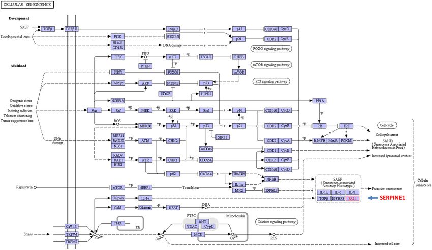
Higgins et al.                                                                                                                             TGF-β1 in Renal Fibrosis

  FIGURE 5 | String Network and Gene Ontology. Protein-protein interaction and GO analyses of the SERPINE1/TGF-β1/fibronectin axis indicates that SERPINE1 is a
  significant nodal contributor to various biological processes that impact the global program of normal and maladaptive tissue repair. These data underscore the
  potential clinical utility of SERPINE1 targeting in the therapy of fibrotic disease.

modeling confirmed that TAZ is necessary for TGF-β1-mediated                         knockdown, moreover, reduces levels of both CTGF (CCN2)
fibrogenesis (Anorga et al., 2018). Vector-driven TAZ synthesis in                   and PAI-1 (SERPINE1) while introduction of the constitutively-
human proximal tubular (HK-2) cells, or addition of conditioned                      active YAPS127A construct increased PAI-1 expression (Marquard
medium from TAZ overproducers to control vector-transduced                           et al., 2020). Although the underlying mechanisms remain to
HK-2 cells, mimics certain aspects of the TGF-β1-induced                             be determined, YAP/TAZ apparently do not alter the rate of
phenotype including G2 /M arrest and acquisition of a profibrotic                    SMAD nuclear import or exit nor impact SMAD phosphorylation
program (Anorga et al., 2018). Exposure of HK2 cells to hypoxic                      but may regulate SMAD nuclear levels by functioning, directly
stress similarly promotes G2 /M stalling and PAI-1 induction                         or indirectly, as retention factors and/or by changing TGF-βR
while TAZ overexpression leads to the accumulation of HK-                            activity (Labibi et al., 2020).
2 cells in G2 /M phase. TAZ is, in fact, required for maximal
TGF-β1-mediated PAI-1 synthesis in proximal tubular cells (Liu
et al., 2015; Samarakoon et al., 2015; Anorga et al., 2018; Bessho                   TGF-β/SMAD SIGNALING DRIVES
et al., 2019) and a similar involvement of YAP in TGF-β1-                            FIBROSIS IN OBSTRUCTIVE
induced PAI-1 expression is evident in lung tumor cells (Kong                        NEPHROPATHY
et al., 2021). KEGG analysis confirmed that convergence of the
TGF-β and Hippo signaling pathways regulates transcription of                        Increased expression of the potent profibrotic cytokine TGF-
the profibrotic CCN2 and SERPINE1 genes (Figure 7). YAP                              β1 and the type I/II TGF-β1 receptors is a hallmark feature of

Frontiers in Cell and Developmental Biology | www.frontiersin.org                8                                             July 2021 | Volume 9 | Article 678524
Higgins et al.                                                                                                                                   TGF-β1 in Renal Fibrosis

  FIGURE 6 | KEGG analysis of the highly interactive program of cellular senescence. SERPINE1 and TGF-β1, two prominent activators and members of the
  stress-activated SASP, are key factors in the global and renal programs of proliferative arrest. Several of the involved networks include the p53, ras and TGF-β
  signaling pathways. Collectively, these regulate the expression of a spectrum of cell cycle and growth control elements (e.g., p16, p21, TGF-β1, PAI-1) (Kortlever
  et al., 2006, 2008).

virtually all forms of CKD (Böttinger, 2007). Tubulointerstitial                        to the fibrotic response, importantly, was confirmed using
pathology following experimental UUO appears largely due                                genetic approaches. Conditional overexpression of TGF-β1 in the
to elevated levels of TGF-β1, SERPINE1 and CCN2 in the                                  tubular epithelium of Pax8-rtTA-tet-o-TGF-β1 double transgenic
injured kidney (Figure 8) mimicking the increased TGF-β1                                mice induces extensive peritubular fibrosis, focal nephron
expression in children with UPJ obstruction (Miyajima et al.,                           degeneration (Traykova-Brauch et al., 2008; Koesters et al., 2010)
2000; Inazaki et al., 2004; Ucero et al., 2010). Within hours,                          and TGF-β1-dependent loss of the SMAD phosphatase PPM1A
the occluded kidney exhibits changes in hydrostatic forces and                          (Tang et al., 2020). Similarly, Pax8 promoter-driven expression of
increased oxidative stress (Schreiner et al., 1988; Klahr and                           a ligand-independent constitutively-active TGF-β type I receptor
Morrissey, 2002; Dendooven et al., 2011). Tubular stretch further                       results in the acquisition of features typical of AKI (e.g., epithelial
stimulates TGF-β1 expression (>20-fold), increases the epithelial                       apoptosis, necrosis and dedifferentiation; renal inflammation)
apoptotic index, and leads to the development of an interstitial                        (Gentle et al., 2013). The albumin/TGF-β1 transgenic mouse
inflammatory infiltrate (Miyajima et al., 2000; Rohatgi and                             (Kopp et al., 1996), moreover, recapitulates the pathophysiologic
Flores, 2010). Persistently elevated renal TGF-β1 expression, even                      heterogeneity of CKD progression highlighting their utility in the
after relief of UUO (depending on the duration of obstruction                           discovery of disease progression signatures (Ju et al., 2009).
and extent of pathology) frequently leads to progressive tissue                             Elevated levels of TGF-β1 in the injured kidney direct the
injury, impaired regenerative growth, and eventual loss of organ                        myofibroblastic differentiation of recruited vascular pericytes
function (Chevalier, 1999; Chevalier et al., 2009, 2010).                               and resident fibroblasts while driving a program of pathologic
   TGF-β1 mRNA levels steadily increase in several nephron                              ECM synthesis and advancing fibrosis (Bonventre, 2010; Meng
segments as early as day-1 post-UUO followed by TGF-β1 protein                          et al., 2015, 2016; Sun et al., 2016; Chen et al., 2018;
upregulation (Isaka et al., 2000; Miyajima et al., 2000; Klahr                          Feng et al., 2018; Higgins et al., 2018). Genetic deficiency
and Morrissey, 2002; Yang et al., 2010; Makitani et al., 2020).                         of SMAD3, a major profibrotic effector of TGF-β1 signaling,
TGF-β1 transcripts are most prominent in the tubular epithelia                          or administration of the SMAD3 inhibitor SIS3 immediately
and, to a lesser extent, in a fraction of infiltrating macrophages                      after ureteral ligation, attenuates myofibroblast accumulation
(Kaneto et al., 1993; Fukuda et al., 2001). Attenuation of UUO-                         while suppressing deposition of collagen I and fibronectin (Sato
induced fibrosis upon administration of the anti-TGF-β antibody                         et al., 2003; Inazaki et al., 2004; Zhang D. et al., 2018; Zhang
1D11 or the TGF-β activin-like kinase 5 (ALK5) receptor                                 S. et al., 2018). One mechanism may involve the SMAD3-
signaling inhibitor SB-525334 further highlight involvement                             dependent autoinduction of TGF-β1 by UUO-stimulated TGF-
of the TGF-β pathway in ureteral obstruction-initiated renal                            β1 expression (Sato et al., 2003). This has potential clinical
scarring (Richards et al., 2018). The contribution of TGF-β1                            ramifications since post-injury treatment with SIS3 also blunted

Frontiers in Cell and Developmental Biology | www.frontiersin.org                   9                                               July 2021 | Volume 9 | Article 678524
Higgins et al.                                                                                                                           TGF-β1 in Renal Fibrosis

  FIGURE 7 | Cross-talk between the TGF-β and the YAP/TAZ pathways impact expression of the profibrotic SERPINE1 and CCN2 genes. TGF-β activates a
  canonical signaling network that involves the SMAD2/3-dependent transcription of SERPINE1 and CCN2. It is also evident that non-canonical pathway engagement
  (e.g., Hippo) contributes to maximal TGF-β1 SERPINE1 (PAI-1) and CCN2 (CTGF) expression by stimulating YAP/TAZ nuclear translocation and interaction with the
  TGF-βR-phosphorylated SMAD2/3 transcriptional effectors and the shuttle SMAD4.

the subsequent fibrotic response (Zhang D. et al., 2018; Zhang                      TGF-β1 is, indeed, the key driver of fibrosis in UUO either
S. et al., 2018) suggesting that blockade of TGF-β1→ALK5                            directly by impacting the transcription of disease-relevant genes
signaling to SMAD3 has therapeutic implications. Several pre-                       or indirectly via angiotensin signaling (Ishidoya et al., 1995;
clinical studies, in fact, targeted SMAD3 as one modality for the                   Pimentel et al., 1995; Fern et al., 1999; Satoh et al., 2001;
treatment of UUO-induced renal disease (e.g., Li et al., 2010; Ji                   Inazaki et al., 2004; Shin et al., 2005). Indeed, angiotensin
et al., 2018; Wang et al., 2018).                                                   stimulates the expression of ECM structural elements (e.g.,
    Initial observations did, in fact, support the premise that                     collagen, fibronectin, laminin) as well as inhibitors of ECM
interstitial fibrosis and disease progression in the obstructed                     degradation including PAI-1 (SERPINE1) through TGF-β1-
kidney can be mitigated by blockade of TGF-β1 expression or                         dependent mechanisms, thus promoting tissue fibrogenesis
function via antisense phosphorothioate oligodeoxynucleotides,                      (Kagami et al., 1994; Wolf, 2006). While global TGF-β1-null mice
small interfering RNA (siRNA) or neutralizing antibodies (Isaka                     exhibit no gross abnormalities at birth but die soon thereafter
et al., 2000; Miyajima et al., 2000; Gagliardini and Benigni,                       due to wasting associated with severe multifocal inflammation
2006; Hwang et al., 2006). Overexpression of the latent form                        (Yaswen et al., 1996), carefully focused anti-TGF-β therapies, and
of TGF-β1, to minimize availability of active TGF-β1 in the                         perhaps targeting disease-critical downstream genes or enhancers
tissue microenvironment, decreases the incidence α-smooth                           of TGF-β1 profibrotic signaling, may be a more prudent and
muscle actin-positive cells (presumably myofibroblasts) in the                      translationally-adaptable therapeutic approach. As one example,
UUO-injured kidney and blocks SMAD2/3 activation (Huang                             small molecule (SK-216) pharmacologic inhibition of the activity
et al., 2006, 2008). The peroxisome proliferator-activated receptor                 of the TGF-β1 target PAI-1 attenuates TGF-β1-induced fibroblast
gamma agonist troglitazone similarly reduces development                            to myofibroblast transition and lung fibrosis (Omori et al., 2016).
of UUO-induced renal interstitial fibrosis and inflammation                         Varga and Pasche (2009) suggest, moreover, that neutralizing
through suppression of TGF-β1 expression (Kawai et al., 2009).                      antibodies, pathway antagonists and soluble (i.e., trap) receptors
Collectively, these data are consistent with the concept that                       attenuate excessive (e.g., disease-associated) TGF-β bioactivity

Frontiers in Cell and Developmental Biology | www.frontiersin.org              10                                            July 2021 | Volume 9 | Article 678524
Higgins et al. TGF-β1 in Renal Fibrosis
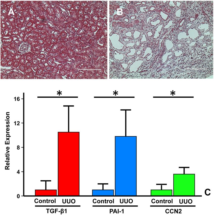
FIGURE 8 | Fibrotic response of the murine kidney to UUO. Compared to the relatively normal histology of the contralateral control or sham-operated kidney (A), a
dysmorphic and flattened epithelium with extensive tubular dilation, expanses of denuded basement membrane and accumulation of connective tissue (blue stain) in
the expanded interstitial regions is evident in the obstructed kidney (B). (A,B), Trichrome stain. Morphometric analyses of immunohistochemical-stained
paraffin-embedded sections of the UUO-injured kidney and the contralateral control 7–14 days post-surgery, revealed significant increases in PAI-1, TGF-β1, and
CCN2 in the obstructed kidney (C). Histograms illustrate the mean ± SD staining intensity (ImageJ threshold analysis) for TGF-β1, PAI-1, and CCN2 between the
two experimental groups. *p < 0.05.
while retaining homeostatic TGF-β signaling functions. Such bonding between LAP and the latent TGF-β binding protein
approaches may avoid the adverse outcomes that result from (LTBP) occurs within the endoplasmic reticulum (Robertson
TGF-β depletion (Yaswen et al., 1996; Yang et al., 2020). and Rifkin, 2016). In the Golgi, LAP is cleaved from the pro-
protein by the subtilisin-like pro-protein convertase furin prior
to extracellular transport of the ternary large latent complex,
MULTIPLE MODES OF TGF-β1 consisting of TGF-β, LAP and the latent TGF-β binding protein
ACTIVATION (TGF-β/LAP/LTBP). The 4 LTBP isoforms (LTBP1-4) then
interact with different structural elements of the ECM including
The tissue response to injury is largely dependent on multi-level fibrillin microfibrils and the fibronectin network (Zilberberg
controls on the persistence of TGF-β isoform expression and et al., 2012; Tsuda, 2018). While the different LTBPs exhibit
activation in the immediate pericellular microenvironment. The some preferences for TGF-β isoform recognition, LTBP-1 has a
transition of TGF-β1 from a latent to bioactive configuration particular affinity for fibronectin and, more specifically, for the
is a critical checkpoint in the fibrogenic response. TGF-β1- extra domain A (EDA) splice variant of fibronectin (FnEDA)
3 pro-proteins are comprised of a dimeric growth factor and (Zilberberg et al., 2012; Tsuda, 2018; Zent and Guo, 2018). FnEDA
N-terminal latency-associated peptide (LAP) domains. Disulfide appears particularly critical in TGF-β1 signaling as interference
Frontiers in Cell and Developmental Biology | www.frontiersin.org 11 July 2021 | Volume 9 | Article 678524Higgins et al. TGF-β1 in Renal Fibrosis
with EDA domain function attenuates both LTBP-1 binding and alternative mode of TGF-β activation that does not necessitate
TGF-β1 activation (Klingberg et al., 2018). Latency, however, is release of dimeric TGF-β from the LAP (Campbell et al.,
strictly dependent on LAP as a LAP mutant that cannot bind 2020). While the existence of multiple mechanisms of TGF-β1
the LTBP effectively retains TGF-β1 in an inactive configuration activation may be cell- and tissue-type dependent, complicating
(Robertson and Rifkin, 2016). the adaption of a universal therapeutic strategy, pharmacologic
Mechanisms underlying release of latent TGF-β1 from the inhibition of RGD-binding integrins attenuates renal fibrosis and
LAP cage include proteases, integrins, other proteins such as improves organ function following injury (Basta et al., 2020)
thrombospondin-1 and various physicochemical factors both and antibody targeting of αvβ6 mitigates bleomycin-induced lung
alone and in combination (Robertson and Rifkin, 2016). Several fibrosis (Horan et al., 2008). Integrin-focused therapies, however,
proteases cleave the hinge region in LAP freeing the TGF-β are not without controversy. Phase 2 clinical trials of antibody
dimer for receptor occupancy, although the physiologic relevance BG00011 (previously known as STX-100), which targets αvβ6
of protease-only liberation is complicated by the considerable was terminated by Biogen due to safety concerns (Freeberg et al.,
redundancy in the various participating enzyme systems. Non- 2021)4 .
proteolytic as well as protease-requiring mechanisms involving Elevated TGF-β1 levels, coupled with loss of tissue elasticity,
αv subunit integrins (e.g., αvβ1, β3, β5, β6, β8), however, also further increases FnEDA expression while promoting LTBP-
activate TGF-β1 particularly in the context of a progressively 1/FnEDA co-localization, facilitating integrin/LAP engagement
fibrosing, increasingly stiff, renal microenvironment (Hysi and and the subsequent creation of tensional strain stimulating
Yuen, 2020). Binding of αv integrins to the LAP N-terminal the generation of bioactive TGF-β1 (Wynn and Ramalingam,
arginine-glycine-aspartic acid (RGD) motif generates Rho/RhoA- 2012; Chang et al., 2017; Freeberg et al., 2021). Progressive
dependent tractional forces with ECM-anchored LTBPs; the ECM stiffness and a TGF-β1-rich microenvironment promotes
resulting distortion of the LAP cage liberates and, thereby, myofibroblast differentiation and survival while activating the
activates the TGF-β1 dimer (Buscemi et al., 2011; Hinz, 2015; Hippo pathway mechanosensitive transcriptional co-activators
Sheppard, 2015; Robertson and Rifkin, 2016; Dong et al., 2017; YAP and TAZ (Liu et al., 2015; Dupont, 2016; Jorgenson et al.,
Nickel et al., 2018; Figure 9). Cooperative involvement of both 2017; Misra and Irvine, 2018; Santos and Lagares, 2018; Totaro
integrins and proteases is an additionally proposed mechanism. et al., 2018). Convergence of YAP/TAZ and TGF-β1 pathways,
One model suggests that tensional strain generated by complex in the context of recurrent or persistent tissue injury, induces
formation between αv integrins and the RGD motif on ECM- expression of several major profibrotic genes including CCN2,
tethered LAP predisposes LAP to cleavage by cell surface- fibronectin and PAI-1 contributing, thereby, to the eventual
proximal proteases (Robertson and Rifkin, 2016). There appears development of fibrotic disease (Kim et al., 2019; Figure 7). These
to be, however, significant differences in the type of strain, the findings suggest a complex mechanism for TGF-β1 involvement
activation of latent TGF-β1 and the amplitude of expression over the course of renal fibrosis in which induction of FnEDA
of the engaged genes. Compared to steady-state shear strain, is a critical element in a TGF-β/FnEDA/αv integrin positive
oscillatory forces generate significantly greater levels of active feed-forward loop. It should be mentioned that there have
TGF-β1 resulting in the increased expression of the profibrotic been some attempts to assess these requirements for TGF-β1
triad PAI-1, collagen 1A1 and periostin (Kouzbari et al., 2019). mobilization in a translational context. Systemic injection of a bi-
Among the αv integrin subtypes, αvβ6 is a major TGF-β1 release specific antibody with FnEDA binding and TGF-β1 neutralizing
trigger and a likely fibrotic effector since renal obstruction in β6- domains confirmed both construct accumulation and reduced
deficient mice is associated with a reduction in TGF-β1 activity fibrosis in the injured kidney providing supporting evidence
and decreases in collagen I, collagen III and PAI-1 expression for such a model (McGaraughty et al., 2017). How such a
(Ma et al., 2003). Regardless of the actual pathway, computational strategy may be adapted for patient treatment, however, remains
modeling suggests that a protease (i.e., plasmin)-dependent to be determined.
bistability mechanism regulates TGF-β1 bioactivity (Li et al.,
2017). It appears that TGF-β1 undergoes a bistable switch in
response to increasing concentrations of plasmin from a high- INVOLVEMENT OF P53 IN
level thrombospondin-1-mediated to lower-level predominantly TGF-β1-INDUCED RENAL FIBROSIS
plasmin-dependent mode of activation; both have implications to
the development and progression of fibrotic disorders. Since TGF-β1 signaling is a major driver of UUO-induced renal
αvβ8 also releases TGF-β1 from the LAP:TGF-β1 complex fibrosis (Richards et al., 2018), clarification of the involved
bound to GARP (glycoprotein A repetitious predominant) intermediates downstream of the activated TGF-β receptors may
on the surface of regulatory T cells (Lienart et al., 2018). have therapeutic implications for patients with UPJ disease.
This mechanism is unique to T-regs; membrane tethered In the canonical pathway, occupancy of a type II receptor
GARP/LAP/TGF-β1 promotes presentation of the LAP RGD (TGF-βRII) by the TGF-β1 dimer drives complex formation
sequence to the αvβ8 integrin on adjacent cells. Tensional strain with, and subsequent phosphorylation of, the ALK5 type I
releases and activates the TGF-β1 dimer in much the same receptor (TGF-βRI) that, in turn, phosphorylates receptor (R)
way as occurs via ECM-anchored LTBP-1. In addition, recent SMADs (predominately SMAD2/3 in fibrotic disease) at the distal
findings using cryo-electron microscopy to probe LAP:TGF-
β complex interactions with the αvβ8 integrin suggests an 4
www.fiercebiotech.com
Frontiers in Cell and Developmental Biology | www.frontiersin.org 12 July 2021 | Volume 9 | Article 678524Higgins et al. TGF-β1 in Renal Fibrosis FIGURE 9 | Tension-dependent release of active TGF-β from the LAP cage. The ternary large latent (LTBP/TGF-β/LAP) complex forms a bridge between an αV integrin bound to the RGD site on the latency-associated peptide and LTBP-1 tethered to the fibronectin-rich ECM. Actinomyosin-based contractility generates mechanical tension within this ternary complex inducing a conformational change in the LAP that releases the now-active TGF-β dimer that, in turn, occupies the TGF-βR to initiate downstream signaling. C-terminal SxS motif (Ser423/425 and Ser465,467 for SMAD3 and PI3K/AKT, GSK3/Twist/FOXO, and PKC/Smurf1/RhoA/Rock SMAD2, respectively) (Matsuzaki, 2013). While early models cascades (Böttinger and Bitzer, 2002; Piersma et al., 2015; suggested that SMAD2 interacts with the SMAD binding Zhang, 2017; Ahmadi et al., 2019; Patel et al., 2019; Finnson domain (SBD) of the SMAD anchor for receptor activation et al., 2020; Labibi et al., 2020). The increasing complexity of (SARA) followed by SARA:SMAD2 delivery to the TGF-βRI participating co-factors in the regulation of TGF-β1-responsive via the C-terminal domain of SARA to facilitate R-SMAD genes likely reflects the comparatively low affinity of DNA- phosphorylation, the actual involvement of SARA in TGF-β SMAD interactions. signaling is controversial (Rozés-Salvador et al., 2018, 2020). One such important co-activator is the tumor-suppressor p53. Regardless of the precise mechanism, pR-SMADs complex with The involvement of p53 in renal disease was initially defined in the shuttle SMAD4 and translocate to the nucleus to impact a rat model of ischemia-reperfusion injury (Kelly et al., 2003). transcription of a rather large slate of TGF-β1 responsive genes p53 induction and increased p53 serine 15 phosphorylation (Massagué, 2000; Massague, 2012). Identification of differentially is also evident in the kidney following nephrotoxin (e.g., expressed genes (DEG), using an unbiased microarray analysis, cisplatin, aristolochic acid) administration or UUO, particularly at two time points post-UUO disclosed 606 upregulated in the dysmorphic epithelium (Zhou et al., 2010; Wei et al., (including 430 annotated) and 485 downregulated (including 2007; Samarakoon et al., 2013a,b), and renal allograft rejection 251 annotated) genes (Higgins et al., 2003). More than 70 such (Higgins et al., 2019). Recent studies, furthermore, link tubular DEG partitioned to the ECM/cytoskeletal cluster indicative of the epithelial dysfunction in response to both acute (e.g., ischemia- breath of targets that may well impact the fibrogenic phenotype. reperfusion, nephrotoxins) and more protracted (UUO) injury KEGG analysis of the transcriptome of diabetic and non-diabetic to the progression of renal fibrosis via the p53 and JNK mice indicated, in fact, that significant differentially-expressed pathways with the retention of TGF-β signaling (Yang et al., genes closely associate with the p53 signaling network, as well as 2010). p53 is activated in the injured renal epithelium initiating the MAPK and TGF-β pathways (Wang et al., 2016). cell cycle arrest at the G1 and G2 /M checkpoints depending The growing number of non-canonical (i.e., non-SMAD) on the participating effectors (e.g., ATM, ATR, CK1, CK2, elements and their associated pathways in the TGF-β1 network, p21, TGF-β1) and extent of tissue hypoxia (Thomasova and however, suggests a more significant level of mechanistic diversity Anders, 2015; Tang et al., 2018, 2019, 2020; Liu et al., in the control of gene expression and the potential existence of 2019). TGF-β1 signaling in the damaged kidney increases p53 an expanding repertoire of regulated sequences (Zhang, 2017). levels and phosphorylation, particularly at p53S9/15 , promoting Appropriately recognized as the master regulator of fibrosis p53 stabilization and triggering p53-SMAD2/3 interactions (Meng et al., 2016; Lodyga and Hinz, 2020), the TGF-β1 signaling resulting in transcription of the growth inhibitor p21 and apparatus, including the downstream SMAD effectors, cross-talk subsequent p21-dependent G1 arrest (Higgins et al., 2019). with an extensive and highly interactive system that includes While p21 is a major p53 responsive gene, p53 upregulation the Raf/MEK/ERK, JAK/STAT, Wnt, Notch, Hippo/YAP/TAZ, in hypoxic tubular cells also suppresses CDK1, cyclin B1 , Frontiers in Cell and Developmental Biology | www.frontiersin.org 13 July 2021 | Volume 9 | Article 678524
You can also read