Clean Bioprinting - Fabrication of 3D Organ Models Devoid of Animal Components
←
→
Page content transcription
If your browser does not render page correctly, please read the page content below
Review Article
Clean Bioprinting – Fabrication of 3D Organ
Models Devoid of Animal Components
Johanna Berg and Jens Kurreck
Institute of Biotechnology, Technische Universität Berlin, Berlin, Germany

      Abstract
      Bioprinting is a rapidly developing technology that enables the exact positioning of living cells embedded in bio-
      materials in precise spatial arrangements to fabricate engineered tissues and organs. While the ultimate goal of bio-
      printing approaches is to produce organs for transplantation purposes, bioprinted organ models also hold great potential
      for research purposes to serve as alternatives to animal experiments. By using human cells, humanized organ models
      can be generated that may produce more relevant results for human (patho-)physiology than animal models. However,
      standard bioprinting procedures currently use numerous hidden animal components. Virtually all studies published in the
      field to date make use of cells grown in media with fetal bovine serum (FBS). In addition, Matrigel, the extracellular matrix
      (ECM) harvested from Engelbreth-Holm-Swarm sarcoma grown in mice, is widely employed to cultivate stem cells and
      3D organ models. Finally, most bioinks currently in use contain gelatin or comparable animal components to improve cell
      viability and adhesion. The present review will give an introduction to the potential of bioprinting to fabricate 3D models
      that may be substituted for animal experiments and will go on to describe strategies to replace animal components cur-
      rently included in standard procedures of bioprinting. These approaches comprise the adaptation of cells to FBS-free
      media, the use of bioinks composed of synthetic or plant material, and the replacement of animal ingredients by materials
      of human origin. We propose denoting bioprinting strategies devoid of animal components as clean bioprinting.

1 Introduction                                                               ences become particularly apparent is infection biology. Many
                                                                             pathogens have a narrow host tropism and only infect a single or
The current biomedical research paradigm is based on initial stud-           a small number of species. A common strategy to cope with this
ies in 2D cell culture followed by animal experiments. Conven-               problem is to adapt a human-pathogenic virus to the test animal,
tional cell culture, however, does not reflect the three-dimension-          which, however, often results in a different course of disease in
al architecture of natural organs that influences cell features, such        animals, as will be outlined in more detail below.
as gene expression patterns. Animal models provide the oppor-                   Furthermore, the high failure rate of drug candidates in clinical
tunity to study (patho-)physiological phenomena in a function-               testing can, at least to some degree, be ascribed to differences in
al biological system. Their major scientific drawbacks are spe-              animal and human physiology. Although pre-clinical testing in-
cies-specific differences that limit the relevance of animal studies         volves multiple animal models to evaluate efficacy and toxicity
to humans. The degree of this problem is highly controversial.               of a substance, approximately 90% of the candidates fail during
A prominent example is the discussion centered around the pre-               clinical development, a number that varies substantially between
dictivity of animal models in inflammation research. While the               different indications and is as high as 97% in oncology (Wong
initial study by Seok et al. (2013) came to the conclusion that              et al., 2019a). The main reasons for failure in clinical develop-
genomic responses to inflammatory stimuli in mice poorly cor-                ment are low efficacy and unexpected toxicity, which is to a cer-
relate with human inflammatory diseases, a subsequent study an-              tain extent due to species-specific differences in physiology be-
alyzing the same dataset came to the opposite conclusion (Takao              tween test animals and humans. Protein-based biologics, such as
and Miyakawa, 2015). A more recent publication suggested that                monoclonal antibodies and recombinant proteins, are among the
some mouse models can provide predictive insights, while oth-                most advanced therapeutics. Their immunogenicity in humans,
ers cannot (Weidner et al., 2016). A field in which species differ-          however, is particularly difficult to predict in animal models. In

Received September 15, 2020; Accepted November 27, 2020;                     This is an Open Access article distributed under the terms of the Creative Commons
Epub December 2, 2020; © The Authors, 2021.                                  Attribution 4.0 International license (http://creativecommons.org/licenses/by/4.0/),
                                                                             which permits unrestricted use, distribution and reproduction in any medium, provi-
ALTEX 38(2), 269-288. doi:10.14573/altex.2009151                             ded the original work is appropriately cited.
Correspondence: Prof. Dr Jens Kurreck
Institute of Biotechnology, Technische Universität Berlin
TIB 4/3-2, Gustav-Meyer-Allee 25, 13355 Berlin, Germany
(jens.kurreck@tu-berlin.de)

ALTEX 38(2), 2021                                                                                                                                           269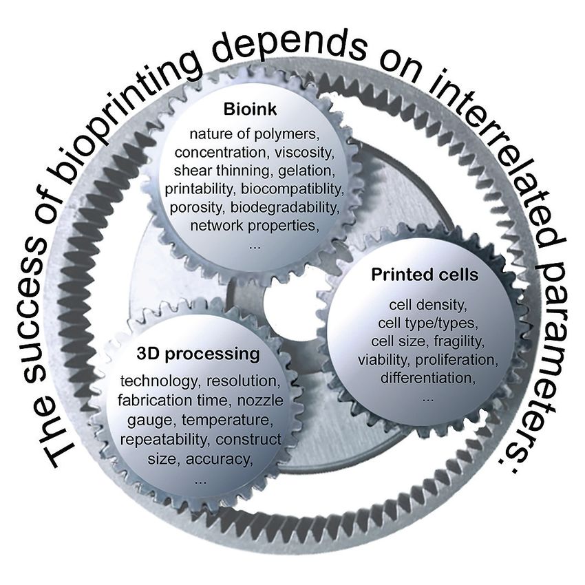
Berg and Kurreck

addition to failure in clinical trials, a large number of drugs has     at high 3D resolution that can subsequently be populated with
to be withdrawn from the market after their approval. As of             cells. The present review, however, will focus on approaches that
2016, a database comprised 578 withdrawn drugs, almost half of          work with cell-laden bioinks, i.e., include living cells during the
which were discontinued due to adverse reactions and toxic ef-          printing process.
fects (Siramshetty et al., 2016).                                          Multiple 3D printing technologies are available, but the major-
   Due to the uncertainties involved in predicting human toxicity,      ity of current bioprinters make use of material deposition tech-
our current approach to investigate properties of drug candidates       niques such as extrusion bioprinting, inkjet bioprinting or mod-
in pre-clinical development with animal experiments is under de-        ern light-based techniques, including laser-assisted and stereo-
bate (Van Norman, 2019). Recent progress in the development of          lithography bioprinting (Fig. 1). These major technologies will
3D organ models may help to overcome the problems inherent in           only be briefly introduced here, and the reader is referred to ex-
predicting the efficacy and toxicity of drug candidates (Weinhart       cellent and exhaustive review articles on general approaches to
et al., 2019). As human cells can be used for ex vivo experiments,      bioprinting published recently that go into depth with the tech-
these models can be expected to reflect the human (patho-)phys-         nology for further details (Heinrich et al., 2019; Matai et al.,
iology better than animal models. Conventional 2D cell cultures         2020; Mota et al., 2020; Sun et al., 2020). It should be noted that
with human cells provide some insight, but their significance is        each of the bioprinting technologies has its specific advantages
limited, as cells behave differently in natural organs with 3D cell-    and disadvantages, and the appropriate method needs to be cho-
cell contacts and interactions with different cell types. Bioprint-     sen for each biological application.
ing is a particularly promising technology for the generation of           The most widely used bioprinting technology is extrusion
organ models with high spatial precision (Crook, 2020). Com-            bioprinting, in which a viscous bioink is extruded through a noz-
pared to other 3D technologies, a key feature of bioprinting is         zle and then remains localized upon deposition (Fig. 1A). For
its high accuracy and reproducibility. However, bioprinting is a        the pneumatic systems, air pressure extrudes highly viscous
highly sophisticated technology, and extensive training and ex-         bioinks as seamless filaments that are then cross-linked by light,
pertise are required to fully exploit the potential of the technolo-    enzymes, chemicals or temperature to form mechanically dura-
gy. A major issue that will be discussed here in detail, is that vir-   ble structures. The mechanical systems are usually controlled
tually all bioprinting studies reported so far (as well as all other    by a piston or a screw. The continuous extrusion of the bioink
methods to produce 3D tissue models) include the use of animal          without interruptions is advantageous to maintain the integri-
components such as fetal bovine serum (FBS), animal extracellu-         ty of the printed constructs in comparison to dropwise methods
lar matrix (ECM, e.g., Matrigel™) or gelatin.                           described below. The extrusion process also allows processing
   The present review will summarize the current state of the bi-       of highly viscous bioinks and high cell numbers; however, the
oprinting field to produce organ models that have the potential to      printing speed is relatively low, and the resolution of the printed
replace animal experiments. We will then identify components of         constructs is lower than with laser-based methods. In addition,
animal origin that are widely used in bioprinting and discuss al-       cells may experience harmful sheer stress on extrusion, so the
ternatives for their replacement with synthetic or plant-derived        bioink and printing conditions must be optimized to maintain
materials. We suggest denoting approaches completely devoid of          high cell viability.
components of animal origin as clean bioprinting. Most impor-              The technology of inkjet bioprinting is derived from conven-
tantly, we will argue that this concept will only have a chance to      tional 2D inkjet printers (Li et al., 2020). It is a non-contact
become widely established in the scientific community if the ad-        process during which picoliter-sized droplets are deposited in a
vantages of avoiding human-animal chimeric systems for obtain-          computer-controlled manner (Fig. 1B). The liquid is dispensed
ing human-relevant research results can be demonstrated, rath-          by temporal deformation of the internal space within the noz-
er than just citing the singular consideration of improved animal       zle due to piezoelectric or digitized thermal actuation. Advan-
welfare.                                                                tages of inkjet bioprinting include the simplicity of the method
                                                                        and its low cost as well as comparatively good resolution and
                                                                        high cell viability. Major disadvantages are the low cell density
2 Bioprinting technologies                                              in the bioprinting process and the restriction to bioinks of low
                                                                        viscosity.
Three-dimensional printing refers to a process of building 3D ob-          Laser-based technologies differ fundamentally from the noz-
jects by successively adding material in a layer-by-layer manner.       zle-based bioprinting approaches described above and allow
It is often referred to by the technically more precise term addi-      very high spatial resolution. Bioprinters based on laser-induced
tive manufacturing (AM). The process is normally controlled by          forward transfer (LIFT) usually consist of a pulsed laser, whose
computer-aided design (CAD) programs (Fay, 2020). Bioprint-             beam is absorbed by a layer below which the bioink is located in
ing is a specific variant of the AM process, which is character-        a donor ribbon (Fig. 1C). When the focused laser beam reaches
ized by the inclusion of living cells, biocompatible materials and,     a desired site of the energy-absorbing layer, the corresponding
in many cases, biologically active factors. The procedure aims          location of the supporting donor layer is vaporized, which caus-
at fabricating multi-cellular tissues or organ equivalents with         es ejection of a droplet of the bioink that falls onto the collector
high spatial precision. In a broader sense, bioprinting also in-        platform. Another light-based bioprinting technology is stereoli-
cludes printing of biocompatible materials to produce scaffolds         thography (SLA). The basic concept of this method is to selec-

270                                                                                                                         ALTEX 38(2), 2021
Berg and Kurreck
Fig. 1: Schematic representation
of the most commonly
used bioprinting technologies
(A) Extrusion bioprinting, (B) inkjet
bioprinting, (C) laser-induced forward
transfer (LIFT), and (D) stereo-
lithography. SLA, stereolithography;
DLP, digital light projection
tively cure a cell-laden bioink in a layer-by-layer process to build tive components and biomaterials” (Groll et al., 2018). Bioinks
up materials (Fig. 1D). A laser-beam cross-links photosensitive need to combine multiple properties: They must initially be flu-
bioinks by projecting a 2D pattern of the plane of interest onto id to be printable but then rapidly transition to the solid state to
the bioink reservoir. Digital light projection (DLP) operates sim- form and maintain the printed structure. At the same time, they
ilarly to SLA bioprinting. The main difference between the two must be biocompatible to guarantee high viability of the printed
is the source of light. In contrast to SLA, where a laser beam so- cells. Common currently used bioinks are cell-laden hydrogels,
lidifies the material of each layer in a point by point-like man- i.e., cells in cross-linked polymeric substances capable of ab-
ner, in DLP a complete layer is solidified at once using a digi- sorbing and retaining large quantities of water. Innumerable dif-
tal micro-mirror device chip. The laser-based systems are usu- ferent types of bioinks have been developed, and it is of utmost
ally fast and cell-friendly, as there is no direct contact between importance to match the bioink, the printing technology used,
the dispenser and the bioink; however, the laser beam and the and the cell types, as their interplay determines the outcome of
cross-linking photo-initiators may harm the cells or their genet- the bioprinting approach (Fig. 2). For the most widely applied
ic material. The instrumentation is also comparatively complex, method, extrusion bioprinting, the bioink flows through the noz-
making the costs of the technology high. zle in a low viscosity state and then rapidly gels by cross-linking,
either induced by chemical treatment or irradiation.
It should be noted that the description of natural and synthet-
3 Bioinks ic materials used for bioink development given below is far from
complete. We have focused on the most commonly used materi-
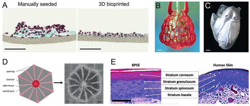
In addition to the bioprinter hardware, the material used during als and their origin, as well as their strengths and disadvantages.
the bioprinting process determines the ultimate outcome of the For further details on these substances as well as on bioinks with
process. It is commonly denoted as bioink and can be defined as additional components such as hyaluronic acid, silk and oth-
“a formulation of cells suitable for processing by an automated er materials, the reader is referred to excellent and exhaustive
biofabrication technology that may also contain biologically ac- reviews published in recent years (Gopinathan and Noh, 2018;
ALTEX 38(2), 2021 271
Berg and Kurreck

                                                                       atively low immunogenicity (Hospodiuk et al., 2017). Further-
                                                                       more, it facilitates adhesion of cells and enhances their growth.
                                                                       On the downside, the mechanical properties of collagen pose
                                                                       some challenges to its use in bioinks. Type I collagen molecules
                                                                       are mainly acid-soluble and remain in a liquid state at low tem-
                                                                       peratures. They start to gel when pH and temperature values are
                                                                       adjusted to near physiological conditions. Collagen’s slow gela-
                                                                       tion kinetics, however, make its use in bioprinting of 3D con-
                                                                       structs difficult. A solution to this problem that is described in
                                                                       more detail in Section 4.2 is the use of a support bath into which
                                                                       the collagen construct is printed (Lee et al., 2019). Following
                                                                       complete gelation of the collagen structure, the support material
                                                                       is removed and the stable form is set free.
                                                                          Gelatin, widely used in food and pharmaceutical industries, is
                                                                       the denatured form of collagen and can also be used as the basis
                                                                       of bioink. Disruption of the typical collagen triple helical struc-
                                                                       ture and its degradation result in lower immunogenicity of gel-
                                                                       atin compared to collagen (Su and Wang, 2015). A great advan-
                                                                       tage is that gelatin still supports cell adhesion, as it retains the
                                                                       arginine, glycine and aspartate (RGD) motifs present in its pre-
                                                                       cursor. Gelatin is water soluble, and the behavior of its solution is
Fig. 2: Parameters determining the outcome of a                        determined by certain factors, including concentration, tempera-
bioprinting approach                                                   ture, pH as well as the method of preparation. Gelatin has a com-
A basic classification of bioinks can be made into natural materials   paratively low gelation temperature (Wang et al., 2017b). During
derived from living organisms and synthetic materials. The             gelation, non-covalent cross-links are formed that are thermo-re-
present review aims at establishing the concept of clean bioprinting   versible, i.e., the gelatin can easily liquefy at 37°C, so the gel dis-
free of animal components to avoid chimeric systems for                solves completely. This can be utilized by employing gelatin as a
scientific reasons and for the sake of animal welfare. In this         structure-maintaining hydrogel component, also known as sacri-
respect, it is important to sub-classify the natural bioinks further   fice material, that can be flushed out during cultivation.
into materials of animal and non-animal origin, which is                  The thermo-sensitivity of gelatin enables a broad spectrum
discussed in more detail in Section 6.                                 of applications; however, this property, at the same time, lim-
                                                                       its structural integrity of printed models during culture. It may
                                                                       thus be advisable to stabilize gelatin-based hydrogels by chem-
Gungor-Ozkerim et al., 2018; Hospodiuk et al., 2017; Sun et al.,       ical cross-linking. One of the most widely used functionalized
2020). None of these articles, however, specifically investigated      variants is methacrylated gelatin, known as GelMA (Nichol et
the use of animal materials in humanized organ models as we do         al., 2010), in which methacrylate groups are conjugated to side
below.                                                                 groups of the protein. In the presence of a photo-initiator, this
                                                                       functionalization enables covalent cross-linking of the gelatin by
3.1 Natural bioinks                                                    irradiation, thereby enabling rapid and stable polymerization of
                                                                       the hydrogel. GelMa provides an aqueous cell environment and
3.1.1 Collagen and gelatin                                             supports cellular growth, adhesion as well as proliferation, and
Collagen is the main structural protein in the ECM in various          combines biological properties of the natural gelatin molecule
connective tissues, making it the most abundant protein in mam-        and controllable mechanical properties due to chemical modifi-
mals. Approximately 30 types of collagen are commonly differ-          cation, resulting in higher stability at physiological temperatures.
entiated, type I collagen being the most common form by far. It
belongs to the fibril-forming collagens and consists of three poly-    3.1.2 Matrigel™
peptide chains that form a triple-helical structure. Collagen is a     Matrigel is a murine ECM that has been widely used in advanced
widely used component of hydrogels in bioprinting applications,        cell culture technologies (Benton et al., 2014). The gelatinous pro-
as stated in recent reviews: “Collagen-containing hydrogels are        tein mixture is harvested from murine Engelbreth-Holm-Swarm
currently the most popular cell scaffold and material for tissue       (EHS) sarcoma, where it constitutes the basal membrane. It
engineering” (Osidak et al., 2020) and “the use of collagen-based      is commercialized under the name Matrigel™ by the company
bio-ink is prevalent in skin bioprinting” (Ng et al., 2016). Colla-    Corning, Inc., but is also available from other companies under
gen for research purposes is usually of bovine or porcine origin       other names. The thin basal membrane sheets of ECM surround
or derived from rat tails.                                             most animal tissues and have an essential function, serving as a
   Collagen has various desirable properties for its application in    barrier to separate different tissue types. They comprise the ma-
tissue engineering, including high biocompatibility and compar-        trix of most tumors that are of epithelial origin.

272                                                                                                                          ALTEX 38(2), 2021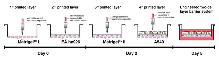
Berg and Kurreck

Fig. 3: Timeline for bioprinting of an air-blood barrier system
Endothelial cells are printed on top of a Matrigel layer. Another layer of Matrigel is printed to ensure adhesion of the top layer of epithelial
cells. Taken from Horvath et al. (2015) in accordance with the Creative Commons Public License (https://creativecommons.org/licenses/
by-nc-nd/4.0/).

   The complex composition of Matrigel has been analyzed in                sue engineering” (Fan et al., 2016). The advantageous proper-
depth by mass spectrometry (Hughes et al., 2010). Main compo-              ties of Matrigel have been confirmed in many studies (Berg et
nents are laminin-111, collagen IV, entactin, and heparan sulfate          al., 2018; Schiele et al., 2010; Schmidt et al., 2019; Snyder et al.,
proteoglycan. In addition, it includes numerous growth factors,            2011; Swaminathan et al., 2019).
which are largely responsible for the ability of Matrigel to en-
hance cell proliferation.                                                  3.1.3 Alginate
   Matrigel is frequently utilized in cell biology in vitro and in         Alginate is one of the most popular non-animal materials that is
vivo (Benton et al., 2014). It is commonly used as a basement              well-suited for 3D bioprinting, particularly in extrusion-based
membrane matrix for stem cells, as it retains the stem cells in an         approaches (Abasalizadeh et al., 2020; Axpe and Oyen, 2016).
undifferentiated state. Furthermore, it is frequently included as a        The polyanionic linear polysaccharide is obtained from brown
substrate in 3D cell culture and in suspension cultures of spher-          algae and is composed of (1-4)-linked β-D-mannuronic (M) and
oids. A field that makes intensive use of Matrigel is cancer re-           α-L-guluronic acids (G), which are ordered in mannuronic or
search, for example in angiogenesis, invasion and dormancy as-             guluronic blocks, separated by regions in which both acids are
says. In animal models, it is also used for angiogenesis assays and        mixed. Alginate is a biocompatible material that does not inten-
to promote growth of xenografts and patient-derived biopsies.              sively interact with cellular surfaces. Water and small molecules
   More recently, Matrigel has been explored as a bioink com-              are trapped in its matrix, but they are still able to diffuse and can
ponent for bioprinting approaches. The material has interesting            provide cells with a sufficient supply of nutrients.
physicochemical properties, as it is liquid at ambient tempera-               For bioprinting approaches, alginate is extruded in its low vis-
ture and reversibly solidifies at elevated temperature, forming            cosity state and is subsequently cross-linked by treatment with
a hydrogel at 37°C. It facilitates the creation of strong, 3D bio-         divalent cations such as Ca2+. The divalent cations form ionic
printed constructs with high cell survival rates. An example of            bridges between the G-blocks of adjacent polymer strands. Pre-
the potential of Matrigel in bioprinting is its application to gen-        cross-linking of alginate during the printing process by mixing
erate an air-blood barrier (Horvath et al., 2015). Production of           with low calcium concentrations can be used to achieve good
the two-cell layer barrier system starts with a layer of Matrigel          printing properties, followed by strengthening the printed con-
printed on porous membranes, on top of which a layer of endo-              struct with higher concentrations of the cross-linker. However,
thelial cells is printed (Fig. 3). On day two, a second Matrigel           a balance must be found, as high viscosity during the printing
layer is printed onto the endothelial cells to ensure the adhesion         procedure requires high pressure during the extrusion process,
of the next printed layer of epithelial cells. In another study, a         which can cause damage to the cells as a result of shear stress,
3D printable hydrogel of Matrigel and agarose was developed                whereas, if the viscosity is too low, slow gelation will hamper
to support the growth of intestinal epithelial cells and cell-ma-          structural reproducibility and resolution of the printed model.
trix interactions (Fan et al., 2016). Here, a particular combina-             Alginate encapsulates the cells of the bioink, which has both
tion of Matrigel with agarose was found to overcome disadvan-              advantages and disadvantages. The encapsulation substantially
tages of individual hydrogels. The authors conclude, “Given that           reduces shear stress in extrusion bioprinting and thereby increas-
Matrigel is used extensively for 3D cell culturing, the developed          es cell viability; however, following the printing process, encap-
3D-printable Matrigel-agarose system will open a new way to                sulation prevents cell proliferation and proper formation of cell-
construct Matrigel-based 3D constructs for cell culture and tis-           cell contacts as desired in 3D models. A way to solve this problem

ALTEX 38(2), 2021                                                                                                                              273
Berg and Kurreck

is to incubate the cross-linked alginate with sodium citrate (Wu et     the Pluronic F-127 fugitive ink is deposited as a branching net-
al., 2016). Citrate is a chelator for divalent cations and thus medi-   work in a gel reservoir that can be cross-linked by photo-poly-
ates slow and controllable degradation of alginate hydrogels.           merization after printing is completed. The fugitive ink, which is
   While the weak interaction of alginate with human cells is           not chemically modified, can be removed by liquefaction at 4°C
desirable to have an inert scaffold that does not influence cel-        and modest vacuum extraction to yield the desired vascular net-
lular behavior, it is at the same time disadvantageous, as cell         work within the matrix.
attachment to the bioink is minimal and therefore cells tend to
sediment in the printed constructs. This problem may be solved
by modifying the alginate surface with RGD motifs that pro-             4 Bioprinted organ models
vide binding sites for the cells and strengthen their attachment
(Daly et al., 2016a).                                                   Models for all major human organs have been produced by
   For many applications, it has been advisable to combine dif-         3D bioprinting during the last few years. A recent review thor-
ferent biopolymers and make use of the desirable characteristics        oughly covers publications related to bioprinting for each or-
of each material. Blends of alginate and gelatin are frequently         gan (Mota et al., 2020). Here, only some exemplary and very
used for extrusion-based bioprinting to combine the thermo-sen-         recent studies for selected organs (lung, heart, liver, and skin)
sitive properties of gelatin with the chemical cross-linking capa-      will be discussed in more detail in order to keep the focus on the
bilities of alginate (Berg et al., 2018; Han et al., 2020; Mondal et    current status of bioprinting aiming at generating new tools for
al., 2019). In these blends, the gelatin component confers good         the replacement of animal experiments by humanized 3D organ
printability to the bioink and ensures rapid, temperature-in-           models. Researchers use the term “organ” somewhat loosely, as
duced gelation immediately after the printing process to provide        it usually only contains one tissue type, which, in the best case,
the initial stability of the printed construct. The slower Ca2+ -in-    consists of several cell types, rather than truly representing an
duced gelation of alginate can then occur, the gelatin dissolves        organ in all its natural complexity. Still, we will use the term or-
over time during cultivation at 37°C, and only the alginate com-        gan or organ model for bioprinted constructs in accordance with
ponent remains to maintain the structural integrity. However,           general use.
while the remaining alginate provides desirable biocompatibil-
ity and high mechanical stability, it has poor biomimetic proper-       4.1 Lung
ties due to the above-mentioned lack of cell adhesion motifs. In        The lungs are part of the lower respiratory tract, which begins at
addition to the already described approach to link RGD motifs to        the trachea and branches into the bronchi and bronchioles. The
the alginate, the bioink can also be blended with suitable protein      respiratory bronchioles divide into alveolar ducts that give rise
mixtures such as Matrigel (Berg et al., 2018) or human ECM              to the alveolar sacs, which finally contain the alveoli, where gas
(Hiller et al., 2018).                                                  exchange takes place. The gold standard for ex vivo lung models
                                                                        was developed by the group of Donald E. Ingber at Harvard Uni-
3.2 Synthetic bioinks                                                   versity’s Wyss Institute in 2010 (Huh et al., 2010), without using
                                                                        bioprinting technologies. It consists of endothelial and epitheli-
3.2.1 Poly(ethylene glycol) (PEG)                                       al cells separated by a porous membrane, which is part of a de-
One of the most widely-used synthetic bioink components is              vice that recreates physiological breathing movements by apply-
poly(ethylene glycol) (PEG), a hydrophilic polymer that is re-          ing vacuum to two-sided chambers, thereby causing cyclic me-
sistant to protein adsorption. While this is a desirable property in    chanical stretching of the membrane. Despite the unquestioned
some respects, it also means that many cell types require cell ad-      value of this model, bioprinting may help to overcome some of
hesion components, such as RGD peptides, to strengthen the in-          its inherent limitations. In addition to the use of immortalized
teractions between the cells and the scaffold. The advantage of         cell lines instead of primary cells, the comparatively thick and
a synthetic material is that its mechanical properties can be ad-       artificial membrane does not reflect the organization of natural
justed through variation of its chemistry. PEG-based hydrogels          alveoli particularly well. This shortcoming was addressed with
can be used with photo-cross-linking in the presence of a pho-          a valve-based bioprinting approach to create an air-blood barri-
to-initiator. Methacrylate can be added to increase the mechani-        er consisting of alveolar epithelial type II cells (A549) and en-
cal strength of the printed construct (Cui et al., 2012).               dothelial cells (EA.hy926) that were separated by a thin layer
                                                                        of Matrigel (Horvath et al., 2015). This experimental strategy is
3.2.2 Pluronic F-127                                                    described in more detail in Section 3.1.2 and illustrated in Fig-
Another synthetic polymer used in bioprinting approaches is             ure 3. Compared to a manually seeded co-culture that formed
Pluronic® F-127, which belongs to the class of poloxamers, i.e.,        overgrowing multi-layered clusters, the layer-by-layer bioprint-
nonionic triblock co-polymers composed of a central hydro-              ed construct spread over the entire surface to form confluent thin
phobic chain of polyoxypropylene, flanked by two hydrophilic            monolayers (Fig. 4A).
chains of polyoxyethylene. Its most interesting feature is its abil-       A recently developed bioprinting method named SLATE (ste-
ity to undergo reverse gelation, as it starts to cross-link with in-    reolithographic apparatus for tissue engineering) allows the pro-
creasing temperature. This behavior can, for example, be used to        duction of functional intravascular topologies in biocompatible
produce a vascular network (Wu et al., 2011). For this approach,        hydrogels consisting of photo-cross-linked GelMA derived from

274                                                                                                                         ALTEX 38(2), 2021
Berg and Kurreck
Fig. 4: Examples of bioprinted organ models
(A) Comparison of manually seeded and bioprinted air-blood barriers consisting of endothelial and epithelial cells separated by a Matrigel
layer (Horvath et al., 2015). Scale bar: 100 µm. (B) Bioprinted vascular network using SLATE (stereolithographic apparatus for tissue
engineering) to mimic a pulmonary alveolus (Dasgupta and Black, 2019). Scale bar: 1 mm. (C) High-resolution replica of the heart made of
collagen and produced by the FRESH (freeform reversible embedding of suspended hydrogels) technology (Dasgupta and Black, 2019).
Scale bar: 5 mm. (D) Liver organoid generated by stereolithographic bioprinting. The computer 3D model is translated into a construct
consisting of a cell-laden multi-material hydrogel. The hexagonal structure including channels reproduces the biological topology of a liver
lobule (Grix et al., 2018). Scale bar: 500 µm. (E) Histological comparison of bioprinted full skin equivalent and native human skin (Derr et
al., 2019). H&E staining shows the layers of the dermis. Scale bar: 100 µm. Copyrights: (A) Taken from Horvath et al. (2015) in accordance
with the Creative Commons Public License (https://creativecommons.org/licenses/by-nc-nd/4.0/). (B, C) Reproduced with permission
from The American Association for the Advancement of Science. (D, E) Taken from Grix et al. (2018) and Derr et al. (2019), respectively, in
accordance with the Creative Commons Public License (https://creativecommons.org/licenses/by-nc-nd/4.0/).
porcine skin tissue (Dasgupta and Black, 2019). The technolo- (Bejleri et al., 2018). Another active field of personalized bio-
gy was used to generate bio-inspired alveolar air sacs (Fig. 4B) printing approaches is the generation of heart valves that can be
(Grigoryan et al., 2019). The complex network, composed of 185 used to replace defective valves in patients (Duan et al., 2014).
vessel segments and 113 fluidic branch points, was then perfused Figure 4C shows a high-resolution replica of the complex
with deoxygenated erythrocytes at the blood vessel inlet. Cyclic structure of the human heart that was produced by an advanced
ventilation of the airways with humidified oxygen led to efficient extrusion bioprinting technology called FRESH (freeform re-
oxygenation of the erythrocytes. versible embedding of suspended hydrogels) (Lee et al., 2019).
The special feature of this technology is the use of a thermally
4.2 Heart reversible, viscous gelatin support bath into which the 3D con-
The heart is a highly compartmentalized organ that is composed struct of interest consisting of a bovine collagen type I hydrogel
of numerous cell types and has a sophisticated architecture, as is printed. The support bath maintains the structure of the con-
well as a complex vascular system. Attempts at using bioprint- struct during the printing process and is melted away by incuba-
ing technologies for the generation of cardiac patches or whole tion at 37°C to set free the actual workpiece. Using this approach,
hearts mainly aim at producing transplantable tissue rather than an unprecedented resolution was achieved with an extrusion bio-
organ models for biomedical research and drug development. printer. When cardiac ventricles were printed with human cardio-
For example, human cardiac progenitor cells were printed with myocytes using this advanced technology, they showed synchro-
cardiac ECM and GelMA, and the resulting cardiac patches were nized beating, directional action potential propagation, and wall
implanted into rat hearts, where they were retained and showed thickening during peak systole.
vascularization over 14 days, thus demonstrating the potential Another important step towards the use of bioprinted organs
of bioprinted patches for the repair of damaged myocardium is the use of patient-specific materials. In a recent study, Noor et
ALTEX 38(2), 2021 275Berg and Kurreck
al. (2019) produced personalized perfusable cardiac patches and stem cells. The main components of the bioink were methacry-
hearts. They used primary human omental tissue to reprogram lated porcine skin gelatin and glycidal methacrylate-hyaluronic
cells into induced pluripotent stem cells (iPSCs) that were then acid. Compared to a monolayer culture or a 3D hepatocyte-only
differentiated into cardiomyocytes and endothelial cells, while model, the 3D multi-cell type model showed improved morpho-
the ECM was processed into a personalized hydrogel. The bio- logical organization, higher liver-specific gene expression levels,
ink was used to produce vascularized cardiac patches according and increased metabolic product secretion. Key drug-metaboliz-
to the patient’s anatomy and whole hearts with a natural archi- ing enzymes were significantly induced upon treatment with ri-
tecture, which were, however, only the size of a rabbit heart and fampicin, a bactericidal antibiotic drug with potential risk of hep-
did not beat. Still, the study demonstrates that bioprinting may atotoxicity. A comparably significant increase was not observed
eventually produce personalized tissues for drug-screening and in cells in 2D monolayer culture or in a 3D monoculture in the ab-
transplantable artificial organs to overcome the shortage of natu- sence of HUVECs and adipose-derived stem cells.
ral grafts from human donors.
4.4 Skin
4.3 Liver A combination of consumer pressure and the ban on animal test-
The largest organ in the human body is the liver, which con- ing in the cosmetics sector in many countries, e.g., in the EU in
sists of multiple cell types. It radiates from the central vein and 2013, initiated intensive efforts to develop sophisticated 3D skin
is surrounded by the portal vein, hepatic arteries, and bile ducts. models for research purposes and toxicity testing (Dellambra et
The liver fulfills multiple vital functions including the produc- al., 2019). As a result, skin models are among the most advanced
tion and secretion of important proteins, the metabolization of 3D tissue culture systems. The skin has a multi-layer organiza-
nutrients, and the bioconversion of many drugs and toxins. It tion that is integral to its barrier function, so specific cell-cell and
does not come as a surprise, therefore, that bioprinting technol- cell-matrix interactions and precise positioning of the cell lay-
ogy has been used to produce sophisticated liver models. The ers are important. These requirements can be met by bioprint-
high resolution of stereolithographic bioprinting allowed repro- ing technologies. Advanced procedures include a multi-step bio-
duction of the natural hexagonal structure of the liver with high printing approach to produce the different layers of the skin (Derr
precision (Fig. 4D) (Grix et al., 2018). The bioink consisted of et al., 2019). This complex model required a sophisticated bioink
a multi-component hydrogel containing porcine GelMA, modi- consisting of rat tail collagen and porcine skin gelatin in addition
fied PEG, and a photo-initiator that was mixed with a co-culture to human plasma-derived fibrinogen. It was cultured under stan-
of the hepatic cell line HepaRG and human stellate cells. Com- dard conditions including 10% FBS. As commonly done for 3D
pared to 2D cell cultures, the bioprinted tissue expressed higher skin models, the constructs were initially cultured submerged and
levels of liver markers and tight junction proteins. Furthermore, were then lifted to the air-liquid interface (ALI) to reflect their
perfusable channels printed into the organoid reproduced the mi- biological function. Histological analysis of the model demon-
cro-vascularization of the natural organ. strated a morphological multi-layer organization that is similar
The goal of producing 3D liver models that mimic the biologi- to normal human skin (Fig. 4E). Furthermore, various markers
cal physiology exemplifies another general challenge of organ re- for tight junctions and epidermal differentiation were expressed
production, i.e., the choice of adequate cells. HepaRG cells are comparably to those present in natural skin.
widely used and acknowledged to exhibit a hepatocyte-like phys- The given examples demonstrate the potential of bioprinting
iology; however, they still have the general limitations of a hepa- technologies to reproduce biological organ structures with high
toma-derived cell line, such as the expression of some typical liv- precision. However, ongoing efforts aim at improving current-
er markers at unphysiological levels. To reflect the natural liver ly available models so that they better reflect normal physiology.
physiology even better, models consisting of primary hepatocytes Major challenges are the inclusion of multiple cell types and the
or hepatocyte-like cells derived from human iPSCs are desirable. modeling of their natural interactions, as well as the precise spa-
The latter would even allow the generation of patient-specific liv- tial arrangements of all components of the model organs.
er models by using iPSCs from the respective individual. How-
ever, the limited cell numbers available from primary isolates or
cells differentiated from iPSCs still prohibit their widespread ap- 5 Application fields for bioprinted organ models
plication in bioprinting approaches. In addition, cell lines are usu-
ally more robust than primary cells or iPSC-derived cells, and Despite the need to further improve physiological features of bi-
strains during the printing process (such as shear stress, UV treat- oprinted organ models, some studies have already shown that
ment, and the formation of initiator radicals, depending on the these biomimetic systems can be used to study processes of bio-
technology used) may have a greater impact on the viability of medical relevance. Some selected examples in the fields of toxi-
primary and iPSC-derived cells. Despite these challenges, some cology, cancer research and infection biology are outlined in this
reports have already demonstrated the suitability of iPSC-de- section.
rived hepatocyte-like cells for bioprinted liver models. For ex-
ample, Ma et al. (2016) used DLP-based 3D bioprinting and he- 5.1 Toxicology
patocyte-like cells derived from iPSCs in co-culture with human The high failure rate of drug candidates in clinical trials is large-
umbilical vein endothelial cells (HUVECs) and adipose-derived ly due to the inability of animal models to predict human toxici-
276 ALTEX 38(2), 2021Berg and Kurreck ty, underscoring the need for test systems that better recapitulate ic due to nephrotoxicity (Sanchez-Romero et al., 2016), there is in vivo human biology. Bioprinted tissues and 3D culture mod- substantial interest in producing a kidney model to study adverse els, in particular, are promising tools for toxicity studies in hu- effects of new candidates. Human renal proximal tubules were manized systems (Nguyen and Pentoney, 2017). The liver is the generated by 3D bioprinting, and the tubular structure could be major organ that metabolizes a large fraction of endogenously maintained for more than two months (Homan et al., 2016). The produced or exogenously applied substances, including almost model allowed investigating adverse drug effects as exemplified all drugs in clinical use, and drug-induced liver toxicity (DILT) for the known nephrotoxin cyclosporine A, which disrupted the is one of the major reasons for failure of candidate substances. epithelial barrier in a dose-dependent manner. Several typical liver enzymes, such as albumin or cytochrome By virtue of its biological function, the skin is exposed to en- P450 enzymes (CYPs), are expressed at physiological levels on- vironmental agents in general and cosmetics in particular. Due ly in 3D arrangements, but not in conventional 2D cultures of to the previously mentioned ban on animal testing for cosmetic hepatocytes. Furthermore, non-parenchymal cells, including si- agents, the latter can only be tested in non-animal test systems. nusoidal endothelial cells, phagocytic Kupffer cells and hepatic The success of this approach can be seen in a comprehensive stellate cells, are essential for the correct function of hepatocytes. study, in which Wei et al. (2020) tested the toxicity and irrita- As there are substantial species-specific differences in liver phys- tion potential of 451 topical-use compounds in various two-di- iology, animal models produce results that are of limited rele- mensional cellular and three-dimensional bioprinted skin mod- vance for humans when studying liver toxicology of exogenous- els. The study identified toxic compounds and defined the con- ly applied substances. centration ranges in which they had irritant potential or allergic To overcome these shortcomings, the Organovo team pro- potential without causing irritation. duced a bioprinted, humanized multi-cell type liver model and The predictive value of animal studies in the pre-clinical de- demonstrated its capacity to investigate the toxicity of phar- velopment of new drug candidates is particularly limited with re- macological substances (Nguyen et al., 2016). The model con- spect to potential drug-induced toxicity. This is mainly due to dif- sisted of primary human hepatocytes that were co-printed with ferent expression levels of factors involved in uptake, distribu- human stellate and HUVEC cells. Unlike 2D cultures, the bio- tion and metabolization of xenobiotic substances. The examples printed tissue maintained high levels of liver-specific markers, discussed in this section demonstrate the potential of bioprinted including albumin and CYPs. The liver model was then used to models consisting of human cells to provide reliable predictions assess the toxicity of two clinically approved drugs, levofloxa- of adverse effects of a substance of interest. cin and trovafloxacin. The latter had to be withdrawn from the market due to hepatotoxic side effects in humans that remained 5.2 Cancer research undetected with standard pre-clinical models. In the bioprinted While 2D cultures of tumor cells have increased our knowledge liver model, but not in conventional 2D cell culture, dose-de- of genetic alterations that contribute to cell proliferation and the pendent toxicity of trovafloxacin was observed at clinically rel- induction of tumor phenotypes, they cannot investigate the im- evant doses, while it was absent in levofloxacin. The study thus portant interactions between a tumor and its microenvironment. demonstrated that such tissue models can predict liver toxicity These shortcomings may be overcome with the help of 3D bi- in humans better than standard pre-clinical models and can dis- oprinting and bridge the gap between conventional 2D cultures tinguish between highly related substances with a differential and animal models by generating a tumor environment with hu- toxicity profile. A recent study on the toxicity of aflatoxin B1 man physiology (Liu et al., 2019; Oztan et al., 2020). By using showed that liver cells in a 3D culture survived longer and were patient-derived cells and materials, it may even support the aim less susceptible to drug-induced toxicity than those cultured in of personalized medicine, i.e., the adaptation of a treatment to the 2D, making the bioprinted organ model suitable for long-term disease of a specific individual (e.g., by adjusting the treatment to studies (Schmidt et al., 2020). the specific mutations of a tumor) rather than the standard ther- In addition to metabolization in the liver, absorption and first- apy. It is difficult to model the vascular network of tumors with line metabolism are crucial steps that determine the level and thus conventional methods for 3D tissue engineering, though this as- the efficacy and toxicity of a pharmaceutical substance. Oral de- pect is considered very important in the development of effective livery is the most common method for drug administration, and treatments. Using bioprinting technologies, microchannels can the intestine is the primary organ for absorption of a drug, in ad- be produced with a sacrificial material that is removed after the dition to being a site of off-target toxicity for certain compounds. printing process, followed by populating the interior surfaces of A bioprinted intestinal tissue composed of human primary intes- the microchannels by endothelial cells (Liu et al., 2019). Alter- tinal epithelial cells and myofibroblasts with an architecture re- natively, blood vessel formation can also occur by self-organiza- flecting the native intestine was shown to develop a physiologi- tion when endothelial cells (HUVECs) are printed together with cal barrier function that was disrupted in response to the known lung fibroblasts in a bioink consisting of gelatin, alginate and fi- toxicants indomethacin and tumor necrosis factor-α (TNF-α) brinogen (Han et al., 2020). The microenvironment of blood ves- (Madden et al., 2018). The bioprinted model has thus the poten- sels and fibroblasts had a strong influence on proliferation, an- tial to support safety assessment and absorption, distribution, giogenesis, and epithelial-mesenchymal transition when seeding metabolism, excretion and toxicity (ADMET) studies in drug de- glioblastoma spheroids onto the vascularized tissue. Treatment velopment. As approximately 7% of new drugs fail in the clin- of a tumor grown on vascularized tissue with a combination of an ALTEX 38(2), 2021 277
Berg and Kurreck

anti-cancer drug (temozolomide) and an angiogenesis inhibitor           adenovirus, which can cause fatal liver failure in immunocom-
(sunitinib) was more efficacious than the anti-cancer substance         promised patients. In addition, adeno-associated virus (AAV)
alone. Importantly, these results were similar to those obtained in     vectors, which are promising delivery tools in gene therapeutic
mouse models.                                                           interventions, efficiently transduced the liver model, indicating
   In another study aiming at analyzing the signaling between the       that the model can support the further development of gene trans-
tumor and surrounding cells, Langer et al. (2019) modelled tu-          fer strategies.
mor phenotypes by 3D bioprinting. They printed the core with              Taken together, these studies demonstrate the potential of bi-
cancer cells, including patient-specific tumor tissue, surround-        oprinted organs to serve as humanized models for the study of
ed by several stromal cell types. Importantly, the hydrogel used        infection with human-pathogenic viruses. The ongoing coro-
as the bioink, consisting of alginate and gelatin, was removed          na pandemic is demonstrating the need for readily available re-
during subsequent culture so that the cells deposited ECM and           search tools to develop new antivirals. It can therefore be expect-
self-organized. Extrinsic signals and therapies were found to al-       ed that bioprinted tissue models will soon be used more frequent-
ter the tumor phenotypes, proliferation and migration. This study       ly to study infection processes. In addition, they can be used to
demonstrates that bioprinted tumor models can be used to inves-         determine the transduction efficiency of viral vectors.
tigate the interaction between cancer cells and their microenvi-
ronment and to study the effects of anti-cancer therapeutics on
both the tumor and the stroma.                                          6 Animal components in bioprinting and
   To investigate the suitability of 3D printed constructs in char-     their alternatives
acterizing anticancer drugs, breast cancer cells and adipose-de-
rived mesenchymal stem/stromal cells were co-cultured in 2D             6.1 Fetal bovine serum
and 3D and treated with doxorubicin (Wang et al., 2018). Treat-         Fetal bovine serum (FBS), also known as fetal calf serum (FCS),
ment induced less apoptosis in the 3D printed constructs. In addi-      is a commonly added supplement used in cell culture media in
tion, treatment with an inhibitor of lysyl oxidase helped to over-      virtually all life science laboratories around the world. Conse-
come drug resistance. Altogether, the study demonstrated that a         quently, it is also a standard component used to supply cells in
3D bioprinted breast cancer model reproduces the biological sys-        bioprinted 3D models with nutrients and factors that ensure high
tem better than a conventional 2D culture.                              cell viability and growth. FBS was introduced as a stimulant of
                                                                        cell growth in the late 1950s (Puck et al., 1958). It is harvested
5.3 Infection biology                                                   by means of cardiac puncture of calf fetuses discovered when
Although in vivo infection studies are usually comparatively            slaughtering pregnant cows. It is controversial whether this pro-
harmful to the test animals, as they cause fever, pain, weight loss     cedure is distressing and painful to the animals. While distrib-
and further symptoms, approximately 10% of all animal exper-            uters of FBS claim that the blood is collected from dead fetuses
iments fall into this category. This is even more startling, as the     (Nielsen and Hawkes, 2019), critics state that the blood is ob-
transferability of results from animal experiments to human pa-         tained from living calf fetuses (van der Valk et al., 2018). Fur-
thology is questionable. Many viruses have a narrow host tro-           thermore, critics point to the problem that FBS is mainly pro-
pism. For example, mice are not natural hosts of Influenza A vi-        duced in countries with less restrictive rules on animal welfare
rus (IAV) and are therefore not susceptible to infection (Radigan       than, for example, the EU, which is not a major producer of FBS.
et al., 2015). As the majority of IAV strains replicate poorly in the   The exact volume of FCS used worldwide is unknown, but has
murine respiratory tract, they are usually adapted to the mouse by      been estimated to be in the range of 500,000 to 800,000 liters an-
serial passaging (Matsuoka et al., 2009). This procedure, howev-        nually, which accounts for 1-2 million bovine fetuses, and glob-
er, leads to ambiguous results, and the course of disease differs       al demand is steadily increasing (Gstraunthaler et al., 2013; van
between humans and rodents (Bouvier and Lowen, 2010), for ex-           der Valk et al., 2018). Commercialization of FBS has a long his-
ample the induction of different pathways by pathogens in human         tory of scandals and abuse, including sales of volumes of FBS
lung tissue and mice (Berg et al., 2017; Cakarova et al., 2009).        that are higher than the officially reported amount collected in
   To close the gap, we recently investigated the potential of hu-      the specified area and contamination of the product (Gstraun-
manized bioprinted organ models to serve as tools for infection         thaler et al., 2013).
studies. Using a bioink composed of alginate, gelatin and Matri-           Apart from animal welfare, the use of FBS also adds compli-
gel for optimal growth of the alveolar basal epithelial cell line       cations from a strictly scientific point of view. Blood is a com-
A549, we were able to demonstrate efficient replication of IAV          plex mixture. A proteomic analysis of human plasma discovered
in infected constructs and observed a clustered pattern of virus        far more than 1,000 proteins (Anderson et al., 2004), and a meta-
distribution that is also found in lungs, but not in 2D cell cul-       bolomic study found more than 4,000 metabolites to be present
ture (Berg et al., 2018). Furthermore, infected cells in the lung       in human serum (Psychogios et al., 2011). To complicate mat-
model released the proinflammatory interferon IL-29. In a sub-          ters further, biological material is subject to substantial fluctua-
sequent study using a bioprinted liver model, Matrigel was re-          tions. Batch-to-batch variations for FBS are a well-known phe-
placed by human ECM (Hiller et al., 2018) to overcome the lim-          nomenon in mammalian cell culture, and it is therefore common
itations associated with the use of Matrigel discussed in Section       practice to test new lots used in research projects. The outcome
6.3. The bioprinted liver model promoted replication of human           of a project may vary depending on the specific FBS lot used.

278                                                                                                                         ALTEX 38(2), 2021
Berg and Kurreck In fact, the reproducibility crisis, according to which more than The 3D constructs were initially printed into an alginate support 70% of researchers have tried and failed to reproduce another bath, from which they were removed when the cross-linking re- scientist’s experiments (Baker, 2016), has – at least partly – been action was completed. Human adipose tissue-derived stem cells attributed to the use of different batches of FBS (van der Valk et were cultured in the printed constructs without additional ani- al., 2018). In addition, safety issues have been brought up for the mal-derived media supplementation. In contrast to cells printed use of FBS. A metagenomic analysis of 26 bovine serum samples in widely used alginate-based and GelMA bioinks, cells in the from 12 manufacturers detected viruses in all samples except one newly developed bioink readily spread, proliferated and pro- (Toohey-Kurth et al., 2017). Except for the virus-free sample, the duced ECM. This work can thus be regarded as a substantial step other samples contained up to 11 different viruses. in the direction of clean bioprinting. For the sake of animal welfare, as well as for scientific reasons, Although a promising alternative to the use of FBS, hPL is still there is a long tradition of efforts to at least reduce the amount of connected to the general problem associated with all materials of FBS required or even replace it with alternatives (Gstraunthal- biological origin, in that there are substantial variations between er, 2003). A widely used component in serum-free media is bo- different lots. Thus, the use of fully synthetic media has been vine serum albumin (BSA). This protein, which is the most com- proposed to solve the FBS-associated problems described above mon protein in FBS, has multiple intracellular functions and in- (van der Valk and Gstraunthaler, 2017). Unlike media of biologi- teracts with numerous ligands or bioactive factors and serves as cal origin, their composition is chemically well-defined and con- an extracellular transport protein (Francis, 2010). These features trolled so that the reproducibility of experimental results is high- help improve cell growth and survival. The use of BSA has pri- er and the risk of contamination with pathogens can be almost marily been driven by the need, for reasons of safety, to culture eliminated. Interestingly, a comprehensive review found that mammalian cells in serum-free media for biopharmaceutical ap- synthetic media are available for many industrially relevant cell plications, i.e., the production of recombinant proteins. However, lines to avoid the problem of batch-to-batch variations, whereas in the context discussed here, replacement of FBS by BSA does serum-containing media are still in general use in basic research not contribute to avoiding chimeric systems composed of human (Yao and Asayama, 2017). cells cultivated in hydrogels or media containing substances of Currently, a major hurdle is that there is no universal chem- animal origin. Furthermore, the substitution of FBS by albumin ically-defined medium, so that a synthetic medium needs to be of bovine origin does not contribute to improving animal wel- optimized for each cell line individually. While this occasionally fare. Recombinant human albumin offers a solution to both prob- may be desirable, for example, to prevent primary cells from be- lems but is substantially more expensive than BSA. ing overgrown by fibroblasts, in most cases it is a laborious and A promising alternative to the use of FBS is human platelet ly- costly step that prevents wide-spread use of serum-free media. sate (hPL). It is commonly obtained from donated human throm- The development of FBS-free media supporting the growth of bocyte units that are past their date of expiration. It is therefore multiple cell lines would greatly facilitate its widespread adop- clinically tested, xeno-free, i.e., free of animal components, and tion. For example, the commercially available medium Neu- contains factors that support cell growth and proliferation (van ro-Pure™ has been used to maintain and differentiate various der Valk et al., 2018). A comparative study confirmed that can- cell types, including neuronal lineages, fibroblasts and prima- cer cells cultured in media supplemented with FBS or outdat- ry cancer stem cells (Usta et al., 2014). For some cell types, se- ed hPL grow very similarly and have practically identical pro- rum-free media are commercially available; however, they are teomes (Pons et al., 2019). Furthermore, they responded equal- usually substantially more expensive than standard media with ly to different drugs and stress conditions that were tested in the FBS. Further support can be obtained from databases such as the study, demonstrating that hPL can substitute for FBS in various FCS-free database1 that collects formulations of serum-free me- experimental settings. dia for individual cell lines. Adequate in vitro reproduction of biological organs requires For the development of a new serum-free, chemically defined the co-cultivation of multiple cell types, which is often compli- cell culture medium, a basal medium of a 50:50 (v/v) mixture of cated by the fact that requirements for media composition may Ham’s F12 with Dulbecco’s modified Eagle’s medium (DMEM) differ between cell lines. An example is the co-culture of pri- is recommended that is supplemented with insulin, transferrin mary human macrophages and human mesenchymal stem cells and selenium (van der Valk et al., 2018). A further common rec- (MSC). While macrophages are commonly cultured in media ommendation is to transfer cells cultured in FBS-containing me- with human serum, media for MSCs are usually supplement- dium to a synthetic medium supplemented with the same amount ed with FBS. To solve this problem, hPL was tested as a sup- of FBS. Then, the concentration of FBS is progressively reduced, plement and found to be the best option to co-culture both cell while monitoring cell growth and viability, until the cell line is types, maintaining their phenotypes, expression profiles, and the completely weaned off FBS. This procedure was exemplarily phagocytosis activity of macrophages (Tylek et al., 2019). demonstrated for the human monocytic cell line THP-1 (Mari- In a recent bioprinting study, Medes et al. (2019) developed gliani et al., 2019). For cells that have successfully been adapted a platelet lysate-based bioink containing cellulose nanocrystals. to FBS-free conditions, it is also important to adjust the freezing 1 https://fcs-free.org ALTEX 38(2), 2021 279
You can also read