2019 HONOURS RESEARCH PROJECTS - DEPARTMENT OF BIOCHEMISTRY & GENETICS SCHOOL OF CANCER MEDICINE - La Trobe University
←
→
Page content transcription
If your browser does not render page correctly, please read the page content below
2019 HONOURS RESEARCH PROJECTS
DEPARTMENT OF BIOCHEMISTRY & GENETICS
           SCHOOL OF CANCER MEDICINE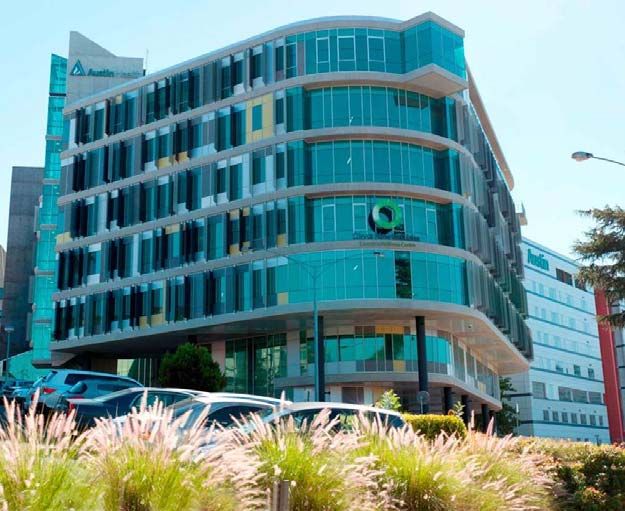
TABLE OF CONTENTS
WHY DO HONOURS? ................................................................................................................................. 2

COURSE STRUCTURE ................................................................................................................................. 3

PROJECT ALLOCATION PROCESS ................................................................................................................ 4

THE DEPARTMENT OF BIOCHEMISTRY & GENETICS .................................................................................... 5
PROFESSOR MARILYN ANDERSON .................................................................................................................. 7
DR SUZANNE CUTTS ..................................................................................................................................... 9
DR DAVID DOUGAN................................................................................................................................... 11
ASSOCIATE PROFESSOR MICK FOLEY .............................................................................................................. 13
DR DAVID GREENING................................................................................................................................. 14
ASSOCIATE PROFESSOR CHRIS HAWKINS ........................................................................................................ 16
DR BEGOÑA HERAS .................................................................................................................................... 17
PROFESSOR ANDREW HILL ............................................................................................................................ 19
DR MARK HULETT ..................................................................................................................................... 21
PROFESSOR PATRICK HUMBERT ................................................................................................................... 23
ASSOCIATE PROFESSOR MARC KVANSAKUL .................................................................................................... 25
DR MIHWA LEE .......................................................................................................................................... 27
ASSOCIATE PROFESSOR SURESH MATHIVANAN .............................................................................................. 29
ASSOCIATE PROFESSOR ROBYN MURPHY ........................................................................................................ 31
DR JACQUELINE ORIAN ............................................................................................................................... 33
DR BELINDA PARKER .................................................................................................................................. 35
ASSOCIATE PROFESSOR HELENA RICHARDSON ................................................................................................ 37
PROFESSOR RICHARD SIMPSON .................................................................................................................... 39
DR TATIANA SOARES DA COSTA ................................................................................................................. 41
DR LAKSHMI WIJEYEWICKREMA ................................................................................................................. 43

OLIVIA NEWTON‐JOHN CANCER RESEARCH INSTITUTE............................................................................. 45
DR ANDREAS BEHREN ................................................................................................................................ 47
DR ASHWINI CHAND .................................................................................................................................. 49
PROFESSOR MATTHIAS ERNST ...................................................................................................................... 50
DR DOUG FAIRLIE ...................................................................................................................................... 51
DR ERINNA LEE .......................................................................................................................................... 52
PROFESSOR JOHN MARIADSON ................................................................................................................... 53
DR DELPHINE MERINO................................................................................................................................ 54
DR LISA MIELKE......................................................................................................................................... 55
DR BHUPINDER PAL .................................................................................................................................... 56
DR NORMAND POULIOT ............................................................................................................................. 57
PROFESSOR ANDREW SCOTT ........................................................................................................................ 59

RANKED ENTRY PROJECT SELECTION FORM............................................................................................. 61

BIOCHEMISTRY & GENETICS                                                                                                                     Page | 1
WHY DO HONOURS?
Honours in Biochemistry and Genetics will kick-start your career in science. The
Biochemistry and Genetics Honours program equips graduates with technical, analytical,
communication and time management skills
demanded by employers in diverse scientific
fields ranging from biotechnology to
biomedicine. Our students work within teams
to undertake novel research projects, under
the supervision of leading scientists.
Honours projects are carried out in state-of-
the-art research facilities, either within the La
Trobe Institute for Molecular Science (LIMS)
on the La Trobe University Bundoora
campus, or at the Olivia Newton John
Cancer Research Institute (ONJCRI) in
Heidelberg.
Objectives
Extend knowledge of biochemistry, molecular genetics and medical biology
Obtain an immersive scientific experience in an authentic research laboratory
Plan and perform cutting edge experimental procedures
Pursue an original research project to generate new knowledge
Skills you will learn
Analyse and integrate research findings from numerous sources to formulate
hypotheses
Design and perform experiments using multiple advanced techniques to investigate
complex scientific questions
Critically interpret experimental data in the context of other results and prior literature
Develop excellent time management skills
Develop advanced scientific oral and written communication skills
Work collaboratively as part of a cohesive and productive research team
Past student testimonials
Honours was an incredibly stimulating learning experience
It was an amazing year which provided me with real research skills
The lab experience was invaluable
The independent research project was intellectually challenging
Working in a biomedical research lab gave me a sense of contribution to society
My honours project lead to a research publication, which set up my career
BIOCHEMISTRY & GENETICS Page | 2
COURSE STRUCTURE
Honours projects are available in the disciplines of Biochemistry, Genetics or Biomedical
Science, and can be performed either at the La Trobe Institute for Molecular Science (LIMS),
La Trobe University, (Bundoora campus) or within the Olivia Newton-John Cancer Research
Institute (ONJCRI), Austin Hospital (Heidelberg) which is affiliated with the School of Cancer
Medicine. All students, regardless of the location of their chosen laboratory, must attend
compulsory sessions (e.g. training, assignments) on the Bundoora campus at La Trobe
University.
The course will commence on Thursday 7th February 2019, with a series of compulsory
induction lectures. After this, students will write a literature review that summarises relevant
prior knowledge and present an introductory seminar to the department (accounting for 8%
of the year’s assessment). The majority of the year is dedicated to performing novel
research, under the supervision of the laboratory head and other senior staff. Throughout
the year, students will learn about diverse research topics through attending research
seminars delivered by invited experts (1%). In May, Honours students will complete a
module introducing them to advanced biotechnology techniques (10%) and in August they
will create and present a poster outlining the goals of their project and their progress to that
point (8%). In September, students will complete their experiments to focus exclusively on
writing their theses, which will be submitted in mid-October (40%). Supervisors will
continuously assess students’ laboratory performance throughout the year (10%). The year
culminates in late October, with students delivering seminars summarising their research
findings to the department (8%) and undertaking oral examinations (15%).
At the end of the course we expect our students to be able to enter the work force, and
perform competently at whatever tasks they are given. Indeed, graduates from our Honours
program are highly sought after by universities (both nationally and internationally), research
institutes, hospitals and biotech companies alike.

Choosing an Honours Project
There are a number of things to consider before deciding which supervisor/laboratory is best
for you. These include:
Research topic: Which research questions and techniques do you find most interesting and
appealing? To what extent will the background knowledge you gained through your
undergraduate studies prepare you for each of the research topics?
Supervisor: What supervisory style suits you best? Would you prefer to receive day-to-day
supervision and feedback regarding practice presentations and thesis drafts predominantly
from the laboratory head, or from a team that may include postdoctoral fellows and research
assistants?
Career opportunities: How will each of the possible projects equip you for your chosen
career, including possible postgraduate research (if applicable).
These questions can be answered through discussions with prospective supervisors,
postgraduate students and current Honours students. Don’t be afraid to ask!

BIOCHEMISTRY & GENETICS                                                               Page | 3
PROJECT ALLOCATION PROCESS
Eligibility: students must complete at least one of the following subjects MED3PRJ,
MED3LAB or GEN3LAB. Some projects have more restricted pre-requisites, so check the
project descriptions carefully. However, mere completion of a pre-requisite subject will not
guarantee entry into the course, as there are a limited number of places available. Students
MUST discuss projects with prospective supervisors.

To organise an Honours research project and supervisor:
1.   Read this booklet carefully.
2.   Attend the Project Pitching presentations in the RL Reid Building Seminar Room (RLR-
     101): 10am – 12.30pm, Tuesday 11th September, 2018, to hear a brief presentation
     about each of the projects being offered.
3.   Email any prospective supervisors who have offered projects that you find interesting
     and would consider undertaking, to organise appointments to discuss the proposed
     projects and your suitability. You can explore the possibility of securing “select” entry
     into your preferred laboratory during these meetings.
Note: Students MUST meet with a supervisor in order to be placed in their laboratory. To
     maximise your chance of being accepted into the Honours program, we therefore
     recommend that you meet with AT LEAST FIVE supervisors.

There are two allocation routes: “select” (by negotioation) and “ranked” (based on marks).
If your meeting with a supervisor leads him/her to offer you a “select” place in his/her
laboratory, and you accept, ensure that the supervisor notifies the course co-ordinator
(c.hawkins@latrobe.edu.au) (cc’ing you) by Friday 26th October, 2018.
If you do not arrange “select” entry by Friday 26th October, you can apply to gain “ranked”
entry into the course, by ranking the supervisors you met with on the attached nomination
form (page 60 of this booklet).
“Ranked” entry will be determined by your final average marks in third year biochemistry,
molecular genetics or biomedical science subjects. This means students with better marks
have a higher chance of being assigned to their preferred supervisor.
To apply for “ranked” entry into Honours, fill out the form on page 61 of this booklet,
specifying your ranking for each supervisor with whom you have met.
Submit the form to c.hawkins@latrobe.edu.au by 5pm Friday 9th November, 2018.
Provisional offers will be made via email in late November (subject to ratification by the
University of semester 2 marks) so make sure you check your university email account
during this period.
If you have any questions about the allocation process, of the Honours course, please e-
mail c.hawkins@latrobe.edu.au to make an appointment.

BIOCHEMISTRY & GENETICS                                                             Page | 4
THE DEPARTMENT OF BIOCHEMISTRY & GENETICS
The Department of Biochemistry and Genetics is engaged in both fundamental and
commercially driven research. It has a strong record of attracting competitive funding from
National (i.e. the Australian Research Council (ARC), the National Health and Medical
Research Council (NHMRC)) and international (i.e. National Institutes of Health) funding
agencies. The Department has published numerous papers in highly influential journals such
as Cell, Science and Nature. Research conducted within the Department has led to the
generation of valuable intellectual property. The Department currently houses three
biotechnology companies involved in product development and marketing.
Many of our Honours students progress to undertake PhDs, and our students often receive
awards in recognition of their research excellence. The following prizes were awarded in
2017 to previous Honours graduates who are currently PhD students:
Alyce Mayfosh (Honours 2014) received the Best Student Poster at the Victorian
Infection & Immunity Network Young Investigator Symposium
Katie Owen (Honours 2014) was awarded
the Best Overall Presenter Award at the 8th
Annual Australian Society for Medical
Research symposium (see photo)
Georgia Atkin-Smith (Honours 2015) was
awarded an ASBMB fellowship
Linda Brain (Honours 2016) and Georgia
Atkin-Smith (Honours 2015) were awarded
ASBMB poster prizes at the ComBio2017
conference
Our graduates are highly employable, being sought after by other universities and research
institutes, as well as biotechnology companies. Many of our Honours/PhD graduates
undertake postdoctoral research, within Australia or overseas. Others, including those listed
below, pursue diverse roles in academia, biotechnology or medicine:
After occupying a number of senior roles within La Trobe University Damian Spencer
(Honours 2001, PhD 2007) recently accepted a position as Dean (Teaching and
Learning) at Cambridge International College, Australia.
Dr Nicole van der Weerden (Honours 2003, PhD 2007) is now the Chief Executive
Officer of Hexima (a successful biotechnology company).
David Bloomer (Honours 2012, PhD 2017) is also working in the biotechnology industry,
within the recombinant proteins sector at CSL.
Mark Miles (Honours 2013, PhD 2018) is now employed as Associate Lecturer within
the department and is currently coordinating MED2BMS.
Stephanie Paone (Honours 2013, PhD 2018) works as Clinical Research Coordinator
at Nucleus Network.
BIOCHEMISTRY & GENETICS Page | 5
Research at LIMS is
The Department of
aimed at generating
Biochemistry and Genetics
Translatable
contains 29 research
Molecular Discoveries
laboratories, teaching
and encompasses
facilities and administration
research in six
across two buildings (LIMS1
thematic areas:
and 2). These buildings
Cancer
contain state-of-the-art
Infection and
facilities with 10,000 m2 of
Immunity
usable space including 18
Neurobiology
new research and support
Molecular Imaging
laboratories, an equipment
Molecular Sensing
barn, and ~ 3,000 m2 of
& Molecular Design
teaching facilities.
Facilities include:
Confocal and widefield microscopy (for imaging fluorescently labelled biomolecules in fixed
and live cells)
FACS/flow cytometry (analyses or sorting of cells based on surface markers or other features)
Fluorescence spectroscopy (for sensitive detection of intrinsically and extrinsically labelled
fluorescent biomolecules)
Gel doc and Chemidoc systems (for stain free analysis and Western blotting)
Gel electrophoresis equipment (for separating proteins, DNA and RNA)
Histology preparation equipment (staining of biological tissue samples)
Ion S5 next generation sequencer (for small RNA profiling and targeted DNA sequencing)
Liquid chromatography systems (for protein purification)
Mass spectrometry (for protein/peptide sequencing, and identification/quantification of
proteins in complex samples)
Protein interaction facility (including Analytical ultracentrifugation, Isothermal Titration
microcalorimetry and Surface Plasmon Resonance) for measuring biomolecular interactions
Microscale thermophoresis instrumentation (for quantification of protein-protein and protein-
ligand interactions)
Plate readers (for UV/Vis, luminescence and fluorescence assays in 96- and 384-well plate
formats)
qRT-PCR systems (for quantification of gene expression) and automated liquid handling
instruments
Tissue culture facilities (for bacterial, insect, mammalian, plant cell & viral culturing)
UV/Vis spectroscopy (for quantifying DNA, RNA, peptides & proteins and performing enzyme
kinetics assays)
X-ray crystallography (for determining the three-dimensional structure of proteins and
biomolecular complexes).
BIOCHEMISTRY & GENETICS Page | 6
Professor Marilyn ANDERSON
                        ANTIFUNGAL AND INSECTICIDAL PROTEINS
                        Office: LIMS 2, Room 407
                        Phone: 9479 1255
                        E-mail: m.anderson@latrobe.edu.au

 Subject prerequisites: MED3PRJ, MED3LAB or GEN3LAB             Theme: Infection & Immunity

 Project 1: The link between fungal EV release and cell wall stress.
 Co-supervisor: Dr Mark Bleackley
 Fungi, like all organisms, secrete extracellular vesicles (EVs) as a
 component of their communication systems. Once secreted, fungal
 EVs can be taken up by other fungi or, in the case of fungal
 pathogens, host organisms. EVs contain a variety of cargo
 including proteins, lipids, nucleic acids and small molecules.
 Upon uptake by target cells the EV cargo can induce a variety
 of changes in the physiology of the target cell. Much of the
 focus on the effect of fungal EV uptake has been on
 increasing virulence by sending signals between fungal cells
 or increasing host susceptibility as a result of uptake of
 virulence factors by host cells.
 The fungal cell wall is a unique feature of fungal EV secretion
 that differentiates it from EV secretion in mammalian cells, the best
 characterized system. The fungal cell wall is a dynamic organelle made
 up of predominantly of carbohydrates and glycoproteins that serves as a
 physical and chemical barrier that protects against biotic and abiotic stresses. It also serves
 as a barrier for the release of EVs. However, the cell wall is able to locally remodel to allow
 secretion and uptake of EVs. We have demonstrated that chemically or genetically
 decreasing the thickness of the fungal cell wall leads to an increase in EV secretion (Fig. 1).

 BIOCHEMISTRY & GENETICS                                                              Page | 7
This project will focus on determining whether this is due to an upregulation of EV biogenesis
and secretion as a component of the fungal cell wall stress response. A link between EV
uptake and resistance to cell wall stress has also been demonstrated. Understanding the
mechanism by which EVs protect against cell wall stress will also be a key aim of this project.
The bulk of the work in this project will be performed using the model yeast Saccharomyces
cerevisiae. We have a collection of S. cerevisiae deletion mutants available that will facilitate
analysis of the link between cell wall stress and EV release/uptake.

Project 2: The role of histidine-rich defensins in the tolerance of metals by plants.
Co-supervisors: Drs Rohan Lowe and Mark Bleackley
Plant defensins are a family of proteins that are well known for their ability to protect plants
against fungal diseases. These proteins have a conserved structural motif stabilised by four
disulphide bonds but exhibit a massive amount of variation at the sequence level. This
diversity has led to the evolution of additional functions, including roles in reproduction,
enzyme inhibition and metal tolerance.
The Anderson Lab has recently identified a class of plant defensins that are enriched in
histidine residues (Fig. 1). Histidine-rich proteins often bind metal ions. Recombinant
expression and purification of the histidine-rich defensins by the Anderson lab has confirmed
in vitro metal binding activity in this new class of defensins. However, it is unclear what the
function of these defensins is in planta.

We have identified histidine-rich defensins in the genome of the Australian native tobacco
Nicotiana benthamiana, a plant found in the Australian outback where soils are metal-rich,
and the dry environment means there is little threat from microbes. We hypothesise that the
histidine-rich defensins have evolved to deal with toxic levels of metals found in the outback
soil, rather than defend against microbial threats. This kind of specialisation has been
previously described for N. benthamiana, which sacrificed its viral defence system in favour
of seedling vigour and increased survival in extreme environments. This project will examine
the expression of histidine-rich defensins in N. benthamiana and compare the metal
tolerance phenotype of N. benthamiana to closely related species that lack histidine-rich
defensins.

BIOCHEMISTRY & GENETICS                                                                 Page | 8
Dr Suzanne CUTTS
MECHANISM OF ACTION OF ANTICANCER DRUGS
Office: LIMS 2, Room 423b
Phone: 9479 1517
E-mail: s.cutts@latrobe.edu.au
Subjects prerequisites: MED3LAB or MED3PRJ Theme: Cancer
The anticancer drugs doxorubicin (an "anthracycline") and mitoxantrone (an
"anthracenedione") are widely used in cancer chemotherapy, and are classified as inhibitors
of topoisomerase II. We strive to develop new therapeutic strategies for cancer treatment by
understanding the specific mechanism of action of these currently used anticancer drugs,
and building on this information to restrict their killing properties to cancerous cell types. In
this way, the toxic side effects of these drugs can be minimised.
It is now well established that anthracyclines such as doxorubicin can bind covalently to DNA
to form DNA adducts when activated by the simple molecule formaldehyde. We can activate
doxorubicin to bind covalently to DNA in this manner by supplementing with low toxicity
formaldehyde-releasing prodrugs. These lesions provide a more lethal death-inducing signal
in cells than damage which occurs in the absence of formaldehyde (ie topoisomerase II-
mediated damage). The combination of anthracyclines with formaldehyde-releasing agents
may prove clinically beneficial.
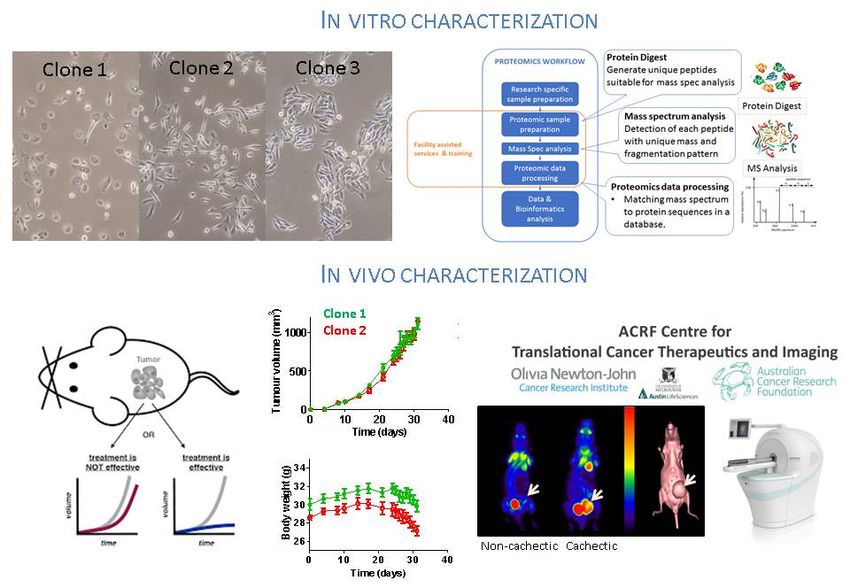
Project example: Which classes of drugs are promising for killing highly metastatic cancer
cells?
Mitoxantrone and doxorubicin are anticancer drugs that mainly function by poisoning the
nuclear enzyme topoisomerase II. Poisoning of topoisomerase II leads to lethal double
stranded DNA breaks and also depletion of cellular topoisomerase II levels, both of which
can culminate in cell death. The DNA damage that is generated by topoisomerase II
poisoning is measurable by H2AX induction.
Green, γH2AX Red, P Ser1981 ATM Merge Bright Field
Mito 2 (12.5 nM) induces DNA damage (shown by H2AX foci that colocalise with
phosphorylated ATM) at growth inhibitory concentrations. Image courtesy of Dr Ben Evison
BIOCHEMISTRY & GENETICS Page | 9For over 80% of the patients who die of cancer, metastasis is the cause of death. The ability to effectively treat metastases is limited and new therapies are urgently needed. We have access to a panel of cell lines which vary in their capacity to undergo metastases in in vivo models. These human and mouse cell lines therefore range from non-metastatic to highly metastatic. We will examine the ability of different classes of drugs to induce DNA damage and subsequent apoptosis in each of these cell lines. Some of these drugs will be prepared in nanoparticle formulations. We will also examine if switching the mechanism of action from topoisomerase II inhibition to other forms of DNA damage alters the potency of the treatments. In the first instance these treatments will be investigated in cells in culture, and then finally selected cell lines and treatments will be analysed further in in vivo models including patient-derived xenografts. The types of techniques to be utilised include mammalian cell culture, MTT cell viability assays, nanoparticle preparation & analysis, apoptosis assays, genomic DNA extraction & quantitation of drug-DNA complexes, Comet DNA damage assays, quantitative PCR, immunohistochemistry, westerns and use of a variety of drugs and chemical inhibitors. Equipment used includes microplate absorbance reader, FACS machine, fluorescence microscope, & scintillation counter. Training in the use of several software packages will also be required. BIOCHEMISTRY & GENETICS Page | 10
Dr David DOUGAN
                     PROTEOSTASIS IN HEALTH & DISEASE
                     Office: LIMS 1, Room 417
                     Phone: 9479 3276
                     E-mail: d.dougan@latrobe.edu.au

Subject prerequisites: MED3PRJ, MED3LAB or GEN3LAB                Theme: Infection & Immunity
Proteostasis is the maintenance of a functional proteome within a cell. Our lab is particularly
interested in the cellular machines that are responsible for maintaining “proteostasis”. A
major focus of the group is understanding how large ATP-dependent machines contribute
to the overall “health” of a cell. We are interested in the biological function of these machines,
their mechanism of action and their role in disease. We are also interested in the
identification and development of novel compounds that dysregulate these machines as a
means generate novel therapeutics or antibiotics.

We focus on the role of molecular chaperones and proteases in the maintenance of the
cellular proteome, not only under normal conditions, but also under condition of cellular
stress. An ultimate goal of the lab is to develop novel drugs, either chemical chaperones that
can stabilise vulnerable proteins in the cellular proteome during times of stress or chemical
dysregulators of these machines that serve as novel antibiotics to kill pathogenic bacteria.

Project 1: Investigating the role of the putative sporulation transcription factor (WhiA) in
Mycobacterium smegmatis proteostasis.
Our lab uses Mycobacterium smegmatis (Msm) as a non-pathogenic model organism to
understand the role of M. tuberculosis in Tuberculosis (TB) and identify any potential
weaknesses this organism may have. The proteostasis network is an important target for the
development of novel drugs against mycobacteria. Recently, we identified the transcriptional
regulator that controls sporulation – WhiA as a putative interacting protein of a novel
component of the Clp protease, and hence is a crucial component of the proteostasis
network in Msm. We hypothesize that the metabolic stability of WhiA is regulated by its
interaction with this novel proteolytic system and as such this proteolytic system may
regulate Msm sporulation. This project will use a range on molecular, biochemical and
genetic approaches to study the interaction of WhiA with the novel Clp protease and analyse
the consequences of this interaction.

Reference: Alhuwaider AAH & Dougan DA (2017). AAA+ Machines of Protein Destruction
in Mycobacteria. Front Mol Biosci. 4: 49 doi: 10.3389/fmolb.2017.00049.

BIOCHEMISTRY & GENETICS                                                                Page | 11
Project 2: Identifying the physiological role on the N-end rule in Mycobacterium smegmatis. Co-supervisor: Dr Kaye Truscott (LIMS) Previously our lab identified the recognition component of the N-end rule pathway in Escherichia coli – ClpS (Erbse et al., 2006 Nature 439: 753-6) and the physiological substrates of this pathway (Ninnis et al., 2009, EMBOJ 28: 1732-44). This project aims to identify novel components of the N-end rule pathway in Msm and as a result elucidate the physiological role of this pathway in mycobacteria. The project will examine the metabolic stability of several ClpS-interacting proteins that we have previously identified in M. smegmatis. It will involve the use of several Msm gene deletion strains, which lack specific components of the putative N-end rule pathway. The student involved in this project will gain experience in molecular biology, biochemistry and genetics. Reference: Kirstein et al., (2009) Adapting the machine: adaptor proteins for Hsp100/Clp and AAA+ proteases Nature Rev Micro. 7: 589-599. BIOCHEMISTRY & GENETICS Page | 12
Associate Professor Mick FOLEY
                  SHARK ANTIBODIES AS HUMAN THERAPIES
                  Office: LIMS 2, Room 217
                  Phone: 9479 2158
                  E-mail: m.foley@latrobe.edu.au

Subjects prerequisites: MED3LAB or MED3PRJ                      Theme: Infection & Immunity

Shark antibodies as therapeutic agents of disease
Co-supervisor: Dr Kate Griffiths (LIMS)
Shark antibodies (IgNARs) are a subset of antibodies found in
sharks, rays and other cartilaginous fish. Some IgNARs have
been shown to possess an elongated CDR3 loop, that is
significantly larger those of human and murine antibodies. The
IgNAR extended CDR3 loop is considered to be ideal for
targeting cleft-type epitopes such as enzyme active sites and
surface receptors which are otherwise inaccessible to
conventional antibodies. Using Plasmodium falciparum as a
model system we have identified peptides and shark antibodies
that block invasion of malaria parasites into host erythrocytes.
The structure of the complex of this IgNAR and its target
revealed that the IgNARs penetrate a hydro- phobic trough on
the malarial protein.
Recently we have created a humanized version of these antibodies and have identified
antibodies from this library that bind to the chemokine receptor CXCR4. This molecule is
up-regulated in many cancer cells and is an important target in fibrosis. We are therefore
exploring the use of these antibodies in both cancer and fibrosis. These antibodies can bind
                                            to and block the growth of cancer cells as well as
                                            block inflammatory cells from migrating towards
                                            the site of inflammation. Moreover these
                                            antibodies can prevent the development of
                                            fibrosis in an animal model. This honours project
                                            will examine the mechanism of action of these
                                            antibodies in either cancer or fibrosis with a view
                                            to developing improved molecules to progress
                                            towards human clinical trials.

BIOCHEMISTRY & GENETICS                                                             Page | 13
Dr David GREENING
EXTRACELLULAR VESICLES, EXOSOMES &
IMPLANTATION
Office: LIMS 1, Room 423
Phone: 9479 5031 E-mail: d.greening@latrobe.edu.au
Subjects prerequisites: MED3LAB or MED3PRJ Theme: Cancer
Extracellular vesicles in implantation
Extensive evidence suggests that the release of membrane enclosed compartments, more
commonly known as extracellular vesicles (EVs), is a potent newly identified mechanism of
cell-to-cell communication both in normal physiology and in pathological conditions. EVs
contain diverse cargo including cell surface receptors, lipids, messenger RNAs (mRNAs),
miRs, proteins and even DNA, and are identified by their size and the presence of cell
surface markers such as the tetraspanins, CD9, CD81 and CD63. EVs are increasingly
recognised as an important mode of cell-to-cell communication as they can transfer their
contents to other cells thereby altering the recipient's behaviour.
Exosomes are a particular subtype of EVs that are secreted from a wide range of cells,
including placental and endometrium cells. Exosomes are very stable vesicles that contain
a broad spectrum of molecules, including proteins, mRNAs and miRNAs. Very little is known
about this form of cell-to-cell communication in the context of ovarian follicular biology and
implantation, but emerging data suggest that exosomes secreted by the blastocyst could
influence gene expression and receptivity of endometrial cells thereby controlling its own
implantation. Implantation involves intricate communication between embryos and the
maternal endometrium. Increasing interest is centred on EVs and their contained cargo,
particularly microRNAs (miRs), as important mediators of this dialogue. Recently, we have
established a role for exosomes in cell-to-cell communication.
Project 1: Functional insights into EVs during human implantation
Co-supervisor: Dr Alin Rai (LIMS)
Actively released from cells, exosomes are tiny vesicles (50-150nm diameter) that transport
cargo consisting of mRNA, miRNA and proteins, to recipient cells. These can be at a
distance from the source of the exosomes which are taken up and their contents released to
alter the behaviour of the recipient cells. Exosomes have been prepared from an human
endometrial epithelial cell model and shown to contain select protein and miRNA cargo,
some of which are not detectable in the cells of origin. We propose that these exosomes
BIOCHEMISTRY & GENETICS Page | 14play an important role in embryo-maternal communication at implantation. This project will utilise co-culture systems to further understand the important role that EVs play in this environment, how they target recipient cells, are internalised, and importantly alter the function of the recipient cell system to facilitate changes in implantation. EV (or EV specific cargo) is expected to functional modulate adhesive or invasive capacity of the trophectodermal cells Project 2: Contribution of EV protein complexes to cell-cell communication Co-supervisor: Dr Rong Xu, Prof Richard Simpson (LIMS) Exosomes, small membrane vesicles of endocytic origin, are secreted by most cell types. Although functioning as powerful intercellular communicators, the identity of exosomal protein complexes (EPCs) and their specific components, together with the molecular mechanisms underlying their functions in recipient cells, remain unknown. This project focuses on the hypothesis that multiprotein complexes contained in cancer cell-derived exosomes play a crucial role in cell-cell communication and that perturbation of EPCs may affect the functionality of stromal target cells. This project will identify exosome protein complexes, their specific protein components and insights into their structural organization (i.e., core subunit interactions) and functionality in the context of cancer biology. Such complexes contained in cancer cell-derived exosomes play a crucial role in cell-cell communication and that perturbation of EPCs may affect target cell functionality and impact on exosome-targeted drug design. Use of innovative specialised techniques including immunoaffinity enrichment coupled to high-resolution mass spectrometry, cryo-lysis, nano- particle tracking, and high-resolution imaging will be employed. References [1] Extracellular Vesicles in Human Reproduction in Health and Disease. Endocrine Rev. (2018) doi: 10.1210/er.2017-00229 [2] Extracellular vesicles in cancer—implications for future improvements in cancer care Nature Reviews Clinical Oncology (2018) doi:10.1038/s41571-018-0036-9 BIOCHEMISTRY & GENETICS Page | 15
Associate Professor Chris HAWKINS
                        APOPTOSIS & NECROPTOSIS RESEARCH
                        Office: LIMS 1, Room 514
                        Phone: 9479 2399
                        E-mail: c.hawkins@latrobe.edu.au

Subjects prerequisites: MED3LAB, MED3PRJ or GEN3LAB                        Theme: Cancer

Our research focusses on two pathways through which surplus or dangerous cells can be
eliminated: “apoptosis” and “necroptosis”. Research indicates that agents which selectively
stimulate these pathways in malignant cells can effectively treat some cancers that do not
respond to conventional therapies. Furthermore, direct engagement of apoptotic or
necroptotic pathways may spare patients some unpleasant and sometimes lethal side
effects associated with chemotherapy or radiotherapy. The Honours projects outlined below
seek to enhance our understanding of cell death signalling pathways, and to exploit this
knowledge to improve cancer treatment.
Project 1: Evaluating potential new drugs for sarcomas
We have recently obtained exciting data suggesting that members of two classes of drugs
(“IAP antagonists” and “proteasome inhibitors”) may be useful for treating the bone cancer
osteosarcoma. Experiments using cell culture techniques and mouse models will reveal
whether these drugs may also be effective against other types of connective tissue cancers
(sarcomas), that develop in the bones, muscle and neuronal tissues. Biochemical assays
will examine the molecular mechanisms that determine sensitivity/resistance of sarcoma
cells to these agents.
Project 2: Defining necroptotic signalling
Necroptosis is a relatively newly-identified
form of programmed cell death, which can
be triggered by immune cytokines or
molecules derived from pathogens, and
often occurs when apoptosis is blocked.
Although the key effectors of this pathway
have been identified, mechanisms
regulating their activity are poorly
understood. Microbiological and cell
biology techniques will be used to define
factors that determine necroptotic
pathway activity.

BIOCHEMISTRY & GENETICS                                                         Page | 16
Dr Begoña HERAS
BACTERIAL VIRULENCE FACTORS: STRUCTURE
& FUNCTION
Office: LIMS 1, Room 517
Phone: 9479 3185 E-mail: b.heras@latrobe.edu.au
Subjects prerequisites: MED3LAB or MED3PRJ Theme: Infection & Immunity
Bacterial virulence factors: structure and function
Bacterial resistance to antibiotics is increasing at an alarming pace
with fears that we could return to the pre-antibiotic era where bacterial
infections were virtually untreatable. There is an urgent need to
increase our understanding of the mechanisms underlying bacterial
pathogenesis to identify new targets for therapeutic intervention. In
the Heras laboratory we are investigate the virulence mechanisms in
Gram-negative bacteria in order to develop antibacterial drugs with
novel modes of action.
Serine protease autotransporters of enterobacteriaceae (SPATEs): structure and function
Pathogens rely on an arsenal of virulence factors, to attach and infect their host.
Autotransporter (AT) proteins are a major group of virulence proteins that bacteria use to
establish highly persistent infectious diseases
In this project we will investigate a key AT subgroup, the Serine
Protease Autotransporters of Enterobacteriaceae (SPATEs).
These secreted trypsin-like serine proteases are associated with
virulence functions such as colonisation, invasion and toxicity.
Two important SPATE proteins are SigA and Sat. Both are
internalized cytotoxins present in pathogens like Shigella and
Escherichia coli which cause diarrheal diseases and urinary tract
infections and are among the most common infectious diseases of
humans. Crystal structure of the
The outcomes of this research will contribute to a better under- autotransporter
standing of the biology of Gram-negative pathogens. Furthermore, adhesin Ag43
this work will provide valuable structure - function information that
will be the all-important basis for the development of new and more effective antibacterial
therapeutics, a subject of major significance given that these pathogens are becoming
increasingly resistant to current antibiotics.
BIOCHEMISTRY & GENETICS Page | 17Application of a fragment based drug discovery approach for developing anti-neisserial
agents
Collaborator: A/Prof Martin Scanlon (MIPS)
MDR pathogens include Neisseria gonorrhoeae the causative agent of sexually transmitted
gonorrhoea, which was recently classified as one of the ‘top urgent threats to global health,
due to their increasing resistance to antimicrobials. The overall goal of this work is to develop
narrow spectrum anti-neisserial therapeutics.
Bacteria contain
periplasmic Disulfide Bond
(Dsb) forming enzymes to
catalyze the folding of many
virulence proteins. These
Dsb catalysts are are
currently being investigated
as potential targets for the
development of
antivirulence agents.
DsbD,a member of the Dsb
family is an essential Fragment-based drug discovery (FBDD) approach:
enzyme for the viability of Involves screening small fragments for the development of
Neisserial pathogens. drugs
In this project a fragment-based drug design approach that combines NMR spectroscopy,
X-ray crystallography and electron transfer assays will be employed to identify small
molecules that bind to the catalytic domains of DsbD.
The binding mode and potency of these fragments will be investigated and their inhibitory
activity tested using electron transfer in in vitro assays. Elaboration of these hit fragments
will also be performed to obtain potent
Neisserial DsbD inhibitors.
The ultimate outcome of this work is the
development DsbD inhibitors as
potential narrow spectrum antibiotics
against Neisserial infections.
BIOCHEMISTRY & GENETICS Page | 18Professor Andrew HILL
NEURODEGENERATIVE DISEASES
Office: LIMS 1, Room 502
Phone: 9479 1224
E-mail: andrew.hill@latrobe.edu.au
Subjects prerequisites: MED3LAB, MED3PRJ or GEN3LAB Theme: Neurobiology
Neurodegenerative diseases such as Alzheimer’s and Parkinson’s disease have an
increasing prevalence amongst our ageing population. Recent estimates suggest the
numbers of Australians suffering from dementia is set to double to 500,000 individuals by
the year 2030. Many of these diseases are associated with the misfolding of certain proteins
into aberrant forms that are found in the brain tissues of individuals with these diseases.
The Hill lab uses a combination of Biochemistry, Molecular and Cell Biology to investigate
these diseases. We are interested in understanding the molecular mechanisms by which
these proteins exert their neurodegenerative properties and in the case of prions, gain their
infectious properties.
We are also interested in extracellular vesicles such as exosomes and microvesicles as
vehicles for the transfer of misfolded proteins between cells. We also investigate the RNA
content of these vesicles using next generation sequencing and have used this to develop
potential diagnostics for prion and Alzheimer’s diseases.
How do highly conserved regions of PrP control prion formation?
We are interested in understanding how the
normal cellular isoform of the prion protein
undergoes structural changes to become the
disease associated form. We have developed
several approaches to study this aspect of prion
biology using cellular, molecular and
biophysical techniques. We have refined a
model based around a highly conserved region
of the prion protein. This project will use a
combination of structural and cell biological approaches to investigate the mechanism of
prion inhibition through this conserved region of the prion protein.
BIOCHEMISTRY & GENETICS Page | 19Molecular mechanisms of Aβ toxicity in Alzheimer’s disease ‐ a role for PrP?
PrP can act as a receptor for Aβ oligomers which are implicated in the
pathogenesis of Alzheimer’s disease. Aβ is derived from a larger protein
called the amyloid precursor protein (APP). We have well established cell
lines expressing different forms of PrP and APP (which generate Aβ). These
will be used to investigate the interactions of different forms of Aβ with PrP
using a combination of biochemical and cell biological assays to determine the molecular
mechanisms underlying this interaction.
Investigating the role of microRNA (miRNA) in neurodegenerative disorders
The role of microRNA in regulating the expression of key genes
and pathways involved in neurodegenerative diseases are being
investigated in both cell and in vivo models of these disorders.
Using human clinical samples, we have identified a subset of
serum microRNA biomarkers associated with various
neurodegenerative diseases such as Alzheimer’s disease.
microRNA are enriched in exosomes and may regulate gene
expression in target neuronal cells of the brain. These novel sequences are being tested for
their functional effects and potential as disease biomarkers using cell models. This project
will also determine whether exosomes assist in transporting microRNA through the tightly
regulated blood-brain barrier to the peripheral system where they can be detected as a liquid
biopsy. This project uses next-generation sequencing technologies, bioinformatics
analyses, advanced molecular biology and cell biology techniques. These projects will be
undertaken with co-supervision by Dr. Lesley Cheng, a Postdoctoral Researcher within Prof.
Andrew Hill’s Laboratory.
Extracellular vesicles in processing of proteins involved in neurodegenerative diseases
This project will investigate the mechanisms of
exosome uptake by cells using modifiers of key cellular
pathways and live cell imaging. This project will also
investigate the role of modifiers in exosome
biogenesis by altering their expression with RNAi,
using a lentiviral delivery system in neuronal cells. We
are also using high-resolution cryo-electron
microscopy to study the structure of exosomes isolated from neuronal cells.
Are extracellular vesicles spreading toxicity in Parkinson’s disease?
The synaptic protein α-synuclein is expressed throughout the brain, however genetic
mutations in α-synuclein cause cell death beginning in dopamine-containing cells, which
spread to other cell types late in the disease. This project will investigate if proteins, such as
mutant α-synuclein, can be modified in one cell type, secreted in exosomes, and taken-up
by other cells to spread toxicity in Parkinson’s and other neurodegenerative diseases.
BIOCHEMISTRY & GENETICS Page | 20Dr Mark HULETT
INHIBITING TUMOUR PROGRESSION &
INFLAMMATION
Office: LIMS 1, Room 520
Phone: 9479 6567 E-mail: m.hulett@latrobe.edu.au
Subjects prerequisites: MED3LAB or MED3PRJ Theme: Cancer
Co-supervisors: Dr Ivan Poon, Dr Fung Lay, Dr Marc Kvansakul (LIMS)
Our research focuses on defining the molecular basis of tumour progression and
inflammatory disease to develop novel anti-cancer and anti-inflammatory drugs. In
consultation with prospective honours students we will “tailor” design projects that are
appropriate to your interests under the following themes.
Heparanase function in tumour metastasis and inflammatory disease
The ability of malignant tumour cells to escape from primary tumour sites and spread
through the circulation to other sites in the body (metastasis) is what makes cancer such
a deadly disease. An essential process in metastasis is cell invasion - where tumour cells
move into and out of the vasculature.
Cell invasion is also a critical event in
the migration of white blood cells of
the immune system (leukocytes) to
sites of inflammation to combat
infections. The heparan-sulphate
(HS)-degrading enzyme has been
shown to play a key roll in the
degradation of extracellular
matrices and its activity strongly
correlates with the metastatic
capacity of tumour cells and the
migratory capacity of leukocytes. We have shown that heparanase is the dominant HS-
degrading enzyme in mammalian tissues, making it an attractive drug target. We are
currently working towards (i) further understanding the molecular basis of heparanase
function at the structural level, (ii) defining the dysregulation of heparanase gene expression
in cancer and inflammatory disease, and (iii) using heparanase conditional knockout
mice in disease models to define the precise role and contribution of heparanase in tumour
progression and inflammation. Our overall goal is to better understand both the biology and
BIOCHEMISTRY & GENETICS Page | 21structure of heparanase to enable the development of specific inhibitors of the enzyme, which will lead to new drugs for the treatment of tumour metastasis and inflammatory diseases. Innate defense molecules as anti-cancer agents Defensins are innate immunity proteins involved in host protection against pathogens. We have identified a subfamily of defensins that show promise as anti-cancer agents and have an extensive programs dedicated to (i) defining the molecular basis of the anti-cancer activity of defensins using a range of biochemical and biophysical methods including live cell imaging, electron microscopy, X-ray crystallography and small-angle X-ray scattering; and (ii) in vivo testing and pharmacokinetic properties of defensins in mouse models of tumour growth and progression. See Elife (Cambridge). 2014; 3:e01808 Histidine-rich glycoprotein in necrotic cell/pathogen clearance and autoimmunity Histidine-rich glycoprotein (HRG) is an abundant multi-functional plasma protein of vertebrates. We have shown HRG is a novel pattern recognition molecule that forms an adaptor complex with other innate immunity molecules to mediate the clearance of necrotic cells through phagocytes. Based on these observations we propose that HRG plays a key role in maintaining efficient clearance of necrotic cells from the circulation, a critical process of the innate immune system for the elimination of self-antigens to prevent autoimmune disease. In addition, we have observed striking similarities between the recognition of necrotic cells and pathogens by the innate immune system. We propose that the same molecular mechanisms are used to clear these potentially harmful materials and promote the resolution of tissue injury. We are currently working on (i) defining the in vivo role of HRG in necrotic cell clearance using HRG deficient mice, (ii) investigating the role of HRG in pathogen recognition and clearance, (iii) defining the HRG complex for the recognition of necrotic cells and pathogens. BIOCHEMISTRY & GENETICS Page | 22
Professor Patrick HUMBERT
                        CELL POLARITY & TISSUE ARCHITECTURE
                        Office: LIMS 1, Room 410
                        Phone: 9479 5155
                        E-mail: p.humbert@latrobe.edu.au

 Subjects prerequisites: MED3LAB or MED3PRJ                                     Theme: Cancer

 Topic 1: Cell Polarity and Cancer
 In the Humbert Laboratory, researchers investigate the fundamental role of tissue
 organisation and asymmetry on cancer progression with the aim of identifying new
 therapeutic strategies.
                                                            Loss of the proper orientation of
                                                            cells within a tissue, known as
                                                            cell polarity, is one of the
                                                            hallmarks of breast cancer and is
                                                            correlated with more aggressive
                                                            and invasive cancers. However
 how loss of cell polarity occurs and how it contributes at the molecular level to tumour
 formation remains unknown. Using a number of approaches such as RNAi screening, our
 laboratory has identified a network of cell polarity tumour suppressor genes that share a
 common function in modulating key oncogenic pathways.

 Project Description: Cell Polarity Proteins and Cancer
 In this project, you will characterize how key polarity regulators from this
 gene network can supress oncogenic signalling at the proteome and
 phospho-proteome level. Using quantitative phospho-proteomics, you will
 reveal the transcriptional and phosphorylation events that are critical for
 tumour suppression by polarity genes. A variety of biochemical, cell
 biological and functional assays set up in our laboratory will allow you to
 delineate the precise molecular mechanisms by which these protein
 modifications can impact on cancer signalling and cell transformation.
 These studies include gene knockdown studies in 3D mammary organoid
 cultures and analysis of established genetically engineered mouse
 models of breast cancer.
 The above experiments will provide essential information as to the requirement for intact

 BIOCHEMISTRY & GENETICS                                                            Page | 23
polarity signalling in breast cancer development and the signalling pathways regulated by the genes that control tissue organization to suppress invasion and tumour growth. Techniques: Cell culture, RNAi, Proteomics, Mass Spectrometry, 3D organotypic cultures, immunostaining and histological analysis. Topic 2: How the Red Blood Cell lost its Nucleus Erythroid enucleation is the process by which the future red blood cell (RBC) disposes of its nucleus prior to entering the blood stream. Although the process of enucleation has been recognized for more than a century, the molecular and cellular programs governing it are still poorly understood. With a large proportion of cancer and surgical patients undergoing blood transfusions as part of their treatment, a major challenge for transfusion medicine is the constant difficulties in obtaining sufficient supplies of specific RBC subtypes. Despite exciting advances in the in vitro production of human red blood cells from hematopoietic, embryonic and induced pluripotent stem cells, the reduced ability of these cultured cells to fully enucleate remains a major hurdle. A better understanding of the enucleation process should lead to improved strategies for the efficient and rapid production of RBCs for autologous (i.e. self generated) patient transfusion. Project Description: Exploring the role of CDK9 in erythroid enucleation Our lab has identified a novel role for the serine/threonine kinase CDK9 in the enucleation process. How CDK9 regulates nuclear extrusion is completely unknown. This project aims to identify the phosphorylation targets and binding partners of CDK9 during erythroid enucleation using mass spectrometry approaches. You will initially use recombinant purified CDK9 protein to perform pull down experiments on protein lysates obtained from enucleating erythroblasts. Binding partners of CDK9 will be identified by mass spectrometry. In addition, phosphoprotein profiles will be established following inhibition and activation of CDK9 kinase activity in enucleating erythroblasts using mass spectrometry. Follow up work will include using confocal microscopy, ex vivo cell biology assays and biochemical techniques to validate potential CDK9 targets and binding partners. Together your studies will provide insights into how CDK9 regulates erythroid enucleation and help develop strategies to enhance the production of red blood cells in vitro for patient transplantation purposes. BIOCHEMISTRY & GENETICS Page | 24
Associate Professor Marc KVANSAKUL
                      HOST PATHOGEN INTERACTIONS
                      Office: LIMS 1, Room 516
                      Phone: 9479 2263
                      E-mail: m.kvansakul@latrobe.edu.au

 Subjects prerequisites: MED3LAB or MED3PRJ                        Theme: Molecular Imaging

 Virus-mediated inhibition of programmed cell death
 Programmed cell death or apoptosis is a critical process that allows
 removal of infected, damaged or otherwise unwanted cells. Failure to
 correctly control apoptosis plays an important role during pathogenic
 infections,    autoimmune diseases and cancer. Using X-ray
 crystallography we aim to understand at the atomic level how certain
 viruses hijack the host cell’s apoptotic machinery to ensure their own
 survival and proliferation. We are particularly interested in certain
 poxviruses as well as tumour viruses such as those from the herpesviridae. All projects in
 this area aim to express milligram quantities of these proteins and biochemically
 characterize them using isothermal calorimetry, surface plasmon resonance or additional
 functional assays. Once this is completed, we aim to crystallize these proteins in complex
 with ligands identified during the initial biochemical studies.
 The role of LgL, a cell polarity gene, in tumour formation and metastasis
 Collaborator: A/Prof Helena Richardson, Prof Patrick Humbert (LIMS)
 Lethal giant larvae (Lgl) is a polarity gene that controls cell orientation and whose
 dysregulation contributes to the development of certain cancers such as prostate, lung, liver,
 colorectal, breast, ovarian, melanoma and glioblastoma tumours. Lgl works in concert with
 the three other proteins called Scribble (Scrib), Discs Large (Dlg) and Gukholder (GukH) as
 part of a multi-protein module called the Scribble module to define the polarity of a cell.
 Previous work by our collaborator A/Prof Richardson has shown that Lgl may function to
 control epithelial tissue formation. We now need to establish how Lgl and its partners
 contribute in this process and clarify their molecular mechanism of action. The Scribble
 polarity module has previously been shown to play a prominent role in tumour development,
 and an improved understanding of how the components of this module work together to
 control cell polarity may enable targeting of these proteins for therapeutic purposes. To
 achieve this, we aim to biochemically characterize the interactions between Lgl and Scrib,
 Dlg and GukH, and using X-ray crystallography, show in atomic detail how they perform their
 function.

 BIOCHEMISTRY & GENETICS                                                            Page | 25
You can also read