Mouse and Rat Monoclonal Antibodies 2021 - CNIO
←
→
Page content transcription
If your browser does not render page correctly, please read the page content below
Mouse and Rat
Monoclonal Antibodies
2021

            1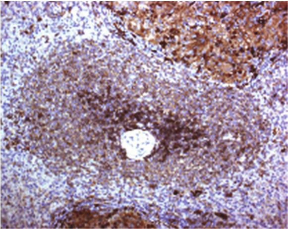
CNIO Monoclonal Antibodies Unit

The Monoclonal Antibodies (mAb) Unit provides CNIO Research Groups with the “à la
carte”, generation of mAbs which can then be used as tools to characterise new
pathways involved in cancer development. We are highly specialised in mouse and rat
monoclonal antibodies production. The Unit also offers mAbs production in gene-
inactivated mice, mAb characterisation and validation, medium-scale mAb production
and a service of Mycoplasma testing for the cell culture facility. The Unit is highly
specialised in the characterisation and validation of antibodies. The mAbs generated
by the Unit are extensively validated using a large set of tissue samples (specifically
designed tissue-microarrays) and cell lines. After validation the mAbs are then tested
in several applications. This work helps CNIO investigators save valuable research
funds and effort, providing reliable reagents for use in their research projects.

Techniques Available

-    Monoclonal antibodies production in mice
-    Monoclonal antibodies production in gene-inactivated mice
-    Rat monoclonal antibodies
-    Antibody characterization
-    Development of double and triple immunostaining techniques
-    Mycoplasma testing
-    Genetic immunization

Monoclonal Antibodies Staff

Unit Leader                 Giovanna Roncador       (groncador@cnio.es)

Technicians                 Lorena Maestre          (lmaestre@cnio.es)
                            Ana Isabel Reyes        (aireyes@cnio.es)
                            Sherezade Jiménez       (sjimenez@cnio.es)
                            Alvaro García           (agarcia@cnio.es)

Secretary                   Celia Ramos             (cmramos@srv.cnio.es)

    mAbs Datasheet are available at:
    https://www.cnio.es/investigacion-e-innovacion/servicios/anticuerpos-monoclonales/

                                           2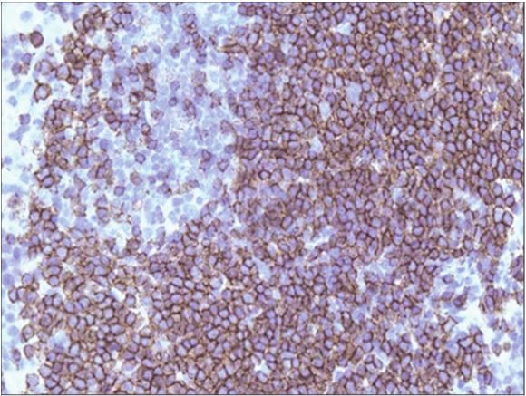
INDEX

AID .................................................................................................... 5       IgG (Fc specific) ........................................................................ 46
A-myb/B-myb/C-myb ............................................................... 5                              KLH ................................................................................................. 48
ANX4 ............................................................................................... 6           KLHL-6 .......................................................................................... 49
BCL-2 ............................................................................................... 7          LCN-2............................................................................................. 50
BCL-6 .............................................................................................. 8           LKB1 ............................................................................................... 51
BCL-6 (Mouse specific) ............................................................. 9                           LMO2 ............................................................................................. 52
BCL7A ........................................................................................... 10             Luciferase ..................................................................................... 52
Beta-Galactosidase .................................................................. 11                         MALT-1 ........................................................................................... 53
BPTF .............................................................................................. 11           MAP-17 .......................................................................................... 54
BSA ................................................................................................ 12          MASTL ........................................................................................... 55
BUB-1 (Xenopus specific) ....................................................... 12                              MBP ................................................................................................ 56
Cas9 (S. Pyogenes) .................................................................. 13                         MCAK (Xenopus) ....................................................................... 56
CD115 (mouse specific) (CSF1R) ......................................... 22                                      MCRS1 (Microspherule protein 1) ......................................... 57
CD115 (CSF1R) ............................................................................. 21                   MNDA ............................................................................................ 58
CD138 (Syndecan-1).................................................................. 22                          NANOG (Mouse specific) ....................................................... 59
CD147 (Basigin) .......................................................................... 23                    NKX2-3 .......................................................................................... 60
CD15 (FUT4) ................................................................................ 15                  NOMO............................................................................................ 59
CD162 (PSGL1) ............................................................................ 24                    NSE2 (NSMCE2) ........................................................................ 61
CD1d (CD1) ................................................................................... 13                p15 (CDKN2B) (Mouse specific) ........................................... 62
CD20 (Mouse specific) ............................................................ 16                            p15/p16 .......................................................................................... 64
CD229 (Ly-9) .............................................................................. 25                   p16 (CDKN2A) (Mouse specific) .......................................... 63
CD271 (NGFR) ............................................................................. 25                    p19 (CDKN2A-ARF) (Mouse specific) ................................ 64
CD271 (NGFR) (mouse specific) .......................................... 26                                      p21 (Mouse specific) ................................................................. 65
CD272 (BTLA) ............................................................................. 27                    p27(Mouse specific) ................................................................. 66
CD279 (PD1) ................................................................................ 28                  p53 (Mouse specific) ................................................................ 66
CD30 (TNFRSF8) ...................................................................... 17                         PDGF-β.......................................................................................... 67
CD370 (CLEC9A) ...................................................................... 29                         PIM2 ............................................................................................... 68
CD38 (Cyclic ADP ribose hydrolase) .................................. 18                                         PLK1 ................................................................................................ 69
CD43 (Leukosialin) .................................................................... 18                       PLK5 ............................................................................................... 69
CD63 (lamp-3) ............................................................................ 19                    Poly PR ......................................................................................... 72
CD85A (LILRB3) ........................................................................ 19                       POT1a ............................................................................................. 70
CD85A/B (LILRB3/LILRA6) ................................................... 20                                   PRDM1/Blimp-1 ........................................................................... 71
CD8a .............................................................................................. 14           PRIMPOL ...................................................................................... 73
CD8a (Mouse specific) ............................................................ 15                            PSF1 ................................................................................................ 73
CDK6 ............................................................................................. 30            PSF2 ............................................................................................... 74
CENP-C1 ....................................................................................... 30               PSF3 ............................................................................................... 74
C-Myb ............................................................................................ 31            RAF1 ............................................................................................... 75
CSF1................................................................................................ 31          RTN4 .............................................................................................. 76
CTCF .............................................................................................. 32           SA-1 (STAG1) ............................................................................... 76
E2F1 ................................................................................................ 32         Saa3 (Mouse specific) .............................................................. 77
E4F1 ................................................................................................ 33         SPIB ................................................................................................ 78
EED ................................................................................................ 34          SUZ12 ............................................................................................. 79
EFNB2 ........................................................................................... 35             Syncytin-1/ERVW-1 ................................................................... 77
Estrogen Receptor (Mouse specific) .................................. 36                                         TdT .................................................................................................. 81
FOXP3 ........................................................................................... 37             TET2 ............................................................................................... 80
FOXP3 (Mouse specific) ......................................................... 38                              Thyroglobulin .............................................................................. 81
FRA2 (Mouse specific) ............................................................ 39                            Timp2/Timp3 ............................................................................... 82
GAPDH .......................................................................................... 40              TOX ................................................................................................ 82
GASDERMIN (GSDMB) ............................................................ 39                                TOX2 .............................................................................................. 83
GCET1 ............................................................................................ 41            TRF-1 (Mouse specific) ............................................................ 84
GFP ................................................................................................. 42         TVA ................................................................................................. 85
GST ................................................................................................. 43         VPREB3 ........................................................................................ 85
Hes1 (Mouse specific) .............................................................. 44                          WAPL ............................................................................................. 86
HIF1A .............................................................................................. 45          WRN (RecQ protein like 2) .................................................... 87
HJURP ........................................................................................... 47             XBP-1s ............................................................................................ 88
HLA, DP-DR ................................................................................. 45                  XCR1 ............................................................................................... 89
HP-1 alpha .................................................................................... 46

                                                                                                             3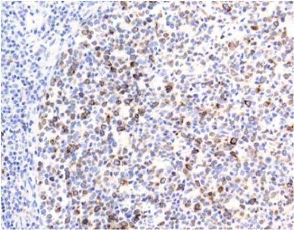
Mouse Monoclonal Antibody

                                                                               AID
                                      TYPE:                   Mouse monoclonal
                                      CLONE:                  JUA51E
                                      CONTROL:                Ramos Cell Line
                                      LOCALIZATION:           Nuclear and cytoplasmic
                                      ISOTYPE:                IgG1
                                      REACTS WITH:            Human

 Maturation of the antibody repertoire is mediated by two different mechanisms: class-switch recombination (CSR) and
 somatic hypermutation (SHM). CSR leads to the production of antibodies of different isotypes whereas SHM leads to
 the selection of B cells expressing a BCR with high affinity for antigen. The activation-induced cytidine deaminase
 (AID) was recently shown to play a key role in these two mechanisms.

  APPLICATIONS                                      DILUTION                 ANTIGEN RETRIEVAL
                         IHC-F                      Not working
                         IHC-P                      1:2 supernatant          15 min R2 Novolink
                         Elisa                      1:1000
                         IF                         Not working
                         WB                         1:2
                         IP                         1:2

REFERENCES

Kanellis G, Roncador G, Arribas A, Mollejo M, Montes-Moreno S, Maestre L, Campos-Martin Y, Martinez-Torrecuadrada JL, Sanchez-
Verde L, Pajares R, Cigudosa JC, Martin MC and Piris MA. Identification of MNDA as a new marker for Nodal Marginal Zone Lymphoma.
Leukemia. 2009 Oct;23(10):1847-57.

de Bont CM, Eerden N, Boelens WC, Pruijn GJM. Neutrophil proteases degrade autoepitopes of NET-associated proteins. Clin Exp
Immunol. 2020 Jan;199(1):1-8. doi: 10.1111/cei.13392. Epub 2019 Nov 15.

Rat Monoclonal Antibody

                                                           A-myb/B-myb/C-myb
                                      TYPE:                    Rat monoclonal
                                      CLONE:                   Dani51
                                      CONTROL:                 Tonsil
                                      LOCALIZATION:            Nuclear
                                      ISOTYPE:                 IgG2a
                                      REACTS WITH:             Human

 The myb gene family consists of three members, named A, B and C-myb which encode nuclear proteins that function
 as transcriptional transactivators. A-myb is predominantly expressed in the male germ cells, although low levels of A-
 myb expression was also detected in ovaries, brain as well as B-cells at the germinal centers. In contrast to the tissue-
 specific expression of A-myb, the B-myb gene expression seems to be ubiquitous. C-myb is predominantly expressed
 in immature hematopoietic cells.

 APPLICATIONS                                     DILUTION                  ANTIGEN RETRIEVAL
                        IHC-F                     Not done
                        IHC-P                     1:2 supernatant           20 min R2 Novolink
                        Elisa                     1:1000
                        IF                        Not done
                        WB                        1:2
                        IP                        Not done

                                                                4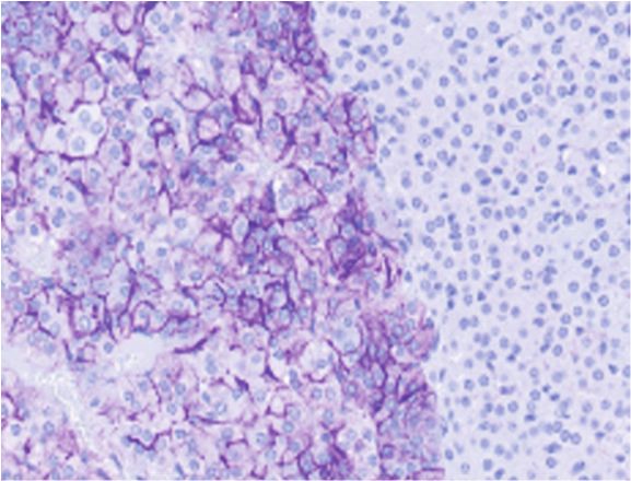
Mouse Monoclonal Antibody

                                                                 ANX4
                               TYPE:                Mouse monoclonal
                               CLONE:               ANX47
                               CONTROL:             Pancreas
                               LOCALIZATION:        Cytoplasmic
                               ISOTYPE:             IgG1
                               REACTS WITH:         Human

Annexin IV (ANX4) belongs to the annexin family of calcium-dependent phospholipid binding proteins.
Although their functions are still not clearly defined, several members of the annexin family have been
implicated in membrane-related events along exocytotic and endocytotic pathways. Isolated from human
placenta, ANX4 encodes a protein that has possible interactions with ATP, and has in vitro anticoagulant
activity and also inhibits phospholipase A2 activity. ANX4 is almost exclusively expressed in epithelial cells.

APPLICATIONS                              DILUTION               ANTIGEN RETRIEVAL
                    IHC-F                 Not done
                    IHC-P                 1:10 supernatant       20 min R2 Novolink
                    Elisa                 1:1000
                    IF                    Not done
                    WB                    1:2
                    IP                    Not done

Commercialized by: Sigma Aldrich

                                                       5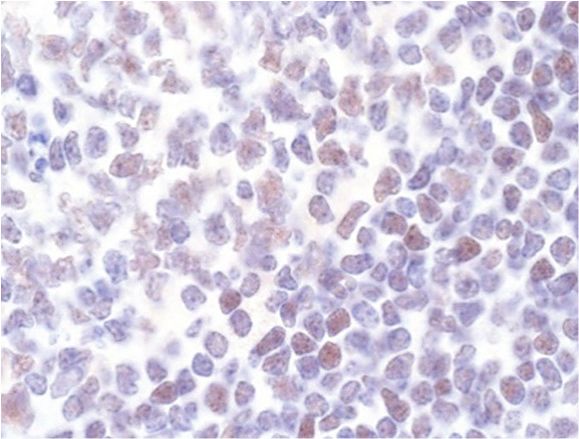
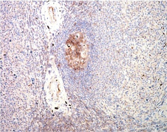
Rat Monoclonal Antibody

                                                             BCL-2
                             TYPE:              Rat monoclonal
                             CLONE:             NOR 235J
                             CONTROL:           Tonsil
                             LOCALIZATION:      Cytoplasmic
                             ISOTYPE:           IgG2b
                             REACTS WITH:       Human

BCL-2 is a human proto-oncogene located on chromosome 18. Its product is an integral membrane protein
(called Bcl-2) located in the membranes of the endoplasmic reticulum, nuclear envelope, and in the outer
membranes of the mitochondria. Suppresses apoptosis in a variety of cell systems including factor-
dependent lymphohematopoietic and neural cells. Regulates cell death by controlling the mitochondrial
membrane permeability.

APPLICATIONS                          DILUTION              ANTIGEN RETRIEVAL
                  IHC-F               Neat
                  IHC-P               1:5 supernatant       30min R1 Novolink
                  Elisa               1:1000
                  IF                  Not done
                  WB                  Neat
                  IP                  Not done

Commercialized by: Abcam

                                                   6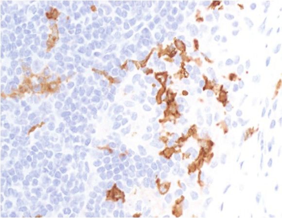
Mouse Monoclonal Antibody

                                                                              BCL-6
                                      TYPE:                    Mouse monoclonal
                                      CLONE:                   IG191E
                                      CONTROL:                 Tonsil
                                      LOCALIZATION:            Nuclear
                                      ISOTYPE:                 IgG1
                                      REACTS WITH:             Human and mouse

 Bcl-6 is a transcriptional regulator gene, which codes for a 706-amino-acid nuclear zinc finger protein.
 Antibodies to this protein stain the germinal centre cells in lymphoid follicles, the follicular cells and
 interfollicular cells in Follicular Lymphoma, Diffuse Large B-Cell Lymphomas, and Burkitt’s lymphoma, and
 the majority of the Reed-Sternberg cells in Nodular Lymphocyte Predominant Hodgkin’s Disease. In
 contrast, anti-BCL-6 rarely stains Mantle Cell Lymphoma and MALT Lymphoma.

 APPLICATIONS                                      DILUTION                   ANTIGEN RETRIEVAL
                         IHC-F                     1:150
                         IHC-P                     1:300 supernatant          20 min R2 Novolink
                         Elisa                     1:1000
                         IF                        1:100                      EDTA
                         WB                        1:10
                         IP                        1:10

 Commercialized by: Cell Marque, Biolegend, Active Motif, eBioscience, Biocare and Millipore.

 REFERENCES
Dhar SS1, Zhao D2, Lin T3, Gu B1, Pal K1, Wu SJ4, Alam H1, Lv J2, Yun K5, Gopalakrishnan V6, Flores ER7, Northcott PA8, Rajaram V9,
Li W10, Shilatifard A11, Sillitoe RV3, Chen K12, Lee MG13. MLL4 Is Required to Maintain Broad H3K4me3 Peaks and Super-Enhancers at
Tumor Suppressor Genes. Mol Cell. 2018.

Klymenko T1, Bloehdorn J2, Bahlo J3, Robrecht S3, Akylzhanova G1, Cox K4, Estenfelder S2, Wang J1, Edelmann J1, Strefford JC4,
Wojdacz TK4,5, Fischer K3, Hallek M3, Stilgenbauer S2, Cragg M4, Gribben J1, Braun A1. Lamin B1 regulates somatic mutations and
progression of B-cell malignancies. Leukemia. 2018.

Béguelin W1, Rivas MA2, Calvo Fernández MT2, Teater M2,3, Purwada A4, Redmond D3,5, Shen H2, Challman MF2, Elemento O3, Singh
A6,7, Melnick AM8. EZH2 enables germinal centre formation through epigenetic silencing of CDKN1A and an Rb-E2F1 feedback loop.
Nat Commun. 2017.

J Cao, X Zhang, Q Wang, G Qiu, C Hou, J Wang, Q Cheng, Y Lan, H Han, H Shen, Y Zhang, X Yang, B Shen, J Zhang. Smad4 represses
the generation of memory-precursor effector T cells but is required for the differentiation of central memory T cells. Cell Death &
Disease on 20 November 2015.

Hsiao-Wei Tsao, Tzong-Shyuan Tai, William Tseng, Hui-Hsin Chang, Roland Grenningloh, Shi-Chuen Miaw, I-Cheng Ho. Ets-1 facilitates
nuclear entry of NFAT proteins and their recruitment to the IL-2 promoter. Proceedings of the National Academy of Sciences of the
United States of America. 24 September 2013.

Ramsey JE1, Fontes JD. The zinc finger transcription factor ZXDC activates CCL2 gene expression by opposing BCL6-mediated
repression. Mol Immunol. 2013.

Marion Travert, Yenlin Huang, Laurence de Leval, Nadine Martin-Garcia, Marie-Helene Delfau-Larue, Françoise Berger, Jacques Bosq,
Josette Brière, Jean Soulier, Elizabeth Macintyre, Teresa Marafioti, Aurélien De Reyniès, Philippe Gaulard. Molecular features of
hepatosplenic T-cell lymphoma unravels potential novel therapeutic targets. Blood. 14 June 2012.

Nam-Cha SH, Montes-Moreno S, Salcedo MT, Sanjuan J, Garcia JF and Piris MA. Lymphocyte-rich classical Hodgkin's lymphoma:
distinctive tumor and microenvironment markers. Modern Pathology 2009, 22, 1006–1015.

Kmieciak M1, Gowda M, Graham L, Godder K, Bear HD, Marincola FM, Manjili MH. Human T cells express CD25 and Foxp3 upon activation
and exhibit effector/memory phenotypes without any regulatory/suppressor function. J Transl Med. 2009.

Thakral D1, Dobbins J, Devine L, Kavathas PB. Differential expression of the human CD8beta splice variants and regulation of the M-2
isoform by ubiquitination. J Immunol. 2008 Jun 1;180(11):7431-42.

Verdes-Montenegro JF, Garcia JF, Maestre L, Lucas-Campeno E, Sanchez-Verde L, Romero-Chala S, Piris MA, Roncador G. Genetic
Immunization: a New Monoclonal Antibody for the Detection of BCL-6 Protein in Paraffin Sections. J Histochem Cytochem. 2006
                                                                  7
Mouse Monoclonal Antibody

                                                        BCL-6 (Mouse specific)
                                     TYPE:                  Mouse monoclonal
                                     CLONE:                 42B
                                     CONTROL:               Tonsil
                                     LOCALIZATION:          Nuclear
                                     ISOTYPE:               IgG1
                                     REACTS WITH:           Mouse and human

 Bcl-6 is a transcriptional regulator gene, which codes for a 706-amino-acid nuclear zinc finger protein.
 Antibodies to this protein stain the germinal centre cells in lymphoid follicles, the follicular cells and
 interfollicular cells in Follicular Lymphoma, Diffuse Large B-Cell Lymphomas, and Burkitt’s lymphoma, and
 the majority of the Reed-Sternberg cells in Nodular Lymphocyte Predominant Hodgkin’s Disease. In
 contrast, anti-BCL-6 rarely stains Mantle Cell Lymphoma and MALT Lymphoma.

 APPLICATIONS
                                                  DILUTION                 ANTIGEN RETRIEVAL
                        IHC-F                     1:4 supernatant
                        IHC-P                     1:4 supernatant          Omnimap Multimer (Ventana)
                                                  1:200 purified
                        Elisa                     1:1000
                        IF                        Neat supernatant
                        WB                        Neat supernatant
                        IP                        Not done

REFERENCES

Verdes-Montenegro JF, Garcia JF, Maestre L, Lucas-Campeno E, Sanchez-Verde L, Romero-Chala S, Piris MA, Roncador G. Genetic
Immunization: a New Monoclonal Antibody for the Detection of BCL-6 Protein in Paraffin Sections. J Histochem Cytochem. 2006.

                                                               8
Mouse Monoclonal Antibody

                                                                         BCL7A
                                   TYPE:                   Mouse monoclonal
                                   CLONE:                  15C
                                   CONTROL:                Tonsil
                                   LOCALIZATION:           Nuclear
                                   ISOTYPE:                IgG1
                                   REACTS WITH:            Human and mouse

This gene is directly involved, with Myc and IgH, in a three-way gene translocation in a Burkitt lymphoma
cell line. As a result of the gene translocation, the N-terminal region of the gene product is disrupted, which
is thought to be related to the pathogenesis of a subset of high-grade B cell non-Hodgkin lymphoma.

APPLICATIONS
                                                 DILUTION            ANTIGEN RETRIEVAL
                      IHC-F                  1:40
                      IHC-P                  1:40 supernatant        20min R2 Novolink
                      Elisa                  1:1000
                      IF                     Not done
                      WB                     Neat
                      IP                     Not done

Commercialized by: Millipore, Abcam and BD Biosciences

REFERENCES

Ramos-Medina R, Montes-Moreno S, Maestre L, Cañamero M, Rodríguez-Pinilla M, Martínez-Torrecuadrada J, Piris MA, Majid A, Dyer
MJ, Pulford K, Roncador G (2012). BCL7A protein expression in normal and malignant lymphoid tissues. Br J Haematol. 2013
Jan;160(1):106-9.

                                                              9
Rat Monoclonal Antibody

                                                    Beta-Galactosidase
                               TYPE:               Rat monoclonal
                               CLONE:              3A9A
                               CONTROL:            Beta-gal expressing tissues
                               LOCALIZATION:       Cytoplasmic
                               ISOTYPE:            IgG2b
                               REACTS WITH:        N/A

Beta galactosidase is coded by a gene (lac z) in the lac operon of Escherichia coli. It is a metalloenzyme that
splits lactose into glucose and galactose. It hydrolyzes terminal, non-reducing beta-D-galactose residues in
beta-D-galactosides.

APPLICATIONS
                                        DILUTION            ANTIGEN RETRIEVAL
                   IHC-F                Neat
                   IHC-P                1:125               Omnimap Multimer (Ventana)
                   Elisa                1:1000
                   IF                   Not done
                   WB                   Neat
                   IP                   Not done

Mouse Monoclonal Antibody

                                                                  BPTF
                               TYPE:               Mouse monoclonal
                               CLONE:              PAC 33A, TOR102G and TOR249C
                               CONTROL:            Tonsil
                               LOCALIZATION:       Nuclear
                               ISOTYPE:            IgG2a
                               REACTS WITH:        Human

Nucleosome-remodeling factor subunit BPTF is a protein that in humans is encoded by the BPTF gene.
Analysis of the original protein (fetal Alz-50 reactive clone 1, or FAC1), identified as an 810 aa protein
containing a DNA-binding domain and a zinc finger motif, suggested it might play a role in the regulation of
transcription. High levels of FAC1 were detected in fetal brain and in patients with neurodegenerative diseases.

APPLICATIONS
                                          DILUTION             ANTIGEN RETRIEVAL
                   IHC-F                  Not done
                   IHC-P                  1:100 supernatant    15 min R2 Novolink
                   Elisa                  1:1000
                   IF                     Not done
                   WB                     1:50 supernatant
                   IP                     Not done

                                                      10
Mouse Monoclonal Antibody
BSA
TYPE: Mouse monoclonal
CLONE: 63A
CONTROL: BSA protein
LOCALIZATION: N/A
ISOTYPE: IgG1
REACTS WITH: Bovine
Serum albumin, the main protein of plasma, has a good binding capacity for water, Ca2+, Na+, K+, fatty acids,
hormones, bilirubin and drugs. Its main function is the regulation of the colloidal osmotic pressure of blood.
Major zinc transporter in plasma, typically binds about 80% of all plasma zinc.
APPLICATIONS
DILUTION ANTIGEN RETRIEVAL
IHC-F Not done
IHC-P Not done
Elisa 1:1000
IF Not done
WB Not done
IP Not done
Mouse Monoclonal Antibody
BUB-1 (Xenopus specific)
TYPE: Mouse monoclonal
CLONE: 31D
CONTROL: Xenopus tissue
LOCALIZATION: Nuclear
ISOTYPE: IgG1
REACTS WITH: Xenopus tissue
Serine/threonine protein kinase xBub1is present in resting oocytes and its protein level increases slightly
during oocyte maturation and early embryogenesis. In Xenopus oocytes, Bub1 is localized to kinetochores
during both meiosis I and meiosis II, and the electrophoretic mobility of Bub1 upon SDS-PAGE decreases
during meiosis I, reflecting phosphorylation and activation of the enzyme.
APPLICATIONS
DILUTION ANTIGEN RETRIEVAL
IHC-F 1.2
IHC-P Not done
Elisa 1:1000
IF Not done
WB 1:2
IP 1:2
11Rat Monoclonal Antibody
Cas9 (S. Pyogenes)
TYPE: Rat Monoclonal
CLONE: KANI345B
CONTROL: CAS9 positive tissue or cell line
LOCALIZATION: membrane, nuclei and cytoplasm
ISOTYPE: IgG2a
REACTS WITH: S. Pyogenes
Cas9 (S. Pyognes) is a useful mAb that allow researcher to: a) check by WB the transfection success of the
Cas9 protein b) check by IF and IHC that the Cas9 protein was delivered to the nucleus c) check by WB the
Cas9 expression level.
APPLICATIONS
DILUTION ANTIGEN RETRIEVAL
IHC-F 1:1000
IHC-P 1:200 purified Ab OminMap Standard RiboCC (Ventana)
Elisa 1:1000
IF Not done
WB 1:200 purified Ab
IP Not done
Commercialized by: Abcam
Mouse Monoclonal Antibody
CD1d (CD1)
TYPE: Mouse monoclonal
CLONE: RITA80C
CONTROL: Tonsil
LOCALIZATION: Celular membrane
ISOTYPE: IgG1
REACTS WITH: Human
CD1d is a MHC-like, type I transmembrane protein, member of the CD1 family and the immunoglobulin
superfamily. On the cell surface, CD1d forms a heterodimer with β2-microglobulin. CD1d is expressed by
antigen-presenting cells such as B cells, monocytes/macrophages, dendritic cells, and some non-lymphoid
cells. Cortical thymocytes express CD1d but the expression is lost in mature T cells.
APPLICATIONS DILUTION ANTIGEN RETRIEVAL
IHC-F Not done
IHC-P Neat supernatant 20min R2 Novolink
Elisa 1:1000
IF Not done
WB Neat supernatant
IP Not done
12Rat Monoclonal Antibody
CD8a
(T-cell surface glycoprotein CD8 alpha)
TYPE: Rat Monoclonal
CLONE: NOR132H
CONTROL: Tonsil
LOCALIZATION: Membrane
ISOTYPE: IgG2a
REACTS WITH: Human
CD8 (cluster of differentiation 8) is a transmembrane glycoprotein that serves as a co-receptor for the T cell
receptor (TCR). Like the TCR, CD8 binds to a major histocompatibility complex (MHC) molecule, but is specific
for the class I MHC protein. CD8 antigen is strongly expressed on human cytotoxic T cells and thymocytes, and
is also expressed on a subset of NK cells.
APPLICATIONS
DILUTION ANTIGEN RETRIEVAL
IHC-F 1:20
IHC-P 1:20 supernatant 30min R1 Novolink
Elisa 1:1000
IF Not done
WB Neat
IP Not done
REFERENCES
Maestre L, García-García JF, Jiménez S, Reyes-García AI, García-González Á, Montes-Moreno S, Arribas AJ, González-García P, Caleiras
E, Banham AH, Piris MÁ, Roncador G. High-mobility group box (TOX) antibody a useful tool for the identification of B and T cell
subpopulations. PLoS One. 2020 Feb 27;15(2):e0229743. doi: 10.1371/journal.pone.0229743. eCollection 2020.
13Rat Monoclonal Antibody
CD8a (Mouse specific)
TYPE: Rat Monoclonal
CLONE: OTO94A
CONTROL: Mouse thymus
LOCALIZATION: Membrane
ISOTYPE: IgG2c
REACTS WITH: Mouse
Mouse CD8a is a integral membrane glycoprotein that plays an essential role in the immune response and
serves multiple functions in responses against both external and internal offenses. In T-cells, functions
primarily as a coreceptor for MHC class I molecule:peptide complex.
APPLICATIONS DILUTION ANTIGEN RETRIEVAL
IHC-F Not done
IHC-P 1:200 Omnimap Multimer (Ventana)
Elisa 1:1000
IF Not done
WB Neat supernatant
IP Not done
Mouse Monoclonal Antibody
CD15 (FUT4)
TYPE: Mouse monoclonal
CLONE: 153B
CONTROL: Tonsil
LOCALIZATION: Cytoplasmic
ISOTYPE: IgM
REACTS WITH: Human
This monoclonal antibody reacts with the CD15 antigen, a triasaccharide structure, 3-fucosyl-N-
acetyllactosamine, also known as X-hapten. This antigen is expressed by Reed-Sternberg Cells, granulocytes
and is also found in a number of normal tissue and cell types.
APPLICATIONS
DILUTION ANTIGEN RETRIEVAL
IHC-F 1:5
IHC-P 1:2 supernatant 30 min R1 Novolink
Elisa 1:1000
IF 1:2 Citrate, EDTA
WB 1:2
IP 1:2
Commercialized by: Abcam
REFERENCES
Nam-Cha SH, Montes-Moreno S, Salcedo MT, Sanjuan J, Garcia JF and Piris MA. Lymphocyte-rich classical Hodgkin's lymphoma:
distinctive tumor and microenvironment markers. Modern Pathology (2009) 22, 1006–1015.
14Rat Monoclonal Antibody
CD20 (Mouse specific)
TYPE: Rat Monoclonal
CLONE: GOT214A
CONTROL: Mouse Tonsil
LOCALIZATION: Membrane
ISOTYPE: IgG2a
REACTS WITH: Mouse
CD20 is a B lymphocyte-specific cell-surface molecule involved in the regulation of transmembrane Ca2+
conductance and cell-cycle progression during B cell activation. CD20 protein is expressed at high levels
on B cells and is downregulated during terminal differentiation into plasma cells.
APPLICATIONS
DILUTION ANTIGEN RETRIEVAL
IHC-F Not done
IHC-P 1:50 supernatant Omnimap Multimer (Ventana)
Elisa 1:1000
IF Not done
WB Neat supernatant
IP Not done
Commercialized by: Abcam
15Mouse Monoclonal Antibody
CD30 (TNFRSF8)
TYPE: Mouse monoclonal
CLONE: CON6D
CONTROL: Hodgkin’s Lymphoma
LOCALIZATION: Membrane
ISOTYPE: IgG2a
REACTS WITH: Human
This antibody reacts with a 595 amino acid transmembrane glycoprotein with a molecular weight of 120kD.
This antibody reacts with mononuclear cells in Hodgkin lymphoma, Reed-Sternberg cells and most
Anaplastic Large Cell Lymphomas. It stains large cells localized around the B cell in the lymphoid tissues.
This antibody stains the cell surface and the golgi region in the cells.
APPLICATIONS DILUTION ANTIGEN RETRIEVAL
IHC-F 1:2
IHC-P Neat supernatant 30 min R1 Novolink
1: 100 purified
Elisa 1:1000
IF 1:2 Citrate, EDTA
WB 1:2
IP 1:2
Commercialized by: Abcam, Santa Cruz and Biocare
REFERENCES
Maestre L, García-García JF, Jiménez S, Reyes-García AI, García-González Á, Montes-Moreno S, Arribas AJ, González-García P, Caleiras
E, Banham AH, Piris MÁ, Roncador G. High-mobility group box (TOX) antibody a useful tool for the identification of B and T cell
subpopulations. PLoS One. 2020 Feb 27;15(2):e0229743. doi: 10.1371/journal.pone.0229743. eCollection 2020.
Lu TX, Liang JH, Miao Y, Fan L, Wang L, Qu XY, Cao L, Gong QX, Wang Z, Zhang ZH, Xu W, Li JY. Epstein-Barr virus positive diffuse large
B-cell lymphoma predict poor outcome, regardless of the age. Sci Rep. 2015 Jul 23;5:12168.
Lau SK1, Thomas P, Weiss LM. Immunohistochemical evaluation of CON6D/B5: a new CD30 monoclonal antibody. Appl
Immunohistochem Mol Morphol. 2010 May;18(3):273-7.
Nam-Cha SH, Montes-Moreno S, Salcedo MT, Sanjuan J, Garcia JF and Piris MA. Lymphocyte-rich classical Hodgkin's lymphoma:
distinctive tumor and microenvironment markers. Modern Pathology. 2009 22, 1006–1015.
Göran Mattsson, Soo Yong Tan, David J P Ferguson, Wendy Erber, Susan H Turner, Teresa Marafioti, David Y Mason. Detection of genetic
alterations by immunoFISH analysis of whole cells extracted from routine biopsy material. The Journal of Molecular Diagnostics: JMD. 1
September 2007.
Garcia JF, Roncador G, Garcia JF, Sanz AI, Maestre L, Lucas E, Montes-Moreno S, Fernandez Victoria R, Martinez-Torrecuadrara JL,
Marafioti T, Mason DY, Piris MA. PRDM1/BLIMP-1 expression in multiple B and T-cell lymphoma. Haematologica. 2006. Apr; 91:467-74.
16Mouse Monoclonal Antibody
CD38 (Cyclic ADP ribose hydrolase)
TYPE: Mouse monoclonal
CLONE: MIX187A
CONTROL: Tonsil
LOCALIZATION: Membrane
ISOTYPE: IgG1
REACTS WITH: Human
CD38 (cluster of differentiation 38), also known as cyclic ADP ribose hydrolase is a glycoprotein found on
the surface of many immune cells (white blood cells), including CD4+, CD8+, B lymphocytes and natural
killer cells. CD38 also functions in cell adhesion, signal transduction and calcium signaling.
APPLICATIONS
DILUTION ANTIGEN RETRIEVAL
IHC-F Neat supernatant
IHC-P Neat supernatant 30 min R1 Novolink
Elisa 1:1000
IF Not Done
WB Neat supernatant
IP Neat supernatant
Mouse Monoclonal Antibody
CD43 (Leukosialin)
TYPE: Mouse monoclonal
CLONE: 93F
CONTROL: Tonsil
LOCALIZATION: Membrana, Cytoplasmic
ISOTYPE: IgG1
REACTS WITH: Human
Recognise a 110kD protein, identified as CD43. CD43 is the major sialoglycoprotein on thymocytes, T cell
and neutrophils. It is also present on activated B cell, plasma cell, NK cells, granulocytes, monocytes,
macrophages, platelets, and bone marrow haematopoietic stem cells
APPLICATIONS
DILUTION ANTIGEN RETRIEVAL
IHC-F 1:100
IHC-P 1:400 supernatant 20 min R2 Novolink
Elisa 1:1000
IF 1:400 Citrate, EDTA
WB 1:10
IP 1:10
17Mouse Monoclonal Antibody
CD63 (lamp-3)
TYPE: Mouse monoclonal
CLONE: KILL150A
CONTROL: Tonsil
LOCALIZATION: Membrana, Cytoplasmic
ISOTYPE: IgG1
REACTS WITH: Human
CD63 is a cell surface glycoprotein that is known to complex with integrins. It may function as a blood platelet
activation marker. Deficiency of this protein is associated with Hermansky-Pudlak syndrome. Also this gene
has been associated with tumor progression.
APPLICATIONS
DILUTION ANTIGEN RETRIEVAL
IHC-F Not done
IHC-P 1:30 supernatant 20min R2 Novolink
1:200 purified Ab (1mg/ml)
Elisa 1:1000
IF Not done
WB Neat supernatant
IP Not done
Commercialized by: Abcam
Rat Monoclonal Antibody
CD85A (LILRB3)
TYPE: Rat monoclonal
CLONE: FRAS92B
CONTROL: Monocytes
LOCALIZATION: membrane
ISOTYPE: IgG1
REACTS WITH: Human
LILRB3 (also called CD85A, ILT5, LIR3, HL9) contains 4 extracellular immunoglobulin domains, a
transmembrane domain, and 4 cytoplasmic ITIMs. Expression of LILRB3 has been reported on monocytes,
monocyte-derived osteoclasts, neutrophils, eosinophils, basophils, osteoclasts. LILRB3 is also proposed to be
an inhibitor of allergic inflammation and a contributor to uncontrolled immune responses and autoimmunity.
APPLICATIONS
DILUTION ANTIGEN RETRIEVAL
Flow Cytometry 1:100 purified Ab 1mg/ml
IHC-P 1:30 20min R2 Novolink
1:100 purified Ab 1mg/ml
Elisa 1.1000
IF Not done
WB 1:100 purified 1mg/ml
IP Not done
Commercialized by: Abcam
18Mouse Monoclonal Antibody
CD85A/B (LILRB3/LILRA6)
TYPE: Mouse monoclonal
CLONE: FANI179B
CONTROL: Monocytes
LOCALIZATION: membrane
ISOTYPE: IgG1
REACTS WITH: Human
The leukocyte Ig-like receptor subfamily B (LILRB) is a group of type I transmembrane glycoproteins with
extracellular Ig-like domains that bind ligands and intracellular ITIMs. LILRBs were reported to be
predominantly expressed in hematopoietic lineage cells and to suppress activation of various types of
immune cells.
APPLICATIONS
DILUTION ANTIGEN RETRIEVAL
Flow Cytometry 1:2 supernatant
IHC-P Not done
Elisa 1:1000
IF Not working
WB Not working
IP Not done
19Mouse Monoclonal Antibody
CD115 (CSF1R)
TYPE: Mouse monoclonal
CLONE: : FER216
CONTROL: Tonsil
LOCALIZATION: Cytoplasmic
ISOTYPE: IgG1
REACTS WITH: Human
The protein encoded by this gene is the receptor for colony stimulating factor 1, a cytokine which controls
the production, differentiation, and function of macrophages. This receptor mediates most if not all of the
biological effects of this cytokine. Ligand binding activates the receptor kinase through a process of
oligomerization and transphosphorylation.
APPLICATIONS DILUTION ANTIGEN RETRIEVAL
IHC-F 1:2 supernatant
IHC-P 1:10 supernatant 20 min R2 Novolink
Elisa 1:1000
IF Not done
WB 1:2 supernatant
IP 1:2 supernatant
Commercialized by: Millipore
REFERENCES
Martín-Moreno AM, Roncador G, Maestre L, Mata E, Jiménez S, Martínez-Torrecuadrada JL, Reyes-García AI, Rubio C, Tomás JF, Estévez
M, Pulford K, Piris MA, García JF. CSF1R Protein Expression in Reactive Lymphoid Tissues and Lymphoma: Its Relevance in Classical
Hodgkin Lymphoma. PLoS One. 2015 Jun 12;10(6).
20RAT Monoclonal Antibody
CD115 (CSFR1-mouse specific)
TYPE: Rat monoclonal
CLONE: YEYE311D
CONTROL: Mouse lymph node
LOCALIZATION: Membrane and Cytoplasm
ISOTYPE: IgG1
REACTS WITH: mouse
Tyrosine-protein kinase that acts as cell-surface receptor for CSF1 and IL34 and plays an essential role in the
regulation of survival, proliferation and differentiation of hematopoietic precursor cells, especially
mononuclear phagocytes, such as macrophages and monocytes.
APPLICATIONS DILUTION ANTIGEN RETRIEVAL
IHC-F Not done
IHC-P 1:4 supernatant Anti Rat + Novolink polymer
Elisa 1:1000
IF Not done
WB 1:4 supernatant
IP 1:2 supernatant
Mouse Monoclonal Antibody
CD138 (Syndecan-1)
TYPE: Mouse monoclonal
CLONE: CRUZ180A
CONTROL: Tonsil
LOCALIZATION: membrane
ISOTYPE: IgG1
REACTS WITH: Human
Syndecan-1 also known as SDC1 and CD138, is the most extensively studied member of the syndecan family.
It is found mainly in epithelial cells, but its expression is developmentally regulated during embryonic
development. Syndecan-1/SDC1/CD138 has been shown to mediate cell adhesion to several ECM molecules,
and to act as a coreceptor for fibroblast growth factors, potent angiogenic growth factors involved also in
differentiation.
APPLICATIONS
DILUTION ANTIGEN RETRIEVAL
IHC-F Not done
IHC-P 1:40 supernatant 30 min R1 NovolinK
Elisa 1:1000
IF Not done
WB Not working
IP Not Done
REFERENCES
Maestre L, García-García JF, Jiménez S, Reyes-García AI, García-González Á, Montes-Moreno S, Arribas AJ, González-García P, Caleiras
E, Banham AH, Piris MÁ, Roncador G. High-mobility group box (TOX) antibody a useful tool for the identification of B and T cell
subpopulations. PLoS One. 2020 Feb 27;15(2):e0229743. doi: 10.1371/journal.pone.0229743. eCollection 2020.
21Mouse Monoclonal Antibody
CD147 (Basigin)
TYPE: Mouse monoclonal
CLONE: NK99C
CONTROL: Tonsil
LOCALIZATION: Celular membrane
ISOTYPE: IgG1
REACTS WITH: Human
CD147 (EMMPRIN or basigin) is a tumor-related glycosylated protein that belongs to the immunoglobulin
superfamily and exists both in transmembrane and soluble forms. CD147 expression is increased in many
types of tumors, and its elevation is often associated with aggressive disease and poor prognosis.
APPLICATIONS DILUTION ANTIGEN RETRIEVAL
IHC-F Not done
IHC-P Neat 20min R2 Novolink
Elisa 1:1000
IF Not done
WB Neat supernatant
IP Neat supernatant
22Mouse Monoclonal Antibody
CD162 (PSGL1)
TYPE: Mouse monoclonal
CLONE: FLEG
CONTROL: Tonsil
LOCALIZATION: Membrane
ISOTYPE: IgG2a
REACTS WITH: Human
P-selectin glycoprotein ligand-1 (PSGL-1, CD162) is a dimeric mucin-like 120-kDa glycoprotein on leukocyte
surfaces that binds to P- and L-selectin and promotes cell adhesion in the inflammatory response. PSGL-1
mediates leukocyte-endothelial and leukocyte-platelet adhesion by binding to P-selectin expressed on
activated endothelium and platelets and PSGL-1 mediates leukocyte-leukocyte adhesion by binding to L-
selectin expressed on apposing leukocytes. PSGL-1 is unique in that it is the only selectin glycoprotein ligand
that has been directly demonstrated to mediate cell-cell adhesion in vitro and in vivo.
APPLICATIONS
DILUTION ANTIGEN RETRIEVAL
IHC-F 1:400
IHC-P 1:500 supernatant 30min R1 Novolink
Elisa 1:1000
IF 1:400 Citrate
WB 1:200
IP 1:200
Commercialized by: eBioscience.
REFERENCES
Rathod KS, Kapil V, Velmurugan S, Khambata RS, Siddique U, Khan S, Van Eijl S, Gee LC, Bansal J, Pitrola K, Shaw C, D'Acquisto F, Colas
RA, Marelli-Berg F, Dalli J, Ahluwalia A. Accelerated resolution of inflammation underlies sex differences in inflammatory responses in
humans. . J Clin Invest. 2017 Jan 3;127(1):169-182.
Yorihiro Nishimura, Masayuki Shimojima, Yoshio Tano, Tatsuo Miyamura, Takaji Wakita, Hiroyuki Shimizu. Human P-selectin glycoprotein
ligand-1 is a functional receptor for enterovirus 71. Nature Medicine 2009, 15;7.
K L Davenpeck, M E Brummet, S A Hudson, R J Mayer, B S Bochner. Activation of human leukocytes reduces surface P-selectin
glycoprotein ligand-1 (PSGL-1, CD162) and adhesion to P-selectin in vitro. The Journal of Immunology 2000, 165; 5.
J P Levesque, A C Zannettino, M Pudney, S Niutta, D N Haylock, K R Snapp, G S Kansas, M C Berndt, P J Simmons. PSGL-1-mediated
adhesion of human hematopoietic progenitors to P-selectin results in suppression of hematopoiesis. Immunity 1999, 11;3.
23Rat Monoclonal Antibody
CD229 (Ly-9)
TYPE: Rat monoclonal
CLONE: PIZCU426A
CONTROL: Human Tonsil
LOCALIZATION: membrane
ISOTYPE: IgG2b
REACTS WITH: Human
T-lymphocyte surface antigenan approximately LY9 is a 120-kDa member of the Signaling Lymphocyte
Activation Molecule (SLAM) immunoglobulin superfamily. This type I transmembrane glycoprotein is
expressed on a variety of immune cell types, including thymocytes, T and B lymphocytes, and natural
killer cells. Expression of CD229 is also detected on T and B cell malignancies.
APPLICATIONS
DILUTION ANTIGEN RETRIEVAL
IHC-F Not done
IHC-P 1:4 Supernatant 20min R2 Novolink
1:640 purified Ab 1mg/ml
Elisa 1:1000
IF 1:4 Supernatant
WB 1:4 Supernatant
IP Not done
Rat Monoclonal Antibody
CD271 (NGFR)
TYPE: Rat monoclonal
CLONE: NORI138B
CONTROL: Human tonsil
LOCALIZATION: Membrane
ISOTYPE IgG2a
REACTS WITH: Mouse and human
The nerve growth factor receptor (NGFR/p75) (CD271), which belongs to the TNF receptor (TNFR) family, is
a multifunctional cell surface receptor that exerts diverse functions, such as the stimulation of cell survival
and differentiation during neuronal development. The expression of NGFR has been observed in several
human cancers, such as thyroid carcinoma, stomach cancer, and liver cancer.
APPLICATIONS
DILUTION ANTIGEN RETRIEVAL
IHC-F Not done
IHC-P 1:10 Supernatant 20min R2 Novolink
Elisa 1:1000
IF 1:200 purified antibody
WB 1: 1000 purified antibody
IP Not done
24Rat Monoclonal Antibody
CD271 (NGFR) (mouse specific)
TYPE: Rat monoclonal
CLONE: NORI146C
CONTROL: Mouse Lymph node
LOCALIZATION: Membrane
ISOTYPE IgG2a
REACTS WITH: Mouse
Ngfr (CD271) is a low affinity receptor which can bind to NGF, BDNF, NT-3, and NT-4. Can mediate cell survival
as well as cell death of neural cells. Plays a role in the regulation of the translocation of GLUT4 to the cell
surface in adipocytes and skeletal muscle cells in response to insulin, probably by regulating RAB31 activity,
and thereby contributes to the regulation of insulin-dependent glucose uptake.
APPLICATIONS
DILUTION ANTIGEN RETRIEVAL
IHC-F Not done
IHC-P 1:10 supernatant Omnimap Multimer (Ventana)
Elisa 1:1000
IF 1:200 purified Ab
WB 1:500 purified Ab
IP Not done
25Mouse Monoclonal Antibody
CD272 (BTLA)
TYPE: Mouse monoclonal
CLONE: FLO67B
CONTROL: Tonsil
LOCALIZATION: Cytoplasmic
ISOTYPE: IgG1
REACTS WITH: Human
B and T lymphocyte attenuator (BTLA) has been recently identified as a new inhibitory receptor of the CD28
superfamily, with similarities to cytotoxic T lymphocyte activation antigen (CTLA-4) and programmed death
(PD-1). Engagement of BTLA on T lymphocytes can profoundly reduce the T cell receptor TCR-mediated
activation.
APPLICATIONS
DILUTION ANTIGEN RETRIEVAL
IHC-F Neat
IHC-P 1: 40 supernatant 20min R2 Novolink
Elisa 1:1000
IF Not working
WB 1:2
IP Not Done
Commercialized by: Millipore and Abcam.
REFERENCES
Carreras J, Lopez-Guillermo A, Kikuti YY, Itoh J, Masashi M, Ikoma H, Tomita S, Hiraiwa S, Hamoudi R, Rosenwald A, Leich E, Martinez A,
Roncador G, Villamor N, Colomo L, Perez P, Tsuji NM, Campo E, Nakamura N. High TNFRSF14 and low BTLA are associated with poor
prognosis in Follicular Lymphoma and in Diffuse Large B-cell Lymphoma transformation. J Clin Exp Hematop. 2019;59(1):1-16
Philippe Trougouboff and Hila Kreizman Shefer. B and T lymphocyte attenuator expression in mature B cell lymphomas. Journal of
Hematopathology, June 2013, Volume 6, Issue 2, pp 57–63.
26Mouse Monoclonal Antibody
CD279 (PD1)
TYPE: Mouse monoclonal
CLONE: NAT105
CONTROL: Tonsil
LOCALIZATION: Membrane
ISOTYPE: IgG1
REACTS WITH: Human
Programmed death-1 (PD-1) is a member of the immunoglobulin superfamily. It contains the immunoreceptor
tyrosine-based inhibitory motif (ITIM) and plays a key role in peripheral tolerance and autoimmune disease. It
is expressed by germinal center-associated T cells in reactive lymphoid tissue.
APPLICATIONS DILUTION ANTIGEN RETRIEVAL
IHC-F 1:5
IHC-P 1:5 supernatant 20 min R2 Novolink
1:100 purified
Elisa 1:1000
IF 1:2
WB 1:100
IP 1:100
Commercialized by: Cell Marque,Dianiva, Abcam, Biolegend and Biocare
REFERENCES (more than 100 citations)
Christopher H Cogbill, Steven H Swerdlow, Sarah E Gibson. Utility of CD279/PD-1 immunohistochemistry in the evaluation of benign and
neoplastic T-cell-rich bone marrow infiltrates. American Journal of Clinical Pathology. 1 July 2014.
Vladimir M Liarski, Natalya Kaverina, Anthony Chang, Daniel Brandt, Denisse Yanez, Lauren Talasnik, Gianluca Carlesso, Ronald Herbst,
Tammy O Utset, Christine Labno, Yahui Peng, Yulei Jiang, Maryellen L Giger, Marcus R Clark. Cell distance mapping identifies functional
T follicular helper cells in inflamed human renal tissue. Science Translational Medicine. 2 April 2014.
Jung Ryul Kim, Young Jae Moon, Keun Sang Kwon, Jun Sang Bae, Sajeev Wagle, Kyoung Min Kim, Ho Sung Park, Ho Lee, Woo Sung
Moon, Myoung Ja Chung, Myoung Jae Kang, Kyu Yun Jang. Tumor infiltrating PD1-positive lymphocytes and the expression of PD-L1
predict poor prognosis of soft tissue sarcomas. PLoS ONE. 18 December 2013.
Myoung Jae Kang, Kyoung Min Kim, Jun Sang Bae, Ho Sung Park, Ho Lee, Myoung Ja Chung, Woo Sung Moon, Dong Geun Lee, Kyu Yun
Jang. Tumor-infiltrating PD1-Positive Lymphocytes and FoxP3-Positive Regulatory T Cells Predict Distant Metastatic Relapse and
Survival of Clear Cell Renal Cell Carcinoma. Translational Oncology. 1 June 2013.
Jena D French, Gregory R Kotnis, Sherif Said, Christopher D Raeburn, Robert C McIntyre, Joshua P Klopper, Bryan R Haugen.
Programmed death-1+ T cells and regulatory T cells are enriched in tumor-involved lymph nodes and associated with aggressive features
in papillary thyroid cancer. The Journal of Clinical Endocrinology and Metabolism. 1 June 2012
Rodríguez Pinilla SM, Roncador G, Rodríguez-Peralto JL, Mollejo M, García JF, Montes-Moreno S, Camacho FI, Ortiz P, Limeres-González
MA, Torres A, Campo E, Navarro-Conde P, Piris MA. Primary cutaneous CD4+ small/medium-sized pleomorphic T-cell lymphoma
expresses follicular T-cell markers. Am J Surg Pathol. 2009 Jan;33(1):81-90.
Rodríguez-Pinilla SM, Atienza L, Murillo C, Pérez-Rodríguez A, Montes-Moreno S, Roncador G, Pérez-Seoane C, Domínguez P, Camacho
FI, Piris MA. Peripheral T-cell Lymphoma With Follicular T-cell Markers. Am J Surg Pathol. 2008. Dec;32(12):1787-99.
Nam-Cha SH, Roncador G, Sanchez-Verde L, Montes-Moreno S, Acevedo A, Domínguez-Franjo P, Piris MA. PD-1, a follicular T-cell marker
useful for recognizing nodular lymphocyte-predominant Hodgkin lymphoma. Am J Surg Pathol. 2008 Aug;32(8):1252-7
Roncador, G., Verdes-Montenegro, J.F.G., Tedoldi, S., Paterson, J.C., Klapper, W., Ballabio, E., Maestre, L., Pileri, S., Hansmann, M.L., Piris,
M.A., Mason, D.Y., Marafioti, T. Expression of two markers of germinal center T cells (SAP and PD-1) in angioimmunoblastic T-cell
lymphoma. Haematologica. 2007 Aug;92(8):1059
27Rat Monoclonal Antibody
CD370 (CLEC9A)
TYPE: Rat Monoclonal
CLONE: LEIA256A
CONTROL: Spleen
LOCALIZATION: membrane
ISOTYPE: IgG2b
REACTS WITH: Human
Clec9a (C-Type Lectin Domain Containing 9A) functions as an endocytic receptor on a small subset of myeloid
cells specialized for the uptake and processing of material from dead cells. Recognizes filamentous form of
actin in association with particular actin-binding domains of cytoskeletal proteins, including spectrin, exposed
when cell membranes are damaged, and mediate the cross-presentation of dead-cell associated antigens in
a Syk-dependent manner.
APPLICATIONS
DILUTION ANTIGEN RETRIEVAL
IHC-F Not done
IHC-P 1:1200 purified Novolink kit 20min R2
Elisa 1:1000
IF Not done
WB Not done
IP Not done
28Mouse Monoclonal Antibody
CDK6
TYPE: Mouse monoclonal
CLONE: 98D and 19G
CONTROL: Tonsil
LOCALIZATION: Nuclear and cytoplasmic
ISOTYPE: IgG1
REACTS WITH: Human and mouse
This antibody recognizes a protein of 40 kDa, identified as cyclin-dependent kinase-6 (cdk6, also known as
p40cdk6 and PLSTIRE). Cyclin-dependent kinases (cdk) are the catalytic subunits of the cyclin/cdk
complexes, which phosphorylate substrates on threonine/serine residues. Cdk6 associates with the D-type
cyclins and is important in the progression of cells from the G1-phase to the S-phase of the cell cycle.
APPLICATIONS
DILUTION ANTIGEN RETRIEVAL
IHC-F 1:15
IHC-P 1:30 supernatant 20min R2 Novolink
Elisa 1:1000
IF Not done
WB 1:50
IP Not done
Commercialized by: Abcam
Mouse Monoclonal Antibody
CENP-C1
TYPE: Mouse monoclonal
CLONE: AL61A and AL159A
CONTROL: Hela cell line
LOCALIZATION: Centromere
ISOTYPE: AL61(IGg1) and AL159(IGg2b)
REACTS WITH: Human
Centromere Protein C (CENP-C) is a component of the CCAN (constitutive centromere associated network),
a group of proteins that localize to the centromeres both in interphase and mitosis. This protein appears to
play a central role both in centromere specification and in kinetochore-microtubule dynamics.
APPLICATIONS
DILUTION ANTIGEN RETRIEVAL
IHC-F 1:10 supernatant
IHC-P Not done
Elisa Not done
IF 1:10 supernatant
WB 1:2 supernatant
IP Not done
Commercialized by: Millipore
29Rat Monoclonal Antibody
C-Myb
TYPE: Rat monoclonal
CLONE: ANA236B
CONTROL: Thymus
LOCALIZATION: Nuclear
ISOTYPE: IgG2a
REACTS WITH: Human
The c-Myb proto-oncogene is a 75 kDa protein involved in growth regulation and differentiation in many
different cell types but it is predominantly expressed in immature hemopoietic cells where it plays an
important role in cell proliferation. c-Myb is a short-lived sequence specific DNA-binding protein that
regulates transcription of several important genes involved directly in cellular processes such as
proliferation, differentiation, and apoptosis.
APPLICATIONS DILUTION ANTIGEN RETRIEVAL
IHC-F Not done
IHC-P 1:2 supernatant 30 min R2 Novolink
Elisa 1:1000
IF Not working
WB 1:2 supernatnat
IP 1:2 supernatant
Commercialized by: Abcam.
Mouse Monoclonal Antibody
CSF1
TYPE: Mouse monoclonal
CLONE: MAR179A
CONTROL: Human spleen
LOCALIZATION: Extracelullar
ISOTYPE: IgG1
REACTS WITH: Human
The colony stimulating factor 1 (CSF1), also known as macrophage colony-stimulating factor (M-CSF), is a
secreted cytokine which influences hematopoietic stem cells to differentiate into macrophages or other
related cell types. Eukaryotic cells also produce M-CSF in order to combat intercellular viral infection.
APPLICATIONS
DILUTION ANTIGEN RETRIEVAL
IHC-F Not done
IHC-P Not done
Elisa 1:1000
IF Not done
WB 1:2 supernatant
IP 1:2 supernatant
30RAT Monoclonal Antibody
CTCF
TYPE: RAT monoclonal
CLONE: MARS327C
CONTROL: Tonsil
LOCALIZATION: Nuclear
ISOTYPE: IgG2c
REACTS WITH: Human
Chromatin binding factor that binds to DNA sequence specific sites. Involved in transcriptional regulation
by binding to chromatin insulators and preventing interaction between promoter and nearby enhancers and
silencers. Acts as transcriptional repressor binding to promoters of vertebrate MYC gene and BAG1 gene.
Plays an essential role in oocyte and preimplantation embryo development by activating or repressing
transcription. Seems to act as tumor suppressor. Plays a critical role in the epigenetic regulation.
APPLICATIONS
DILUTION ANTIGEN RETRIEVAL
IHC-F Not done
IHC-P 1:5 supernatant 20 min R2 Novolink
Elisa 1:1000
IF Not done
WB 1:5 supernatant
IP Not done
RAT Monoclonal Antibody
E2F1
TYPE: RAT monoclonal
CLONE: AGRO368C
CONTROL: Tonsil
LOCALIZATION: Nuclear
ISOTYPE: IgG1
REACTS WITH: Human
Transcription factor E2F1 is a protein that in humans is encoded by the E2F1 gene The E2F family plays a
crucial role in the control of cell cycle and action of tumor suppressor proteins and is also a target of the
transforming proteins of small DNA tumor viruses.
APPLICATIONS
DILUTION ANTIGEN RETRIEVAL
IHC-F Not done
IHC-P 1:7 supernatant 20 min R2 Novolink
Elisa 1:1000
IF Not done
WB 1:5 supernatant
IP Not done
31Mouse Monoclonal Antibody
E4F1
TYPE: Mouse monoclonal
CLONE: 272G
CONTROL: Tonsil
LOCALIZATION: Nuclear
ISOTYPE: IgG1
REACTS WITH: Human
E4F1 is a ubiquitously expressed 120 kDa zinc-finger protein of the GLI/Kruppel family that was first identified
as a cellular target of the viral oncoprotein E1A13S, required for both transcriptional activation and repression
of adenoviral genes. Several recent observations suggest that E4F1 also plays important roles during normal
cell proliferation and survival.
APPLICATIONS
DILUTION ANTIGEN RETRIEVAL
IHC-F Not done
IHC-P 1:50 supernatant 20min R2 Novolink
Elisa 1:1000
IF Not done
WB 1:20
IP 1:20
32Mouse Monoclonal Antibody
EED
TYPE: Mouse monoclonal
CLONES: 163C
CONTROL: Tonsil
LOCALIZATION: Nuclear
ISOTYPE: IGg2a (163C) and IgG2a (41D)
REACTS WITH: Human and mouse
This gene encodes a member of the Polycomb-group (PcG) family. PcG family members form multimeric
protein complexes, which are involved in maintaining the transcriptional repressive state of genes over
successive cell generations.
APPLICATIONS
DILUTION ANTIGEN RETRIEVAL
IHC-F 1:100
IHC-P 1.100 supernatant 20min R2 Novolink
Elisa 1:1000
IF Not done
WB 1:50
IP 1:50
Commercialized by: Active Motif and Abcam
REFERENCES
Cho YJ, Kim SH, Kim EK, Han JW, Shin KH, Hu H, Kim KS, Choi YD, Kim S, Lee YH8, Suh JS8, Ahn JB9, Chung HC9, Noh SH5, Rha SY9,
Jung ST10, Kim HS11. Prognostic implications of polycomb proteins ezh2, suz12, and eed1 and histone modification by H3K27me3 in
sarcoma. BMC Cancer. 2018 Feb 7;18(1):158.
Jiali Li, Ronald P Hart, Elyse M Mallimo, Mavis R Swerdel, Alexander W Kusnecov, Karl Herrup. EZH2-mediated H3K27 trimethylation
mediates neurodegeneration in ataxia-telangiectasia. Nature Neuroscience. 1 December 2013.
33Mouse Monoclonal Antibody
EFNB2
TYPE: Mouse monoclonal
CLONE: EFR163M
CONTROL: Skin
LOCALIZATION: Cytoplasmic
ISOTYPE: IgG1
REACTS WITH: Human
EphrinB2, a transmembrane ligand, is expressed by arteries but not veins, whereas one of its receptors, the
tyrosine kinase EphB4, is more abundantly expressed by veins than by arteries. The genes ephrinB2 and
ephrinB4 are also essential for proper development of the cardiovascular system. Targeted null mutations in
these genes cause embryonic lethality, accompanied by defects in angiogenic remodelling of the peripheral
vasculature and defective myocardial trabeculation in the heart.
APPLICATIONS DILUTION ANTIGEN RETRIEVAL
IHC-F 1:2
IHC-P 1:2 supernatant 30min R1 Novolink
Elisa 1:1000
IF Not done
WB 1:2
IP Not done
Commercialized by: Sigma and Abcam
REFERENCES
Thomas MG, Saldanha M, Mistry RJ, Dexter DT, Ramsden DB, Parsons RB. Nicotinamide N-methyltransferase expression in SH-SY5Y
neuroblastoma and N27 mesencephalic neurones induces changes in cell morphology via ephrin-B2 and Akt signalling. Cell Death &
Disease, June 2013.
Andrew C McClelland, Sean I Sheffler-Collins, Matthew S Kayser, Matthew B Dalva. Ephrin-B1 and ephrin-B2 mediate EphB-dependent
presynaptic development via syntenin-1. Proceedings of the National Academy of Sciences of the United States of America. 1 December
2009
Yuichi Oike, Yasuhiro Ito, Koichi Hamada, Xiu-Qin Zhang, Keishi Miyata, Fumio Arai, Tomohisa Inada, Kimi Araki, Naomi Nakagata,
Motohiro Takeya, Yaz Y Kisanuki, Masashi Yanagisawa, Nicholas W Gale, Toshio Suda. Regulation of vasculogenesis and angiogenesis by
EphB/ephrin-B2 signaling between endothelial cells and surrounding mesenchymal cells. Blood. 15 August 2002.
N W Gale, G D Yancopoulos. Growth factors acting via endothelial cell-specific receptor tyrosine kinases: VEGFs, angiopoietins, and
ephrins in vascular development. Genes and Development. 1 May 1999.
E Stein, A A Lane, D P Cerretti, H O Schoecklmann, A D Schroff, R L Van Etten, T O Daniel. Eph receptors discriminate specific ligand
oligomers to determine alternative signaling complexes, attachment, and assembly responses. Genes and Development. 1 March 1998.
34You can also read