Microenvironmental Regulation of Tumor Progression and Therapeutic Response in Brain Metastasis - Frontiers
←
→
Page content transcription
If your browser does not render page correctly, please read the page content below
REVIEW
                                                                                                                                                 published: 24 July 2019
                                                                                                                                        doi: 10.3389/fimmu.2019.01713

                                            Microenvironmental Regulation of
                                            Tumor Progression and Therapeutic
                                            Response in Brain Metastasis
                                            Michael Schulz 1,2† , Anna Salamero-Boix 1† , Katja Niesel 1† , Tijna Alekseeva 1† and
                                            Lisa Sevenich 1,3,4*
                                            1
                                              Institute for Tumor Biology and Experimental Therapy, Georg-Speyer-Haus, Frankfurt, Germany, 2 Biological Sciences,
                                            Faculty 15, Goethe University, Frankfurt, Germany, 3 Frankfurt Cancer Institute, Goethe University, Frankfurt, Germany,
                                            4
                                              German Cancer Consortium (DKTK, Partner Site Frankfurt/Mainz) and German Cancer Research Center (DKFZ),
                                            Heidelberg, Germany

                          Edited by:        Cellular and non-cellular components of the tumor microenvironment (TME) are
                         Leila Akkari,
         Netherlands Cancer Institute
                                            emerging as key regulators of primary tumor progression, organ-specific metastasis,
                  (NKI), Netherlands        and therapeutic response. In the era of TME-targeted- and immunotherapies,
                        Reviewed by:        cancer-associated inflammation has gained increasing attention. In this regard, the brain
                    William K. Decker,      represents a unique and highly specialized organ. It has long been regarded as an
          Baylor College of Medicine,
                         United States      immunological sanctuary site where the presence of the blood brain barrier (BBB) and
                       Egídio Torrado,      blood cerebrospinal fluid barrier (BCB) restricts the entry of immune cells from the
         University of Minho, Portugal
                                            periphery. Consequently, tumor cells that metastasize to the brain were thought to be
                 *Correspondence:
                      Lisa Sevenich
                                            shielded from systemic immune surveillance and destruction. However, the detailed
        sevenich@gsh.uni-frankfurt.de       characterization of the immune landscape within border-associated areas of the central
     † These   authors have contributed
                                            nervous system (CNS), such as the meninges and the choroid plexus, as well as the
                    equally to this work    discovery of lymphatics and channels that connect the CNS with the periphery, have
                                            recently challenged the dogma of the immune privileged status of the brain. Moreover, the
                   Specialty section:
                                            presence of brain metastases (BrM) disrupts the integrity of the BBB and BCB. Indeed,
         This article was submitted to
Cancer Immunity and Immunotherapy,          BrM induce the recruitment of different immune cells from the myeloid and lymphoid
               a section of the journal     lineage to the CNS. Blood-borne immune cells together with brain-resident cell-types,
              Frontiers in Immunology
                                            such as astrocytes, microglia, and neurons, form a highly complex and dynamic TME that
               Received: 15 May 2019
               Accepted: 09 July 2019
                                            affects tumor cell survival and modulates the mode of immune responses that are elicited
               Published: 24 July 2019      by brain metastatic tumor cells. In this review, we will summarize recent findings on
                          Citation:         heterotypic interactions within the brain metastatic TME and highlight specific functions
Schulz M, Salamero-Boix A, Niesel K,
                                            of brain-resident and recruited cells at different rate-limiting steps of the metastatic
  Alekseeva T and Sevenich L (2019)
    Microenvironmental Regulation of        cascade. Based on the insight from recent studies, we will discuss new opportunities
 Tumor Progression and Therapeutic          and challenges for TME-targeted and immunotherapies for BrM.
       Response in Brain Metastasis.
           Front. Immunol. 10:1713.         Keywords: brain metastases, tumor microenviroment, microglia, astrocytes, immune system, immunotherapy,
    doi: 10.3389/fimmu.2019.01713           neurons

Frontiers in Immunology | www.frontiersin.org                                      1                                                 July 2019 | Volume 10 | Article 1713
Schulz et al. Brain Metastasis-Associated Inflammation
INTRODUCTION tumor cells. Myelinating glial cells and oligodendrocytes are also
functionally compromised in this tumor-reactive milieu and thus
The stepwise process in which cancer cells disseminate from further contribute to neuronal dysfunction (10). Interestingly,
the primary tumor site to colonize distant organs is biologically glial dysfunction and its effect on myelin sheath development
a highly inefficient process, yet metastasis accounts for 90% are implicated in common side effects of chemotherapy. Those
of cancer related deaths (1). In particular, metastasis to the characteristic cognitive symptoms are collectively referred to
brain represents a considerable burden and is associated with as chemobrain (11). Moreover, a recent study by Seano et al.
high morbidity and unfavorable prognosis for patients (2). shed additional light on the cause of neuronal cell death in the
A central question in the biology of metastasis remains the presence of BrM. The authors demonstrated that mechanical
preference of certain tumor types to colonize individual organs, compressive stress from a solid tumor leads to indirect neuronal
such as the brain. Gene signatures that mediate the preferential malfunction and blood vessel degeneration in the peri-tumor
organ tropism have been identified (3). Differentially expressed area thereby causing neuronal cell death by critical deformation
genes in tumor cell variants with high tropism for a specific of the neuronal bodies (Figure 1; Boxes 6, 7). Intriguingly, the
organ are often associated with factors that assist tumor cells authors were able to show that common neuroprotective lithium
to overcome tissue specific barriers, e.g., the blood brain barrier medication was effective in preventing neuronal damage and
(BBB), or to generate a cancer permissive niche in potentially alleviate in part negative cognitive symptoms (12).
hostile environments (4, 5). In addition to tumor cell intrinsic While the niche cells in the CNS have to cope with the
traits, the ability of tumor cells to rapidly co-opt niche cells in arrival and expansion of tumor cells, also metastatic cancer
foreign organs to exploit their functions and to block or evade cells have to adapt to the brain microenvironment, which
anti-tumor activity is a key determinant for successful metastatic differs considerably from the tissue of origin (6). The extent
colonization (6, 7). of this adaptation has been demonstrated by Neman et al.
Upon entry into the central nervous system (CNS), tumor (13). The authors show that breast cancer cells are capable to
cells are confronted with the highly complex and specialized change their metabolic machinery and to mimic the reciprocal
brain tissue environment that is fundamentally different from relationship between neurons and astrocytes by expressing all
the primary site with respect to cellular constituents, matrix major genes of a GABAergic phenotype, a feature attributed
composition, metabolism, and immune landscape (6). The to neurons (13) (Figure 1; Box 6). This adaptive mechanism
cellular composition of the brain is represented by the main allows cancer cells to utilize a novel energy source, glutamate,
functional cells, including neurons and auxiliary cell types, prevalent in the normal brain. A follow up study by Schnepp
macroglia (astrocytes and oligodendrocytes), and microglia. In et al. has shown that this feature is not exclusive to breast
addition to brain resident cell types, blood-borne immune and cancer cells. The authors unveiled the mechanism of this genetic
inflammatory cells have recently gained attention as potent shift, implicating increased GABA synthesis by metastatic cancer
mediators of brain metastasis-associated inflammation. While cells via methylation-dependent upregulation of glutamate
the presence of tumor-infiltrating lymphocytes is often correlated decarboxylase 1 (GAD1) expression (14). Interestingly, Schnepp
with better prognosis and is indicative for higher response et al. have shown that this precise feature can be used to
rates to immunotherapy, high content of myeloid cells is explore novel treatment options, such as GABA antagonists,
associated with immune suppression, tumor promotion, and frequently used for seizure treatment. While it is increasingly
therapy resistance (8). In this review, we highlight the complex recognized that tumor cells have to adopt to the unique
interactions between tumor cells and tumor-associated niche metabolism of the brain in order to thrive, it is less well-
cells and discuss current knowledge on cell type-specific pro- characterized to which extent metastatic tumor cells that arise
or anti-tumor functions of cells in the tumor microenvironment from epithelial origin can benefit from neuronal growth factors
(TME) in brain metastases (BrM). Based on this knowledge, we as previously demonstrated for primary brain cancers. Glioma,
will discuss opportunities and challenges for TME-targeted or as primary brain cancer, arise from different neuronal or glial
immunotherapies against BrM. cell lineages (i.e., neural stem/progenitor cells or oligodendroglial
lineage) (15) and hence originate from cells that are known
Neurons in Brain Metastases—Innocent to be influenced by neuronal activity (16, 17). Indeed, it has
Victims or Critical Mediators? been shown that neuronal excitation and subsequent release of
Neurons, as highly specialized cells responsible for cell-to-cell synaptic adhesion protein Neuroligin-3 (NLGN3), Brain Derived
signal propagation, are certainly one of the most critical and Neurotrophic Factor (BDNF), and neurotransmitter such as
highly abundant cell types in the CNS (9). However, to date little dopamine and serotonin are utilized by glioma cells to promote
is known about their contribution to BrM. Currently, astrocytes tumor growth (18–20). Moreover, it has been shown that glioma
and microglia, as well as recruited peripheral immune cells, are cells can influence neuronal excitation in the vicinity of tumors
within the main focus of research in the context of BrM. Neurons through secretion of glutamate, thus ensuring the supply of
are mostly regarded as passive bystanders and neuronal cell proliferative factors (18, 21). Interestingly, although breast-to-
death and dysfunction are rather thought to result from collateral brain metastatic tumor cells are of epithelial origin, there is
damage in the process of BrM progression and/or treatment. evidence that breast cancer cells express receptors for two major
Neuronal cell death results from persistent neuro-inflammation neurotrophic growth factors, neuronal growth factor (NGF)
caused by reactive microglia and astrocytes in response to and BDNF (22). Moreover, a recent transcriptome analysis of
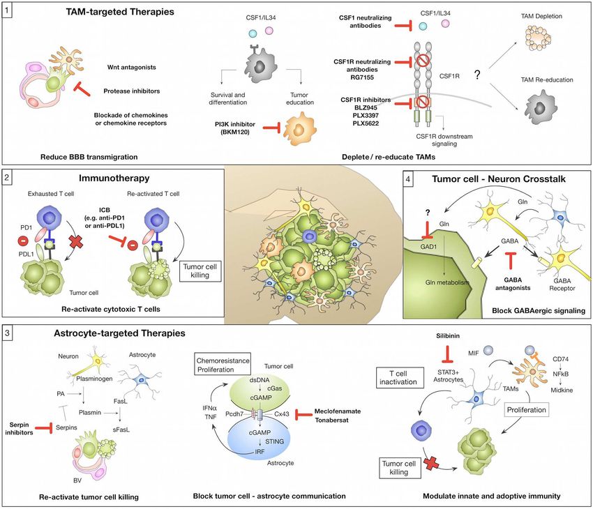
Frontiers in Immunology | www.frontiersin.org 2 July 2019 | Volume 10 | Article 1713Schulz et al. Brain Metastasis-Associated Inflammation FIGURE 1 | Microenvironmental regulation of the metastatic cascade. The tumor microenvironment of brain metastasis comprises different brain-resident and recruited cell types with cell-type and/or stage-dependent pro- or anti-tumor functions. (1) Different microglial-derived factors including proteases (e.g., Ctss, Mmp3, and Mmp9), Wnt signaling components or chemokines (e.g., Cxcl12) have been implicated in assisting tumor cells to cross the blood brain barrier (BBB) and colonize the brain parenchyma. (2) In contrast, astrocytes were shown to prevent early stages of metastatic colonization by inducing soluble (s)-FasL-mediated tumor cell killing. Tumor cell-derived serpins can block this effect by inhibiting astrocyte-derived plasminogen activator (PA), therefore preventing the generation of active plasmin that converts FasL into sFasL. (3) While the initial tumor cell—astrocyte contact leads to tumor cell killing, close interactions between tumor cells and astrocytes via gap junctions foster tumor cell proliferation and protect tumor cells from chemotherapy. This process was linked to the transfer of cGAMP from tumor cells to astrocytes that triggers cGas-STING-mediated IRF activation leading to production of IFNα and TNF. (4) Cytotoxic T cells represent an important component of the adoptive immune response against brain metastasis by executing tumor cell killing. (5) However, T cell activity is efficiently blunted by the immune-suppressive milieu in brain metastasis. T cell activity is modulated through interaction with several cell types including tumor cells, tumor-associated macrophages/microglia (TAM-MG and TAM-BMDM) and astrocytes by expressing immune checkpoint molecules or by secreting immune-suppressive cytokines (e.g., IL10, TGFβ or IL6). Moreover, astrocytes with high STAT3 expression were shown to activate tumor-promoting TAMs via the MIF-CD74-NfkB-Midkine axis. (6) Tumor cells that colonize the brain were shown to adopt to the neuro-glial niche by acquiring neuronal gene signatures that induce specific metabolic programs (e.g., GABAergic signaling and the expression of neurotrophic factors). (7) Tumor expansion leads to neuronal damage by mechanical compression of neurons. tumor- and stromal signatures in BrM revealed an enrichment of and use growing axons as migratory tracts for cancer cell neuronal differentiation pathways in the tumor cell population dissemination (19, 25). (23). Further exploration of neuronal mimicry revealed that To date it remains unknown to which extent neurons play GABAergic signaling is not limited to the CNS, but has also an active role in BrM onset and progression. However, given emerged as a tumor signaling molecule in cancers of peripheral the recently demonstrated role of neurons in glioma together organs such as breast, liver, pancreas, and colon (24). Hence, it with the observation that highly innervated tumors (i.e., prostate is possible that tumor cells are primed for GABAergic signaling or head and neck cancer) are more aggressive than their less already at the primary tumor site providing an advantage for innervated counterparts (18, 19, 25), it might be premature to rapid adaptation to metabolic conditions in the brain. Moreover, exclude neurons as active players in BrM. Future studies will it was demonstrated that prostate cancer cells induce axonogensis hopefully provide more detailed insight into the role of neurons Frontiers in Immunology | www.frontiersin.org 3 July 2019 | Volume 10 | Article 1713
Schulz et al.                                                                                               Brain Metastasis-Associated Inflammation

in BrM and potentially open new therapeutic avenues against                 repair mechanisms (43). However, it should be noted that
BrM by targeting interactions between tumor cells and neurons.              the M1/M2 and A1/A2 nomenclature reflects functional and
                                                                            phenotypic extremes within a spectrum of activation states
                                                                            and that along the continuum of activation states mixed
Astrocytes in Brain Metastases—Versatile                                    phenotypes have been reported (47). The high phenotypic
Players in Mediating Distinct Steps Within                                  plasticity of astrocytes was also reported in pre-clinical and
the Brain Metastatic Cascade                                                clinical studies on different neurodegenerative disorders. For
Astrocytes belong to the glial cell types and represent the                 example, Liddelow et al. demonstrated that microglia induce
most abundant cell population within the CNS (26). Originally               A1 astrocytes with neurotoxic properties. The presence of A1
described as star-like “glue” cells of the CNS, the variety                 astrocytes was demonstrated in various human malignancies
and complexity of astrocyte function in health and disease is               from the spectrum of neurodegenerative diseases (10). On the
increasingly recognized. Under normal conditions their role in              contrary, microglia were shown to exert crucial functions in
tissue homeostasis includes maintenance of the blood brain-                 activating neuroprotective astrocytes in a model of Spinal Cord
barrier (BBB), immune signaling, regulation of extracellular                Injury (SCI) (48), further underpinning the context-dependent
ion, and fluid homeostasis, as well as control and maintenance              outcome of cellular interactions. In line with this finding,
of a broad range of functions implicated in modulating                      microglia-mediated blockade of an A1 astrocyte conversion was
neuronal networks, such as regulation of synaptogenesis,                    shown to be neuro-protective in a mouse model of sporadic
synaptic plasticity, and elimination, neurotransmitter clearance,           Parkinson’s Disease (49). There is also evidence that RA are
and neurotrophin secretion (27–30). To fulfill this functional              regulated by distinct T cell subsets in neuro-inflammatory
diversity, it is now widely accepted that astrocytes represent              conditions such as stroke, which then potentiates neurological
a highly heterogeneous cell population (28, 31–33). With                    recovery (50).
the advent of high-throughput single cell sequencing and                       While our understanding of astrocyte function in
other “omic” approaches, the existence of several astrocyte                 neurodegenerative disorders is steadily increasing, we are
subpopulations was revealed in rodents (31, 34, 35). An even                just at the beginning to decipher the underlying mechanisms
higher heterogeneity was found within the human brain (36, 37).             of pro- or anti-tumor functions of astrocytes in BrM (51, 52).
Interestingly, neuronal stimuli have been shown to determine                Induction of astrogliosis is an early event during metastatic
distinct features of astrocytes (38). Moreover, it was shown                colonization and outgrowth. This early reaction is attributed
that during aging, astrocytes change their transcriptomes in                to neuro-protection by delineating metastatic foci from the
different regions of the murine brain (39), which is in part                normal brain parenchyma. Valiente et al. proposed that early
orchestrated by interacting with local microglia (40). Given                contacts between tumor cells and astrocytes lead to tumor cell
the phenotypic and functional diversity of astrocytes, it is not            death and clearance of the majority of tumor cells that enter
surprising that astrocytes play a central role in maintaining tissue        the brain. In order to successfully colonize the brain, tumor
homeostasis and in regulating neuro-glial communication under               cells have to acquire traits to block pro-apoptotic stimuli from
physiological conditions. Consequently, astrocytes are also often           astrocytes (53) (Figure 1; Box 2). On the other hand, there is
found to be involved in disease progression of different CNS                accumulating evidence that astrocytes promote distinct steps
malignancies (30). Moreover, malignantly transformed astrocytes             of the metastatic cascade, including initial seeding and support
are the cell of origin for astrocytoma, the most common form                of tumor outgrowth (54–56). Moreover, astrocytes have been
of glioma (41). Astrocytes respond to disease-associated stimuli            shown to protect tumor cells from chemotherapy (57). This
by undergoing morphological and functional changes, which are               process was shown to be dependent on gap junction formation
collectively referred to as reactive astrogliosis (30, 42, 43). A key       (57, 58). The importance of direct cellular connections between
feature of reactive astrocytes (RA) is the formation of a glial             astrocytes and breast- or lung brain metastatic tumor cells
scar that confines pathological foci from the healthy parenchyma            via gap junctions was further demonstrated by Chen et al.
(27). Interactions between astrocytes and other brain-resident or           (59). In this context, gap junction formation was mediated by
recruited cells have been investigated in different disease models.         connexin43 (Cx43) and protocadherin (Pcdh7) and activated
It was shown that every cell type in the CNS can release factors            the innate immune response pathway cGAS-Sting (Cyclic
that induce astrogliosis (27), and the outcome is regulated in a            GMP-AMP synthase-stimulator of interferon genes) leading to
time- and context-dependent manner (44).                                    secretion of tumor-supportive cytokines such as IFNα and TNF
    A particularly close connection between astrocytes and                  (Figure 1; Box 3). Functional co-option of RA by melanoma
microglia was observed in the diseased CNS (10). Depending on               cells was further exemplified by Schwartz et al. (60). The authors
the environmental stimuli, astrocytes acquire different activation          demonstrated in a melanoma brain metastasis model that
states that are referred to as A1 and A2 following the previously           astrogliosis is exploited by the tumor cells to support their
defined nomenclature to classify macrophage polarization                    growth (60). Astrocytes are also emerging as critical modulators
states into pro-inflammatory, anti-tumor M1 macrophages, and                of immune responses in BrM by interacting with brain-resident
immune-suppressive, tumor-promoting M2 macrophages (45,                     and recruited inflammatory cells. Priego et al. recently proposed
46). A1 astrocytes are regarded as neuro-inflammatory, while                an important role of astrocytes in the modulation of innate
A2 astrocytes are associated with neuro-protective features by              and acquired immunity in BrM (61). The authors identified a
promoting survival and growth of neurons and by inducing                    subpopulation of RA with high STAT3 activation levels associated

Frontiers in Immunology | www.frontiersin.org                           4                                        July 2019 | Volume 10 | Article 1713
Schulz et al. Brain Metastasis-Associated Inflammation
with BrM of different primary origin. STAT3 activation was microglia, largely due to higher expression of MHCII (74),
shown to affect microglia and T cell functions, likely leading to however their contribution to BrM progression remains to be
the establishment of an immunosuppressive microenvironment elucidated. Detailed insight into transcriptional programs of
(Figure 1; Box 5). CD74+ TAMs were previously shown to microglia revealed a remarkable plasticity in response to a wide
generate an immunosuppressive milieu by reducing the secretion variety of stimuli, such as regional differences in the brain, aging,
of IFNγ in glioma (62). More recently it was demonstrated sex, or the composition of the microbiome and gene signatures
in BrM that CD74+ TAMs depend on pSTAT3+ astrocytes reflect cellular functions during developmental stages (75–78).
that secrete macrophage migration inhibitory factor (MIF), In addition to in depth analysis of microglial heterogeneity,
the ligand for CD74. In response to ligand binding, CD74 dissecting gene signatures of disease-associated microglia
acts as a transcription factor and promotes the expression provides detailed insight into lineage-dependent functions and
of NFkB downstream targets, such as midkine, a factor that cellular dynamics (79–82). In this regard, the identification of a
promotes cell viability (61). MIF inhibition by ibudilast led to disease-associated signature in microglia (DAM) by single cell
a reduction of BrM in organotypic cultures (61). Moreover, sequencing in Alzheimer’s disease, aging, multiple sclerosis, and
genetic and pharmacological inhibition of STAT3 resulted in amyotrophic lateral sclerosis models significantly contributed
impaired viability of tumor cells and reduced outgrowth of to our understanding how different pathological conditions
brain metastasis (61). Heiland et al. recently confirmed the shape the molecular identity of disease-associated cells and
findings on STAT3+ astrocytes in primary brain tumors and how the respective subpopulation might enhance or ameliorate
demonstrated that astrocyte-microglia interactions generate a disease progression. Upregulation of phagocytosis components
strong immune-suppressive environment due to up-regulation and neurodegenerative markers such as Trem2 and ApoE and
of PD-L1 on tumor-associated astrocytes and production of the downregulation of microglia homeostatic markers such as
cytokines such as IL10 and TGFβ (63). Cx3cr1 and Tmem119 were shown to be characteristic for the
Taken together, astrocytes are emerging as one of the key DAM signature (71, 79–82). Remarkably, single cell analysis
regulators of brain metastatic colonization and outgrowth. of human microglia from multiple sclerosis patients revealed
Owing to their high phenotypic and functional heterogeneity, an even higher heterogeneity, as seven different populations
astrocytes exert pro-tumor as well as anti-tumor functions. of microglia were identified. Three of those populations
Detailed insights into the existence of different astrocyte represented homeostatic genes, one population showed an
subpopulations or stimuli that polarize astrocytes at distinct upregulation of chemokine and cytokine signaling, whereas the
stages of the brain metastatic cascade are required to develop three other populations correlated with the clusters associated
astrocyte-targeted therapies. with demyelination and remyelination in mice (83). Although
the BrM field currently lacks detailed insight into the molecular
identity of disease-associated macrophages/microglia compared
Myeloid Cells in Brain Metastases—Origin to neurodegenerative diseases or primary brain tumors, a
and Location Matters series of pre-clinical studies shed light into tumor-associated
Myeloid cells in brain malignancies comprise a highly abundant macrophage (TAM) functions during distinct steps of the
and heterogeneous cell population and consist of brain resident metastatic cascade. Invasion of metastasizing tumor cells is
myeloid cells as well as recruited cells including monocytes, bone rapidly sensed by microglia and the presence of single tumor
marrow-derived macrophages (BMDM), and granulocytes (64). cells is sufficient to recruit and to activate microglia (84, 85).
Brain-resident microglia are the major representatives of the Given the role of microglia in immune surveillance and host
innate immune system in the CNS and exert critical functions in defense, it is tempting to speculate that the initial contact
immune surveillance and host defense. In addition to functions between tumor cells and microglia at sites of extravasation leads
related to neuro-inflammation, microglia are also responsible to clearance of invading tumor cells. However, Chuang et al.
for synapse pruning and remodeling (65). Microglia represent demonstrated that tumor cells block pro-apoptotic functions
a unique cell type among the glial cells with respect to their of microglia and exploit tissue damage responses to increase
ontological origin. In contrast to other glial cells, microglia are their invasive capacity (86). The role of microglia in tumor
of mesodermal origin and arise from primitive hematopoietic cell extravasation was further confirmed by Qiao et al. using
progenitors (erythromyeloid progenitors) that are present in the a CSF1R inhibitor to deplete microglia in prevention trial
yolk sac during embryonic development (66–68). In addition settings in a mouse model for melanoma BrM (84). The authors
to parenchymal microglia, the CNS harbors myeloid cell also found that Mmp3 expression by microglia was negatively
populations that reside in specific regions of the CNS including correlated with ZO-1 expression on endothelial cells. Moreover,
the choroid plexus, the interphase between blood and meninges, the incidence of melanoma BrM was decreased by Mmp3
and the perivascular space of vessels (69–71). Border-associated inhibition (84). Co-option of microglial functions and adoption
macrophages (BAMs) derive from erythro-myeloid precursors of leukocytic characteristics to increase the capacity of tumor
that arise from the yolk sac and the fetal liver. Interestingly, cells to colonize the brain parenchyma was previously proposed
bone marrow-derived monocytes also contribute to the choroid (5) (Figure 1; Box 1). Interestingly, it was observed that tumor
plexus macrophage population (70–72). Moreover, monocytes cells increase the expression of cathepsin S, a protease that is
have been shown to reside within the meninges (73). BAMs pre-dominantly expressed by leukocytes, to cleave junctional
are believed to have a higher antigen presenting capacity than adhesion molecules that maintain the BBB integrity and thus
Frontiers in Immunology | www.frontiersin.org 5 July 2019 | Volume 10 | Article 1713Schulz et al. Brain Metastasis-Associated Inflammation
assist tumor cells to breach the BBB. Importantly, only the BBB has to be diminished in order for blood-borne cells to
combined depletion of cathepsin S in the tumor and stroma efficiently breach the BBB. It was shown that the BBB in BrM
compartment was efficient to reduce BrM burden (5). The co- is not fully disrupted but rather remodeled into a blood-tumor-
option of leukocyte characteristics by tumor cells is also evident barrier (BTB) due to alterations in the pericyte subpopulation
in the role of the C-X-C chemokine receptor type-4 (Cxcr4) (106). While this is not sufficient to allow free penetration
along with its ligand Cxcl12 that are involved in lymphocyte of therapeutic antibodies or chemical compounds that are not
chemotaxis. Cxcr4 expression has been detected in BrM tumor BBB permeable (107), it is possible that vessel structures of
cells (87, 88). Remarkably, the inhibition of this pathway the BTB lose their capability of restricting the entry of blood-
decreased breast cancer cell migration (89) and impaired BrM borne immune cells and at the same provide the necessary
establishment (90). molecular structures such as adhesion molecules for efficient
In established BrM, tumor-associated macrophages and transmigration of peripheral leukocytes. Cell-tracing techniques
microglia are the most abundant non-cancerous cell type based on the transplantation of genetically labeled HSCs
and constitute up to 30% of the total tumor mass (5). In into mice following whole-body irradiation or head protected
primary brain cancer, TAMs tend to be pro-tumorigenic and irradiation have been used to decipher the origins of TAMs
accumulate with higher tumor grade (91, 92). As revealed in primary brain tumors (108). By means of transplantation
by immunohistochemistry of BrM sections, microglia and and lineage-tracing models, numbers for peripheral macrophages
macrophages showed signs of intratumoral activation and range between 25 and 75% in glioma and 25% in BrM (64,
formed a boundary between the tumor mass and normal brain 92, 108). Similar to neurodegenerative disorders, the bulk FACS
tissue (93–95). They were identified as foamy cytoplasmatic cells sorted TAMs showed a different expression profile compared
with shortened cell processes and immunoreactive to CD68. to normal microglia and monocytes in a mouse glioma model
However, there is currently no clinical evidence in BrM for (64). More importantly, the profile of tumor-associated microglia
a correlation between microglia density and activation marker and macrophages was different, confirming the functional
expression with treatment modality, anatomic brain regions or impact of their different ontological origin. While TAM-MG
necrosis (96). Despite the lack of clinical correlation between showed profiles rich in cytokines, chemokines, and complement
TAM content and BrM patient prognosis, pre-clinical data components, TAM-BMDM signatures were associated with
indicate tumor-promoting functions of TAMs in BrM. The wound healing, antigen presentation and immune suppression
crosstalk between microglia and melanoma BrM is evident from (64, 92). Another evidence for the intrinsic differences within
the alteration of JNK and p38 components in microglia, which the macrophage/microglia population is the lack of impact
may attenuate their phagocytic response, as well as ERK and of anti-CSF1 treatment on microglia compared to monocyte-
STAT3 in melanoma cells, which are linked to angiogenesis. derived cells, which may be due to the presence of the CSF1R
The authors also provided evidence for a metastasis-supportive alternate ligand IL34 (109). This observation is also supported
niche, as secretion of vascularization factors was reshaped and by the fact that in multiple sclerosis dendritic cells (DC)
proliferation of both cell types was increased (97). Correlating and monocyte-derived cells are the major antigen presenting
with the latter finding, anti-inflammatory microglia depletion cells (APCs). Indeed interactions of microglia with infiltrating
by mannosylated clodronate liposomes decreased the growth T cells were found to be transient (110). Importantly, in
of intracranially implanted breast cancer cells (98). Another order to unravel the molecular pathways and functional pre-
evidence from the interplay between cancer cells and microglia is dominance in every cell population, it is mandatory to properly
that XIST-deficient-breast cancer cells led to an increased amount distinguish them. Under physiological conditions, the different
of M2-markers in microglia (99). However, the genetic programs expression profiles of macrophages and microglia, residing in
that lead to an induction of tumor-promoting functions in their respective environment enables their differentiation (69,
TAMs in BrM are not well-characterized to date. Detailed 110–112). The identification of novel markers for microglia such
analysis of signaling pathways and transcription factor activity as Tmem119, P2ry12, Sall1, SiglecH (113–116) is important
is required to evaluate if similar mechanisms lead to the to unravel their specific role in health and disease. However,
induction of a TAM gene signature in BrM as proposed for other these expression patterns are less well-defined in TAMs, as e.g.,
tumor types including glioma (64, 100). For example, Blazquez homeostatic Tmem119 is upregulated in TAM-BMDM, while
et al. recently proposed the importance of PI3K signaling as it is downregulated in TAM-MG (64). Remarkably, CD49d has
a master regulator of tumor promoting activation states of been described as a differential marker between blood-borne
macrophages/microglia (101). macrophages and microglia in brain malignancy (64). However,
Previous studies that interrogated the role of TAMs in BrM did to date gene expression signatures of TAM-MG in comparison to
not discriminate between cells originating from brain-resident TAM-BMDM have not been investigated.
microglia or from bone marrow-derived macrophages. As In summary, within the myeloid compartment in BrM,
mentioned earlier, under steady-state conditions, bone marrow- TAMs constitute the most abundant cell population. Based
derived myeloid precursors do not contribute to the microglia on their ontological origin and localization within the brain
pool. However, damage to the blood-brain barrier as described parenchyma and border regions of the CNS, they represent
for BrM (102–104) allows the recruitment of such progenitors a highly heterogeneous cell population. Until now, most pre-
that supplement the microglial population (105). In this context clinical studies that aimed to unravel the role of TAMs in
it is important to evaluate to which extent the integrity of the BrM did not discriminate between different subpopulations.
Frontiers in Immunology | www.frontiersin.org 6 July 2019 | Volume 10 | Article 1713Schulz et al. Brain Metastasis-Associated Inflammation
The identification of lineage-restricted markers that allow to the latter might be polarized in the TME under certain conditions
distinguish TAM-MG and TAM-BMDM as well as single cell resulting in tumor promoting rather than anti-tumor functions,
sequencing approaches will help to unravel gene signatures so they will not only be inhibited in their anti-tumor functions
of individual subpopulations and provide insight into their but promote tumor growth as discussed for macrophages and
functional contribution in BrM. astrocytes in the sections above. Furthermore, it remains unclear
what dictates the number of TILs in BrM. Generally, T cells are
primed in peripheral lymph nodes (e.g., cervical lymph nodes).
Tumor-Infiltrating Lymphocytes in Brain Extravasation and T cell homing to sites of inflammation or tissue
Metastases—Can Activity be Unleashed injury is dependent on binding of VLA-1 and LFA-1 expressed
Without Inducing Neurotoxicity? by T cells to endothelial cellular adhesion molecules (CAMs)
Traditionally, the brain has been regarded as an immune such as VCAM-1 or ICAM-1 (130–132). The exact entry route
privileged organ, with lack of peripheral immune surveillance for T cells into BrM is not yet fully understood, however there
through blood-borne immune cells such as T cells owing to the is evidence that expression of CAMs on endothelial cells plays
blood-brain-barrier (BBB) and the lack of effective lymphatic a central role in the homing process of T cells. For example,
drainage (117, 118). However, this view has recently changed, it was shown that vessels in the proximity of tumor lesions
as it is recognized that while the brain might be privileged show expression of VCAM-1, ICAM-1, and other CAMs in
to some extent, this does not mean total exclusion of blood- different BrM models (133–136). Interestingly, it was proposed
borne immune cells. Clearly, the entry to the parenchyma is that tumor cells exploit this mechanism to breach the BBB
strictly controlled to prevent fatal neurotoxicity, but patrolling and home to the brain parenchyma (134). Serres et al. could
leukocytes, such as bone marrow-derived antigen-presenting DC also detect VCAM-1 on human BrM samples, while healthy
as well as CD4+ and CD8+ T cells, have been identified in the controls showed only minimal expression (133). Additionally, it
meninges and choroid plexus (70, 71, 77, 119, 120). DC have a was demonstrated that VCAM-1 expression increases with tumor
higher capacity of antigen presentation and T cell stimulation progression in a BrM mouse model (133). Taggert et al. proposed
than microglia (121). Furthermore, it has been demonstrated that increased CD8+ T cell trafficking to BrM is dependent on
that afferent antigen sampling from the brain parenchyma does VCAM-1 and ICAM-1 expression induced by IFNγ produced
take place and CNS-derived antigens can lead to peripheral by BMDMs, microglia and NK cells (135). Hence, TIL numbers
priming of T cells (122). Moreover, recent studies unveiled can at least in part be determined by IFNγ levels and CAM
the existence of lymphatic vessels in the meninges, which can expression. However, other determinates of TIL numbers in
transport antigens, derived from the brain parenchyma via BrM such as mutational load and presence of tumor antigens
the cerebrospinal fluid/lymphatic system into the deep cervical are expected to affect T cell infiltration. It was demonstrated
lymph nodes (123–125), indicating that the brain has a functional for primary melanoma, a highly immunogenic tumor with high
draining lymphatic system. However, the details about anatomy TIL content, that the density of antigens did not correlate with
and composition, as well as the efferent route of T cells into the presence or absence of TILs (137). Additionally, Mansfield
the brain, specifically in the scenario of BrM, require further et al. applied TCR profiling of patient derived samples and could
investigation. As immunotherapies gain increasing attention, show that the mutational burden is higher in BrM of NSCLC
the infiltration of BrM with T cells and their function in the than in the respective primary tumor and is correlated with T
context of brain tumors comes into focus. By now, several studies cell richness (138). In this study the authors also observed a
demonstrated that tumor infiltrating T lymphocytes (TILs) are contraction of T cell clones compared to the primary site, with
present in BrM of different primary cancers such as Non- the 10 most abundant T cell clones being more heavily expanded
Small Cell Lung Cancer (NSCLC), Small Cell Lung Cancer compared to the primary tumor site (138). This expansion hints
(SCLC), renal cell cancer (RCC), melanoma, or breast cancer. toward an immune response in BrM with the involvement of
Of these RCC and melanoma show the highest CD3+ numbers antigen specific T cells, even though in a more restricted manner
and highest CD8+/CD3+ ratio. The infiltration patterns of than in primary tumors. The fact that BrM still represents a
TILs seem to be diverse, ranging from a diffuse spreading highly aggressive, fatal disease and monotherapies with immune
through the metastases to an accumulation in the stroma and modulatory agents only show modest effects indicates that this
around vessels, depending on primary tumor type (126). The adaptive immune response is not strong enough to halt tumor
prognostic value of TIL numbers is currently being disputed, growth. Comparison with the respective primary tumors led to
with several studies indicating favorable outcome on survival the observation that BrM, e.g., derived from breast cancer, have a
(96, 127, 128), while Harter et al. could not find a significant lower content of TILs than the primary counterpart, with 5 and
correlation (126). Moreover, Mustafa et al. demonstrated that 20%, respectively (139). While the extent of T cell exclusion is
T cells promote breast cancer BrM. This is due to a direct lower in BrM compared to many primary brain tumors, the TME
interaction of T cells with tumor cells, leading to increased is still highly immune suppressive. Current research investigates
guanylate binding protein 1 (GBP)-1 expression by the latter, strategies to increase the inflammatory response against BrM, to
which in turn enables them to cross the BBB (129). These render the tumors more prone to immune-modulating agents.
conflicting data indicate that further research is necessary to Using adoptive T cell transfer is only one strategy, which has
elucidate the complex function of T cells in BrM as well as demonstrated encouraging results in melanoma BrM patients
possible influence of the TME on T cells. It is conceivable, that (140). However, not only the number of T cells plays an
Frontiers in Immunology | www.frontiersin.org 7 July 2019 | Volume 10 | Article 1713Schulz et al. Brain Metastasis-Associated Inflammation
important role in the immune response against brain metastatic immunogenic (144–147). For example, a limited number of
cells, but also their activation status is relevant. The latter is retrospective and prospective clinical trials indicate intracranial
dependent on many factors and the cell composition in the response rates of 16–25% following ipilimumab treatment in
TME. The brain naturally constitutes an immune suppressive melanoma patients (148, 149) and 50–55% in trials combining
microenvironment to prevent fatal neurotoxicity, potentially ipilimumab and nivolumab (ABC trial and CheckMate 204) (150,
resulting in the exhaustion and inactivation of T cells in primary 151). Therefore, other strategies are being explored to increase
brain tumors (141). Moreover, it has been demonstrated with T cell immunity e.g., the combination of checkpoint inhibition
different BrM mouse models, that the number of FOXP3+ T with radiotherapy (RT). The latter has the potential to sensitize
regulatory cells (Tregs) is increased during BrM progression. for immune modulation by inducing immunogenic cell death
These results have also been recapitulated on patient samples resulting in secretion of inflammatory cytokines, upregulation
from melanoma and NSCLC BrM (142). Additionally, not only of MHCI and therefore increased trafficking of T cells to the
the tumors themselves are infiltrated with Tregs, but also the BrM, as shown for other cancers (152, 153). Taggart et al.
blood of patients bearing BrM contains an increased percentage could show in a mouse model of melanoma BrM that successful
of Tregs compared to healthy donors (143). Those inhibitory immunotherapy depends on enhanced trafficking of CD8+ T
T cells can hypothetically contribute to the exhaustion of anti- cells, activated in peripheral lymphoid organs, to the brain
tumor effector T cells. parenchyma (135). Additionally, RT can potentially increase the
Taken together, insights into the complex and dynamic tumor mutational load thereby broadening the immune response
interplay between different cell types of the TME in BrM, (154). Indeed, radio-immunotherapies show promising results
in which the activity of individual cell populations is tightly and are currently being tested in clinical trials also for BrM
controlled by other cell types, underpins the challenges (155). Nevertheless, T effector cell activity in the brain is not only
in developing effective therapies against BrM. However, a dependent on Treg infiltration or immune suppressive cytokines
comprehensive view on the complex interactions provides in the TME, but also on the presence of APCs. As mentioned
opportunities for the development of improved therapeutic earlier, in the brain this role can be fulfilled by DC, BMDM, and
intervention strategies as discussed in the following paragraph. to a lesser extent by microglia (100, 121). The presence of those
cell types in the brain tumor context is not questioned anymore,
Tumor Microenvironment-Targeted and but there are no detailed reports about specific interaction with
Immunotherapies Against APCs and TILs that lead to antigen-specific T cell activation in
Brain Metastases BrM. It is important to further investigate this in the future to
The development of effective therapies against BrM is one of improve response rates of patients to immunotherapy and to find
the most challenging aspects of cancer research. Intervention new strategies against BrM by exploiting the full potential of T
strategies developed for extracranial tumors cannot easily be cell immunity in this context. Using DC vaccines to boost T cell
translated into effective therapeutic avenues for brain cancers. responses is only one of many potential treatment possibilities,
Instead, approaches have to be tailored to the unique brain which could be explored in this context and is under current
environment to breach tissue-specific restrictions of therapeutic investigation in brain tumors (156). Another strategy of applying
efficacy, but at the same time consider the protection of delicate T cells for BrM treatment is the delivery of genetically engineered
anatomical structures that control higher cognitive functions. CAR T cells directed against known tumor antigens, which led
Detailed insights into the critical cellular and molecular drivers to reduced tumor growth in a xenograft mouse model (157).
of BrM are necessary to provide a scientific rationale for Currently, this approach is investigated in a clinical trial for
the development of improved intervention strategies. Recent breast cancer patients with BrM (158). However, it remains
research efforts shed light on the complexity of the tumor- questionable if T cell-directed therapies can be successful in the
stroma crosstalk in BrM and indicate potential therapeutic targets presence of a highly immune suppressive myeloid compartment.
for immune- or tumor microenvironment targeted therapies Alternatively, one could argue that myeloid-targeted therapies
(Figure 2). might be more promising.
Immunotherapies Against Brain Metastases Modulating the Myeloid Compartment in
The introduction of immunotherapy has recently revolutionized Brain Metastases
treatment options for a range of extracranial primary tumor types Cells of the myeloid compartment represent the most abundant
including melanoma and NSCLC that frequently metastasize non-malignant cell type in the BrM microenvironment. Pre-
to the brain. Hence, it appears logical to test the efficacy of clinical data indicate a critical role in mediating distinct steps
immunotherapy against BrM, even though the brain tissue within the metastatic cascade leading to the generation of a
environment represents one of the most immune suppressed cancer-permissive, immune suppressive environment (Figure 1).
milieus. One arm of immunotherapy aims at re-activating T Different strategies have been employed to target TAMs in BrM to
effector cells via immune checkpoint inhibition (Figure 2; Box 2). evaluate therapeutic efficacy. Blocking macrophage survival and
Indeed monoclonal antibodies, which block immune checkpoints differentiation by disrupting CSF1-CSF1R signaling represents
(e.g., anti-CTLA4, anti-PD1, or anti-PDL1), demonstrate efficacy one of the most promising strategies (Figure 2; Box 1). Since
in individual BrM patients, but the overall response rates are there are two cognate ligands that bind to CSF1R, targeting the
modest, even in melanoma BrM, which is thought to be highly receptor rather than the ligand, leads to efficient blockade of
Frontiers in Immunology | www.frontiersin.org 8 July 2019 | Volume 10 | Article 1713Schulz et al. Brain Metastasis-Associated Inflammation FIGURE 2 | Novel concepts of tumor microenvironment-targeted therapies or immunotherapies (1) Tumor-associated macrophages/microglia (TAMs) represent a highly abundant cell type in BrM with known roles in mediating tumor cell BBB transmigration and tumor-supportive functions that foster metastatic outgrowth. Strategies for TAM-targeted therapies include the reduction of tumor cell BBB transmigration (e.g., by Wnt antagonists, protease inhibitors, or blockade of chemokines/chemokine receptors). Blockade of CSF1-CSF1R signaling represents another strategy to target TAMs by inhibiting a central pathway for macrophage differentiation and survival. The CSF1-CSF1R signaling axis can be inhibited by (i) CSF1 blocking antibodies (with no effects on IL34 mediated CSF1R activation), (ii) CSF1R blocking antibodies, or (iii) ATP competitive small molecule inhibitors. Consequences of CSF1R inhibition on TAMs in established BrM (depletion vs. re-education) remain to be elucidated. An alternative strategy might be the inhibition of Pi3K by BKM130 to prevent the activation of pro-tumor TAMs. (2) Tumor-infiltrating T cells in BrM show signs of T cell exhaustion mediated by immune checkpoints (e.g., PD1-PDL1) or immune-suppressive cytokine milieus. Blockade of immune checkpoints e.g., by anti-PD1 or anti-PDL1 reactivates T cells and reinstates tumor cell killing by cytotoxic T cells. (3) Astrocytes represent a highly plastic cell type in BrM and their function was associated with pro- and anti-tumor activity. Inhibition of serpins could re-activate sFasL-mediated tumor cell killing and thereby prevent early metastatic colonization. Blockade of gap junctions by meclofenamate or tonabersat was shown to inhibit tumor cell-astrocyte crosstalk that supports proliferation and protects tumor cells from chemotherapy. Targeting of STAT3+ astrocytes by silibinin represents a strategy to block the induction of pro-proliferative functions of TAMs and reduce astrocyte-mediated inactivation of T cells. (4) Brain metastatic tumor cells adopt neuronal features to integrate into the neuro-glial niche and to exploit brain specific energy sources e.g., glutamate (Gln). GABA antagonists were shown to reduce GABAergic signaling in tumor cells. Furthermore, blockade of Gln influx into tumor cells by GAD1 inhibition could represent a promising therapeutic strategy. CSF1R downstream signaling. CSF1R inhibition can be achieved PLX3397 in a prevention trial setting and demonstrated that by CSF1R blocking antibodies (e.g., RG7155) (159) or ATP microglia depletion reduced tumor cell transmigration potential competitive small molecule inhibitors (e.g., BLZ945, PLX3397, of melanoma brain metastatic cells (84). This is in line with or PLX5622) (91, 160) (Figure 2; Box 1). Qiao et al. employed previous findings that demonstrated that clodronate liposome Frontiers in Immunology | www.frontiersin.org 9 July 2019 | Volume 10 | Article 1713
Schulz et al.                                                                                                Brain Metastasis-Associated Inflammation

mediated microglia depletion resulted in a reduction of the                  approach in the clinic has to be carefully evaluated. Given the
BrM burden (98). Given the promising results of TAM-targeted                 physiological importance of gap junctions for tissue integrity as
therapies with the CSF1R inhibitor BLZ945 in a mouse model                   well as normal brain function (162), potential adverse effects
of pro-neural glioblastoma (91) it remains to be elucidated                  have to be taken into account. Moreover, approaches that target
whether CSF1R inhibition in established BrM shows anti-tumor                 the formation of gap junctions between astrocytes and tumor
activity. Importantly, analyses in two independent glioblastoma              cells are expected to be most efficient at initial stages of brain
models revealed that conditions in which CSF1R inhibition                    colonization, when the majority of tumor cells is in direct
leads to TAM depolarization show higher efficacy compared                    contact with astrocytes, while at later stages only tumor cells
to TAM depletion (91, 161). Consequently, research effort                    at the tumor-stroma interface are in close vicinity to astrocytes
should be put on the identification of gene signatures that                  (30, 38, 51). Indeed, the formation of gap junctions between
determine tumor-promoting vs. anti-tumor characteristics in                  tumor cells and astrocytes was detected in subpopulations but
TAMs to specifically target tumor supportive traits of TAMs                  not ubiquitously (59).
but spare physiologically important functions. Blazquez et al.                  Another promising approach was recently described by
recently proposed Pi3K signaling as a master regulator of tumor-             targeting STAT3 signaling in RAs via the inhibitor Silibinin
promoting functions of BrM-associated macrophages/microglia                  (61) (Figure 2; Box 3). Clinical data from lung cancer BrM
and demonstrated that BKM120, a pan-PI3K inhibitor, reduced                  patients treated with Silibinin showed significantly increased
tumor-promoting features of macrophages/microglia (101).                     overall survival in response to STAT3 inhibition (61). However,
However, it is important to note that clinical data revealed                 some patients did not respond and the progression of extra-
better overall survival for patients with high PI3K activity,                cranial disease was not affected, providing the possibility for
while patients with moderate or low PI3K activity showed                     BrM relapse. It remains to be shown how patients with
worse prognosis (101). Hence, inhibiting PI3K signaling in                   BrM derived from other primary tumor entities respond to
BrM might have opposing effects depending on which cell type                 this treatment approach, and if variability of the outcome
is targeted.                                                                 is due to tumor heterogeneity, differences in the TME
    Given the importance of the myeloid compartment to                       and/or different patient histories. It is also unclear why
establish an immune suppressive environment to protect the CNS               only a subset of astrocytes activates STAT3 signaling, which
from neuro-inflammation, myeloid-targeted therapies should                   requires deeper understanding, especially with respect to
be taken into account carefully. Blocking an integral part of                other immune cells (e.g., macrophages, microglia) and how
a tissue protective mechanism might unleash unwanted pro-                    different cellular and also molecular (e.g., different cytokine
inflammatory responses that lead to detrimental tissue damage.               milieus) microenvironments influence the outcome of impaired
Detailed knowledge in disease-associated effector functions of               STAT3 signaling.
different myeloid cell populations is therefore needed to block                 Targeting astrocytes in the context of BrM is a promising
tumor-promoting functions but maintain critical functions in                 approach, since these cells are highly susceptible to tumor cell-
host defense and neuro-protection.                                           mediated education within the brain, thus promoting BrM.
                                                                             However, it remains to be investigated how distinct astrocyte
Astrocyte-Targeted Therapies                                                 subpopulations support BrM formation to develop strategies
Astrocytes are emerging as one of the key regulators of BrM                  that block tumor-promoting or enhance anti-tumor functions
(51). However, pre-clinical studies revealed high functional                 of astrocytes.
heterogeneity with tumor-promoting and anti-tumor functions.
Therefore, it will be critical to gain detailed mechanistic insight          Prevention of Neuronal Mimicry of Tumor Cells
into functional subpopulations or conditions that favor the                  Tumor cells that successfully colonize the brain fulfill certain
induction of anti- vs. pro-tumor functions. Pre-clinical studies             criteria that allow them to integrate into the neuronal niche
provided critical insight into potential therapeutic targets for             to evade immune destruction and to exploit brain specific
astrocyte-targeted therapies. Valiente at al. demonstrated that              energy sources to propagate their growth (Figure 1; Box 6).
tumor cells successfully block Fas- mediated cell killing by                 Strategies that prevent tumor cells from functionally integrating
blocking the activity of plasminogen activator via serpins (53).             into the neural niche and to exclude them from important
Neutralizing tumor-derived serpins could therefore reinstate                 energy sources are expected to have critical clinical impact.
tumor cell killing during early metastatic colonization (Figure 2;           For example, blockade of GABAergic signaling with GABA
Box 3). However, from a clinical perspective, strategies that                antagonists was proposed as a promising strategy to block the
control established disease are more urgently needed. One                    availability of glutamate as an energy source (13, 14) (Figure 2;
possibility is the blockade of astrocyte-tumor cell crosstalk via gap        Box 4). However, strategies that target traits that tumor cells
junctions to block tumor promotion. Chen et al. demonstrated                 acquire to hijack the tissue environment bear the risk of adverse
that shRNA-mediated knockdown of Cx43 or Pcdh7 reduced                       effects by targeting physiologically highly relevant pathways.
the tumor burden and pharmacological intervention with the                   Future studies are therefore needed to understand mechanisms
gap junction inhibitors meclofenamate and tonabersat decreased               used by tumor cells to adopt to the neuronal-glial niche to
growth kinetics of BrM in pre-clinical trials (59) (Figure 2;                interfere with the acquisition of neuronal-like features, rather
Box 1). Although targeting of gap junctions shows promising                  than blocking cell-cell communication or metabolic pathways
results in pre-clinical disease models, the applicability of this            within the CNS.

Frontiers in Immunology | www.frontiersin.org                           10                                        July 2019 | Volume 10 | Article 1713
You can also read