Oxidative Stress in Non-alcoholic Fatty Liver Disease. An Updated Mini Review - Frontiers
←
→
Page content transcription
If your browser does not render page correctly, please read the page content below
REVIEW
                                                                                                                                              published: 26 February 2021
                                                                                                                                          doi: 10.3389/fmed.2021.595371

                                              Oxidative Stress in Non-alcoholic
                                              Fatty Liver Disease. An Updated Mini
                                              Review
                                              Anna Pia Delli Bovi 1†‡ , Francesca Marciano 1,2† , Claudia Mandato 3*, Maria Anna Siano 1 ,
                                              Marcella Savoia 2 and Pietro Vajro 1
                                              1
                                                Pediatrics Section, Department of Medicine and Surgery, Scuola Medica Salernitana, University of Salerno, Baronissi, Italy,
                                              2
                                                Department of Molecular Medicine and Medical Biotechnologies, University of Naples Federico II, Naples, Italy,
                                              3
                                                Department of Pediatrics, Santobono-Pausilipon Children’s Hospital, Naples, Italy

                                              Non-alcoholic fatty liver disease (NAFLD) is a challenging disease caused by multiple
                           Edited by:
                  Daniel E. Francés,
                                              factors, which may partly explain why it remains still orphan of an adequate therapeutic
       CONICET Instituto de Fisiología        strategy. Herein we focus on the interplay between oxidative stress (OS) and the
       Experimental (IFISE), Argentina        other causal pathogenetic factors. Different reactive oxygen species (ROS) generators
                       Reviewed by:           contribute to NAFLD inflammatory and fibrotic progression, which is quite strictly linked
                       Marcelo Roma,
       CONICET Instituto de Fisiología        to the lipotoxic liver injury from fatty acids and/or a wide variety of their biologically
       Experimental (IFISE), Argentina        active metabolites in the context of either a two-hit or a (more recent) multiple parallel
           Ana J. Fernández-Alvarez,
       IIBBA-CONICET Leloir Institute
                                              hits theory. An antioxidant defense system is usually able to protect hepatic cells from
              Foundation, Argentina           damaging effects caused by ROS, including those produced into the gastrointestinal
                    *Correspondence:          tract, i.e., by-products generated by usual cellular metabolic processes, normal or
                      Claudia Mandato         dysbiotic microbiota, and/or diet through an enhanced gut–liver axis. Oxidative stress
               cla.mandato@gmail.com
                                              originating from the imbalance between ROS generation and antioxidant defenses is
     † These  authors have contributed        under the influence of individual genetic and epigenetic factors as well. Healthy diet
    equally to this work and share first
                            authorship        and physical activity have been shown to be effective on NAFLD also with antioxidant
                   ‡ Present address:         mechanisms, but compliance to these lifestyles is very low. Among several considered
                   Anna Pia Delli Bovi,       antioxidants, vitamin E has been particularly studied; however, data are still contradictory.
     Residency Program in Pediatrics,
       University of Siena, Siena, Italy
                                              Some studies with natural polyphenols proposed for NAFLD prevention and treatment
                                              are encouraging. Probiotics, prebiotics, diet, or fecal microbiota transplantation represent
                    Specialty section:        new therapeutic approaches targeting the gut microbiota dysbiosis. In the near future,
          This article was submitted to
                                              precision medicine taking into consideration genetic or environmental epigenetic risk
                      Gastroenterology,
                a section of the journal      factors will likely assist in further selecting the treatment that could work best for a
                   Frontiers in Medicine      specific patient.
          Received: 16 August 2020
                                              Keywords: non-alcoholic fatty liver disease, oxidative stress, antioxidants, obstructive sleep apnea syndrome, gut
        Accepted: 01 February 2021
                                              microbiota, obesity, metabolic syndrome
        Published: 26 February 2021

                              Citation:
Delli Bovi AP, Marciano F, Mandato C,
      Siano MA, Savoia M and Vajro P
                                              INTRODUCTION
              (2021) Oxidative Stress in
Non-alcoholic Fatty Liver Disease. An
                                              The term non-alcoholic fatty liver disease (NAFLD) was originally coined by Ludwig et al. (1). It
                  Updated Mini Review.        indicated a hepatopathy similar to that of alcohol abuse without alcohol consumption history, and
                 Front. Med. 8:595371.        it is now reputed as the hepatic component of metabolic syndrome (2, 3). It affects approximately
      doi: 10.3389/fmed.2021.595371           a quarter of the population, mostly obese, and has no approved drug therapy. Although NAFLD

Frontiers in Medicine | www.frontiersin.org                                          1                                           February 2021 | Volume 8 | Article 595371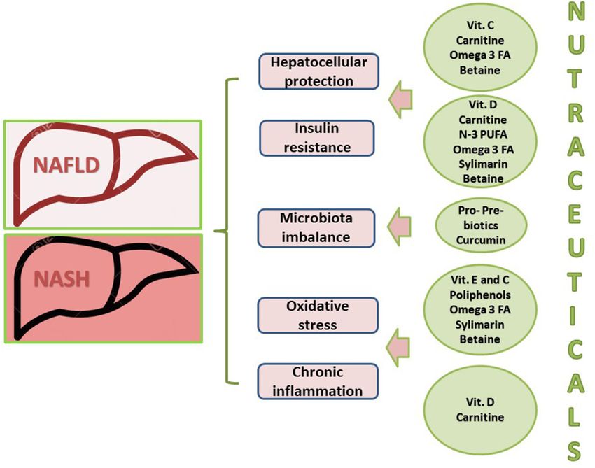
Delli Bovi et al.                                                                                                        Oxidative Stress in NAFLD

is generally benign, ∼20–30% of patients develop liver                     include SOD2 gene, coding for the manganese-dependent
inflammation, fibrosis/cirrhosis (non-alcoholic steatohepatitis,           superoxide dismutase (MnSOD); UCP3, coding for the
NASH), and, in some cases, hepatocellular carcinoma                        uncoupling protein 3, a mitochondrial transporter that enhances
(4, 5). Moreover, patients with NAFLD are at higher risk of                the proton leak of mitochondrial inner membrane and unhooks
cardiovascular diseases. Because of the lack of valid therapies            the oxidative phosphorylation; uncoupling protein 2 (UCP2),
and of the obesity pandemic, NAFLD is one of rapidly growing               regulating oxidative metabolism and mitochondrial lipid efflux;
indications for liver transplantation (6).                                 and MARC1 (A165T), which codes for the mitochondrial
    Most NAFLD patients are obese and present a mild                       amidoxime reducing component 1, a protein involved in the
systemic inflammation, which hampers insulin signaling [insulin            neutralization of reactive oxygen species (ROS) (19, 20).
resistance (IR)], playing a relevant role in the pathomechanism of            The NAFLD story is even more complex than this, as it may
liver damage (7, 8). Recently, in consideration of this association,       start even before conception and pregnancy. Epigenetic changes,
an international group of experts highlighted the poor coherence           comprising microRNA features, may cause fetal reprogramming
of the term non-alcoholic fatty liver disease and proposed                 during the pregnancy of an obese mother and transgenerational
that of metabolic (dysfunction)–associated fatty liver disease (9).        transmission of the susceptibility to NAFLD in childhood and
The reason why some patients with simple steatosis show a                  progression to NASH across the lifetime. Moreover, improving
progression to more severe hepatic injury, whereas others do               obese mothers’ diet reduces fetal hypoxemia and counteracts
not, was in part simplified by the so-called “two-hit” model,              metabolic pathways able to generate OS, liver injury precursors,
founded on IR, and the deposits of relatively inert triglycerides          and lipotoxicity in non-human primates (21–23).
(TGs) within the liver as initial damage. This first event was                On the basis of the most recent literature, herein we will
thought to be due to a “second hit” generated by oxidative                 focus especially on OS because the understanding of a main
stress (OS) or depletion of ATP (10) with the activation of an             role for OS in NAFLD development and progression can have
inflammatory cytokine cascade contributing to the development              important preventive and therapeutic implications for possible
of NASH necroinflammation and fibrosis (10–12).                            novel treatments.
    However, it has been found that hepatic lipid accumulation in
NAFLD occurs mostly as relatively inert TGs droplets, and this
is nowadays regarded as a protective rather than a deleterious
mechanism, by impeding the storage of free fatty acids (FFAs),             OXIDATIVE STRESS AND ITS ROLE IN
which are the actual harmful agents in this hepatopathy. Most              NAFLD PATHOLOGY
recent evidences underline that inflammation may even precede
fat accumulation, which would become only a response (12,                  OS is caused by a discrepancy between ROS generation and
13). As schematically shown in Figure 1, hepatic FFAs originate            antioxidant defenses, which lead to DNA and tissue damage
from lipolysis in adipose tissue and dietary lipids. Moreover,             (24, 25). It may occur both for the increasing production of pro-
particularly in conditions of IR, they may also be synthesized             oxidant products and the dysfunction of the antioxidant system.
de novo (so-called de novo lipogenesis) from carbohydrates in                  Although it is essential to tissue repair, it may conceal
the liver and be deposited as TG droplets (hepatic steatosis),             also negative features implying the development and/or
or exported contributing to the very low-density lipoprotein               exacerbation of several systemic diseases and conditions [e.g.,
pool (14).                                                                 mental/neurological diseases (26, 27), inflammatory bowel
    The previous “two-hit theory” has therefore led the way to             diseases (28), cardiovascular disease (29), and cancer (30)].
the “multiple parallel hits theory” (12), with the contribution of         Starting from these premises, one can therefore easily predict
a number of “multiple parallel (and not sequential)” offenders             that OS represents an important mediator triggering low-grade
acting with different combinations, at times synergistically, to           inflammation also in metabolic syndrome and in the progression
generate NAFLD. These offenders include, in addition to IR (3)             of NAFLD into NASH (31–35).
and OS, hormones secreted from the adipose tissue, intestinal                  ROS, in fact, appear tightly involved in those processes that
dysbiosis, increased intestinal permeability, and also exposure            lead to hepatic fibrosis (36). Multiple interlaced pro-oxidative
to environmental agents such as endocrine disruptors (15) and              triggers operate together with the mitochondrial dysfunction as
particulate matter (PM) (16, 17) interacting among themselves in           a likely common denominator of OS (37). In NASH, there are
individuals predisposed by genetic and epigenetic factors.                 more evidences of mitochondrial DNA and protein abnormalities
    Genes that modulate hepatic fat accumulation and retinol               being responsible for the increase of OS (38, 39). A decreased
metabolism [i.e., transmembrane 6 superfamily member 2                     oxidative capacity of the electron transport chain (ETC) and
(TM6SF2), variants of patatin-like phospholipase domain                    mutations in complex II could also lead to a condition of
which contain protein 3 (PNPLA3), membrane-bound O-                        “electron leakage” (40), meaning that the electron normal flow
acyltransferase domain containing 7 (MBOAT7), hydroxysteroid               could be interrupted, binding with oxygen to produce superoxide
17β-dehydrogenase (HSD17B13), and glucokinase regulator                    or hydrogen peroxide. Moreover, the levels of glutathione (GSH)
(GCKR)] (9) and the deregulation of microRNAs are known to                 peroxidase, MnSOD, and catalase seem to be low in NASH, so
influence NAFLD development and progression (18).                          that the capability of the mitochondria to reduce ROS levels
    In addition, also genetic variants involved in OS regulation           is reduced. In NASH patients, an increased activity of CYP2E1
play an important role in NAFLD pathogenesis. These genes                  (41) has been also observed, an important microsomal source

Frontiers in Medicine | www.frontiersin.org                            2                                  February 2021 | Volume 8 | Article 595371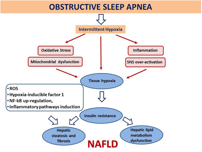
Delli Bovi et al.                                                                                                                                 Oxidative Stress in NAFLD

  FIGURE 1 | Simplified flow of pathogenetic events in non-alcoholic fatty liver disease. The figure shows the crosstalk between systems and metabolisms in the
  pathogenetic events leading to fatty liver and its progression to NASH. In the upper part (yellow lane), one can note that hepatic FFAs derive from lipolysis in adipose
  tissue, dietary lipids, and DNL from COH in the liver. These FFAs may either be stored in the liver as TG droplets (hepatic steatosis) or be exported as VLDL to adipose
  tissue. FFA overload may concur in the hepatic IR (vertical light azur lane), which interplays with the JNK, PKC system where the activation of JNK1 may impair insulin
  signaling via serine phosphorylation of IRS1. The UPR/ER stress is a source of ROS and of lipotoxic species and plays a link between the OX stress and IR. Upon
  disruption of mitochondria-associated membranes (MAM) integrity, miscommunication directly or indirectly disrupts Ca2+ homeostasis and increases ERS (brown box)
  and OS, leading to defective insulin secretion and accelerated lipid droplet formation in hepatocytes. Inflammatory mediators (adipokines, cytokines) in large part
  arrange the progression from NAFLD to NASH (red boxes) in case of shortage of endogenous antioxidant molecules. These mediators are variously triggered by
  oxidative hepatic environment [ROS, lipid peroxidation] and bacterial overgrowth (pink boxes) after the infraction of the gut barrier (gut leakage) by bacterial Eth and
  enhanced intestinal permeability, which allows lipopolysaccharides (a) to activate PRR–LRs—NLRs–DAMPS—PAMPS and (b) to concur with ROS/PUFA in the
  inhibition of the mitochondrial respiratory chain. Lipotoxic lipid species lead to hepatic stress and subsequent release of extracellular vesicles, cytokines, chemokines,
  and DAMPs from hepatocytes. This results in enrolment of bone marrow immune cells. As shown in the lower part of the figure, liver-resident stellate/KCs are
  activated by several triggers (mainly ROS, gut microbiota), resulting in the release of chemokine (C-C motif) ligand 2 (CCL2) and other proinflammatory cytokines (i.e.,
  TNF-α, IL-1, and IL-6). The oxidative hepatic environment also stimulates transcription programs (STAT-1 and STAT-3) promoting T-cell recruitment and hepatic
  disease progression. Overall, the scenario ultimately leads to the recruitment of bone marrow–derived monocytes and neutrophils that further contribute to the
  inflammatory response and a rebound ROS production. A number of genetic variants are implicated in NAFLD development, and progression is shown. BPA,
  bisphenol A; CYP, cytochrome; COH, carbohydrates; DAMPS, damage-associated molecular patterns; DNL, de novo lipogenesis; EDC, endocrine-disrupting
  chemicals; ETC, electron transport chain; Eth, ethanol; FFA, free fatty acids; FIAF, fasting-induced adipose factor; HNE, hydroxynonenal; IKKB, inhibitor of nuclear
  factor κB kinase subunit β; IL, interleukin; IRS, insulin receptor substrate; JNK, c-Jun N-terminal kinase; LPL, lipoprotein lipase; LPS, lipopolysaccharide; LRs, lectin
  receptors; MAM, mitochondria-associated membrane; MDA, malondialdehyde; mRNA, microRNA; MTTP, microsomal triglyceride transfer protein; NAFLD,
  non-alcoholic fatty liver disease; NASH, non-alcoholic steatohepatitis; NLRs, NOD-like receptors; NF-κB, nuclear factor κ-light-chain enhancer of activated B cells;
  NTC non-electron transport chain; PAMPs, pathogen-associated molecular patterns; PKC, protein kinase; PNPLA3, patatin-like phospholipase domain-containing
  protein 3; PRR, pattern recognition receptor; PPAR, peroxisome proliferator-activated receptor; PUFA, polyunsaturated fatty acids; ROS, reactive oxygen species; TG,
  triglyceride; TNF, tumor necrosis factor; UPR, unfolded protein response; VLDL, very low-density lipoprotein; , increase.

of OS, especially together with C47T polymorphisms of SOD2                                insult. In the liver, actually, these conditions trigger lipid
(encoding MnSOD) (41–45).                                                                 peroxidation by specific polyunsaturated fatty acids (PUFAs),
   In the development of NASH, OS probably occurs not only                                along with the formation of highly reactive aldehyde products
due to the saturation of the antioxidant machinery secondary                              [e.g., malondialdehyde (MDA) and 4-hydroxy-2-non-enal (4-
to the increased pro-oxidant species production and its direct                            HNE)]. Overall, these events appear involved in the diffusion of

Frontiers in Medicine | www.frontiersin.org                                           3                                           February 2021 | Volume 8 | Article 595371
Delli Bovi et al.                                                                                                       Oxidative Stress in NAFLD

ROS and reactive nitrogen species (RNS) into the extracellular           • calcium leakage from ER, which increases its flow through
space, perpetuating intracellular and tissue damage. Moreover,             mitochondrial membranes leading to proapoptotic
hepatic OS may result from gut microbiota (GM)–related                     mitochondrial membrane permeabilization (55);
inflammation and the disturbance in the normal functions of              • GSH depletion (56), altering GSH–oxidized glutathione
endoplasmic reticulum [so-called ER stress (ERS)] (see below)              balance, which is essential to redox homeostasis; and
[(37, 46); Figure 1].                                                    • inhibition of nuclear factor, erythroid 2–related factor 2, a
                                                                           factor encoding for antioxidant proteins (57).
                                                                         The cross talk between ERS and ROS (Figure 1) appears relevant
IMPLICATION OF THE OXIDATIVE STRESS                                      in the pathogenesis of NAFLD (58). Mitochondria-associated
IN HEPATIC INJURY                                                        membranes (MAMs) represent a physical junction between ER
                                                                         and mitochondria, allowing Ca2+ , lipids, and ROS exchange.
ROS/RNS (i.e., hydrogen peroxide, superoxide anion radical,              Because normal communication between mitochondria and ER
peroxynitrite, and hydroxyl radical), and not lipid peroxidation         depends on MAM structural and functional integrity, lack of
byproducts, are the responsible for cytokine elevations (47)             calcium homeostasis may lead to ERS and OS increase, defective
such as tumor necrosis factor α (TNF-α), transforming growth             insulin secretion, and accelerated lipid droplet formation in
factor β, interleukin 8 (IL-8), and Fas ligand. The sum of               hepatocytes. The steps involve apoB misfolding, impaired
these events results in NAFLD development (25). The oxidative            lipoprotein secretion, and lipogenesis stimulation. On these
hepatic environment in obesity furthermore promotes the signal           bases, protecting the ER via the administration of antioxidants or
transduction and activation of transcription programs (STAT-1            activation of peroxisome proliferator-activated receptor (PPAR)
and STAT-3) that promote T-cell recruitment and liver damage             has been suggested as promising avenues against hepatic steatosis
with disease progression up to its malignant transformation              (59, 60).
[(48); Figure 1].                                                           Studies in rodents show the existence of a link between
                                                                         ERS and regulation of hepatic iron metabolism both in ASH
                                                                         and NASH models mainly due to the capacity of ferrous
OS and Hepatic Injury: Possible                                          iron to catalyze the production of hydroxyl radical (OH− )
Implications in NAFLD Progression                                        from H2 O2 , deriving by peroxisomal β-oxidation (52, 61, 62).
Hepatocyte damage involves a cascade of events leading                   Interestingly, iron deficiency too may reduce the cell antioxidant
to NAFLD progression into NASH and cirrhosis: damage-                    capability by inhibiting heme oxygenase-1 by Bach1 (63). Beyond
associated molecular patterns, discharged from damaged                   doubt, it is not always simple to study the progression of a
hepatocytes, lead to the release of chemokines and cytokines             disease, especially in humans in vivo, and establish if a certain
from Kupffer cells (KCs) and the recruitment of monocyte-                factor is exactly the cause or the effect of NASH. Moreover, a
derived macrophages. ROS directly and indirectly contribute to           disagreement often happens between animal models and clinical
stellate cell activation and to chronic inflammatory response with       studies due to several factors such as gut microflora differences
up-regulation of proinflammatory cytokines (TNF-α, IL-6, and             and patient inclusion criteria/ethnicity–related predisposition,
IL-1), apoptosis, and development of hepatic fibrosis [(49–51);          respectively (37).
Figure 1].                                                                  In order to assess the redox state in NAFLD/NASH, some
   In conditions of progressive NAFLD, OS can also result from           markers of OS and antioxidants have been studied in NAFLD and
increased ROS generation due to impairment of mitochondria               NASH models, both clinical and experimental. OS biomarkers
caused by an overload of FFAs and an increase of their                   include nitric oxide, lipid damage products (lipid peroxides,
metabolism, lipotoxicity, and hypoxia, as well as ROS production         thiobarbituric acid reactive substances/MDA), hydroperoxides,
through NADPH-oxidase isoforms associated to ligand–receptor             8-isoprostane, 4-HNE, DNA oxidation product [CYP2E1 and
link or by activated inflammatory cells (49).                            8-hydroxydeoxyguanosine (8-OH-dG)], and protein oxidation
   Evidences suggest that lipotoxicity mediated by FFAs (52)             products (nitrotyrosine, protein carbonyl). All these had
may induce disruption of ER homeostasis, known as “unfolded              increased activities in most NAFLD/NASH clinical models
protein response,” an intracellular signaling activated by the           evaluated. On the contrary, antioxidant markers (superoxide
accumulation of unfolded/misfolded proteins. Thanks to it, ER            dismutase, catalase, glutathione peroxidase, reduced glutathione)
can communicate the folding status of its proteins to the rest           measured in rodent models showed decreased activities mainly in
of the cell, particularly to the nucleus, and so activate genes          NASH (64).
transcription. As a result, the ERS, a term that includes also
several other mechanisms conducing to ROS generation, occurs
                                                                         GUT MICROBIOTA AND INTESTINE
(37), and this leads to
                                                                         PERMEABILITY AS A CAUSE OF
• increased endoplasmic reticulum oxidoreduction-1 (ERO-1)               OXIDATIVE STRESS
  activity, the enzyme that catalyzes disulfide bond formation
  (53) with H2 O2 production;                                            Gut Microbiota as a Source of ROS
• upregulation      of   CCAAT/enhancer-binding         protein          Human commensal microbiota (Figure 1) generates
  homologous protein (Chop), a proapoptotic mechanism (54);              physiological ROS levels in intestinal human epithelial

Frontiers in Medicine | www.frontiersin.org                          4                                   February 2021 | Volume 8 | Article 595371
Delli Bovi et al. Oxidative Stress in NAFLD
cells. Basically, aerobic cell systems are exposed to oxygen molecular patterns) are recognized and bound by PRRs, inducing
free radicals (65, 66), and their damaging role relies on their mitochondrial ROS production and nuclear gene expression.
concentrations. When the levels of ROS exceed antioxidant PRR classes sensitive to the microbiota’s factors are Toll-like
defenses, harmful effects on cells may occur, conducing to receptor (TLR), Rig-1-like receptor, Nod-like receptor, and C-
uncontrolled proliferation, inflammation, and/or apoptosis type lectin receptor. They induce the NF-κB pathway activation
(67, 68). This is what happens also in obesity and its related and enhance the inflammatory response when proinflammatory
hepatometabolic comorbidities, including NAFLD progression to cytokines and antibacterial factors are released (81, 82).
NASH (see below). ROS can also operate as second messengers in Differently, small formylated peptides produced and released by
intracellular signaling stimulated by proinflammatory cytokines commensal bacteria are recognized by another kind of receptor,
and growth factors and by the quick and reversible oxidative known as formylated peptide receptors. These are G-proteins
inactivation of proteins having thiol groups sensitive to oxidants linked to surface receptors of neutrophils and macrophages,
(69). In inflammation and obesity, ROS generation is probably stimulating ROS synthesis in phagocytes and epithelial cells
strictly related with activation of nuclear factor κ-light-chain (83). In particular, their activation stimulates superoxide anion
enhancer of activated B cells (NF-κB) and degradation of NF-κB production by NADPH oxidase 1, increasing ROS levels in cell
inhibitor (IκB), making NF-κB more transcriptionally active cytoplasm that lead to an inflammatory response and increase of
(70–72). As shown in Figure 1, a quantitative or qualitative (in cell OS (84). As a consequence of cell stress, mitochondrial and
term of dysbiosis) bacterial alteration (small intestine bacterial bacterial DNA may be integrated in the nuclear genome causing
overgrowth) is also concatenated with OS through the inhibition the alteration of cellular gene expression.
of mitochondrial respiratory chain. Intestinal mucosa permeability has an important role in
modulating how GM can influence also other parts of the body.
An alteration of its barrier function consents to the GM and its
endotoxins to cross the intestinal epithelium and the endothelial
Interaction Between Gut Microbiota, OS, barrier (85) traveling into systemic circulation and reaching
and Intestinal Permeability in NAFLD different target organs (75, 86).
Gut mucosal barrier separates, functionally and physically, There are many evidences according to which gut bacteria
the luminal content from the underlying compartment that, are involved in the pathogenesis of liver injury induced by
in addition to gut epithelia, includes immune, vascular, and alcohol, and gut leakiness promotes proinflammatory bacterial
structural elements in the lamina propria. The intestinal mucosa products reaching the liver, thus initiating the proinflammatory
is constantly exposed to oxidants and carcinogens taken in cascade that causes alcoholic steatohepatitis (ASH). Alcohol
from diet and/or bacteria, whose chronic exposure may cause impairs intestinal epithelial cell permeability in vitro through a
production of free radicals leading to redox imbalance and mechanism mediated by OS (87), supporting therefore the idea
subsequent DNA damage, disturbing the intestinal metabolic that OS may be the main cause of alcohol-induced intestinal
equilibrium (73). leakage (88, 89).
GM plays an important role in different processes (metabolic, In NAFLD, with a quite similar mechanism, endogenous
nutritional, physiological, and immunological) involved ethanol produced by some microbial species [e.g., Escherichia
in maintaining a healthy status (69, 74). Its qualitative genus members of the Proteobacteria phylum induced by high-
and quantitative composition differs in the distinct parts fat diet; (90)] is able to induce the formation of ROS by HSC
of gastrointestinal (GI) tract because of the influences by cells and impair intestinal integrity. The latter allows LPS to
different conditions [e.g., age, dietary habits, ethnicity, delivery reach hepatic TLRs activating and further enhancing oxidative,
mode, exposure to therapies, pathogens, and contact with inflammatory, and fibrogenetic mechanisms (75, 90, 91, 91–94).
several environmental stimuli (75–78)]. Perturbation of GM GM seems to mediate the progression from simple steatosis
composition, called “dysbiosis,” has been recognized in diseases to NASH. In particular, increased Gram-negative bacteria expose
associated not only with the GI tract [e.g., inflammatory KCs to an elevated amount of LPS and upregulation of PRRs
bowel disease (79)] but also with systemic conditions such (37). It has been hypothesized that the endocytosis of LPS by KC
as obesity, diabetes mellitus, autism, depression, and NAFLD could induce upregulation of cytokine receptors, especially the
(80). While a quite clear causal role of a specific GM has been TNF-α receptor, which seems to be also involved in the increased
demonstrated in murine models of NAFLD (e.g., unhealthy diet ROS production (95). Activated KCs have a role in IR, fibrosis
dependent shift from Bacteroidetes to Firmicutes), recognition development, and inflammation amplification.
of a corresponding human microbiome signature is more Also, the association between obstructive sleep apnea
difficult. In fact, it may be hindered by the components of syndrome (OSAS) and NASH severity seems to correlate
associated metabolic syndrome and several other confounding with endotoxemia increase and gut barrier function alteration,
factors. Anyway, Gram-negative harmful bacteria release conducing to increased hepatic susceptibility to endotoxemia
lipopolysaccharide (LPS), lipoteichoic acid, flagellin, lipoprotein, mediated by TLR-4 (96). An alarming 60% OSAS incidence
or other toxins recognized by the pattern recognition receptors has been reported in pediatric NAFLD (97, 98). This disorder
(PRRs) expressed on the surface of innate immune system of breathing during sleep has been associated with fatty acid
cells. Similarly, structurally conserved motifs present on the accumulation in the liver and inflammation caused by frequent
surface of different types of pathogens (pathogen-associated nocturnal hypoxia (NH), IR, OS, and adipokine dysregulation
Frontiers in Medicine | www.frontiersin.org 5 February 2021 | Volume 8 | Article 595371Delli Bovi et al. Oxidative Stress in NAFLD
(99). Growing experimental evidences link the alternation of NH with Non-alcoholic Steatohepatitis) (127) and the TONIC
with normoxia (so-called chronic intermittent hypoxia) caused (Treatment of Non-alcoholic Fatty Liver Disease in Children)
by OSAS to NAFLD development and progression (49, 100). A (121) trials. The first showed that both drugs tested in adults
study that compared healthy controls and NAFLD patients (some ameliorated steatosis, lobular inflammation, and hepatocellular
of which with OSA/NH), identified NH as a possible source of OS ballooning, but did not ameliorate fibrosis. Vitamin E but not
in NAFLD. OSA/NH is common in pediatric patients with liver pioglitazone induced a clinical improvement in NASH. The
biopsy-proven NAFLD and is associated with more advanced TONIC trial, which evaluated therapeutic intervention with
liver injury and histological disease (97, 98). Intermittent hypoxia vitamin E vs. metformin in children with NAFLD, showed
conduces to tissue hypoxia and can lead to OS, mitochondrial that both improved hepatocellular ballooning and the NAFLD
malfunction, inflammation, and sympathetic nervous system activity score (NAS), but neither vitamin E nor metformin
hyperactivation. As a consequence, intermittent hypoxia causes decreased alanine aminotransferase (ALT) values or hepatic
IR, impairment of hepatic lipid metabolism pathways (84), and steatosis, inflammation, or fibrosis in NASH. The reasons of
hepatic steatosis and fibrosis, each of which is involved into these disappointing results depend on the need of better patient
NAFLD development and/or progression [(101); Figure 2]. selection and protocols. Interestingly, a most recent systematic
review and meta-analysis (1,317 patients from 15 randomized
THERAPEUTIC STRATEGIES controlled trials) concluded that vitamin E improves biochemical
and histological outcomes in adults and pediatric patients, with
Non-enzymatic Anti-oxidants Defenses a significant negative association between transaminases levels
Antioxidants are substances that inhibit the oxidation of and vitamin E dosage—more satisfactorily ranging between 400
any biomolecule (102), neutralizing the harmful effects of and 800 IU. However, while adults receiving vitamin E improved
oxidation caused by free radicals, maintaining therefore the redox significantly transaminases, fibrosis, and NAS both at early and
homeostasis. Antioxidants are either synthesized endogenously late follow-up, children showed more significant changes at long-
(e.g., GSH, superoxide dismutase) or taken from the diet. term follow-up, which could partly explain the negative results
Anthocyanins, lycopene, coenzyme Q10, flavonoids, β-carotene, obtained by certain short-term studies (128). Some still unsolved
lipoic acid, selenium, lutein, catechins, and vitamins A, C, and safety concerns should be considered as well. Vitamin E, in
E are among the many substances normally present in foods fact, has been suspected to have a dichotomous suppressive
that possess a high antioxidant activity. As reported in the and promoting activity with respect to tumorigenesis [e.g., co-
table, they can be also classified in two large groups on the cancerogenic in prostate cancer; (25, 129)] possibly explainable
basis of the presence/absence of their enzymatic action [(103– by still poorly studied host gene–supplement interactions (130).
114); Table 1]. In our opinion, further carefully designed studies are still
Despite the above premises, antioxidants as potential necessary for substantiating this view and supporting optimum
pharmacological agents have hitherto not appeared extremely procedures in terms of both efficacy and safety profiles.
effective in vivo as either a preventive or therapeutic tool in Results from multiple regression models showed a significant
NAFLD (31, 117–122). Studies that have investigated the role negative association between ALT, AST levels, and vitamin E
of vitamin E as a treatment of NASH confirm that it acts dosage—more favorably between 400 and 800 IU.
against pathogenic mechanisms conducting to liver damage and A quite large number of other nutraceutical antioxidants that
NASH, thanks to its antioxidant and anti-inflammatory activity seem to improve NASH through more than one pathway (Table 2
(123–126). The antioxidant power of vitamin E is due to the and Figure 3) include but are not limited to the following:
hydroxyl group in the tocochromanol ring, which neutralizes
free radicals and ROS by donating hydrogen. The major forms - Curcumin (37, 103), with effects on different amino acids, bile
of tocopherol and tocotrienol are α, β-, γ-, and δ-, with the acids, tricarboxylic acid cycle, and GM (131, 132), although
antioxidant activity of the δ-isoform being weaker than the only few human clinical trials are available (132–134).
others, the vitamin E isoforms are also involved in many other - PUFAs of omega-3 series (PUFA omega-3), which may act as
activities (Table 2). Among the vitamin E isoforms, the α- an antioxidant, have a role in modulating OS improving the
tocopherol, has other different properties independently from defense capacity against an increased oxidative burden (135,
its antioxidant ability: it can inhibit the activity of protein 136).
kinase C, reducing the proliferation of different cell types Thanks to them, the cellular metabolism switch from lipogenesis
(vascular smooth muscle cells, mesangial cells, neutrophils, and triacylglycerol accumulation to fatty acid oxidation thus
monocytes/macrophages, fibroblast, and various cancer cell plays a role in decreasing fatty liver. Furthermore, they have
lines) and the 5-lipoxygenase pathway, inhibiting the release of anti-inflammatory activity and enhance insulin sensitivity (103).
proinflammatory cytokine IL-1β. Clinical trials evaluating the efficacy of n-3 PUFA (including
docosahexaenoic acid and eicosapentaenoic acid) on systemic
Clinical Trials of Vitamin E for NAFLD: OS in NAFLD and NASH have shown controversial results.
Vitamin E in the Clinics While n-3 PUFA supplementation appears useful in NAFLD
Available data are still conflicting. The largest trials with early stages (137), unfortunately, total (enzymatic and non-
vitamin E in NAFLD are the PIVENS (Pioglitazone vs. Vitamin enzymatic) antioxidant capacity is not enough to attenuate
E vs. Placebo for the Treatment of Non-diabetic Patients the hepatic damage (35). Interestingly, dietary antioxidant
Frontiers in Medicine | www.frontiersin.org 6 February 2021 | Volume 8 | Article 595371Delli Bovi et al. Oxidative Stress in NAFLD
FIGURE 2 | Association between obstructive sleep apnea and the development and evolution of non-alcoholic fatty liver disease. Intermittent hypoxia leads to tissue
hypoxia, OS, mitochondrial dysfunction, inflammation, and overactivation of the sympathetic nervous system (SNS). Generated reactive O2 species (ROS) may amplify
liver injury by activating hypoxia-inducible factor 1, a transcriptional activator and master regulator of O2 homeostasis during hypoxia, and by up-regulating nuclear
factor κ-light-chain enhancer of activated B cells (NF-κB), with subsequent downstream induction of inflammatory pathways. As a consequence, this involves insulin
resistance, dysfunction of key steps in hepatic lipid metabolism, atherosclerosis, and hepatic steatosis and fibrosis, each of which is pertinent to the development
and/or progression of non-alcoholic fatty liver disease (NAFLD) (98–101).
intake is significantly lesser in NASH patients than in healthy through dysbiotic microbiota normalization (100, 144, 145) and
controls (138). reducing intestinal OS (146). A recent meta-analysis found
a beneficial effect of probiotics also on hepatic antioxidative
- Among polyphenols, blueberry leaf polyphenols appear to
capacity as mirrored by the increase of SOD and GSH-PX
have a positive effect on hepatic mitochondrial dysfunction
activities and decrease of MDA content (147). A daily LGG
and redox homeostasis, whereas bergamot polyphenolic
treatment in alcohol-fed rats significantly improves severity of
formulation seems to improve IR, hepatocellular ballooning,
ASH and gut leakiness induced by alcohol, decreases intestinal
inflammation, and fibrosis (115, 116).
and liver OS markers and inflammation, and normalizes the
gut barrier task, avoiding to trigger liver disease (148). GM
Probiotics and Prebiotics: Other Tools regulates also the powerful antioxidant glutathione and amino
Improving Defenses Against OS acid metabolism (144). It is not surprising therefore that
Improving defenses against OS through modulation of the GM fecal microbiota transplantation (FMT) from control donors
composition and functionality offers a promising means of in steatotic rats has been found to have beneficial effects
managing or treating metabolic disorders (74). in terms of decrease of portal hypertension through insulin
Probiotics are living microorganisms with beneficial health sensitivity improvement mediated by the endothelial nitric oxide
activity on the host. For example, they are able to improve GM synthase signaling pathway, a pathway clearly involved in the
composition and reduce LPS serum amount and liver TLR4, antioxidant mechanisms (149). A pilot study of FMT in NASH is
delaying liver disease progression (81, 98, 99). Lactobacilli and currently undergoing to evaluate whether restoration of healthful
bifidobacteria are the most commonly used, usually present in GM through FMT from lean donors (FMT-L) ameliorates
dietary supplements or fermented foods such as yogurt and NASH (150).
cultured milk (100). Changing the resident GM composition and Similarly, “prebiotics” are fermentable carbohydrates that
the gut lumen, they create an anti-inflammatory environment, selectively modulate microbiota composition and/or activity,
obtaining decreased proinflammatory bacterial products and resulting in a beneficial effect for the host (146). Finally, also
gut barrier integrity improvement. Lactobacillus rhamnosus synbiotics (i.e., a combination of prebiotics and probiotics) have
GG (LGG) is the subject of numerous studies (139–144); shown a positive effect on GM and have been proposed as a
it has different beneficial effects on the intestinal function support for the treatment of NAFLD (151).
Frontiers in Medicine | www.frontiersin.org 7 February 2021 | Volume 8 | Article 595371Delli Bovi et al. Oxidative Stress in NAFLD
Summing up, the modulation of quality and diversity of every some metabolic anomalies caused by dysbiosis (152). Moreover,
single human microbiota appears therefore an appealing tool it is suggested that changes in GM occurring upon prebiotic
in the management of intestinal ROS, OS, inflammation, and consumption may be due to gut bacterial functions improvement.
In other words, products generated by Lactobacillus and
metabolites derived by microbiota, such as antioxidants and fatty
acids, could be employed for target medicine in the management
TABLE 1 | Antioxidants with and without enzymatic action.
of liver disease including NAFLD (146).
Enzymatic Non-enzymatic antioxidants
antioxidants
OTHER THERAPEUTIC STRATEGIES
Superoxide dismutase Low-molecular-weight compounds
(SOD) Glutathione, thioredoxin, lactoferrin Drugs
Endogenous substances Ursodeoxycholic acid (UDCA) remains one of the most studied
Lipoic acid, melatonin, albumin, bilirubin, uric acid, drugs: in addition to exerting a possible therapeutic effect on
polyunsaturated fatty acids omega 3 NAFLD by modulating autophagy and apoptosis dysregulation,
Catalase (CAT) Flavonoid polyphenols UDCA appears to have also antioxidant properties (153).
Silymarin (104, 105)
Blueberry leaf, bergamot polyphenols (115, 116)
Stilbenes
Resveratrol (106, 107)
TABLE 2 | Activities influenced by vitamin E isoforms.
Glutathione peroxidase Herbs
Erchen decoction, danshen, berberine (108) I. Regulation of the inflammatory response
Carotenoids II. Gene expression
β-Carotene, astaxanthin, lycopene, β-
III. Membrane-bound enzymes
cryptoxanthin, lutein, fucoxanthin, crocetin
IV. Cellular signaling
(109, 110)
V. Cell proliferation
Paraoxonase 1 (PON 1) Phenolic compounds
Açai (111) VI. Regulation of several enzymes involved in signal transduction:
Protein kinase C (PKC), protein phosphate 2A (PP2A), 5-lipoxygenase,
Vitamins cyclooxygenase 2 (COX-2), and monocyte chemoattractant protein 1 (MCP-1)
Ascorbic acid (vitamin C), α-tocopherol [vitamin E, VII. Regulation of several factors in the mitogen-activated protein kinase (MAPK)
vitamin A, vitamin D (112–114)] signal transduction pathway
FIGURE 3 | Multiple targets of nutraceuticals for the treatment of non-alcoholic fatty liver disease. FA, fatty acids; NAFLD, non-alcoholic fatty liver disease; NASH,
non-alcoholic steatohepatitis. Adapted and modified by Del Ben et al. (103).
Frontiers in Medicine | www.frontiersin.org 8 February 2021 | Volume 8 | Article 595371Delli Bovi et al. Oxidative Stress in NAFLD
A number of other drugs that have been tested for conditions, GM may take advantage of the increased intestinal
their influence on hepatic steatosis have still uncertain/elusive permeability and/or impairment of epithelial tight junctions.
molecular mechanisms. There are several innovative agents This results in an enhancement of the gut–liver axis with bacteria
currently undergoing phases II and III clinical trials with different and endotoxin transit through the intestinal and endothelial
targets (154). vascular wall, ending up into hepatic and other systemic diseases
Obeticholic acid, a semisynthetic bile acid analog, is an agonist as well.
of the farnesoid X receptor, which has anti-inflammatory and The above scenario would suggest a therapeutic role of
antioxidant activities (155). antioxidants in patients with fatty liver disease, but this
Silymarin, a botanical product extracted from milk thistle, approach has not been entirely translated yet in human
because of its antioxidant properties appears to improve NAFLD (160), as most studies still derive from murine models with
hypertransaminasemia and reduce liver disease progression in substantial differences in genetic background and in the digestive
NASH, but at present, available results are inconclusive (156). system; the need to perform more human studies appear
Cannabidiol, a chemical without psychotropic effects, has evident (161).
antioxidant and anti-inflammatory properties by acting on the Vitamin E has shown promising data but without significant
endocannabinoid system. After stimulation of the G-protein– benefit in fibrosis improvement (162). Several natural
coupled receptors and their endogenous lipid ligands, it interferes polyphenols and n-3 PUFA supplementation provided with
with progression toward NASH (26, 157). a number of antioxidant, antiobesity, and anti-inflammatory
effects could have potential in NAFLD prevention and treatment
Physical Activity by acting on its multifactorial pathogenetic components, but also
Physical activity (PA) acts favorably in NAFLD primarily by here data either to support or refuse their use are insufficient
reducing intrahepatic fat content with β-oxidation of fatty acids (115, 116).
and lipogenesis regulation, enhancing the expression and activity In addition to healthy diet (e.g., a Mediterranean diet seems
of PPAR-γ, insulin sensitivity, and hepatoprotective autophagy, to reduce OS), probiotics, prebiotics, and fecal transplantation
reducing hepatocyte apoptosis, and inflammation of the liver by appear to be emerging strategies to modulate microbiota quality
decreasing the proinflammatory mediators. PA, moreover, has and diversity, in order to prevent and/or avoid gut damage.
several beneficial effects on NAFLD also with the improvement Avoidance of exposure to endocrine disruptors (15) and to
of several antioxidants activity [e.g., catalase, SOD, glutathione ambient PM (16, 17) also appears strategic to add benefices
peroxidase and reductase, glutathione-S-transferase, thioredoxin to NAFLD.
reductases, NADH cytochrome B5 reductase, and NAD(P)H Last but not least, the accurate assessment of NAFLD-
quinone acceptor oxidoreductase], leading to decreased ROS associated genetic/epigenetic risk factors of diseases and
production and proinflammatory cytokines (158). likelihood of disease progression is going to aid to target
individualized appropriate treatments (163).
CONCLUDING REMARKS
AUTHOR CONTRIBUTIONS
Our review exhibits that OS not counteracted by intact
antioxidant defense system plays an important role in FM and APDB collected literature and prepared the first draft of
NAFLD/NASH with a number of other casual factors. Excessive the manuscript. MS and MAS collected literature on specific areas
FFA β-oxidation due to increased FFA fueling leads to excessive and gave critical suggestions. PV gave critical suggestions, made
ROS formation, which, in turn, downregulates ETC, and non- substantial intellectual contributions to the study design, and
ETC systems, affect insulin sensitivity, hepatic lipid metabolism, manuscript preparation. CM provided a major intellectual input,
and inflammatory responses by interacting with innate immune verified/contributed to data analysis, and took over writing of the
signaling (159). manuscript as and when required. All authors gave substantial
Gut dysbiosis may induce further signaling processes, which contributions to the work and revised critically and approved the
engage the epithelium and immune/inflammatory cells. In these final manuscript.
REFERENCES 4. Tiniakos DG, Vos MB, Brunt EM. Nonalcoholic fatty liver disease:
pathology and pathogenesis. Annu Rev Pathol. (2010) 5:145–71.
1. Ludwig J, Viggiano TR, McGill DB, Oh BJ. Nonalcoholic steatohepatitis: doi: 10.1146/annurev-pathol-121808-102132
Mayo Clinic experiences with a hitherto unnamed disease. Mayo Clin Proc. 5. Baffy G, Brunt EM, Caldwell SH. Hepatocellular carcinoma in non-alcoholic
(1980) 55:434–8. fatty liver disease: an emerging menace. J Hepatol. (2012) 56:1384–91.
2. Abenavoli L, Greco M, Milic N, Accattato F, Foti D, Gulletta E doi: 10.1016/j.jhep.2011.10.027
et al. Effect of Mediterranean diet and antioxidant formulation in non- 6. Mikolasevic I, Filipec-Kanizaj T, Mijic M, Jakopcic I, Milic S, Hrstic
alcoholic fatty liver disease: a randomized study. Nutrients. (2017) 9:870. I, et al. Nonalcoholic fatty liver disease and liver transplantation
doi: 10.3390/nu9080870 - where do we stand? World J Gastroenterol. (2018) 24:1491–506.
3. Buzzati E, Pinzani M, Tsochatzis EA. The multiple hit-pathogenesis of doi: 10.3748/wjg.v24.i14.1491
non-alcoholic fatty liver disease (NAFLD). Metabolism. (2016) 65:1038–48. 7. Sanyal AJ, Campbell-Sargent C, Mirshahi F, Rizzo WB, Contos MJ, Sterling
doi: 10.1016/j.metabol.2015.12.012 RK, et al. Nonalcoholic steatohepatitis: association of insulin resistance
Frontiers in Medicine | www.frontiersin.org 9 February 2021 | Volume 8 | Article 595371Delli Bovi et al. Oxidative Stress in NAFLD
and mitochondrial abnormalities. Gastroenterology. (2001) 120:1183–92. 29. Sinha N, Dabla PK. Oxidative stress and antioxidants in
doi: 10.1053/gast.2001.23256 hypertension-a current review. Curr Hypertens Rev. (2015) 11:132–42.
8. Fabbrini E, Sullivan S, Klein S. Obesity and nonalcoholic fatty liver doi: 10.2174/1573402111666150529130922
disease: biochemical, metabolic, and clinical implications. Hepatology. (2010) 30. Bellot GL, Liu D, Pervaiz S. ROS, autophagy, mitochondria and
51:679–89. doi: 10.1002/hep.23280 cancer: Ras, the hidden master? Mitochondrion. (2013) 13:155–62.
9. Eslam M, Sanyal AJ, George J, International Consensus Panel. doi: 10.1016/j.mito.2012.06.007
MAFLD: a consensus-driven proposed nomenclature for metabolic 31. Mandato C, Lucariello S Franzese A, Licenziati MR, Franzese A, Spagnuolo
associated fatty liver disease. Gastroenterology. (2020) 158:1999–2014. MI, Ficarella R, et al. Metabolic, hormonal, oxidative, and inflammatory
doi: 10.1053/j.gastro.2019.11.312 factors in pediatric obesity-related liver disease. J Pediatr. (2005) 147:62–6.
10. Day CP, James OF. Steatohepatitis: a tale of two “hits”? Gastroenterology. doi: 10.1016/j.jpeds.2005.02.028
(1998) 114:842–5. doi: 10.1016/s0016-5085(98)70599-2 32. Paravicini TM, Touyz RM. Redox signaling in hypertension. Cardiovasc Res.
11. Peverill W, Powell LW, Skoien R. Evolving concepts in the pathogenesis of (2006) 71:247–58. doi: 10.1016/j.cardiores.2006.05.001
NASH: beyond steatosis and inflammation. Int J Mol Sci. (2014) 15:8591– 33. Trachootham D, Alexandre J, Huang P. Targeting cancer cells by ROS-
638. doi: 10.3390/ijms15058591 mediated mechanisms: a radical therapeutic approach? Nat Rev Drug Discov.
12. Tilg H, Moschen AR. Evolution of inflammation in nonalcoholic fatty liver (2009) 8:579–91. doi: 10.1038/nrd2803
disease: the multiple parallel hits hypothesis. Hepatology. (2010) 52:1836–46. 34. Hardwick RN, Fisher CD, Canet MJ, Lake AD, Cherrington NJ.
doi: 10.1002/hep.24001 Diversity in antioxidant response enzymes in progressive stages of human
13. Yamaguchi K, Yang L, McCall S, Huang J, Yu XX, Pandey SK, et al. Inhibiting nonalcoholic fatty liver disease. Drug Metab Dispos. (2010) 38:2293–301.
triglyceride synthesis improves hepatic steatosis but exacerbates liver damage doi: 10.1124/dmd.110.035006
and fibrosis in obese mice with nonalcoholic steatohepatitis. Hepatology. 35. Koek GH, Liedorp PR, Bast A. The role of oxidative stress in
(2007) 45:1366–74. doi: 10.1002/hep.21655 non-alcoholic steatohepatitis. Clin Chim Acta. (2011) 412:1297–305.
14. Saponaro C, Gaggini M, Carli F, Gastaldelli A. The subtle balance between doi: 10.1016/j.cca.2011.04.013
lipolysis and lipogenesis: a critical point in metabolic homeostasis. Nutrients. 36. Rector RS, Thyfault JP, Uptergrove GM, Morris EM, Naples SP, Borengasser
(2015) 7:9453–74. doi: 10.3390/nu7115475 SJ, et al. Mitochondrial dysfunction precedes insulin resistance and hepatic
15. Foulds CE, Treviño LS, York B, Walker CL. Endocrine-disrupting steatosis and contributes to the natural history of non-alcoholic fatty
chemicals and fatty liver disease. Nat Rev Endocrinol. (2017) 13:445–57. liver disease in an obese rodent model. J Hepatol. (2010) 52:727–36.
doi: 10.1038/nrendo.2017.42 doi: 10.1016/j.jhep.2009.11.030333333
16. Xu MX, Ge CX, Qin YT, Gu TT, Lou DS, Li Q, et al. Prolonged PM2.5 37. Tariq Z, Green JC, Hodson L. Are oxidative stress mechanisms the common
exposure elevates risk of oxidative stress-driven nonalcoholic fatty liver denominator in the progression from hepatic steatosis towards non-
disease by triggering increase of dyslipidemia. Free Radic Biol Med. (2019) alcoholic steatohepatitis (NASH). Liver International. (2014) 34:e180-90.
130:542–56. doi: 10.1016/j.freeradbiomed.2018.11.016 doi: 10.1111/liv.12523
17. Ding S, Yuan C, Si B, Wang M, Da S, Bai L, et al. Combined 38. Pérez-Carreras M, Del Hoyo P, Martín MA, Rubio JC, Martín A,
effects of ambient particulate matter exposure and a high-fat diet on Castellano G, et al. Defective hepatic mitochondrial respiratory chain in
oxidative stress and steatohepatitis in mice. PLoS ONE. (2019) 14:e0214680. patients with nonalcoholic steatohepatitis. Hepatology. (2003) 38:999–1007.
doi: 10.1371/journal.pone.0214680 doi: 10.1053/jhep.2003.50398
18. Gjorgjieva M, Sobolewski C, Dolicka D, Correia de Sousa M, Foti M. miRNAs 39. Berson A, De Beco V, Lettéron P, Robin MA, Moreau C, El Kahwaji J,
and NAFLD: from pathophysiology to therapy. Gut. (2019) 68:2065–2079. et al. Steatohepatitis-inducing drugs cause mitochondrial dysfunction and
doi: 10.1136/gutjnl-2018-318146 lipid peroxidation in rat hepatocytes. Gastroenterology. (1998) 114:764–74.
19. Taliento AE, Dallio M, Federico A, Prati D, Valenti L. Novel insights into the doi: 10.1016/s0016-5085(98)70590-6
genetic landscape of nonalcoholic fatty liver disease. Int J Environ Res Public 40. Kawahara H, Fukura M, Tsuchishima M, Takase S. Mutation of
Health. (2019) 16:2755. doi: 10.3390/ijerph16152755 mitochondrial DNA in livers from patients with alcoholic hepatitis and
20. Al-Serri A, Anstee QM, Valenti L, Nobili V, Leathart JB, Dongiovanni P, nonalcoholic steatohepatitis. Alcohol Clin Exp Res. (2007) 31:S54–60.
et al. The SOD2 C47T polymorphism influences NAFLD fibrosis severity: doi: 10.1111/j.1530-0277.2006.00287.x
evidence from case-control and intra-familial allele association studies. J 41. Chalasani N, Gorski JC, Asghar MS, Asghar A, Foresman B, Hall SD,
Hepatol. (2012) 56:448–54. doi: 10.1016/j.jhep.2011.05.029 et al. Hepatic cytochrome P450 2E1 activity in nondiabetic patients
21. Wesolowski SR, Mulligan CM, Janssen RC, Baker PR II, Bergman BC, with nonalcoholic steatohepatitis. Hepatology. (2003) 37:544–50.
D’Alessandro A, et al. Switching obese mothers to a healthy diet improves doi: 10.1053/jhep.2003.50095
fetal hypoxemia, hepatic metabolites, and lipotoxicity in non-human 42. Varela NM, Quiñones LA, Orellana M, Poniachik J, Csendes A, Smok
primates. Mol Metab. (2018) 18:25–41. doi: 10.1016/j.molmet.2018.09.008 G, et al. Study of cytochrome P450 2E1 and its allele variants in liver
22. Baker PR 2nd, Friedman JE. Mitochondrial role in the neonatal injury of nondiabetic, nonalcoholic steatohepatitis obese women. Biol Res.
predisposition to developing nonalcoholic fatty liver disease. J Clin Invest. (2008) 41:81–92.
(2018) 128:3692–703. doi: 10.1172/JCI120846 43. El-Koofy NM, El-Karaksy HM, Mandour IM, Anwar GM, El-Raziky MS,
23. Bertrando S, Vajro P. NAFLD at the interface of the mother-infant dyad. Curr El-Hennawy AM. Genetic polymorphisms in non-alcoholic fatty liver
Pharm Des. (2020) 26:1119–25. doi: 10.2174/1381612826666200122153055 disease in obese Egyptian children. Saudi J Gastroenterol. (2011) 17:265–70.
24. Sies H. Oxidative stress: a concept in redox biology and medicine. Redox Biol. doi: 10.4103/1319-3767.82582
(2015) 4:180–3. doi: 10.1016/j.redox.2015.01.002 44. Namikawa C, Shu-Ping Z, Vyselaar JR, Nozaki Y, Nemoto Y, Ono
25. Nagashimada M Ota T. Role of vitamin E in nonalcoholic fatty liver disease. M, et al. Polymorphisms of microsomal triglyceride transfer protein
IUBMB Life. (2019) 71:516–22. doi: 10.1002/iub.1991 gene and manganese superoxide dismutase gene in non-alcoholic
26. Atalay S, Jarocka-Karpowicz I, Skrzydlewska E. Antioxidative and steatohepatitis. J Hepatol. (2004) 40:781–6. doi: 10.1016/j.jhep.2004.
anti-inflammatory properties of cannabidiol. Antioxidants. (2019) 9:2. 01.028
doi: 10.3390/antiox9010021 45. Nobili V, Donati B, Panera N, Vongsakulyanon A, Alisi A, Dallapiccola
27. Yusuf M, Khan M, Robaian MA, Khan RA. Biomechanistic insights into the B, et al. A 4-polymorphism risk score predicts steatohepatitis in children
roles of oxidative stress in generating complex neurological disorders. Biol with nonalcoholic fatty liver disease. J Pediatr Gastroenterol Nutr. (2014)
Chem. (2018) 399:305–19. doi: 10.1515/hsz-2017-0250 58:632–6. doi: 10.1097/MPG.0000000000000279
28. Kruidenier L, Verspaget HW. Oxidative stress as a pathogenic factor in 46. Rolo AP, Teodoro JS, Palmeira CM. Role of oxidative stress in the
inflammatory bowel disease-radicals or ridiculous? Aliment Pharmacol Ther. pathogenesis of nonalcoholic steatohepatitis. Free Radic Biol Med. (2012)
(2002) 16:1997–2015. doi: 10.1046/j.1365-2036.2002.01378.x 52:59–69. doi: 10.1016/j.freeradbiomed.2011.10.003
Frontiers in Medicine | www.frontiersin.org 10 February 2021 | Volume 8 | Article 595371Delli Bovi et al. Oxidative Stress in NAFLD
47. Pessayre D. Role of mitochondria in non-alcoholic fatty 68. Halliwell B, Whiteman M. Measuring reactive species and oxidative damage
liver disease. J Gastroenterol Hepatol. (2007) 1:S20–7. in vivo and in cell culture: how should you do it and what do the results
doi: 10.1111/j.1440-1746.2006.04640.x mean? Br J Pharmacol. (2004) 142:231–55. doi: 10.1038/sj.bjp.0705776
48. Grohmann M, Wiede F, Dodd GT, Gurzov EN, Ooi GJ, Butt T, et al. Obesity 69. Jones RM, Mercante JW, Neish AS. Reactive oxygen production induced
drives STAT-1-dependent NASH and STAT-3-dependent HCC. Cell. (2018) by the gut microbiota: pharmacotherapeutic implications. Curr Med Chem.
175:1289–306. doi: 10.1016/j.cell.2018.09.053 (2012) 19:1519–29. doi: 10.2174/092986712799828283
49. Mann JP, Raponi M, Nobili V. Clinical implications of 70. Görlach A, Dimova EY, Petry A, Martínez-Ruiz A, Hernansanz-
understanding the association between oxidative stress and pediatric Agustín P, Rolo AP, et al. Reactive oxygen species, nutrition,
NAFLD. Expert Rev Gastroenterol Hepatol. (2017) 11:371–82. hypoxia and diseases: problems solved? Redox Biol. (2015) 6:372–85.
doi: 10.1080/17474124.2017.1291340 doi: 10.1016/j.redox.2015.08.016
50. Day CP. Pathogenesis of steatohepatitis. Best Pract Res Clin Gastroenterol. 71. Tornatore L, Thotakura AK, Bennett J, Moretti M, Franzoso
(2002) 16:663–78. doi: 10.1053/bega.2002.0333 G. The nuclear factor kappa B signaling pathway: integrating
51. De Knegt RJ. Non-alcoholic steatohepatitis: clinical significance metabolism with inflammation. Trends Cell Biol. (2012) 22:557–66.
and pathogenesis. Scand J Gastroenterol Suppl. (2001) (234):88–92. doi: 10.1016/j.tcb.2012.08.001
doi: 10.1080/003655201753265505 72. Kamata H, Manabe T, Oka S, Kamata K, Hirata H. Hydrogen
52. Gentile CL, Pagliassotti MJ. The endoplasmic reticulum as a potential peroxide activates IkappaB kinases through phosphorylation of
therapeutic target in nonalcoholic fatty liver disease. Curr Opin Investig serine residues in the activation loops. FEBS Lett. (2002) 519:231–7.
Drugs. (2008) 9:1084–8. doi: 10.1016/s0014-5793(02)02712-6
53. Gross E, Kastner DB, Kaiser CA, Fass D. Structure of Ero1p, source of 73. Bokoch GM, Gilman AG. Inhibition of receptor-mediated release
disulfide bonds for oxidative protein folding in the cell. Cell. (2004) 117:601– of arachidonic acid by pertussis toxin. Cell. (1984) 39:301–8.
10. doi: 10.1016/s0092-8674(04)00418-0 doi: 10.1016/0092-8674(84)90008-4
54. Song B, Scheuner D, Ron D, Pennathur S, Kaufman RJ. Chop deletion 74. Kamada N, Nunez G. Regulation of the immune system by the
reduces oxidative stress, improves beta cell function, and promotes cell resident intestinal bacteria. Gastroenterology. (2014) 146:1477–88.
survival in multiple mouse models of diabetes. J Clin Invest. (2008) 118:3378– doi: 10.1053/j.gastro.2014.01.060
89. doi: 10.1172/JCI34587 75. Vajro P, Paolella G, Fasano A. Microbiota and gut-liver axis: a mini-review
55. Deniaud A, Sharaf el dein O, Maillier E, Poncet D, Kroemer G, Lemaire C, on their influences on obesity and obesity related liver disease. J Pedeatr
et al. Endoplasmic reticulum stress induces calcium-dependent permeability Gastroenterol Nutr. (2013) 56:461–8. doi: 10.1097/MPG.0b013e318284abb5
transition, mitochondrial outer membrane permeabilization and apoptosis. 76. Musso G, Gambino R, Cassader M. Obesity, diabetes, and gut microbiota;
Oncogene. (2008) 27:285–99. doi: 10.1038/sj.onc.1210638 the hygiene hypothesis expanded? Diabetes Care. (2010) 33:2277–84.
56. Haynes CM, Titus EA, Cooper AA. Degradation of misfolded proteins doi: 10.2337/dc10-0556
prevents ER-derived oxidative stress and cell death. Mol Cell. (2004) 15:767– 77. Pierri L, Saggese P, Guercio Nuzio S, Troisi J, Di Stasi M, Poeta M, et al.
76. doi: 10.1016/j.molcel.2004.08.025 Relations of gut liver axis components and gut microbiota in obese children
57. Sugimoto H, Okada K, Shoda J, Warabi E, Ishige K, Ueda T, et al. Deletion with fatty liver: a pilot study. Clin Res Hepatol Gastroenterol. (2018) 42:387–
of nuclear factor-E2-related factor-2 leads to rapid onset and progression of 90. doi: 10.1016/j.clinre.2018.03.015
nutritional steatohepatitis in mice. Am J Physiol Gastrointest Liver Physiol. 78. Valitutti F, Cucchiara S, Fasano A. Celiac disease and the microbiome.
(2010) 298:G283–94. doi: 10.1152/ajpgi.00296.2009 Nutrients. (2019) 11:2403. doi: 10.3390/nu11102403
58. Ashraf NU, Sheikh TA. Endoplasmic reticulum stress and Oxidative stress in 79. Ni J, Wu GD, Albenberg L, Tomov VT. Gut microbiota and IBD:
the pathogenesis of Non-alcoholic fatty liver disease. Free Radic Res. (2015) causation or correlation? Nat Rev Gastroenterol Hepatol. (2017) 14:573–84.
49:1405–18. doi: 10.3109/10715762.2015.1078461 doi: 10.1038/nrgastro.2017.88
59. Fujii J, Homma T, Kobayashi S, Seo HG. Mutual interaction between 80. Prakash S, Rodes L, Coussa-Charley M, Tomaro-Duchesneau C. Gut
oxidative stress and endoplasmic reticulum stress in the pathogenesis of microbiota: next frontier in understanding human health and development
diseases specifically focusing on non-alcoholic fatty liver disease. World J Biol of biotherapeutics. Biologics. (2011) 5:71–86. doi: 10.2147/BTT.S19099
Chem. (2018) 9:1–15. doi: 10.4331/wjbc.v9.i1.1 81. Poeta M, Pierri L, Vajro P. Gut-liver axis derangement in non-alcoholic fatty
60. Wang J, He W, Tsai PJ, Chen PH, Ye M, Guo J, et al. Mutual interaction liver disease. Children. (2017) 4:66. doi: 10.3390/children4080066
between endoplasmic reticulum and mitochondria in nonalcoholic fatty liver 82. Weissig V, Guzman-Villanueva D. Nanocarrier-based antioxidant therapy:
disease. Lipids Health Dis. (2020) 19:72. doi: 10.1186/s12944-020-01210-0 promise or delusion? Expert Opin Drug Deliv. (2015) 12:1783–90.
61. Tan TC, Crawford DH, Jaskowski LA, Subramaniam VN, Clouston AD, doi: 10.1517/17425247.2015.1063611
Crane DI, et al. Excess iron modulates endoplasmic reticulum stress- 83. Migeotte I, Communi D, Parmentier M. Formyl peptide receptors:
associated pathways in a mouse model of alcohol and high-fat diet-induced a promiscuous subfamily of G protein-coupled receptors controlling
liver injury. Lab Invest. (2013) 93:1295–312. doi: 10.1038/labinvest.2013.121 immune responses. Cytokine Growth Factor Rev. (2006) 17:501–19.
62. Masarone M, Rosato V, Dallio M, Gravina AG, Aglitti A, Loguercio C, et al. doi: 10.1016/j.cytogfr.2006.09.009
Role of oxidative stress in pathophysiology of nonalcoholic fatty liver disease. 84. Zhang L, She ZG, Li H, Zhang XJ. Non -alcholic fatty liver disease: a
Oxid Med Cell Longev. (2018) 2018:9547613. doi: 10.1155/2018/9547613 metabolic burden promoting atherosclerosis. Clin Sci. (2020) 134:1775–99.
63. Evstatiev R, Gasche C. Iron sensing and signalling. Gut. (2012) 61:933–52. doi: 10.1042/CS20200446
doi: 10.1136/gut.2010.214312 85. Spadoni I, Zagato E, Bertocchi A, Paolinelli R, Hot E, Di Sabatino A, et al. A
64. Ore A, Akinloye OA. Oxidative stress and antioxidant biomarkers in clinical gut-vascular barrier controls the systemic dissemination of bacteria. Science.
and experimental models of non-alcoholic fatty liver disease. Medicina. (2015) 350:830–4. doi: 10.1126/science.aad0135
(2019) 55:26. doi: 10.3390/medicina55020026 86. De Goffau MC, Fuentes S, van den Bogert B, Honkanen H, de Vos WM,
65. Hensley K, Robinson KA, Gabbita SP, Salsman S, Floyd RA. Reactive oxygen Welling GW, et al. Aberrant gut microbiota composition at the onset
species, cell signalling and cell injury. Free Radic Biol Med. (2000) 28:1456– of type 1 diabetes in young children. Diabetologia. (2014) 57:1569–77.
62. doi: 10.1016/s0891-5849(00)00252-5 doi: 10.1007/s00125-014-3274-0
66. Valko M, Leibfritz D, Moncol J, Cronin MT, Mazur M, Telser J. Free radicals 87. Banan A, Choudhary S, Zhang Y, Fields JZ, Keshavarzian A. Ethanol-
and antioxidants in normal physiological functions and human disease. Int J induced barrier dysfunction and its prevention by growth factors in human
Biochem Cell Biol. (2007) 39:44–84. doi: 10.1016/j.biocel.2006.07.001 intestinal monolayers: evidence for oxidative and cytoskeletal mechanisms. J
67. Ji LL, Gomez-Cabrera MC, Vina J. Exercise and hormesis: activation of Pharmacol Exp Ther. (1999) 291:1075–85.
cellular antioxidant signaling pathway. Ann N Y Acad Sci. (2006) 1067:425– 88. Varella Morandi Junqueira-Franco M, Ernesto Troncon L, Garcia Chiarello
35. doi: 10.1196/annals.1354.061 P, do Rosário Del Lama Unamuno M, Afonso Jordao A, Vannucchi
Frontiers in Medicine | www.frontiersin.org 11 February 2021 | Volume 8 | Article 595371You can also read