Pediatric Chronic Liver Diseases: A Clinicopathological Study from a Tertiary Care Center - International Journal of ...
←
→
Page content transcription
If your browser does not render page correctly, please read the page content below
http:// ijp.mums.ac.ir
                                                                            Original Article (Pages: 9305-9315)

    Pediatric Chronic Liver Diseases: A Clinicopathological Study
                    from a Tertiary Care Center
                *Ashraf Abou-Taleb1, Ahmed RH Ahmed 2, Ahmed El-Hennawy31
1
  Department of Pediatrics, Faculty of Medicine, Sohag University, Egypt.
2
  Department of Pathology, Faculty of Medicine, Sohag University, Egypt.
3
  Department of Pathology, Faculty of Medicine, Cairo University, Egypt.

Abstract
Background
Chronic liver diseases (CLD) in children represent a growing health problem with significant
morbidity and mortality. This study aimed to define the clinicopathological pattern of pediatric CLD
in Sohag University Hospital, Sohag,Upper Egypt.
Materials and Methods
A total of 151children with CLD were included in a prospective hospital-based study from June 2014
to May 2018. Cases of acute liver illness or hepatic focal lesions were excluded. All patients were
subjected to detailed history and thorough physical examination. Abdominal ultrasonography, CBC,
liver function tests, viral serology, evaluation of autoantibodies for autoimmune hepatitis, and liver
core biopsies were performed for all children.
Results
Pediatric CLD comprised 1.6% of total admissions in pediatric department. Neonatal cholestasis
disorders (NCD), and metabolic liver disorders (MLD) were the leading causes of CLD (41.05% and
35.1%, respectively). NCD comprised neonatal hepatitis (25.1%), extrahepatic biliary atresia (13.2%),
and paucity of interlobular bile ducts (2.7%). MLD included glycogen storage disease (26.5%),
undetermined inborn error of metabolism (5.3%), Gaucher's disease (2.0%), and Niemann Pick
disease (1.3%). Other causes of CLD comprised autoimmune hepatitis (8.6%), congenital hepatic
fibrosis (5.9%), non-alcoholic fatty liver disease (4.0%), chronic hepatitis C infection (2.7%), and
Budd Chiari disease (0.6%). On follow-up of 89 cases, stationary clinical course, clinical
improvement, and clinical deterioration were seen in 52.8%, 34.8%, and 12.3% of them, respectively.
Conclusion
The rate of CLD is growing in Upper Egypt and is mainly caused by neonatal cholestasis and
metabolic liver disorders. In general, the outcome of children is favorable and comparable to other
countries.
Key Words: Children, Chronic liver diseases, Cholestasis, Egypt, Metabolic liver disorders.

*Please cite this article as: Abou-Taleb A, Ahmed ARH, El-Hennawy A. Pediatric Chronic Liver Diseases: A
Clinicopathological Study from a Tertiary Care Center. Int J Pediatr 2019; 7(4): 9305-15. DOI:
10.22038/ijp.2019.37294.3246

*Corresponding Author:
Ashraf Abou-Taleb (M.D), Address: Pediatric Department, Faculty of Medicine, Sohag University, Sohag, PO
82524, Egypt. Fax: +20 934602963.
Email: ashmaabu@yahoo.com AND ashraf_radwan@.med.sohag.edu.eg
Received date: Jul. 20, 2018; Accepted date: Dec.22, 2018

Int J Pediatr, Vol.7, N.4, Serial No.64, Apr. 2019                                                        9305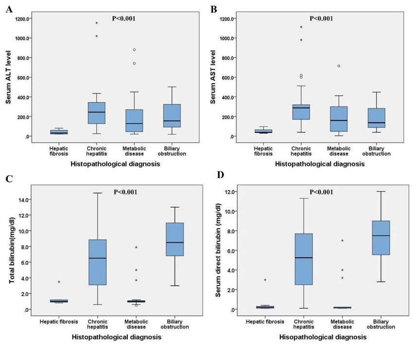
Pediatric Chronic Liver Diseases

1- INTRODUCTION                                      serodiagnosis of hepatotropic viruses,
                                                     autoimmune markers and improved liver
    Chronic liver diseases (CLD) in
                                                     biopsy technique. Although being an
children represent a growing health
                                                     invasive tool, histopathological evaluation
problem with significant morbidity and
                                                     of liver biopsy is still considered the
mortality (1). The exact prevalence of
                                                     cornerstone for diagnosis of CLD (8).
pediatric CLD is unknown , though, in the
                                                     Recently, non-invasive methods have been
United States, it has been reported that
                                                     proposed for the assessment of CLD
CLD is the cause of hospitalization of
                                                     including transient liver elastography and
about 15,000 children every year (2).
                                                     biological markers that could help in
Pediatric liver diseases comprise a wide
                                                     assessment of liver fibrosis (5).
variety of disorders, including infections,
developmental abnormalities, genetic, and            CLD in children are the precursors of adult
metabolic disorders that ultimately result           liver disease. Knowledge of exact burden
in progressive alterations in structure of           of CLD in children is important for health
liver and may end in cirrhosis and its               policy making by the government and its
consequences (3). Liver disorders which              cost effective implementation, especially
affect children are unique and different             in developing countries (4). This study
from those of adults and involve different           aimed to evaluate clinical pattern,
groups of acute and chronic disorders.               pathological pattern and outcome of
                                                     pediatric CLD in a tertiary care hospital in
The spectrum of pediatric liver diseases,
                                                     Upper Egypt.
especially those with genetic and
metabolic etiologies, show variation
                                                     2- MATERIALS AND METHODS
according to geographical locations. This
encourages studies on various features of            2-1. Study design and population
pediatric liver diseases in different                    This was a prospective hospital-based
countries and communities (4). There is              study conducted in Pediatric Department,
age dependent variation in causes of CLD.            Sohag University Hospital, a tertiary care
In the first years of life, biliary atresia,         hospital in Upper Egypt, in the period from
choledochal cysts, congenital hepatic                June 2014 to May 2018. All children
fibrosis, galactosemia, Tyrosinemia, and             presented with diffuse chronic liver
alpha one antitrypsin deficiency are                 diseases were included in the study.
frequent causes of CLD. In older children            Patients were recruited from pediatric
etiologies of CLD include chronic viral              gastroenterology clinic and pediatric
hepatitis C and B, autoimmune hepatitis,             department. Cases with acute liver disease
Wilson’s disease, cystic fibrosis and                and those with solitary or multiple hepatic
Primary sclerosing cholangitis (PSC) (5).            focal lesions were excluded from the
Recently, non-alcoholic fatty liver disease          study.
(NAFLD) has been described as a                      2-2. Ethical Consideration
common cause of CLD with a pooled
mean prevalence of 7.65 % in all children,           The protocol of the study was approved by
and 34% in obese children (6). It is worth           Sohag Faculty of Medicine Ethics
mentioning that the etiology of liver                Committee       in     accordance      with
disease cannot be determined in 5-15 %               international agreements. Written informed
of children with cirrhotic liver (7). The            consent was obtained from parents of all
approach to the diagnosis and management             participants.
of CLD has been revolutionized by the                2-3. Inclusion and exclusion criteria
advent of better radiological diagnostic
techniques, recent advances in the

Int J Pediatr, Vol.7, N.4, Serial No.64, Apr. 2019                                           9306
Abou-Taleb et al.
All children presented to pediatric sections were prepared for pathological
department with manifestations of chronic diagnosis of liver biopsies, and Gomori
liver diseases were included in the study. trichrome-stained sections were prepared
Patients with acute liver disease and those for accurate assessment of hepatic fibrosis.
with solitary or multiple hepatic focal
2-5. Statistical analysis
lesions were excluded from the study.
IBM-SPSS (22.0 for Windows) software
2-4. Methods version was used for data analysis and P-
All patients were subjected to detailed valuePediatric Chronic Liver Diseases stenosis, hydrocephalus, microcephaly, ALT, and AST values above 500 IU/L, and purpura were reported in one patient respectively. Serum total bilirubin was each. Family history of glycogen storage raised in 71 (47%) children, of which 62 disease and previous history of neonatal had direct hyperbilirubinemia and 9 deaths were reported in 5 (3.3%), and 2 children had combined direct, and indirect (1.3%) patients, respectively. Nearly 1/3 of hyperbilirubinemia. Significant rise of the patients (31.8%, n=48) have microcytic serum total bilirubin above 10mg/dl was hypochromic anemia with no other recorded in 20 (13.3%) of the children and significant hematological abnormalities. the serum level of GGT was evaluated in Liver enzymes were raised in 124 (82.1%) 49 cases. of the cases, of which 6 and 8 patients had Table-1: Clinical evaluation and biochemical parameters of investigated cases Parameter Statistic Presentation - Abdominal distention Number (%) 71 (47.0) - Jaundice Number (%) 40 (26.5) - Abdominal distention and jaundice Number (%) 34(22.5) - Acholic Stool Number (%) 32 (21.2) - Epileptic fits Number (%) 5 (3.3) - Dark urine Number (%) 3 (2.0) Clinical evaluation and US findings - Weight (kg) Mean (±SD) and median 9.8 (±5.9) and 8.0 - Hepatomegaly Number (%) 61 (40.4) - Hepatosplenomegaly Number (%) 49 (32.5) - Ascites Number (%) 5 (3.3) - Dilated portal vein Number (%) 1 (0.7) - Liver coarse echo pattern Number (%) 9 (5.9) - Liver fibrosis / cirrhosis Number (%) 3 (2.0) - Non-visualized gall bladder Number (%) 4 (2.6) Laboratory investigations: - Serum ALT (n=151) Mean (±SD) and median 208 (±179) and 185 - Serum AST (n=151) Mean (±SD) and median 217 (±177) and 192 - Serum total bilirubin (n=151) Mean (±SD) and median 4.2 (±3.9) and 1.2 - Serum direct bilirubin (n=151) Mean (±SD) and median 3.2 (±3.6) and 0.2 - Serum GGT (n=57) Mean (±SD) and median 185 (±163) and 90 - Positive anti-smooth muscle antibody Number (%) 13 (8.6%) - Cytomegalovirus positive Number (%) 4 (2.6) - Hepatitis C virus positive Number (%) 3 (2%) SD: Standard deviation; GGT: gamma-glutamyl transpeptidase; ALT: alanine transaminase; AST: aspartate transaminase. Based on liver biopsy, chronic hepatitis one case each (Table.2). Among cases of was the most frequent cause of diffuse hepatitis, idiopathic neonatal hepatitis liver insult in this study, representing tends to present clinically at a significantly 37.1% followed by metabolic liver younger age compared to autoimmune diseases, while Budd Chiari disease and hepatitis (p
Abou-Taleb et al.

  Table-2: Histopathological diagnosis of the investigated cases
                                                                    Age (months)                 Gender
Disease                                          Number (%)
                                                                  Mean (±SD)/median     Male         Female
Chronic hepatitis:                             56 (37.1)
 - Neonatal (idiopathic)                       38 (25.1)          3.7 (±6.1)/2          25           13
 - Autoimmune                                  13 (8.6)           39.7 (±26.5)/36       7            6
 - Viral                                       4 (2.7)            56 (±33.8)/50         2            2
 - Drug induced                                1 (0.7)            NA                    1            0
                                                                        P
Pediatric Chronic Liver Diseases
Fig.1: The main biochemical parameters in different patterns of CLD of investigated cases.
Table-3: Main pathological findings among frequent diffuse liver diseases of this study.
Chronic hepatitis Metabolic liver disease Biliary obstruction P- value
Disease
Number (%) Number (%) Number (%)
- Hepatic necrosis
- Absent 47 (83.9) 52 (98.1) 23 (95.8)
0.02
- Detected 9 (16.1) 1 (1.9) 1 (94.2)
- Associated steatosis
- Absent 52 (92.8) 40 (75.4) 22 (91.6)
0.23
- Detected 4 (7.2) 13 (24.6) 2 (8.4)
- Hepatic inflammation
- No/mild 37 (66) 45 (84.9) 14 (58.3)
0.022
- Moderate/severe 19 (34) 8 (15.1) 10 (41.7)
- Giant cell change
- Absent 35 (62.5) 52 (98.1) 21(87.5)
0.0001
- Detected 21 (37.5) 1 (1.9) 3 (12.5)
- Hepatic fibrosis
- No/mild 48 (85.7) 48 (90.5) 8 (33.3)
0.0001
- Moderate/marked 8 (14.3) 5 (9.5) 16 (66.7)
- Liver architecture
- Preserved 50 (89.2) 50 (94.3) 14 (58.3)
0.001
- Distorted 6 (10.8) 3 (5.7) 10 (41.7)
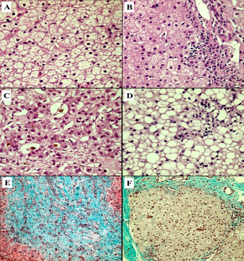
Int J Pediatr, Vol.7, N.4, Serial No.64, Apr. 2019 9310Abou-Taleb et al. Fig.2: Histopathological patterns of investigated cases. Ballooning of hepatocytes, rarified cytoplasm and compressed sinusoids in glycogen storage disease (A), piecemeal necrosis and portal inflammation in chronic hepatitis (B), canalicular bile retention in biliary atresia (C), Macrovesicular hepatic steatosis (D), broad fibrous septum in congenital hepatic fibrosis (E) and regeneration nodules with distorted architecture in end stage biliary cirrhosis (F). H&E stained sections (A, B, C and D) and Gömöri trichrome stained sections (E and F); magnification is x400 for A, B, C and D and x200 for E and F. All children included in this study had surgical intervention, and only 8 of these received supportive treatment in the form cases underwent Kasi operation. Long of vitamin A, D, E, and K. Additional term follow-up was available in 89 individualized treatment based on clinical children with lost follow-up for the condition included: prednisolone, remaining cases. The mean (±SD) follow ursodeoxycholic acid, azathioprine, and up duration was 14.2 (±11.5) months and Inderal in 5, 2, 1 and 1 patients, the median was 12 months. Forty-seven respectively. Children with glycogen children showed stationary clinical course storage disease received uncooked starch. while improvement and deterioration of Children with extrahepatic biliary atresia clinical status were reported in 31 and 11 were re-evaluated for the benefit of children, respectively (Table.4). Int J Pediatr, Vol.7, N.4, Serial No.64, Apr. 2019 9311
Pediatric Chronic Liver Diseases

      Table-4: Outcome of diffuse hepatic liver diseases of investigated children
                                                   Patients` outcome, Number (%)
Disease                                                                                         Total
                                       Improvement             Stationary      Deterioration
  -    Chronic Hepatitis                 23 (74.1)              7(22.5)           1(3.4)         31
  -    Metabolic diseases                 5 (14.3)               28(80)           2(5.7)         35
  -    Biliary obstructive                3 (21.4)              4(28.6)           7(50)          14
  -    Congenital fibrosis                   0                   5(100)              0            5
  -    Hepatic Steatosis                     0                   2(100)              0            2
  -    Budd Chiari disease                   0                   1(100)              0            1
  -    Biliary cirrhosis                     0                    0(0)            1(100)          1
                 Total                       31                    47               11           89

4- DISCUSSION                                               pattern (12). In this series, neonatal
                                                            cholestasis disorders (41.05%), and
    The aim of this study was to define the
                                                            metabolic liver disorders (35.1%) were the
clinicopathological pattern and the
                                                            leading causes of pediatric CLD. Similar
outcome of pediatric CLD in Sohag
                                                            pattern was reported by Akinbami et al.
University Hospital, Upper Egypt.
                                                            (13) in a study of CLD in Omani children
Neonatal cholestasis disorders (NCD), and
                                                            whereas they demonstrated that neonatal
metabolic liver disorders (MLD) were the
                                                            cholestasis constituted 50% of CLD. Also,
leading causes of CLD (41.05% and
                                                            metabolic liver disease accounted for 36%,
35.1%, respectively). In this 4-year study,
                                                            and 36.5% of CLD in 2 studies from
151 infants and children (aged from 15
                                                            Pakistan (8, 14). In addition, Sathe (15), in
days to 12 years) were diagnosed with
                                                            a study from Western India, reported that
CLD. They represent 1.6 % (151/9097) of
                                                            metabolic liver disease constituted 41.2 %
total admissions in pediatric department
                                                            of CLD cases. Contrary to our results,
during the same period. This finding was
                                                            Rajeshwari and Gogia (12) reported that
in agreement with Dhole et al., whereas
                                                            cryptogenic cirrhosis was the main cause
they reported that CLD constitute 1.1 % of
                                                            of CLD in children referred to a tertiary
total admission (9). So, a very high index
                                                            care hospital in New Delhi, India.
of suspicion is required for diagnosis of
CLD. Male predominance (63.5%) in the                       Authors explained this high incidence of
current study was consistent with other                     cryptogenic cirrhosis by referral of
studies that reported male predominance                     undiagnosed cases from other hospitals
from 58% to 69 % in children with CLD                       and by the presence of unrecognized
(8-10). Contrary to our results, Dar et al.                 infective agent or metabolic causes. Also,
reported female predominance (55%) in                       Hanif et al, in a study from Karachi in
children with CLD (11).                                     Pakistan, reported that the main cause of
                                                            chronic liver disease in children was
The main clinical presentations in this
                                                            chronic Hepatitis B. In addition, in a study
study were jaundice, abdominal distension
                                                            from Nigeria, Obafunwa and Elesha
(47%), jaundice (26.5%), acholic stool
                                                            reported that hepatic schistosomaias
(21.2%), hepatomegaly (40.4%), and
                                                            (37.5%) was the commonest etiology of
hepatosplenomegaly      (32.5%).    These
                                                            childhood CLD in tropical countries due to
findings were similar to what has been
                                                            high prevelance of infection by
reported by other studies (9-12). The
                                                            schistosoma (16). In the present study,
etiological spectrum of CLD varies
                                                            neonatal cholestasis disorders comprised
according to patients' age, geographical
                                                            neonatal hepatitis (25.1% of CLD),
location of the study, prevalence of the
                                                            extrahepatic biliary atresia (13.2% of
disease, availability of diagnostic tools,
                                                            CLD), and paucity of interlobular bile
experience of the physicians and referral

Int J Pediatr, Vol.7, N.4, Serial No.64, Apr. 2019                                                    9312
Abou-Taleb et al.
ducts (2.7% of CLD). These results were with glycogen storage disease as the
in agreement with Akinbami et al. (13) as commonest disorder, constituting 26.5% of
they demonstrated that neonatal hepatitis CLD causes. Other metabolic liver
was found in 28.9% of children with CLD diseases accounted for 8.65% of CLD
followed by EHBA in 11.8 % of cases then cases and comprised Gaucher's disease
paucity of interlobular bile ducts in 9.2% (n=3), Niemann Pick disease (n=2), and 8
of cases. Similar incidence of EHBA was cases with undetermined inborn error of
reported by Cheema et al. (14) (13.4%) of metabolism. These findings were in
cases, Narayan et al. (12.8%) of cases (4), agreement with Hashmi et al. (8) who
and Sathe (10.3%) of cases (15). However, reported that metabolic disorders were
lower incidence of EHBA was reported by found in 36.5% of pediatric liver disease
other studies ranging from 1.1% to 8.1% patients, and glycogen storage disease was
of CLD cases (8, 9, 12, 17-19). Also, other the most common metabolic disorder in
studies reported lower prevalence of NH (16.2%) of patients. Also, Cheema et al.
ranging from 7.1% to 10% among causes (14) reported that metabolic disorders
of CLD (4, 9, 17). This variation can be constituted 36 % of histological diagnosis
explained by difference in the age of study of pediatric liver disease with glycogen
groups. storage disorder disease accounting for
13.7% of cases. In the study done by Sathe
Cases with neonatal hepatitis are managed
(15), metabolic liver disease constituted
medically, while in extrahepatic biliary
41.2 % of CLD cases, with the most
atresia (EHBA) early surgical intervention
common being Wilson disease (n=15,
by Kasai portoenterostomy (KPE) is
15.4%) followed by Gaucher's disease
required to improve the outcome of these
(n=10, 10.3%), and glycogen storage
cases (20). In this study almost all cases of
disorder (n =4, 4.1%), and Niemann Pick
NH received medical treatment in the form
disease in one patient.
of vitamin A, D, E, and K, and responded
well to treatment. Nine out of 20 (45%) In this study, patients with glycogen
cases of EHBA underwent KPE. Two of storage disease received treatment with
the operated infants (22.2%) had uncooked starch and showed a stationary
successful Kasi with establishment of bile course on follow-up. Autoimmune
flow, colored stool, and absent jaundice for hepatitis was encountered in 13 (8.6%) of
one year after operation. Out of 7 (77.7%) our cases. All of them were type I
infants with failed Kasi operation, 5 autoimmune hepatitis (had positive anti
developed progressive liver disease, one smooth muscle antibodies [ASMA] and
died 2 months after operation, and one lost negative liver kidney microsomal [LKM]
follow-up. Eleven cases with EHBA (55%) antibodies). This is in agreement with
lost the chance for KPE due to late Dhole et al. (9) who reported that the
presentation, 6 of them had progressive incidence of autoimmune hepatitis among
liver disease and 5 lost follow- up. The patients with CLD was 10.9%, and Zhange
outcome of Kasi operation for EHBA was et al. who demonstrated that autoimmune
compatible with the previously established hepatitis was found in 7.1% of children
records which show that a small with CLD (19). A higher incidence of
proportion of infants with EHBA benefit autoimmune disease (16%) was reported
from early Kasi operation, and by Hanif et al. (10), and a lower incidence
approximately 60% of cases develop (4%) was reported by Rajeshwari and
progressive liver disease (21). In the Gogia (12). In the present study, out of 13
current study, metabolic liver disorders cases with autoimmune hepatitis, 7 cases
were diagnosed in 35.1% of CLD cases improved on treatment with prednisolone,
Int J Pediatr, Vol.7, N.4, Serial No.64, Apr. 2019 9313Pediatric Chronic Liver Diseases
one patient had a stationary course on cases perhaps due to the young age of our
treatment with prednisolone and cases, as the majority of cases were
azathioprine, and 5 cases lost follow-up younger than 2 years of age and Wilson
with unrecognized outcome. Congenital disease is a rare disease and usually
hepatic fibrosis was diagnosed in 5.9% of manifests in older children (5, 15).
our cases. This was in agreement with the
4-1. Limitation
study on Omani children whereas
congenital hepatic fibrosis constituted This study has some limitations as it was a
6.5% of CLD cases (13). However, single center study, and many patients may
Sobhan et al. (22) reported congenital be managed in other centers. Lack of
hepatic fibrosis in 3.3% of CLD cases and investigations for metabolic and genetic
in a Chinese study the frequency was 3.1% diseases in our center is another limitation
of cases (19). Out of 9 cases with factor.
congenital hepatic fibrosis, 5 had
stationary course and 4 lost follow-up with 5- CONCLUSION
unrecognized outcome. The rate of CLD is growing in Upper
Several recent studies have demonstrated Egypt, in this 4-year study, 151 infants and
the rising frequency of NAFLD in children children were diagnosed with CLD,
(23-25).In the current study, NAFLD was representing 1.6 % of total admissions in
diagnosed in 4% of cases. These cases had pediatric department. The most common
bright liver echo pattern by presentations were jaundice and abdominal
ultrasonography and elevated liver distension. Neonatal cholestasis disorders
enzymes. Liver biopsy in these cases and metabolic liver disorders were the
showed hepatic steatosis. Sobhan et al., leading causes of pediatric CLD. Other
reported that NAFLD was diagnosed in etiologies included autoimmune hepatitis,
16.5% of children with CLD (22). The congenital hepatic fibrosis, NAFLD, and
difference in incidence may be explained chronic hepatitis C infection. Budd Chiari
by difference in age of the patients as most disease was the least common etiology in
of our patients were below 2 years. this series. In general, the outcome of
children is favorable and comparable to
It has been estimated that chronic other countries.
hepatitis C infection was found in 3% of
children under 19 years of age in upper 6- CONFLICT OF INTEREST: None.
Egypt and 9% in lower Egypt (26). In this
study hepatitis C infection was the cause 7- REFERENCES
of CLD in 4 (2.7%) patients. Rajeshwari 1. Della Corte C, Mosca A, Vania A,
and Gogia reported only one patient with Alterio A, Alisi A, Nobili V. Pediatric liver
chronic hepatitis C (12), and Hanif et al. diseases: Current challenges and future
reported no case with hepatitis C infection perspectives. Expert Rev Gastroenterol
(10). This incidence was lower than what Hepatol 2016; 10(2):255-65.
has been reported by Tahir et al (31.66%) 2. Arya G, Balistreri WF. Pediatric liver
(18), and (6.4%) reported by Dar et al. (3). disease in the United States: Epidemiology and
Again, the difference may be explained by impact. J Gastroenterol Hepatol
difference in age group, prevalence of the 2002;17(5):521–5.
diseases and referral pattern. In this study, 3. Dar GA, Zarger SA, Jan K, Malik MI,
Budd Chiari disease was diagnosed in one Mir TA, Dar MA. Spectrum of Liver Diseases
patient who was lost to follow-up with among Children in Kashmir Valley. Acad Med
J India 2014;2(3):80–6.
unrecognized outcome. There are no
reported cases of Wilson disease in our 4. Narayan S, Gautam M, Dipankar H.
Int J Pediatr, Vol.7, N.4, Serial No.64, Apr. 2019 9314Abou-Taleb et al. Spectrum of Hepatobiliary Disease Among India. Apollo Med 2016;13(2):108–13. Children in Northeast India. J Dent Med Sci 16. Obafunwa JO, Elesha SO. Childhood 2017;16(7):50–4. liver diseases in Jos, Nigeria: a retrospective 5. Ruzman L, Mikolasevic I, Baraba histopathological study. East Afr Med J Dekanic K, Milic S, Palcevski G. Advances in 1991;68(9):702–6. diagnosis of chronic liver diseases in pediatric 17. Ahmed PA, Ulonnam CC, patients. World J Pediatr 2018;14(6):541–7. Mohammed-Nafiu R, Ballong J NG. Pattern of 6. Anderson EL, Howe LD, Jones HE, liver diseases among children attending the Higgins JPT, Lawlor DA, Fraser A. The National Hospital Abuja , Nigeria. Niger J Prevalence of Non-Alcoholic Fatty Liver Paed 2016;43(1):46–50. Disease in Children and Adolescents: A 18. Tahir A, Malik FR, Ahmad I, Krishin Systematic Review and Meta-Analysis. PLoS J, Akhtar P. Aetiological Factors of Chronic One 2015;10(10):e0140908. doi: Liver Disease in children. J Ayub Med Coll 10.1371/journal.pone.0140908. Abbottabad 2011;23(2):12–4. 7. Pinto RB, Schneider ACR, da Silveira 19. Zhang HF, Yang XJ, Zhu SS, Zhao TR. Cirrhosis in children and adolescents: An JM, Zhang TH, Xu ZQ, et al. Pathological overview. World J Hepatol 2015 ;7(3):392– changes and clinical manifestations of 1020 405. children with liver diseases confirmed by 8. Hashmi SS, Bhatti ABH, Malik MI, biopsy. Hepatobiliary Pancreat Dis Int 2004 Rana A, Nasir H, Dar FS, et al. Spectrum of ;3(3):395-8. histopathological diagnosis in paediatric 20. Verkade HJ, Bezerra JA, Davenport patients with liver disorders in Pakistan. J Pak M, Schreiber RA, Mieli-Vergani G, Hulscher Med Assoc 2017;67(2):266–9. JB, et al. Biliary atresia and other cholestatic 9. Dhole SD, Kher AS, Ghildiyal RG, childhood diseases: Advances and future Tambse MP. Chronic liver diseases in challenges. J Hepatol 2016;65(3):631–42. children: Clinical profile and histology. J Clin 21. Chung PHY, Wong KKY, Tam PKH. Diagnostic Res 2015;9(7):SC04-SC07. Predictors for failure after Kasai operation. J 10. Hanif M, Raza J, Qureshi H, Issani Z. Pediatr Surg. 2015 ;50(2):293-6. Etiology of Chronic Liver Disease in Children. 22. Sobhan PK, Sarojam B, Krishnan R, J Pak Med Assoc 2004;54(3):119–22. Kumar AS, Devadas K. Clinical Profile of 11. Dar GA, Malik MI, Ganie FA, Jan K, Chronic Liver Disease in Children: Experience Abdullah T, Dar MI, et al. Chronic Liver From a Tertiary Care Teaching Hospital in Diseases in Children : Clinical Spectrum and South India. J Clin Exp Hepatol 2017;7:S98. Etiology. Br Med Bull 2014;2(2):406–11. 23. Uppal V, Mansoor S, Furuya KN. 12. Rajeshwari K, Gogia S. The clinical Pediatric Non-alcoholic Fatty Liver Disease. spectrum of chronic liver disease in children Curr Gastroenterol Rep 2016;18(5):24. presenting to a tertiary level teaching hospital 24. Tiniakos DG, Vos MB, Brunt EM. in NewDelhi. Eur Neurol 1998;40(3):173–7. Nonalcoholic fatty liver disease: pathology 13. Akinbami FO, Venugopalan P, and pathogenesis. Annu Rev Pathol Nirmala V, Suresh J AP. Pattern of chronic 2010;5:145–71. liver disease in Omani children: A 25. McPherson S, Hardy T, Henderson E, clinicopatho logical review. West Afr J Med Burt AD, Day CP, Anstee QM. Evidence of 2004; 23(2):162-6. NAFLD progression from steatosis to 14. Cheema HA, Parkash A, Malik HS fibrosing-steatohepatitis using paired biopsies: FZ. Safety of outpatient blind percutaneous implications for prognosis and clinical liver biopsy (OBPLB) in children and to management. J Hepatol 2015 ;62(5):1148–55. document the spectrum of pediatric liver 26. Kamal SM. Hepatitis C virus genotype disease. Pakistan Paediatr J 2015;39(1):12–8. 4 therapy: progress and challenges. Liver Int 15. Sathe KP. Spectrum of pediatric liver 2011;31 Suppl 1:45–52. disease in a tertiary care center in western Int J Pediatr, Vol.7, N.4, Serial No.64, Apr. 2019 9315
You can also read