Frontiers in Celiac Disease: Where Autoimmunity and Environment Meet Marie Robert, M.D - Gastrointestinal Pathology Society
←
→
Page content transcription
If your browser does not render page correctly, please read the page content below
Frontiers in Celiac Disease:
   Where Autoimmunity and
     Environment Meet
  PRESENTED BY
              Marie Robert, M.D.
      Yale University School of Medicine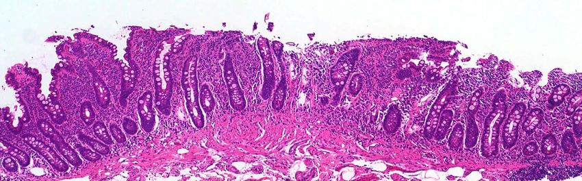
Disclosure of Relevant
                 Financial Relationships
The faculty, committee members, and staff who are in position to control the content of this activity are
required to disclose to USCAP and to learners any relevant financial relationship(s) of the individual or
spouse/partner that have occurred within the last 12 months with any commercial interest(s) whose
products or services are related to the CME content. USCAP has reviewed all disclosures and
resolved or managed all identified conflicts of interest, as applicable.

Marie Robert, M.D. reported the following relevant financial relationship(s) during the content
development process for this activity: Chief Scientific Officer, Beyond Celiac (until 12-30-19)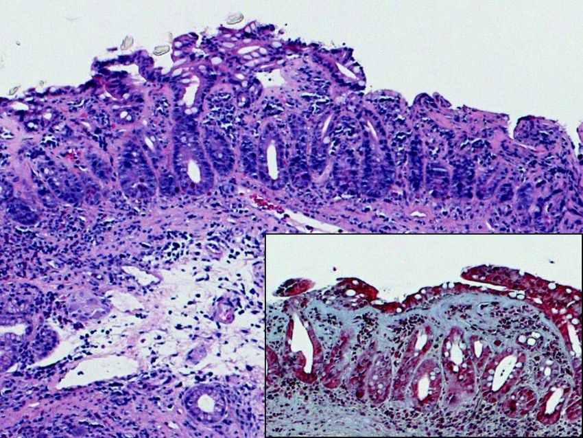
Definitions
Autoimmunity
The system of immune responses of an organism against its own
healthy cells and tissues.

Environment
The complex of physical, chemical, and biotic factors (such as
climate, soil, and living things) that act upon an organism or an
ecological community and ultimately determine its form and
survival.
Environment of a Disease
• Biological environment
   • Genetics
   • Interface with other organisms
      • Symbiotic-microbiome
      • Pathologic- infections
• Dietary, Hygiene and Medication environment
   • Geographic and cultural differences
   • Antibiotic use practices
• Societal environment
   • How society views the importance of the disease
   • Level of education and awareness, rates and accuracy of diagnosis, research
     funding, medical and social support for the condition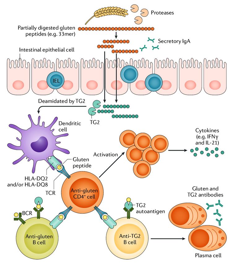
Celiac Disease                 3.2 million

Inflammatory Bowel Disease (IBD)           1.6

            Rheumatoid Arthritis        1.5

                          Lupus         1.5

                 Type I Diabetes    1.25

               Multiple Sclerosis   1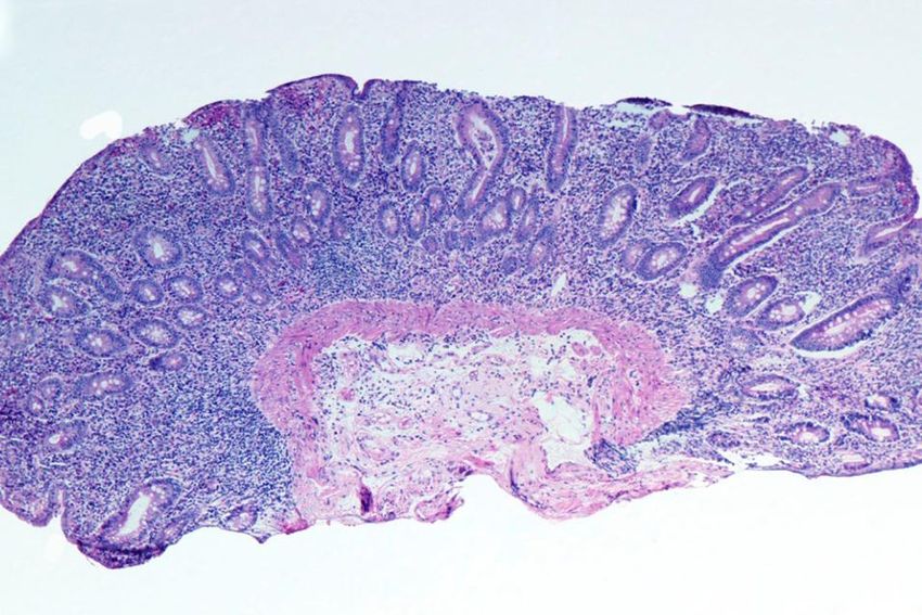
Outline
• Overview celiac disease (CeD)
   • Epidemiology, clinical manifestations, burden of disease
   • Pathophysiology

• Duodenal pathology on biopsy at diagnosis and in follow up

• Clinical trials in celiac disease-a lot happening
What is Celiac Disease
• Chronic, immune-mediated enteropathy driven by dietary gluten
  (wheat, barley and rye)
• More common in females, presents at any age and in almost all
  ethnicities
• Requires genetic susceptibility
   • Almost exclusively in MHC Class II HLA-DQ2 & DQ8 haplotypes
   • Other genetic factors
      • Only a fraction of DQ2/DQ8 develop disease
      • More than 100 non-HLA genetic associations
• Environmental factors
   • Amount and timing of gluten introduction into diet, microbiota, infections

While genetics provide the hard wiring, it is the environment that flips
                           the switch on.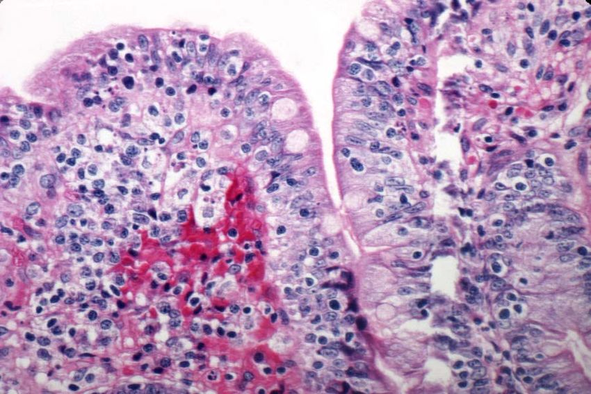
Protean Clinical Manifestations

• May have only
  non-GI symptoms

• May be
  asymptomatic

                    Infertility
                    Miscarraige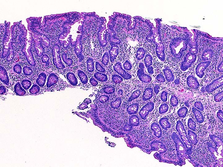
Perceived treatment burden
                     is very high in treated Celiac disease

ESRD patients on

                                                100
                                                                      Perceived Treatment Burden
hemodialysis = 56.4              VAS †

                                                80
Celiac disease = 44.9
                                                      Celiac disease

                                                60
-“Cloud over your head”
-Social anxiety/exclusion
-Lack of spontaneity

                                                40
-Fear of eating out/travel                                                     56.4

-Affects entire family                                44.9

                                                20
                                                                                      41.7   38.4            40.4
-”Not a real disease”
                                                                                                    31.9
-Activities skipped                                           23.5*    21.3*
-Life decisions altered                          0
                                                         CD    HTN     GERD    ESRD    DM    CHF      IBD     IBS

A smaller, limited life      † VAS:   0 = Very Easy
                                  100 = Very Difficult                 *Compared with CD, p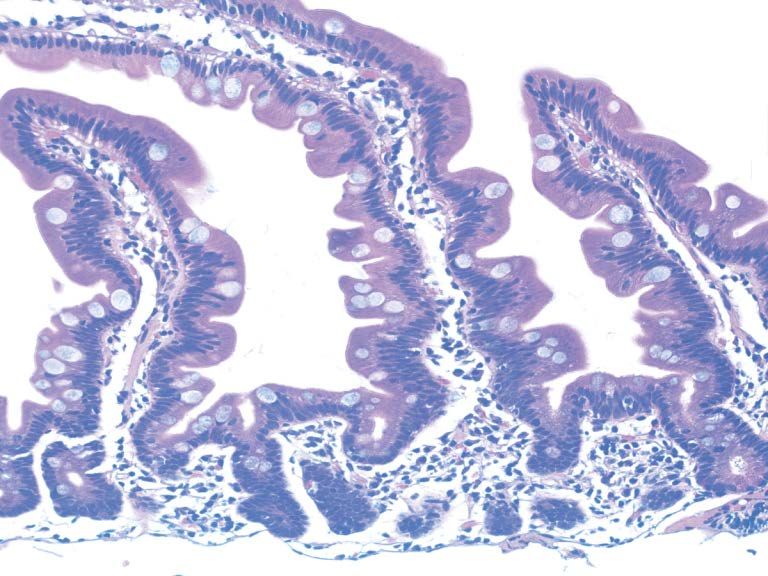
Diagnosis
A significant barrier to diagnosis is the ongoing insufficient awareness among
medical professionals
Adults report average delay of diagnosis of 6-10 years. Delays in children
also occur.
• Serologic tests
• Anti-tissue transglutaminase IgA antibodies
• Quantitative IgA assay
• Anti-deamidated Gliadin Peptide IgG antibodies
• Anti-endomysial antibodies
• HLA typing- >95% harbor HLA DQ2 (90%) or DQ8
• Duodenal biopsies, 2 from bulb and 4 from second duodenum
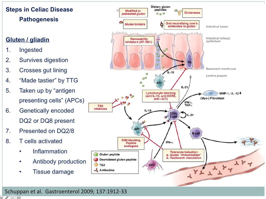
• Recently, may omit biopsy in children if certain criteria metPathogenesis
Adaptive immune                                                            Innate
 responses in CeD                                                           Immune
                                                                            response

                                                                            • IELs induce
                                                                              apoptosis
Characterized by:                                                           • Tregs
• Binding of deamidated                                                       suppressed
  gluten to dendritic APCs
• Gluten-specific mucosal
  CD4+ T-cells
• Anti-gliadin and TG2
  antibodies
• Secrete IFN and IL21
• Increase permeability of
  epithelial barrier

                             Lindfors et al, Nature Reviews/Disease Primers, (2019) 5:3.
Role of Microorganisms in Celiac Disease:
A Simplification
• In vivo and in vitro studies (mostly of bacteria) support association
between gut microbiota and celiac disease
• Taxonomic microbiota composition differs between active CeD, GFD and
normal controls, in oral, duodenal and fecal samples
• Increase in virulent strains (D’Argenio et al. Am J gastroenterol. 111:879-890, 2016)
• Can modify immunogenic food antigens, increasing or decreasing their
immunogenicity
• Use undigested particles as substrates, producing metabolites such as short chain
fatty acids affecting homeostasis
• Contribute to intestinal barrier dysfunction
• Common viral infections (rotovirus and rheovirus) trigger inflammatory
response to gluten antigens by initiating TH1 responses instead of Treg
responses
• Associated with development of symptomatic celiac disease in susceptible hosts.
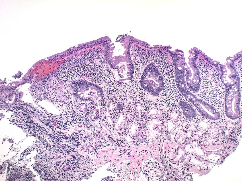
• Bouziat et al. Science 365:44-50, 2017Duodenal Mucosal Pathology Non-Frontier Territory
Villous blunting scores (Marsh
System, 0-4)

Mild blunting, Marsh   Moderate blunting, Marsh 3B   Severe blunting, Marsh 3C
3A
Assessing Intraepithelial Lymphocytes
Upper limit of normal is 20/100 enterocytes
• The villous tip method for use in normal villi
              • Jarvinen et al. Scand J Gastreoenterol 2004; , Biagi et al. J Clin Pathol 2004, Goldstein et
                al. Am J Clin Pathol 2001

• Villous tip to base ratio in 100 enterocytes at each site
              • Mino M et al.AJSP, 2003

• IELs in 50 enterocytes
              • Walker MM et al. Gastroenterol, 2010

• Remember, increased IELs does not prove CD. Need serology!
Evidenced based approach to
duodenal sampling
• Duodenal bulb historically avoided due to common inflammatory
changes attributed to ‘peptic duodenitis’
• Several studies confirm duodenal bulb reliably involved in celiac
disease and may be only site of involvement
• First described in children, then in adults
• Gonzalez et al. Gastrointest Endosc 2010;72:1837-1842.
• Evans et al. Am J Gastroenterol 2011;106:1837-1842.
• Bonamico et al. J pediatr Gastroenterol nutr. 2008;47:618-622.
• AGA: Allen J, Katzka D, Robert M, Leontiadis G. Gastroenterol 2015;149:1088-
1118.
• GIPS/SSCD joint recommendations
• Robert ME, Crowe SE, Burgart L, Yantiss RK, Lebwohl B, Greenson JK, Guandalini
S, and Murray JA. Am J Surgical Pathol. 42:e44-e58, 2018Best Practices for Duodenal Biopsy in CeD:
Robert ME, Crowe SE, Burgart L, Yantiss RK, Lebwohl B, Greenson JK, Guandalini S, and Murray JA. Am J
Surgical Pathol. 42:e44-e58, 2018.
A collaboration between GIPS and SSCD to address pathology component
• Practitioners should obtain at least 2 specimens from the proximal (bulb) and 4 from the
distal duodenum
• The evaluation of villi must occur in well-oriented regions
• Avoid lymphoglandular complexes and regions of where Brunner glands distort
• Serial sections should be employed routinely in order to achieve well oriented villi for evaluation.
• For diagnostic purposes, can decrease interobserver disagreement by considering a three tier
score: ‘normal’, ‘partial’ or ‘complete’ blunting (Vh/Cd ratios in research)
• Intraepithelial lymphocyte quantity should be determined to be either within normal limits
or increased
• In equivocal cases, a quantitative assessment should be performed
• CD3 stain not needed
• No need to include exact number in report outside of research settingLive Content Slide
When playing as a slideshow, this slide will display live content
Poll: How should the duodenum be sampled to evaluate for
the presence of celiac disease?Answer
ARS 1: How should the duodenum be
sampled to evaluate for the presence of
celiac disease?
A. Four biopsies from the distal duodenum, since villous
architecture cannot be reliably assessed in the duodenal bulb.
B. At least two biopsies from the duodenal bulb and four from the
second duodenum.
C. Four biopsies from anywhere in the duodenum, since the
disease can manifest in any segment.
Robert ME, Crowe SE, Burgart L, Yantiss RK, Lebwohl B, Greenson JK,
Guandalini S, and Murray JA. Am J Surgical Pathol. 42:e44-e58, 2018.
Allen J, Katzka D, Robert M, Leontiadis G. Gastroenterol 2015;149:1088-1118Duodenal Pathology: The Frontier Is Follow Up Biopsy Needed in Celiac Disease
What is Proper Follow up for CeD Patients

Follow up often ‘Diagnose and Adios’, in adults
  • Initial consult with dietician, then on your own
  • No requirement of follow up biopsy to confirm return to ‘normal’ villous
    mucosa
  • 10-30% have persistent symptoms despite good faith attempts at GFD
  • Ongoing mucosal injury is associated with nutritional deficiencies,
    osteoporosis and lymphoma
What is Proper Follow up for CeD Patients
Follow up often ‘Diagnose and Adios’, in adults
• Initial consult with dietician, then on your own
• No requirement of follow up biopsy to confirm return to ‘normal’ villous mucosa
• 10-30% have persistent symptoms despite good faith attempts at GFD
• Ongoing mucosal injury is associated with nutritional deficiencies, osteoporosis and
lymphoma
• Are symptom profile, GFD adherence, follow-up tTG titers surrogates for mucosal
healing?
• This premise has not been tested across celiac disease populations
• Hint from clinical trials that mucosal injury persists in some patients
• Follow biopsies are required in clinical trials (drug effect and endpoint)• Aims:
• Define factors associated with mucosal healing and persistent
villous blunting in a multinational cohort
• Stratify populations who may benefit from adjunctive therapies
under development
N Patel1, D Leffler2, G Gan1 ,Y Dan1, A Atsawarungruangkit2, A. Altoma3 C
Mulder4 L Elli5, A Del Gobbo6 , J Goldsmith7, Z Hintze7, C Pacheco8, M Vieth9,
B Melcher9, M Salomao10, R Pai10, J Hart11, A Olivas11, B Naini12, C Meyerson12, W Choi13,
S Kakar13, M Westerhoff14, J Cheng14, P Gopal15, M Moreno16, M Bronner16, M Robert1
239 patients with 478 biopsies from two timepoints
184 patients with biopsy interval ≥ 1 year
Mean interval follow-up: 3.3yrs (range: 1-18yrs)
GFD Adherence:
Strict: 143 (78%)
Partial 34 (19%)
Absent: 5 (3%)Classic Celiac Symptoms: Diarrhea, stool incontinence, nausea, vomiting, fatigue, abdominal pain, bloating, flatus, constipation, dehydration, soft stool, urgency Malabsorptive Symptoms: Weight loss, loss of appetite, anorexia, cachexia, iron deficiency, anemia, vitamin B12 deficiency, steatorrhea, short stature, slow growth,
Initial Take Home Points
In a multinational pathology-based cohort of adults and children
with celiac disease:
• Symptom improvement and decreasing tTG titers are not
  reliable indicators of normalization of duodenal histology in
  celiac disease.

• Duodenal mucosal injury persists in a significant subset of
  celiac disease patients adhering to a strict gluten free diet.

• These findings support the need for:
   • Greater monitoring of mucosal healing in celiac disease
   • Development of disease activity biomarkers that are less dependent on
     patient reported symptoms and diet adherence.
Refractory Celiac Disease
Definition Refractory Celiac
Disease
Persistent or recurrent malabsorptive
symptoms and signs with villous atrophy
despite a strict gluten free diet for more than
12 months in the absence of other causes of
non-responsive treated celiac disease and
overt malignancy.

                           Ludvigsson, Leffler et al, GUT 2012
“Apparent Refractoriness”
Unremitting IL-15 productionTwo Categories of RCD
patients
(defined at RCD referral centers)
Clinical/Pathologic Criteria Disease Category
RCD type RCD type
1 2
Abnormal intraepithelial lymphocyte No Yes
immunophenotype: either >40-50 % by
immunohistochemistry or >20-25% by flow
cytometry
T-cell receptor chains (γ or δ) clonal rearrangement No Yes
by molecular methods
Clinical or histological response to treatment Yes Variable
Lymphomagenesis potential (especially EATL Rare Frequent
development)
Rubio-Tapia/Murray, Gut 2010Prognosis by Refractory Celiac
Disease Classification
Al-toma et al, Gut 2007
Type 1 RCD
Type 2 RCD
De Novo EATL
=
Secondary
EATLAbnormal IELs (CD3ε+ CD8-)
in refractory celiac disease
Type 1
Refractory CeD
Type 2
Refractory CeD
Cellier et al, Lancet 2000 CD3 CD8Problems with Published RCD Diagnostic Criteria: The Yale Experience • Following published guidelines, increase in requests for clonal analysis in follow up biopsies of symptomatic patients • Greater than 60% had monoclonal TCR gene rearrangements by PCR • No correlation with degree of villous blunting • Most patient’s persistent symptoms responded to diet improvement • Alarming result for physicians and patients • Suspicion that test overly sensitive and lacking in clinical relevance • Had not been discussed in RCD literature
Clinical significance of monoclonal T-cell populations by PCR in celiac and non-celiac populations

Patient group         Number        Villous        Clinical F/U of patients with        Clonal T-cell populations
                      patients      architecture   clones                               clinically insignificant unless
                      with T-cell   (some                                               bona fide RCD criteria are met.
                      clone         patients had
                                                                                        Not previously reported.
                                    multiple
                                    biopsies)
                                                                                        Paradigm shift in Practice:
RCD2                  3/3           4 severe       1 died of disease
                                                                                        Resist submitting small biopsies for
                                                   2 clinical trials
                                                                                        PCR analysis until the much more
RCD1                  4/4           6 normal       4 alive and well on GFD              common causes of ‘apparent
                                    1 mild
                                                                                        refractoriness’ are excluded.
                                    2 moderate
                                    1 severe
                                                                                        Most prior literature subject to
CeD-Follow up Bx      7/10          3 normal       1 asymptomatic w/ benign stricture
                                                                                        referral center bias.
                                    2 mild         3 doing well, partial diet
                                    1 moderate     2 not following diet
                                    1 severe       1 died of other disease              Let clinical features guide
                                                                                        approach.
New Dx CeD            2/10          1 normal       1 with diarrhea (IBS-like), on GFD
                                    1 severe       1 well on GFD

                                                                                          Celli, Hui, Triscott, Bogardus, Gibson,
H.Pylori duodenitis   3/5           3 normal       N/A                                    Hwang, Robert. Am J Surg Pathol
                                                                                          2019;43:151–160
Small Intestine Histology RCD
• Subcryptal lymphocytic infiltrates, 10/10
• IBD like
• Thin mucosa/macrocytosis, 3/10
• Small bowel lymphoma, B-cell, 1/10
• Acute inflammation/ulceration, 5/10
• Collagenous sprue, 5/10 patients
• Collagenous colitis in 2/5
Robert ME, Ament M, Weinstein W. Am J Surgical
Pathol;24:676-687, 2000Live Content Slide
When playing as a slideshow, this slide will display live content
Poll: When is it appropriate to initiate a tissue work up for
suspected RCD?Answer
ARS 2: When is it appropriate to initiate a
tissue work up for suspected RCD?
A. When follow up duodenal biopsies show persistent villous
blunting in a celiac disease patient on a strict gluten free diet.
B. When clinician asks for T-cell clonal status and IHC
phenotyping of intraepithelial lymphocytes in a duodenal
biopsy.
C. When clinician has convincingly excluded likely causes
of ‘apparent refractoriness’ in a celiac disease patient.
Celli, Hui, Triscott, Bogardus, Gibson, Hwang, Robert. Am J Surg Pathol
2019;43:151–160Clinical Trials in Celiac Disease
Celiac disease:
Poised for drug development
Common: Revised prevalence estimates
US ~0.02% [1/5000] revised to ~1% (~3 million in US]
Europe ~0.1% [1/1000] revised to 1-2% (7-14 million)
GFD Inadequate:
>10% Persistent / frequent non-responsive disease
1 - 2% Refractory to GFD
~ 30% of adults on GFD for celiac disease have ongoing partial villous atrophy on biopsy
Strict GFD difficult to maintain
Need for lifelong therapy
Pathophysiology well elucidated - Multiple treatment targets
Progress in key areas
Elucidation of acceptable study designs in path to approval
Elucidation of acceptable outcome measures
Success in recruiting volunteers to participate in clinical studies
Robust co-operation by all stakeholders (patients, pharma, healthcare/research community and regulatory authorities (FDA, NIH).
Kelly CP, et al. Gastroenterol. 2015Immune Tolerance • Immunologic Tolerance – antigen-specific inhibition of selective immune responses is the‘holy grail’ and the ‘future’ for the treatment of autoimmune diseases, allergy and tissue transplantation • Goal – ‘cure’ disease by specifically targeting ‘only’ the immunopathologic T cells in autoimmune disease rather than large scale immunosuppressive drugs which can lead to increased rates of infection and neoplasia
Immune Tolerance Therapy for Celiac Disease
       – Nanoparticle Technology –
                      Healthy Individual – Balance between effector T cells
                      (Teff) and regulatory T cells (Treg)
  Teff    Treg

         Gluten Sensitization

                      Celiac Disease – Autoimmune Teff cells outnumber the
                      Treg cells leading to intestinal inflammation
          Treg
  Teff                          Tolerogenic Immune
                                Modifying Particle
                                (TIMP) Infusion

                      Tolerized Celiac Patient – PLG(Gliadin)-induced
                      expansion of gliadin-specific Tregs restores balance
                      between Teff cells and Tregs restoring homeostasis in the
  Teff    Treg        immune system and ameliorating disease symptoms
CNP-101 gliadin nanoparticles infusion
(Phase 2a):
• Met primary study objective of preventing activation of IFN-
gamma producing gliadin-specific cells during gluten challenge
(GC)
• Associated with a trend towards a reduction in the GC induced
villous height:crypt depth ratio deterioration
• Reduced circulating, gut-homing, CD4+ and CD8+ cells during
GC
• First clinical trial to demonstrate induction of antigen specific
immune tolerance in any autoimmune disease
Kelly C, Murray J, Leffler D, Bledsoe A, Smithson G, Podojil J, First R,
Morris A, Boyn M, Elhofy A, Wu T, and Miller S: Cour- TakedaFrontiers of Celiac Disease
Frontiers of Celiac Disease
    Celiac disease is a widely misunderstood genetically based
    autoimmune disorder affecting more than 1% of the world population
Frontiers of Celiac Disease
Celiac disease is a widely misunderstood genetically based
autoimmune disorder affecting more than 1% of the world population
Life with celiac disease includes medical, psychological and social
burdens that, until recently, have been underestimated and even
belittled by physicians and society.Frontiers of Celiac Disease
Celiac disease is a widely misunderstood genetically based
autoimmune disorder affecting more than 1% of the world population
Life with celiac disease includes medical, psychological and social
burdens that, until recently, have been underestimated and even
belittled by physicians and society.
From a scientific standpoint, CeD is poised to be a break out
disorder that will impact advances in a host of other autoimmume
diseases.
Still much to be learned about interface with environmental triggers,
such as microbiome, infections and introducing gluten in diet.Frontiers of Celiac Disease
Celiac disease is a widely misunderstood genetically based
autoimmune disorder affecting more than 1% of the world population
Life with celiac disease includes medical, psychological and social
burdens that, until recently, have been underestimated and even
belittled by physicians and society.
From a scientific standpoint, CeD is poised to be a break out
disorder that will impact advances in a host of other autoimmume
diseases.
Still much to be learned about interface with environmental triggers,
such as microbiome, infections and introducing gluten in diet.
Pathologists are a key part of this evolving story.
The role of biopsy interpretation is likely to increase as
patients get more follow up biopsies, participate in clinical
trials, and in the pursuit of biomarker development.CD3 immunostain
Be careful not to count IELs really in lamina propriaYou can also read