Epstein Barr Virus-Associated Hodgkin Lymphoma - MDPI
←
→
Page content transcription
If your browser does not render page correctly, please read the page content below
Commentary

Epstein Barr Virus-Associated Hodgkin Lymphoma
Antonino Carbone 1,* and Annunziata Gloghini 2
 1 Department of Pathology, Centro di Riferimento Oncologico Aviano, Istituto Nazionale Tumori, IRCCS,
   Via F. Gallini 2, 33081 Aviano, Italy
 2 Department of Diagnostic Pathology and Laboratory Medicine, Fondazione IRCCS, Istituto Nazionale

   Tumori, Via G. Venezian 1, I-20133 Milano, Italy; annunziata.gloghini@istitutotumori.mi.it
 * Correspondence: acarbone@cro.it; Tel.: +39-0434-659-085; Fax: +39-0434-659-370

 Received: 10 May 2018; Accepted: 24 May 2018; Published: 25 May 2018

 Abstract: Classical Hodgkin lymphoma (cHL) is a distinct clinical and pathological entity with
 heterogeneous genetic and virological features, with regards to Epstein–Barr virus (EBV) infection.
 The variable association of cHL with EBV infection is probably related to the different levels of
 patient immunosuppression, both locally in the tumour tissue and at the systemic level. This review
 paper focuses on EBV-related cHL highlighting pathogenetic and pathological features that may
 impact pathobiology-driven treatment for the affected patients.

 Keywords: Hodgkin                 lymphoma;         Epstein-Barr   virus;   immunosuppression;      tumour
 microenvironment

1. Introduction
      Classification of Hodgkin lymphoma (HL) evolved from histologic classifications [1,2], to the
multiparameter current World Health Organization (WHO) classification [3], in which HL has been
classified into classical HL (cHL), and the less common nodular lymphocyte predominant HL
(NLPHL). In 1944, Jackson and Parker called NLPHL as “paragranuloma” to separate it from
Hodgkin “granuloma [1].” In 1966, Lukes and Butler [2] renamed paragranuloma “lymphocytic
and/or histiocytic predominance Hodgkin disease,” recognizing a nodular and a diffuse pattern.
They used the term of lymphocytic and histiocytic (L&H) Reed-Sternberg (RS)-cell variant for the
diagnostic tumour cell [2], now called lymphocyte predominant (LP) cell [3]. At the Rye symposium,
it was decided to combine the nodular and diffuse types of the Lukes and Butler classification under
the term “lymphocytic predominance Hodgkin disease” [4]. A considerable body of evidence has
indicated that NLPHL exhibits features of a B-cell lymphoma, with a characteristic antigen profile
and clinical behaviour [3,5]. According to its cell of origin, phenotype and type of progression to large
B-cell lymphoma, NLPHL should probably be considered as a B-cell lymphoma tout court [6]. LP
cells indeed are antigen-selected mutating germinal centre (GC) B cells, express CD20, CD45, BCL6
and CD40 and are surrounded by CD4+ and PD-1+ T cells in the presence of follicular dendritic cell
meshworks within tumour nodules. Interestingly, multiparametric studies have shown similarities
between NLPHL and T-cell or histiocyte-rich large B-cell lymphoma (THCRLBCL) [7,8]. Moreover,
NLPHL may evolve to a completely diffuse T-cell–rich proliferation resembling a THCRLBCL [9,10].
The designation of these cases as THCRLBCL-like transformation of NLPHL has been recommended
[10]. Classical HL is a distinct entity with heterogeneous pathological, genetic, and virological
features, with regards to Epstein Barr virus (EBV) infection. Based on the morphologic characteristics
of the Hodgkin Reed-Sternberg (HRS) tumour cells (lacunar cells, multinucleated giant cells,
pseudosarcomatous cells) and the composition of the reactive infiltrate of tumour microenvironment,
four histologic subtypes have been distinguished: lymphocyte-rich cHL (LRCHL), nodular sclerosis
(NS) cHL, mixed cellularity (MC) cHL, and lymphocyte depletion (LD) cHL [2,6]. The tumour

Cancers 2018, 10, 163; doi:10.3390/cancers10060163                                www.mdpi.com/journal/cancers
Cancers 2018, 10, 163 2 of 7
microenvironment shows a cellular composition which is characteristic for each histotype. For
example, in MC cHL, microenvironmental cell types include T- and B-reactive lymphocytes,
eosinophils, granulocytes, histiocytes/macrophages, plasma cells, mast cells. In addition, a great
number of fibroblast-like cells and fibrosis are frequently found in NS cHL.
A fraction of patients with advanced stage disease are not cured by conventional first-line
chemotherapy [5] and show either primary refractoriness to chemotherapy or early disease relapse.
Recently, the treatment strategy for relapsed and refractory/relapsed HL patients included
immunotherapy through the use of checkpoint inhibitors.
2. Epstein–Barr Virus and cHL
The immunophenotypic features of HRS cells (CD30+, CD40+, CD15+, IRF4/MUM1+) are identical
in the different histologic subtypes of cHL. Conversely, the association with EBV shows marked
differences: EBV is found in HRS cells preferentially in cases of MC and LD cHL, and less frequently
in NS and LR cHL. Notably, the virologic characteristics of cHL vary according to the
immunocompetence status of the host and cHL subtype [11] (Table 1 and Figure 1), with EBV being
found in HRS cells in nearly all cases of cHL occurring in patients infected with HIV [10,12].
Table 1. EBV infection in Hodgkin lymphoma according to the immunocompetence status of the host.
Host Hodgkin Lymphoma EBV Infection
NLPHL Usually absent
Without known cHL, nodular sclerosis Variably present
immunosuppression cHL, mixed cellularity Usually present
Rare cHL subtypes Variably present
HIV-associated cHL Present
With acquired
Immunodeficiency Post-transplant, cHL type PTLD Present
Iatrogenic (methotrexate) Variably present
cHL, classical Hodgkin lymphoma; NLPHL, nodular lymphocyte predominant Hodgkin lymphoma; PTLD, post-
transplant lymphoproliferative disorder.
A pathogenic role for this herpesvirus in EBV-positive cases, probably as an early event in HL
development, has been suggested [13]. The demonstration of monoclonal EBV genomes in HRS cells
indicates that EBV infection occurred prior to clonal expansion. EBV-positive HRS cells express the
so-called type II latency pattern including a relatively restricted set of viral genes (EBNA-1, LMP-1,
and LMP-2 latent proteins, together with EBERs and BARTs RNAs) [14].Cancers 2018, 10, 163 3 of 7
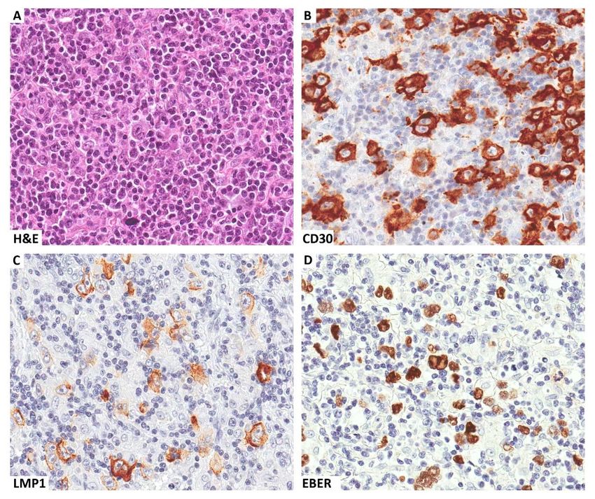
Figure 1. A clinical case of EBV-related Hodgkin lymphoma occurring in a patient with HIV infection.
Several Reed-Sternberg cells are seen within a mixed inflammatory microenvironment (A). These cells
express CD30 (B) and are EBV infected, as demonstrated by LMP1 immunostaining (C) and EBER in
situ hybridization (D). Original magnification ×400 (A–D).
3. Tumour Microenvironment in EBV-Related cHL
EBV-positive cHL tissues are enriched in genes characteristic of T-cell and antiviral responses
[15], suggesting that EBV has a role in influencing the tumour microenvironment. The
microenvironmental cellular infiltrate of EBV-associated cHL is composed either of immune cells,
including cytotoxic T lymphocytes against EBV-infected HRS cells, or of inflammatory cells
supporting the growth and survival of the neoplastic clone [16–18].
Tumour microenvironment of EBV-associated cHLs is also characterized by a significantly
higher numbers of CD68+, CD163+ macrophages than that of EBV-unrelated cHL. Coherently,
overexpression of macrophage-related genes was identified in EBV-positive cHLs by gene expression
profiling of whole tumour tissues [15]. Depending on availability of different microenvironmental
signals, macrophages may undergo polarized activation towards two functional states: the M1
macrophages with a pro-inflammatory phenotype, having the ability to promote Th1 responses and
kill tumour cells and the M2 macrophages with potent tumour-promoting activity, regulatory
functions in tissue repair and remodelling and promotion of Th2 responses [19]. A main M1
polarization of macrophages infiltrating EBV-positive cHL is in keeping with a predominant Th1
microenvironment of EBV-positive cHLs [20], which is similar to, albeit less prominent of that of Th1-
predominant inflammatory disorders.
LMP-1 may directly contribute to the generation of an immunosuppressive microenvironment
through its ability to induce/enhance the production of immunosuppressive cytokines such as IL-6,Cancers 2018, 10, 163 4 of 7
IL-8, and IL-10 [21–23]. In addition, the inflammatory milieu of EBV-positive cHL is enriched in
histiocytes, dendritic cells and endothelial cells.
4. HIV-Related Hodgkin Lymphoma
Classic HL is currently the most common type of non-AIDS-defining cancers. The pathological
spectrum of HIV-related cHL differs from that of HIV-unrelated cHL. In particular, the aggressive
histological subtypes of cHL—mainly MC and LD—predominate among HIV-related cHL. As
observed in HIV-unrelated cHL, the so-called HRS cell is the diagnostic key for assessing this
lymphoma owing to its typical morphology [24,25].
HIV-related HL shows several morphologic differences as compared with cases of the general
population. Among them, the occurrence of large confluent areas of necrosis is a distinctive feature
of HL occurring in HIV-infected patients and underlies the presence of a pro-inflammatory activity
[26]. A “sarcomatoid” pattern, although not specific, is also observed more frequently in these cases
and it has been associated with increased numbers of CD163 + spindle-shaped macrophages [27].
Intriguingly, a significant increase in spindle-shaped cells was observed after differentiation of
monocytes into M1 compared with M2 macrophages [27]. Nevertheless, available data on the
absolute numbers and functional polarization of macrophages infiltrating HIV-related HL are scarce
and controversial. Unlike what it could be expected in an immune compromised setting such as HIV
infection, similar numbers of HRS cells are usually found in HIV-related and HIV-unrelated cHLs
[26]. This could be the result of a partially retained ability of host immune system to control the
expansion of the tumour cell pool, consistently with the observation that cHL generally occurs in
HIV-positive patients with a moderate level of immune deficiency, as indicated by CD4 + T-cell count
[28,29].
In HIV-infected patients, nearly all cases of cHL are associated with EBV infection and express
a type II latency. HIV-associated cHL displays biological peculiarities when compared with cHL of
HIV-uninfected patients. In particular, tumour tissue is characterized by an unusually large
proportion of HRS cells infected by EBV. Moreover, the fact that LMP1 is expressed in virtually all
HIV-associated HL cases suggests that EBV has an aetiological role in their pathogenesis [11].
The combination of cART with better supportive therapy (such as G-CSF use and prophylaxis
of major opportunistic infections) has made standard ABVD (doxorubicin, bleomycin, vinblastine,
dacarbazine) and intensive chemotherapy regimens feasible also in patients with HIV-associated HL
[24]. The outcome and survival of HIV-associated HL are now approaching those of HIV-uninfected
patients.
5. Implications of Immune Evasion for Immunotherapy in EBV-Associated cHL
As for other tumours, cHL has been investigated for the expression of immunomodulatory
molecules including PD-1 on T cells, and its ligands PD-L1 and PD-L2, on HRS tumour cells, which
are involved in tumour cell evasion of host immune system. Classical HL is a neoplasm characterized
by robust inflammatory infiltrates and heightened expression of the immunosuppressive PD-1/PD-
L1 pathway. Antibodies against PD-1 have shown clinical efficacy in patients affected by cHL [30–
35].
In cHL, PD-L1 expression is the result of 9p24.1 amplification and EBV infection [36,37].
Expression of PD-L1 may be up-regulated by LMP-1 in lymphoblastoid B-cell lines through activation
of JAK3 and STAT5 phosphorylation or engagement of c-Jun [37]. EBV-related and EBV-unrelated
cHL cases have a similar frequency of 9p24.1 gains/amplifications [38]. Moreover, EBV infection was
not significantly associated the expression of PD-1, PD-L1, or PD-L2 [39]. However, EBV-related cHL
had higher PD-L1 expression according to a further up-regulation of PD-L1 by viral infection [38].
According to these findings, treatments targeting PD-1 may successfully restore therapeutically
immune responses against EBV-carrying HRS cells [40]. Indeed, anti-PD-1 therapy can be really
effective in patients with refractory cHL. Improved treatment options, however, are needed for CHLs
which are resistant to anti-PD-1 or relapse after this form of immunotherapy. A deeper
understanding of immunologic factors in the cHL microenvironment might support the design ofCancers 2018, 10, 163 5 of 7
more effective treatment combinations based on anti-PD-1. In addition, because the EBV residing in
CHL tumours is strongly immunogenic, characteristics of the tumour immune microenvironment in
EBV-unrelated cHL would be distinct from EBV-related cHL, with specific implications for designing
combination treatment regimens. A recent study has shown that the microenvironmental Th profiles
are strikingly different, with EBV-related cHL demonstrating a T helper 1 (Th1) profile, whereas EBV-
unrelated cHL has a Th17 profile. These results can address potential correlations of tumour response
or resistance with EBV status, and with expression of a pathogenic Th17 profile, in cHL patients
receiving anti-PD-1 monotherapy [41].
6. Conclusions
The virologic characteristics of cHL vary according to immunocompetence status of the host and
cHL subtype (Table 1), while paediatric HLs are often EBV-positive [42]. Different pathogenic
pathways are variably triggered by interactions of HRS cells with critical microenvironmental
components and concomitant EBV viral infection. In EBV-associated cHL, LMP-1 viral oncoprotein
may directly contribute to generation of an immunosuppressive microenvironment. The presence of
enhanced immunosuppressive features, with high numbers of M2 macrophages and elevated
expression levels of PD-L1 should make EBV-related cHL patients more susceptible to checkpoint
blockade [18].
Author Contributions: Conceptualization, A.C.; Writing—Original Draft Preparation, A.C. and A.G.; Writing—
Review and Editing, A.C. and A.G.; Funding Acquisition, A.C.
Funding: This research was funded in part by a grant from Centro di Riferimento Oncologico, Aviano, Italy
(intramural project “Infectious agents and cancer” to A.C.).
Conflicts of Interest: The authors declare no conflict of interest
References
1. Jackson, H.; Parker, F. Hodgkin’s disease. I. General considerations. N. Engl. J. Med. 1944, 230, 1–8.
2. Lukes, R.J.; Butler, J.J. The pathology and nomenclature of Hodgkin’s disease. Cancer Res. 1966, 26, 1063–
1083.
3. Stein, H.; Pileri, S.A.; Weiss, L.M. Classical Hodgkin lymphoma, introduction. In WHO Classification of
Tumours of Haematopoietic and Lymphoid Tissues, Revised 4th ed.; Swerdlow, S.H., Campo, E., Harris, N.L.,
Jaffe, E.S., Pileri, S.A., Stein, H., Thiele, J., Eds.; IARC: Lyon, France, 2017; pp. 424–430.
4. Lukes, R.J.; Craver, L.F.; Hall, T.C.; Rappaport, H.; Ruben, P. Report of the nomenclature committee. Cancer
Res. 1966, 26, 1311.
5. Younes, A.; Carbone, A.; Johnson, P.; Dabaja, B.; Ansell, S.; Kuruvilla, J. Hodgkin’s lymphoma. In De Vita,
Hellman, and Rosenberg’s Cancer: Principles & Practice of Oncology; De Vita, V.T.J., Lawrrence, T.S.,
Rosemberg, S.A., Eds.; Wolters Kluwer Health; Lippincott Williams & Wilkins: Philadelphia, PA, USA,
2014.
6. Carbone, A.; Gloghini, A. Hodgkin lymphoma classification: Are we at a crossroads? Cancer 2017, 123,
3654–3655.
7. Boudová, L.; Torlakovic, E.; Delabie. J.; Reimer, P.; Pfistner, B.; Wiedenmann, S.; Diehl, V.; Müller-
Hermelink, H.K.; Rüdiger, T. Nodular lymphocyte-predominant Hodgkin lymphoma with nodules
resembling T-cell/histiocyte-rich B-cell lymphoma: Differential diagnosis between nodular lymphocyte-
predominant Hodgkin lymphoma and T-cell/histiocyte-rich B-cell lymphoma. Blood 2003, 102, 3753–3758.
8. Brune, V.; Tiacci, E.; Pfeil, I.; Döring, C.; Eckerle, S.; van Noesel, C.J.; Klapper, W.; Falini, B.; von
Heydebreck, A.; Metzler, D.; et al. Origin and pathogenesis of nodular lymphocyte-predominant Hodgkin
lymphoma as revealed by global gene expression analysis. J. Exp. Med. 2008, 205, 2251–2268.
9. De Wolf-Peeters, C.; Delabie, J.; Campo, E.; Jaffe, E.S.; Delsol, G. T cell/histiocyte-rich large B-cell
lymphoma. In World Health Organization Classification of Tumours, Pathology and Genetics of Tumours of
Haematopoietic and Lymphoid Tissues; Swerdlow, S.H., Campo, E., Harris, N.L., Jaffe, E.S., Pileri, S.A., Stein,
H., Thiele, J., Vardiman, J.W., Eds.; IARC Press: Lyon, France 2008; pp. 238–239.Cancers 2018, 10, 163 6 of 7
10. Swerdlow, S.H.; Campo, E.; Pileri, S.A.; Harris, N.L.; Stein, H.; Siebert, R.; Advani, R.; Ghielmini; M.; Salles,
G.A.; Zelenetz, A.D.; et al. The 2016 revision of the World Health Organization classification of lymphoid
neoplasms. Blood 2016, 127, 2375–2390.
11. IARC Monograph on the Evaluation of Carcinogenic Risk to Humans. A Review of Human Carcinogens.
Part B: Biological Agents; IARC: Lyon, France, 2012; Volume 100.
12. Dolcetti, R.; Gloghini, A.; Caruso, A.; Carbone, A. A lymphomagenic role for HIV beyond immune
suppression? Blood 2016, 127, 1403–1409.
13. Shannon-Lowe, C.; Rickinson, A.B.; Bell, A.I. Epstein-Barr virus-associated lymphomas. Philos. Trans. R.
Soc. Lond. B Biol. Sci. 2017, 372, 1732.
14. Grywalska, E.; Rolinski, J. Epstein-Barr virus-associated lymphomas. Semin. Oncol. 2015, 42, 291–303.
15. Chetaille, B.; Bertucci, F.; Finetti, P.; Esterni, B.; Stamatoullas, A.; Picquenot, J.M.; Copin, M.C.;
Morschhauser, F.; Casasnovas, O.; Petrella, T.; et al. Molecular profiling of classical Hodgkin lymphoma
tissues uncovers variations in the tumor microenvironment and correlations with EBV infection and
outcome. Blood 2009, 113, 2765–2775.
16. Aldinucci, D.; Gloghini, A.; Pinto, A.; De Filippi, R.; Carbone, A. The classical Hodgkin’s lymphoma
microenvironment and its role in promoting tumour growth and immune escape. J. Pathol. 2010, 221, 248–
263.
17. Steidl, C.; Connors, J.M.; Gascoyne, R.D. Molecular pathogenesis of Hodgkin’s lymphoma: Increasing
evidence of the importance of the microenvironment. J. Clin. Oncol. 2011, 29, 1812–1826.
18. Carbone, A.; Gloghini, A.; Carlo-Stella, C. Are EBV-related and EBV-unrelated Hodgkin lymphomas
different with regard to susceptibility to checkpoint blockade? Blood 2018, doi:10.1182/blood-2018-02-
833806.
19. Mills, C.D. Anatomy of a discovery: M1 and m2 macrophages. Front. Immunol. 2015, 6, 212.
20. Barros, M.H.; Segges. P.; Vera-Lozada, G.; Hassan, R.; Niedobitek, G. Macrophage polarization reflects T
cell composition of tumor microenvironment in pediatric classical Hodgkin lymphoma and has impact on
survival. PLoS ONE 2015, 10, e0124531.
21. Eliopoulos, A.G.; Stack, M.; Dawson, C.W.; Kaye, K.M.; Hodgkin, L.; Sihota, S.; Rowe, M.; Young, L.S.
Epstein-Barr virus-encoded LMP1 and CD40 mediate IL-6 production in epithelial cells via an NF-kappaB
pathway involving TNF receptor-associated factors. Oncogene 1997, 14, 2899–2916.
22. Nakagomi, H.; Dolcetti, R.; Bejarano, M.T.; Pisa, P.; Kiessling, R.; Masucci, M.G. The Epstein-Barr virus
latent membrane protein-1 (LMP1) induces interleukin-10 production in Burkitt lymphoma lines. Int. J.
Cancer 1994, 57, 240–244.
23. Eliopoulos, A.G.; Gallagher, N.J.; Blake, S.M.; Dawson, C.W.; Young, L.S. Activation of the p38 mitogen-
activated protein kinase pathway by Epstein-Barr virus-encoded latent membrane protein 1 coregulates
interleukin-6 and interleukin-8 production. J. Biol. Chem. 1999, 274, 16085–16096.
24. Carbone, A.; Vaccher, E.; Gloghini, A.; Pantanowitz, L.; Abayomi, A.; de Paoli, P.; Franceschi, S. Diagnosis
and management of lymphomas and other cancers in HIV-infected patients. Nat. Rev. Clin. Oncol. 2014, 11,
223–238.
25. Carbone, A.; Volpi, C.C.; Gualeni, A.V.; Gloghini, A. Epstein-Barr virus associated lymphomas in people
with HIV. Curr. Opin. HIV AIDS 2017, 12, 39–46.
26. Hartmann, S.; Jakobus, C.; Rengstl, B.; Döring, C.; Newrzela, S.; Brodt, H.R.; Wolf, T.; Hansmann, M.L.
Spindle-shaped CD163+ rosetting macrophages replace CD4+ T-cells in HIV-related classical Hodgkin
lymphoma. Mod. Pathol. 2013, 26, 648–657.
27. Cassol, E.; Cassetta, L.; Rizzi, C.; Alfano, M.; Poli, G. M1 and M2a polarization of human monocyte-derived
macrophages inhibits HIV-1 replication by distinct mechanisms. J. Immunol. 2009, 182, 6237–6346.
28. Biggar, R.J.; Jaffe, E.S.; Goedert, J.J.; Chaturvedi, A.; Pfeiffer, R.; Engels, E.A. Hodgkin lymphoma and
immunodeficiency in persons with HIV/AIDS. Blood 2006, 108, 3786–3791.
29. Clifford, G.M.; Rickenbach, M.; Lise, M.; Dal Maso, L.; Battegay, M.; Bohlius, J.; Boffi, E.A.E.; Karrer, U.;
Jundt, G.; Bordoni, A.; et al. Swiss HIV Cohort Study. Hodgkin lymphoma in the Swiss HIV Cohort Study.
Blood 2009, 113, 5737–5742.
30. Tsirigotis, P.; Savani, B.N.; Nagler, A. Programmed death-1 immune checkpoint blockade in the treatment
of hematological malignancies. Ann. Med. 2016, 48, 428–439.Cancers 2018, 10, 163 7 of 7
31. Ansell, S.M.; Lesokhin, A.M.; Borrello, I.; Halwani, A.; Scott, E.C.; Gutierrez, M.; Schuster, S.J.; Millenson,
M.M.; Cattry, D.; Freeman, G.J.; et al. PD-1 blockade with nivolumab in relapsed or refractory Hodgkin’s
lymphoma. N. Engl. J. Med. 2015, 372, 311–319.
32. Younes, A.; Ansell, S.M. Novel agents in the treatment of Hodgkin lymphoma: Biological basis and clinical
results. Semin. Hematol. 2016, 53, 186–189.
33. Bartlett, N.L. Emerging role of novel therapies in Hodgkin lymphoma: Proceed with caution. Hematol. Am.
Soc. Hematol. Educ. Program. 2017, 2017, 317–323.
34. Shanbhag, S.; Ambinder, R.F. Hodgkin lymphoma: A review and update on recent progress. CA Cancer J.
Clin. 2018, 68, 116–132.
35. Carbone, A.; Gloghini, A.; Castagna, L.; Santoro, A.; Carlo-Stella, C. Primary refractory and early-relapsed
Hodgkin’s lymphoma: Strategies for therapeutic targeting based on the tumour microenvironment. J.
Pathol. 2015, 237, 4–13.
36. Green, M.R.; Monti, S.M.; Rodig, S.J.; Juszczynski, P.; Currie, T.; O’Donnell, E.; Chapuy, B.; Takeyama, K.;
Neuberg, D.; Golub, T.R.; et al. Integrative analysis reveals selective 9p24.1 amplification, increased PD-1
ligand expression, and further induction via JAK2 in nodular sclerosing Hodgkin lymphoma and primary
mediastinal large B-cell lymphoma. Blood 2010, 116, 3268–3277.
37. Green, M.R.; Rodig, S.; Juszczynski. P.; Ouyang, J.; Sinha, P.; O’Donnell, E.; Neuberg, D.; Shipp, M.A.
Constitutive AP-1 activity and EBV infection induce PD-L1 in Hodgkin lymphomas and posttransplant
lymphoproliferative disorders: Implications for targeted therapy. Clin. Cancer Res. 2012, 18, 1611–1618.
38. Roemer, M.G.; Advani, R.H.; Ligon, A.H.; Natkunam, Y.; Redd, R.A.; Homer, H.; Connelly, C.F.; Sun, H.H.;
Daadi, S.E.; Freeman, G.J.; et al. PD-L1 and PD-L2 Genetic Alterations Define Classical Hodgkin
Lymphoma and Predict Outcome. J. Clin. Oncol. 2016, 34, 2690–2697.
39. Koh, Y.W.; Jeon, Y.K.; Yoon, D.H.; Suh, C.; Huh, J. Programmed death 1 expression in the peritumoral
microenvironment is associated with a poorer prognosis in classical Hodgkin lymphoma. Tumour Biol.
2016, 37, 7507–7514.
40. Greenough, T.C.; Campellone, S.C.; Brody, R.; Jain, S.; Sanchez-Merino, V.; Somasundaran, M.; Luzuriaga,
K. Programmed Death-1 expression on Epstein Barr virus specific CD8+ T cells varies by stage of infection,
epitope specificity, and T-cell receptor usage. PLoS ONE 2010, 5, e12926.
41. Duffield, A.S.; Ascierto, M.L.; Anders, R.A.; Taube, J.M.; Meeker, A.K.; Chen, S.; McMiller, T.L.; Phillips,
N.A.; Xu, H.; Ogurtsova, A.; et al. Th17 immune microenvironment in Epstein-Barr virus-negative Hodgkin
lymphoma: Implications for immunotherapy. Blood Adv. 2017, 1, 1324–1334.
42. Di Napoli, A.; Al-Jadiri, M.F.; Talerico, C.; Duranti, E.; Pilozzi, E.; Trivedi, P.; Anastasiadou, E.; Alsaadawi,
A.R.; Al-Darraji, A.F.; Al-Hadad, S.A.; et al. Epstein-Barr virus (EBV) positive classical Hodgkin lymphoma
of Iraqi children: An immunophenotypic and molecular characterization of Hodgkin/Reed-Sternberg cells.
Pediatr. Blood Cancer 2013, 60, 2068–2072.
© 2018 by the authors. Licensee MDPI, Basel, Switzerland. This article is an open access
article distributed under the terms and conditions of the Creative Commons Attribution
(CC BY) license (http://creativecommons.org/licenses/by/4.0/).You can also read