Espin gene (ESPN) mutations associated with autosomal dominant hearing loss cause defects in microvillar elongation or organisation
←
→
Page content transcription
If your browser does not render page correctly, please read the page content below
157

   LETTER TO JMG

Espin gene (ESPN) mutations associated with autosomal
dominant hearing loss cause defects in microvillar
elongation or organisation
F Donaudy, L Zheng, R Ficarella, E Ballana, M Carella, S Melchionda, X Estivill, J R Bartles,
P Gasparini
...............................................................................................................................

                                                                      J Med Genet 2006;43:157–161. doi: 10.1136/jmg.2005.032086

                                                                     The phenotype segregates with mutations in ESPN, which
 Background: Espins are actin bundling proteins present in         encodes espin. These findings establish espin as an essential
 hair cell stereocilia. A recessive mutation in the espin gene     protein for hearing and vestibular function in humans. Here
 (Espn) has been detected in the jerker mouse and causes           we describe the identification of new functionally abnormal
 deafness, vestibular dysfunction, and hair cell degeneration.     ESPN alleles associated with dominant forms of hearing loss
 More recently mutations in the human espin gene (ESPN)            without vestibular involvement.
 have been described in two families affected by autosomal
 recessive hearing loss and vestibular areflexia.                  METHODS
 Objective: To report the identification of four additional        We analysed 450 subjects for mutations in the espin gene.
 ESPN mutations (S719R, D744N, R774Q, and delK848) in              Inclusion criteria were: absence of the most common
 patients affected by autosomal dominant hearing loss without      mutations within the GJB2 gene; sensorineural hearing loss;
 vestibular involvement.                                           and normal tympanometric evaluation.6 In all cases, vestib-
 Results: To determine whether the mutated ESPN alleles            ular data were obtained by clinical examination and routine
 affected the biological activity of the corresponding espin       vestibular tests (one or more of the following tests: caloric,
 proteins in vivo, their ability to target and elongate the        rotatory, optokinetic, swinging torsion, statokinesimetric,
 parallel actin bundles of brush border microvilli was             and vestibulo-vegetative). The series includes cases with a
 investigated in transfected LLC-PK1-CL4 epithelial cells. For     variable degree of hearing loss, ranging from mild to
 three mutated alleles clear abnormalities in microvillar length   profound, and with a variable age of onset, from congenital
 or distribution were obtained.                                    to late onset. Familial records were available in most cases.
                                                                   The majority of patients came from central and southern Italy
 Conclusions: The results further strengthen the causative role
                                                                   (200), while 140 were from Spain, 54 from Belgium, and 50
 of the espin gene in non-syndromic hearing loss and add
                                                                   from Israel. After informed consent, peripheral blood was
 new insights into espin structure and function.                   obtained from all subjects, and DNA was isolated from blood
                                                                   leucocytes using standard methods.
                                                                      Thirteen different primer pairs were designed in order to
                                                                   amplify the 13 coding exons of the ESPN gene, including the

E
      spins are a family of multifunctional actin cytoskeletal     splice sites (the polymerase chain reaction (PCR) primer
      regulatory proteins. They are able to influence the          sequences and conditions are available upon request). All
      organisation, dimensions, dynamics, and signalling           amplicons were screened by denaturing high performance
capabilities of the actin-filament-rich, microvillus type          liquid chromatography (DHPLC) on a WAVEH nucleic acid
specialisations that mediate sensory transduction in various       fragment analysis system HSM (Transgenomic Inc, Santa
mechanosensory and chemosensory cells.1 They are asso-             Clara, California, USA) according to the protocols supplied.
ciated with the parallel actin bundles of hair cell stereocilia    DHPLC data analysis was based on a subjective comparison
and are the target of mutations that cause deafness and            of sample and reference chromatograms. PCR products
vestibular dysfunction in mice (jerker) and humans                 showing an abnormal chromatographic profile were directly
(DFNB36).2 3 One striking illustration of espin activity in        sequenced on an automated sequencer (ABI 3100; Perkin
vivo is the ability to cause a dramatic, concentration             Elmer, Norwalk, Connecticut, USA).
dependent lengthening of brush border microvilli and their            Human espin 3A cDNA was obtained by reverse transcrip-
parallel actin bundles in transfected LLC-PK1-CL4 epithelial       tion PCR from human testis RNA (BD Biosciences, Clontech,
cells.4 The espin COOH-terminal peptide, which contains the        Palo Alto, California, USA). Mutations were introduced by
116 amino acid actin bundling module,5 is required for this        PCR. All cDNAs were checked by DNA sequence analysis
microvillar lengthening activity.4 This ability to increase        before introduction into the SmaI site of the pEGFP-C2 vector
microvillar length in transfected epithelial cells has led to      for transfection studies in differentiated LLC-PK1-CL4
the hypothesis that espins play an essential role in               epithelial cells.4 Briefly, cells were cultured at 37˚C in
determining the length of hair cell stereocilia.4 In support       minimum essential medium alpha medium (with L-gluta-
of this hypothesis, espin protein level is positively correlated   mine, without nucleosides) supplemented with 10% fetal
with stereocilium length,4 and abnormally short hair cell          bovine serum (Invitrogen, San Diego, California, USA) and
stereocilia are observed in homozygous jerker mice which           100 U/ml penicillin and streptomycin. Cells cultured on glass
lack espin protein.2 A human deafness recessive locus              cover slips for 8 to 10 days (,75% confluency) were
DFNB36 has recently been mapped to chromosome 1p36.3               transfected for four hours with a fixed amount of plasmid
in two consanguineous families affected by deafness and            DNA using Lipofectamine (Invitrogen) and examined 20 to
vestibular areflexia.3                                             24 hours later. Cells were fixed with 2% paraformaldehyde,

                                                                                                              www.jmedgenet.com
158 Letter to JMG
Figure 1 Amino acid sequence alignments for the peptides that contain the newly identified ESPN mutations. Alignments were undertaken using
ClustalX and WU-Blast 2. The mutated residues are shown in red. Note their conservation across species.
treated briefly with 0.1% Triton X-100, labelled with Texas and leads to a mild to moderate hearing loss in the fourth
Red–phalloidin (Molecular Probes, Eugene, Oregon, USA) to decade (high frequencies mainly involved). No vestibular
detect F-actin, and mounted in 5% n-propylgallate, 90% involvement was detected using standard vestibular tests.
glycerol. Transfected cells were identified by green fluores- Normal members of the family (two additional subjects)
cent protein (GFP) fluorescence; 0.5 mm confocal z sections were completely negative for the presence of the mutation.
were collected at room temperature using a confocal A GRA nucleotide change was detected at position 2230,
microscope and a 1006 1.4 NA oil immersion objective. leading to an amino acid substitution from aspartic acid to
LSM510 imaging software was used to generate stacks from aspargine at position 744 (D744N). This aspartic acid residue
which measurements of microvillar length were made. is highly conserved across species (fig 1). The mutation was
Images were saved in TIF format, transferred to Photoshop detected in an Italian patient affected by severe bilateral
(Adobe Systems), assembled into composites, and converted sensorineural hearing loss involving all frequencies, without
to CMYK colour format with minor adjustments of bright- vestibular involvement. Although there was a family history
ness and contrast. Antibody to purified recombinant rat espin of hearing loss, no additional genetic and instrumental tests
2B, previously called rat Purkinje cell espin 1 (GenBank could be carried out in other family members.
accession number AF540946), was prepared in rabbits and A GRA nucleotide change affecting position 2321 was
affinity purified on columns of rat espin 2B-sepharose 4B. detected in a sporadic Italian case. This change leads to
This antibody has been used extensively to detect espin substitution of a highly conserved arginine residue with
isoforms on western blots and by immunocytochemistry.1 glutamine at position 774 (R774Q) (fig 1). The patient is
Western blotting was carried out using the ECL method affected by late onset (age 42) mild bilateral sensorineural
(Amersham Biosciences, Amersham, UK) on extracts pre- hearing loss involving all frequencies but mainly the high
pared from replicate dishes by boiling (or heating at 100˚C) ones.
for three minutes in SDS gel sample buffer.4
Finally, the sequence from a Spanish sporadic case showed
a three nucleotide deletion (2541-2543delAAG), leading to
RESULTS the loss of a lysine residue at position 848 in the COOH-
Mutational screening of the ESPN gene by DHPLC and
terminal peptide (delK848). This patient (now aged 4) has
sequencing of 450 subjects from different countries allowed
severe bilateral sensorineural hearing impairment without
us to identify one deletion and three missense mutations that
vestibular involvement. Independent analysis of the proband
were associated with autosomal dominant hearing loss
sample confirmed the presence of this mutated allele, which
(table 1). The first such mutation to be identified was an
affects a residue that is highly conserved across species (fig 1)
ARC nucleotide change at position 2155. This replaces a
and must be catalogued as a de novo alteration.
conserved serine residue at position 719 with an arginine
(S719R) (fig 1). (The numbering schemes used are those of Two hundred chromosomes of individuals from the same
prototypical espin (GenBank accession number NM_031475), geographical areas as the patients were examined for the
which is referred to as espin 1 in the revised nomenclature for presence of the mutant alleles described above. None of the
espin isoforms1.) This mutation was detected in a small changes was detected in the control samples. We also
Italian kindred, in which two affected individuals in two detected six synonymous substitutions (table 1) in the cohort
generations show a dominant form of progressive sensor- of hearing subjects. As these latter changes do not create
ineural hearing impairment that starts in the second decade putative cryptic splice sites or did not segregate with deafness
(data not shown), they have been excluded as causative
mutations for deafness in our cohort of patients.
Table 1 Mutations and polymorphisms identified in To determine whether the espin gene mutations associated
ESPN (GenBank accession number NM_031475) with autosomal dominant sensorineural hearing loss had a
discernible effect on espin function in vivo, we compared the
Exon Nucleotide Protein effects of the wild type and mutated espin proteins on the
Mutation microvilli of transiently transfected LLC-PK1-CL4 epithelial
EX10 A2155C S719R cells.4 When maintained as confluent islands for 8 to 10 days
EX10 G2230A D744N in culture, LLC-PK1-CL4 epithelial cells invariably express
EX10 G2321A R774Q
EX13 2541-2543delAAG delK848
uniform brush border microvilli 1–1.5 mm in length.4 7 8
When expressed exogenously in these cells by transient
Polymorphism transfection, wild type espins are efficiently targeted to the
EX2 C390U Silent microvilli and bring about a dramatic elongation of the
EX3 C564G Silent
EX3 A591G Silent
microvilli and the parallel actin bundle scaffold at their core.4
EX3 G627A Silent This activity, which is believed to stem directly from espin
EX5 G909C Silent cross links in the core actin bundle, could explain the positive
EX8 T1476C Silent correlation noted between espin protein level and stereo-
cilium length and the shortening of hair cell stereocilia
observed in the espin deficient homozygous jerker mice.2 4
www.jmedgenet.comLetter to JMG 159
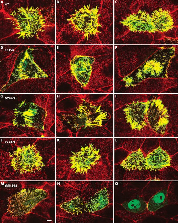
Figure 2 Functional comparisons of wt and mutated human espin 3A using the LLC-PK1-CL4 epithelial cell transfection model. Differentiated LLC-PK1-
CL4 epithelial cells were transfected with GFP-wt human espin 3A or the designated mutated construct, labeled for F-actin with Texas Red-phalloidlin
and examined by confocal microscopy. Multiple examples are shown. A-C, wt. D-F, S719R. G-I, D744N. J-L, R774Q. M-O, delK848. All images are
of the apical (microvillar) surface of the monolayer, except O, which is a z-section through the middle of the cells in N highlighting the nuclear
accumulation of the delK848 construct. The F-actin-rich junctional complexes adjointing the apical-lateral boundaries of neighboring cells are often
evident as red lines. Note that the wt construct (A-C) is colocalized (yellow) in microvilli that are much longer than the brush border microvilli (red) of
surrounding control (untransfected) cells. The R774Q (J-L) is indistinguishable from wt. The S719R (D-F) and D744N (G-I) constructs are targeted to
microvilli and cause microvillar elongation, but the ong microvilli frequently appear in irregular patches that occupy only a small percentage of the
apical surface. The delK848 construct (M-O) is severely impaired in microvillar elongation and shows abnormally high accumulation in the nucleus (O).
The objects within the nucleus that exclude the GFP-delK848 construct are nucleoli. Bar, 5 mm.
www.jmedgenet.com160 Letter to JMG
After 8 to 10 days in culture, LLC-PK1-CL4 cells were peptide, which is shared among all known espin isoforms,1
transfected with plasmids encoding GFP tagged wild type or is necessary and sufficient for potent actin bundling activity
mutated human espin 3A under the control of the cytome- in vitro5 and for microvillar elongation activity in vivo.4
galovirus promoter. Espin 3A is the major espin isoform The delK848 mutation severely impairs microvillar elonga-
found in cochlear hair cells.1 Between 20 and 24 hours after tion and causes abnormally high levels of espin protein to
transfection, the cells were fixed, labelled with Texas Red- accumulate in the nucleus. This mutation is located in the
phalloidin to localise F-actin, and imaged by cofocal COOH-terminal part of the actin bundling module, in a
microscopy. The wild type GFP-human espin 3A was found region believed to encompass one of its two F-actin binding
to be highly concentrated in the microvilli and caused about a sites.5 In fact, the 13 and 19 amino acid COOH-terminal parts
fivefold elongation of the microvilli, from 1.33 (0.04) mm, the of the protein that include the homologous lysine residue in
length in untransfected control cells, to 6.28 (0.09) mm rat espins are known to be required for actin bundling5 and
(mean (SEM), n = 167 to 189 microvilli, 14 to 17 cells) (fig microvillar elongation activity,4 respectively. Thus our func-
2A–C). The long microvilli of the transfected cells appear tional data on this mutated allele are in agreement with the
yellow because the GFP-espin is colocalised with the results of previous studies. Moreover, the severity of the
microvillar F-actin bundles, which are brightly labelled with functional deficit noted with the delK848 mutation in the
the Texas Red–phalloidin. The construct bearing the R774Q LLC-PK1-CL4 transfection model is strongly correlated with
mutation gave results that were not significantly different the severe phenotype of the corresponding patient, who had
from those obtained with the wild type (length = 6.2 bilateral severe sensorineural hearing loss with a very early
(0.1) mm; n = 153 microvilli, 14 cells) (fig 2J–L). In contrast, onset in childhood. The basis for the increased nuclear
constructs bearing the S719R mutation or the D744N accumulation of the delK848 construct is not clear, but this
mutation made long microvilli, as long as or longer than result raises the intriguing possibility that the dominant
wild type, but they often appeared in clumps that occupied effect of this mutation is exerted in the nucleus.
only a relatively small percentage of the apical cell surface Two of the mutated alleles, D744N and S719R, cause a
(fig 2D–I). A simple quantification of the behaviour of these
novel defect in microvillar organisation in the LLC-PK1-CL4
latter two mutations revealed an eight- to 10-fold increase in
transfection model. The D744N mutation maps to the actin
the number of cells showing such a disorganised microvillar
bundling module, whereas the S719R mutation maps to an
phenotype relative to wild type: while only 2–3% of cells
upstream location, which is currently without a known
transfected with wild type human espin 3A showed
function. Although both of these mutated constructs appear
disorganised long microvilli, 25–30% of cells transfected with
capable of elongating microvilli-like wild type espin, they also
the D744N or S719R construct showed the defect (n = 540–
cause a marked increase in the frequency of transfected cells
710 transfected cells for each construct in three independent
showing long microvilli confined to small patches of apical
experiments). The remainder of the cells transfected with the
S719R or D744N construct (70–75%) looked similar to those surface. This peculiar behaviour is noted only rarely in cells
transfected with wild type. Finally, the construct bearing the transfected with wild type espins and has not been detected
delK848 mutation was severely impaired in microvillar previously for espin constructs bearing a wide variety of
elongation (fig 2M, 2N). The delK848 construct caused only different truncation or deletion mutations.4 This defect in
a 1.5-fold elongation, from 1.33 (0.04) mm to 2.02 (0.04) mm microvillar organisation could reflect aberrant espin targeting
(n = 136 microvilli, 13 cells), compared with the nearly or a dominant negative effect of the mutated espin on the
fivefold elongation observed with wild type. In fact, other organising elements at the apical end of the epithelial
compared with the other constructs, the delK848 construct cell. Patients carrying these alleles have moderate to severe
appeared to be less efficiently targeted to microvilli and bilateral hearing loss.
commonly showed higher levels of accumulation in the Finally, the R774Q mutation causes no obvious problems
nucleus. This trend, which was often noticeable as a green in the LLC-PK1-CL4 transfection model. This mutation was
haze beneath the microvilli (fig 2N), was seen to better detected in a sporadic case with a mild form of hearing loss
advantage in z sections positioned below the microvilli (fig and a late onset (over age 40). Some possible conclusions can
2O). Western blots of the transfected cells labelled with be drawn from this case, considering both functional and
affinity purified espin antibody showed a single major band clinical data: first, the mutation is causative but its effect is
at the expected molecular mass (,64 kDa = ,29 kDa for too small to be detected in our functional assay; second, the
GFP and linker + ,35 kDa for human espin 3A) for each mutated allele has a small contribution per se on the
construct, and indicated that there were no major quantita- phenotype but interacts as a modifier with the genetic
tive differences in the levels of the wild type and mutated background contributing to the phenotype itself; third, the
proteins in the transfected cells that could account for their mutation, even if affecting an highly conserved residue and
differing effects on the microvilli (data not shown). never being present in normal controls, is truly a neutral
variant and not a causative allele.
In conclusion, the data presented here show an association
DISCUSSION
between ESPN mutations and dominant forms of hearing
Espins are associated with the parallel actin bundles of hair cell
stereocilia and are the target of mutations that cause deafness loss. The detrimental effects of these mutations noted in the
and vestibular dysfunction in mice and non-syndromic LLC-PK1-CL4 transfection model provide new insight into
recessive hearing loss and vestibular areflexia in humans.2 3 espin structure and function and suggest that the patients
Here, we report that mutations in ESPN also cause dominant carrying these mutated alleles have defects in the elongation
forms of hearing loss without vestibular signs. Moreover, using or organisation of their hair cell stereocilia.
the LLC-PK1-CL4 transfection model, we detected in vivo
functional deficits for three of the new mutations that could ACKNOWLEDGEMENTS
affect the dimensions or organisation of hair cell stereocilia. The work was supported by grants from: Telethon Foundation and
CNR (to PG); the National Institutes of Health (R01 DC004314 to
All of the newly described mutations affect residues JRB); DURSI and the ‘‘Redes Tematica se Investigacion Cooperativa
conserved across species and were never found in normal of the Fondo de Investigaciones Sanitarias’’ (G03/203) (to XE). EB is
chromosomes. Three of four of the mutations—D744N, supported by a fellowship from the Generalitat de Catalunya
R774Q, and delK848—mapped to the distal COOH-terminal (Departament d’Universitats, Recerca i Societat de la Informació,
peptide that includes the actin bundling module.5 This DURSI), 2003-FI066). We thank Dr Patricia A Loomis for her help on
www.jmedgenet.comLetter to JMG 161
confocal microscopy, and Dr Karen Avraham and Dr Guy Van Camp REFERENCES
for providing DNA samples and manuscript revision.
1 Sekerková G, Zheng L, Loomis PA, Changyaleket B, Whitlon DS, Mugnaini E,
Bartles JR. Espins are multifunctional actin cytoskeletal regulatory proteins in
..................... the microvilli of chemosensory and mechanosensory cells. J Neurosci
2004;24:5445–56.
Authors’ affiliations 2 Zheng L, Sekerková G, Vranich K, Tilney LG, Mugnaini E, Bartles JR.
F Donaudy, R Ficarella, P Gasparini, TIGEM-Telethon Institute of The deaf jerker mouse has a mutation in the gene encoding the espin
Genetics and Medicine, Naples, Italy actin-bundling proteins of hair cell stereocilia and lacks espins. Cell
L Zheng, J R Bartles, Department of Cell and Molecular Biology, 2000;102:377–85.
3 Naz S, Griffith AJ, Riazuddin S, Hampton LL, Battey JF, Khan SN,
Feinberg School of Medicine and Institute for Neuroscience, Riazuddin S, Wilcox ER, Friedman TB. Mutations of ESPN cause
Northwestern University, Chicago, Illinois, USA autosomal recessive deafness and vestibular dysfunction. J Med Genet
E Ballana, X Estivill, Genes and Disease Programme, Centre for 2004;41:591–5.
Genomic Regulation (CRG), Pompeu Fabra University (UPF), Barcelona, 4 Loomis PA, Zheng L, Sekerkova G, Changyaleket B, Mugnaini E, Bartles JR.
Espin cross-links cause the elongation of microvillus-type parallel actin bundles
Spain
in vivo. J Cell Biol 2003;163:1045–55.
M Carella, S Melchionda, Servizio di Genetica Medica, IRCCS-Hospital 5 Bartles JR, Zheng L, Li A, Wierda A, Chen B. Small espin: a third actin-
‘‘CSS’’, San Giovanni Rotondo, Italy bundling protein and potential forked protein ortholog in brush border
microvilli. J Cell Biol 1998;143:107–19.
Conflicts of interest: none declared 6 Donaudy F, Snoeckx R, Pfister M, Zenner HP, Blin N, Di Stazio M, Ferrara A,
Lanzara C, Ficarella R, Declau F, Pusch CM, Nurnberg P, Melchionda S,
Correspondence to: Dr Paolo Gasparini, Telethon Institute of Genetics Zelante L, Ballana E, Estivill X, Van Camp G, Gasparini P, Savoia A.
and Medicine, Via P Castellino III, Torino I-71013, Italy; gasparini@ Nonmuscle myosin heavy-chain gene MYH14 is expressed in cochlea and
tigem.it mutated in patients affected by autosomal dominant hearing impairment
(DFNA4). Am J Hum Genet 2004;74:770–6.
7 Hasson T, Mooseker MS. Porcine myosin VI: characterization of a new
Received 15 February 2005 mammalian unconventional myosin. J Cell Biol 1994;127:425–40.
Revised version received 24 May 2005 8 Tyska MJ, Mooseker MS. MYO1A (brush border myosin I) dynamics in the
Accepted for publication 25 May 2005 brush border of LLC-PK1-CL4 cells. Biophys J 2002;82:1869–83.
www.jmedgenet.comYou can also read