Human-Based Advanced in vitro Approaches to Investigate Lung Fibrosis and Pulmonary Effects of
←
→
Page content transcription
If your browser does not render page correctly, please read the page content below
REVIEW
                                                                                                                                                    published: 07 May 2021
                                                                                                                                           doi: 10.3389/fmed.2021.644678

                                              Human-Based Advanced in vitro
                                              Approaches to Investigate Lung
                                              Fibrosis and Pulmonary Effects of
                                              COVID-19
                                              Mirjam Kiener 1,2,3 , Nuria Roldan 3 , Carlos Machahua 1,4 , Arunima Sengupta 5 ,
                                              Thomas Geiser 1,4 , Olivier Thierry Guenat 1,5,6 , Manuela Funke-Chambour 1,4 , Nina Hobi 3*†
                                              and Marianna Kruithof-de Julio 2,3,7†
                                              1
                                               Department of Pulmonary Medicine, Inselspital, Bern University Hospital, University of Bern, Bern, Switzerland,
                                              2
                                               Department for BioMedical Research DBMR, Urology Research Laboratory, University of Bern, Bern, Switzerland, 3 Alveolix
                                              AG, Swiss Organs-on-Chip Innovation, Bern, Switzerland, 4 Department for BioMedical Research DBMR, Department of
                                              Pulmonary Medicine, Inselspital, Bern University Hospital, University of Bern, Bern, Switzerland, 5 Organs-on-Chip
                                              Technologies, ARTORG Center for Biomedical Engineering, University of Bern, Bern, Switzerland, 6 Department of General
                           Edited by:         Thoracic Surgery, Inselspital, Bern University Hospital, University of Bern, Bern, Switzerland, 7 Organoid Core, Department for
              Elena Lopez-Rodriguez,          BioMedical Research, University of Bern, Bern, Switzerland
         Charité – Universitätsmedizin
                      Berlin, Germany
                                              The coronavirus disease 2019 (COVID-19) pandemic has caused considerable
                        Reviewed by:
                   Ilias Papanikolaou,
                                              socio-economic burden, which fueled the development of treatment strategies and
    General Hospital of Corfu, Greece         vaccines at an unprecedented speed. However, our knowledge on disease recovery
                  Sonia Giambelluca,          is sparse and concerns about long-term pulmonary impairments are increasing.
        Charité – Universitätsmedizin
                       Berlin, Germany        Causing a broad spectrum of symptoms, COVID-19 can manifest as acute respiratory
                   *Correspondence:           distress syndrome (ARDS) in the most severely affected patients. Notably, pulmonary
                             Nina Hobi        infection with Severe Acute Respiratory Syndrome coronavirus 2 (SARS-CoV-2), the
                nina.hobi@alveolix.com
                                              causing agent of COVID-19, induces diffuse alveolar damage (DAD) followed by fibrotic
     † These   authors have contributed       remodeling and persistent reduced oxygenation in some patients. It is currently not
                    equally to this work
                                              known whether tissue scaring fully resolves or progresses to interstitial pulmonary
                    Specialty section:        fibrosis. The most aggressive form of pulmonary fibrosis is idiopathic pulmonary
          This article was submitted to       fibrosis (IPF). IPF is a fatal disease that progressively destroys alveolar architecture
                   Pulmonary Medicine,
                                              by uncontrolled fibroblast proliferation and the deposition of collagen and extracellular
                a section of the journal
                   Frontiers in Medicine      matrix (ECM) proteins. It is assumed that micro-injuries to the alveolar epithelium
       Received: 21 December 2020             may be induced by inhalation of micro-particles, pathophysiological mechanical stress
           Accepted: 01 April 2021            or viral infections, which can result in abnormal wound healing response. However,
           Published: 07 May 2021
                                              the exact underlying causes and molecular mechanisms of lung fibrosis are poorly
                              Citation:
    Kiener M, Roldan N, Machahua C,
                                              understood due to the limited availability of clinically relevant models. Recently, the
    Sengupta A, Geiser T, Guenat OT,          emergence of SARS-CoV-2 with the urgent need to investigate its pathogenesis
     Funke-Chambour M, Hobi N and
                                              and address drug options, has led to the broad application of in vivo and in vitro
             Kruithof-de Julio M (2021)
      Human-Based Advanced in vitro           models to study lung diseases. In particular, advanced in vitro models including
       Approaches to Investigate Lung         precision-cut lung slices (PCLS), lung organoids, 3D in vitro tissues and lung-on-chip
     Fibrosis and Pulmonary Effects of
    COVID-19. Front. Med. 8:644678.
                                              (LOC) models have been successfully employed for drug screens. In order to
     doi: 10.3389/fmed.2021.644678            gain a deeper understanding of SARS-CoV-2 infection and ultimately alveolar tissue

Frontiers in Medicine | www.frontiersin.org                                           1                                               May 2021 | Volume 8 | Article 644678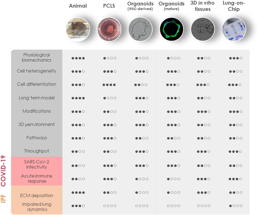
Kiener et al.                                                                                                              Human-Based Fibrosis and COVID19 Models

                                              regeneration, it will be crucial to optimize the available models for SARS-CoV-2 infection
                                              in multicellular systems that recapitulate tissue regeneration and fibrotic remodeling.
                                              Current evidence for SARS-CoV-2 mediated pulmonary fibrosis and a selection of
                                              classical and novel lung models will be discussed in this review.

                                              Keywords: COVID-19, interstitial pulmonary fibrosis, SARS-CoV-2, alveolar regeneration, organoids, lung-on-chip,
                                              precision-cut lung slices, in vitro lung models

INTRODUCTION                                                                                as remdesivir, camostat, imatinib, and Retro-2.1 and have
                                                                                            helped elucidating the molecular mechanisms of host-pathogen
Coronavirus disease 2019 (COVID-19) is a zoonotic disease                                   interactions in more detail (10–15). Increasing knowledge about
caused by the novel Severe Acute Respiratory Syndrome                                       the course of COVID-19 raised concerns regarding its long-
coronavirus 2 (SARS-CoV-2). SARS-CoV-2 is the seventh                                       term consequences. Experts warn that SARS-CoV-2 might cause
coronavirus known to infect humans. Human coronavirus                                       long-lasting or persisting interstitial pulmonary fibrosis, an
strains HKU1, OC43, NL63 and 229E cause mild symptoms                                       incurable clinical condition marked by abnormal fibrogenesis
similar to the common cold, while SARS-CoV and Middle                                       in the alveolar wall resulting in a progressive reduction of
East Respiratory Syndrome coronavirus (MERS-CoV) can result                                 pulmonary function and gas exchange in the lung (16). Recent
in severe viral pneumonia with a high mortality and have                                    studies show that severe or critically ill COVID-19 survivors
been responsible for two epidemic outbreaks in the twenty-                                  have reduced diffusion capacity and oxygenation levels compared
first century (1). Compared to SARS-CoV and MERS-CoV,                                       to mildly or moderately sick patients 4 months after infection
SARS-CoV-2 is more easily transmitted from human to human                                   (17). Whether these impairments resolve, remain or evolve into
which has allowed it to evolve into a worldwide pandemic                                    persisting pulmonary fibrosis is currently unknown.
(2). SARS-CoV-2 enters the human body via the respiratory                                      This review focuses on the clinical course of COVID-19 in
tract and reaches its initial main target organ, the lung.                                  the lung and relates the pathology to the underlying molecular
About one-fourth to one-third of hospitalized patients develop                              biology. Furthermore, we will discuss interstitial pulmonary
severe complications and require treatment in the intensive                                 fibrosis, with idiopathic pulmonary fibrosis (IPF) as the worst
care unit for ∼10 days or longer (3, 4), which risks a global                               example, and how COVID-19 may lead to pulmonary fibrosis.
collapse of the health care system. Countermeasures including                               Finally, we will review available in vivo and in vitro models of lung
curfews to limit the spread of SARS-CoV-2 have caused                                       fibrosis and SARS-CoV-2 infection to propose the most suited
dramatic economic losses (5). Despite improved management                                   advanced in vitro models for studying COVID-19-associated
of critically ill patients (6) this situation can only be resolved                          pulmonary fibrosis.
by effective treatment strategies and COVID-19 vaccines. Four
COVID-19 vaccines have already been approved in Europe
(ema.europa.eu) and various other vaccines are currently being                              PATHOGENESIS OF COVID-19 IN THE
developed or have entered late-phase clinical trials (7). In                                LUNG
parallel, inhibitory compounds are tested for re-purposing (8,
                                                                                            Fundamental Processes of Breathing: The
9). In vitro models of the respiratory tract have significantly
contributed to screening for promising drug candidates such                                 Biology and Regeneration of the Lung
                                                                                            Epithelium
Abbreviations: α-SMA, α-smooth muscle actin; ACE2, angiotensin-converting                   The respiratory tract is continuously exposed to inhaled particles
enzyme 2; ALI, air-liquid interface; AMφ, alveolar macrophage; APN,                         and pathogens. Therefore, it is lined by a highly specialized
aminopeptidase N; ARDS, Acute Respiratory Distress Syndrome; ATI, type                      epithelium, which can be divided into conducting airways and
I alveolar epithelial cell; ATII, type II alveolar epithelial cell; COVID-19,               alveoli based on their location and primary function. The
coronavirus disease 2019; CT scan, computed tomography scan; DAD, diffuse
                                                                                            pseudostratified epithelium in the proximal airways harbors
alveolar damage; DPP4, dipeptidyl peptidase 4; ECM, extracellular matrix; FITC,
fluorescein isothiocyanate; hPSC, human pluripotent stem cell; IFN, interferon; IL,         secretory club and goblet cells, which produce a protective layer
interleukin; ILD, interstitial lung disease; IPF, idiopathic pulmonary fibrosis; LOC,       of mucus toward the lumen. The terminally differentiated ciliated
lung-on-chip; MERS, Middle East Respiratory Syndrome; MERS-CoV, Middle                      cells convey the mucus layer upwards to clear trapped particles.
East Respiratory Syndrome coronavirus; MUC5B, mucin-5B; NRP1, neuropilin-1;                 Basal cells are able to differentiate into secretory or ciliated cells
NRP2, neuropilin-2; PCLS, precision-cut lung slices; PCSK, proprotein convertase
subtilisin kexin; PDGF, platelet-derived growth factor; RBD, receptor-binding
                                                                                            and are therefore considered to represent the progenitor cells
domain; RTC, replication and transcription complex; S protein, spike protein;               of the airway epithelium, though most cell types of the airway
SARS, Severe Acute Respiratory Syndrome; SARS-CoV, Severe Acute Respiratory                 epithelium are highly plastic [(18); Figure 1A]. On the distal end,
Syndrome coronavirus; SARS-CoV-2, Severe Acute Respiratory Syndrome                         the conducting airways branch into bronchioles and ultimately
coronavirus 2; SP-A, surfactant protein A; SP-C, surfactant protein C; TEER,                in the alveoli. These sac-shaped units represent one of the
transepithelial electrical resistance; TERC, telomerase RNA component; TERF-1,
telomeric repeat-binding factor 1; TERT, telomerase reverse transcriptase; TGF-β,
                                                                                            largest body surfaces in constant contact with the environment
transforming growth factor β; TLC, total lung capacity; TMPRSS2, transmembrane              essential for efficient gas exchange. About 95% of the alveolar
protease serine subtype 2; TNF-α, tumor necrosis factor α.                                  surface is covered by highly specialized flattened type I alveolar

Frontiers in Medicine | www.frontiersin.org                                             2                                        May 2021 | Volume 8 | Article 644678
Kiener et al. Human-Based Fibrosis and COVID19 Models
epithelial (ATI) cells (19). They form an ultra-thin epithelial- In summary, repair in the alveolar epithelium is characterized
blood barrier with the pulmonary microvasculature endothelial by an acute inflammatory phase, progenitor differentiation and
cells, supporting efficient oxygen and CO2 passive diffusion (20). migration, wound closure and finally, resolution (33). Upon
Together with ATI cells, type II alveolar epithelial (ATII) cells injury, ATII cells behave as facultative stem cells and activate
are the main constituents of the highly differentiated alveolar their regenerative response becoming hyperplastic. These ATII
epithelium, which closely interacts with surrounding cells in cells will either self-renew, migrate to the site of injury and
the niche including alveolar macrophages (AMφ), microvascular differentiate into ATI cells, or undergo apoptosis. These processes
cells, and fibroblasts (Figure 1B). depend on the balance of different mediators and a complex cell-
ATII cells are cuboidal cells often located at the edges of cell crosstalk in which stromal cells and AMϕ are crucial players
the alveolar sacs and, as opposed to the flat and large ATI (34). Some studies point at the pro-inflammatory and oxidative
cells, account for a small fraction of the alveolar surface. ATII environment as a driving force for differentiation and repair
cells produce pulmonary surfactant, a lipid-protein complex with in the mouse [reviewed in (22)], with Wnt signaling as a key
exceptional surface tension lowering properties (21). By doing regulator for ATII cell differentiation (35). Further, the relevance
so, they sustain the breathing function and protect the delicate of ATII cells in the repair process is highlighted by studies
alveolar structure from collapsing upon exhalation (21). ATII in which ATII-targeted damage or cellular intrinsic alterations,
cells also have a role in innate immunity and take part in rather genetic or due to aging, lead to aberrant tissue remodeling
surfactant recycling. But most importantly, ATII cells are capable (36, 37).
of self-renewal and differentiation into ATI cells, which allows It is also important to consider that the lung is subject
re-epithelization upon injury [reviewed in (22)]. to mechanical stress and deformation which is essential for
Already in 1977, Mason and Williams termed ATII cells as “the several key biological events such as lung development (38)
defender of the alveolus” for their central role in lung homeostasis and pulmonary surfactant secretion (39–41). The correlation
(23). However, what exactly defines an ATII cell has been a between alveolar inflation to the corresponding increase in
matter of discussion for years (24). In vitro, isolated human alveolar surface area is still debatable. Nevertheless, during restful
ATII cells behave as facultative stem cells giving rise to alveolar breathing, also termed as tidal breathing (defined as 40–80%
organoids containing multiple cell types (25, 26). Recent studies of TLC, total lung capacity), alveolar linear strain has been
have suggested that different ATII subtypes may coexist within suggested to go from 4 to 10% (42–45), up to even higher than
what has been classically considered as ATII cells, including the 20% during exercise or deep sighs (42, 43, 45, 46). Hence, local
proposed alveolar epithelial progenitors comprising TM4SF1+ mechanical tension and stiffness changes which occur along the
cells which are highly responsive to Wnt signaling (26). repair process converge with the forces supporting breathing
Rising evidence supports the role of other cell types in (38). In fact, breathing-like cyclic strain has been proven to
alveolar tissue repair together with ATII cells. A subset of influence the regenerative epithelial response as shown by wound
Hopx+ ATI cells has been suggested as a source of ATII closure experiments in vitro (47–51). Mechanical ventilation with
cells via transdifferentiation upon injury (27). Other studies high tidal volumes, on the other hand, has been observed to
have proven that a rare basal-like p63+ Krt5+ epithelial cell amplify lung damage in animal models and in ventilated patients
population migrates to sites of injury in the distal lung to re- suffering from different respiratory pathologies (52, 53). In fact,
create the damaged barrier in the mouse [reviewed in (28)]. mechanical stress has been suggested as an important factor
In humans, such a population has not been found to date, but for fibrogenesis (54). Considering this, protective ventilation
bronchiolization is a common histologic finding after injury. In protocols have been adopted to prevent ventilation-associated
addition, the contribution of basaloid cells in the repair process lung injury in COVID-19 patients (55).
is supported by the finding of basaloid cells in the damaged Besides stretch, the alveolar niche sustains other mechanical
areas of patients suffering from IPF, an aggressive form of forces such as shear stress and surface tension. At the alveolar
progressive interstitial pulmonary fibrosis with unknown cause, epithelium, surface tension, and the so-called interfacial stress
although several risk factors have been identified [reviewed in dominate particularly at low volumes (45). These forces stem
(28)]. Recently, EpCAM+ CD73+ epithelial cells, which localize from the continuous change in area exposed to the air, its
at the basal membrane of the respiratory and alveolar epithelium, associated fluid oscillation and cell-induced deformation (45).
have also been suggested as progenitors for both, pseudostratified Interfacial stress alone has been observed to be deleterious
mucociliary and mature alveolar epithelium in the postnatal and for ATII cells in vitro, however, it has been also proven to
adult human lung (29). constitute a powerful signal for pulmonary surfactant release
Further, the contribution of stromal cells to ATII cell stemness in addition to cyclic stretch (56, 57). Pulmonary surfactant
maintenance and tissue repair cannot be neglected. Lung efficiency in lowering surface tension is tightly associated to its
fibroblasts have been shown to support progenitor ATII cell lipid and protein composition, which adapts very quickly to meet
characteristics in vitro and in vivo in mice (25, 30, 31) and different respiratory demands (58, 59), and has been suggested
human (25), underscoring the relevance of Wnt signals as to be refined along breathing cycles in a mechanism assisted
determinants for ATII cell fate. On the other hand, fibroblasts, by surfactant proteins (60–64). In the context of surfactant
and myofibroblasts are also responsible for extracellular exhaustion, higher surface tension may then act as a trigger
matrix (ECM) deposition and wound closure upon alveolar for further surfactant release to restore alveolar homeostasis.
injury (32). This system fails in pathological conditions in which aberrant
Frontiers in Medicine | www.frontiersin.org 3 May 2021 | Volume 8 | Article 644678Kiener et al. Human-Based Fibrosis and COVID19 Models
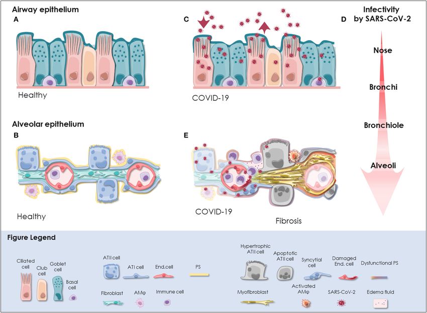
FIGURE 1 | SARS-CoV-2 infection in the respiratory tract. (A) In the pseudostratified epithelium of the airways, secretory goblet, and club cells produce mucus, which
is transported by ciliated cells to clear trapped particles and protect the lung from micro-injuries and infection. Basal cells reside at the lamina propria and comprise
progenitor cells. The composition and frequency of the individual cell types is variable among the distinct anatomical sites in the nose, trachea, bronchi, and
bronchioles. (B) The alveolar epithelium is specialized for gas exchange with flattened ATI cells forming an ultra-thin (∼2 µm) epithelial-endothelial barrier allowing
oxygen and CO2 diffusion. Cuboidal ATII cells are considered as progenitor cells of ATI cells and fulfill vital functions by the production of pulmonary surfactant (PS),
which lowers surface tension and prevents alveolar collapse. Lung fibroblasts are essential to maintain the ATII stem cell niche. Resident alveolar macrophages (AMϕ)
and immune cells defend the epithelium from infection. (C) SARS-CoV-2 initially infects the airway epithelium. The virus can efficiently replicate in ciliated and secretory
cells resulting in the shedding of high viral titers and mild to moderate COVID-19 symptoms. (D) The respiratory epithelium exhibits differential susceptibility to
SARS-CoV-2 infection. In correlation with ACE2 expression, SARS-CoV-2 infection is most efficient in the upper airways, particularly in the nasal epithelium. Infectivity
gradually decreases toward the alveoli. However, when SARS-CoV-2 reaches the alveoli it can result in severe manifestation of COVID-19. (E) Upon reaching the
alveoli, SARS-CoV-2 infects alveolar epithelial cells and endothelial cells and causes viral pneumonia. Cytopathic effects of SARS-CoV-2 are evident as syncytial and
apoptotic alveolar epithelial cells resulting in the breakdown of pulmonary surfactant and barrier integrity. In some patients, alveolar damage culminates in
life-threatening microvascular activation and an imbalanced immune response. Tissue regeneration takes place already during acute COVID-19 as indicated by fibrin
deposition, ATII cell hyperplasia and alveolar wall thickening. Moreover, severely ill COVID-19 patients exhibit radiological signs of fibrosis even months after recovery
indicative for the induction of COVID-19-associated fibrosis. ACE2, angiotensin-converting enzyme 2; ATI cell, type I alveolar epithelial cell; ATII cell, type II alveolar
epithelial cell; COVID-19, coronavirus disease 2019; End. cell, endothelial cell; AMϕ, alveolar macrophage; PS, pulmonary surfactant; SARS-CoV-2, severe acute
respiratory syndrome coronavirus 2.
surfactant composition (65–67) contributes to associated higher to injury and aberrant tissue repair, with severe long-term
surface tension and repetitive tissue damage (54), thus stressing consequences even after disease resolution.
the relevance of surfactant and ATII as a secreting cell in addition
to its role in repair. Molecular Mechanisms of SARS-CoV-2
Altogether, this evidence highlights the complexity of alveolar Infection in the Lung
epithelial repair and the central role played by ATII cells. Hence, The initial step of coronavirus infection involves binding of
we speculate that targeted ATII cell injury such as that caused the viral spike (S) protein to the compatible receptor on the
by SARS-CoV-2 infection may increase alveolar susceptibility surface of the target cell (68). Like the closely related SARS-CoV,
Frontiers in Medicine | www.frontiersin.org 4 May 2021 | Volume 8 | Article 644678Kiener et al. Human-Based Fibrosis and COVID19 Models
SARS-CoV-2 uses angiotensin-converting enzyme 2 (ACE2) as promoting infection. Taken together, it is likely that SARS-CoV-2
an essential entry receptor into human cells. In contrast to initially infects and replicates in the nasal epithelium, particularly
SARS-CoV, the receptor binding domain (RBD) of SARS-CoV- in ciliated cells, achieves high titers in the proximal airways and
2 S protein even exhibits higher binding affinity for human ACE2 reaches the alveoli by aspiration through the airways [(74, 82, 90);
(69–72) but seems to be less exposed possibly enabling immune Figure 1D].
evasion (70). As a consequence, ACE2 affinity of SARS-CoV and In the alveoli, SARS-CoV-2 can be detected in ATI and ATII
SARS-CoV-2 full-length S protein is comparable enabling both cells, endothelial cells and immune cells of deceased COVID-
viruses to attach to human ACE2 but not to other coronavirus 19 patients, which is in line with experimental findings from
entry receptors such as aminopeptidase N (APN) and dipeptidyl 3D in vitro models (82, 95, 98–101). Infection of ATI cells,
peptidase 4 (DPP4) (11, 73, 74). endothelial cells and alveolar immune cells presumably results
Subsequently, proteolytic cleavage of the S protein exposes in a disturbed immune response and persistent inflammation
the fusion domain and enables virus entry into the host (98–100). However, based on the analysis of single cell RNA
cell (75). Multiple proteases can fulfill this function such as sequencing datasets and in vitro infectivity experiments it has
transmembrane protease serine subtype 2 (TMPRSS2) and been suggested that ATII cells represent the primary target of
cathepsin B/L in case of SARS-CoV (76–79) or TMPRSS2, SARS-CoV-2 in the alveoli [(82, 83, 92, 95, 100–102); Figure 1E].
cathepsin L, and Furin for MERS-CoV (80, 81). Strong evidence Notably, increased susceptibility of an ATII cell subpopulation
for SARS-CoV-2 S protein priming by TMPRSS2 and cathepsin L has been consistently reported by in vitro studies (99, 101). Gene
has been gathered in vitro (11, 69, 82). expression profiling revealed an apoptotic signature and a strong
Therefore, cells co-expressing ACE2 and TMPRSS2 can downregulation of ATII-specific genes including surfactant
potentially be infected by SARS-CoV-2. Single cell RNA proteins in heavily infected ATII cell models (101–103). In
sequencing data analysis has revealed ACE2 expressing cells in line with in vitro data, the induction of apoptotic pathways
multiple organs, though it is generally expressed at low levels. paralleled by a significant downregulation of surfactant protein
This suggests that ACE2 expression is the limiting factor for transcripts is also apparent in ATII cells of COVID-19 patients
SARS-CoV-2 infection (83, 84). However, enriched expression (103) suggesting that SARS-CoV-2 infection results in the loss of
of ACE2 protein and co-expression with TMPRSS2 potentially ATII cell identity and function. Ultimately, this potentially leads
renders alveolar epithelial cells and enterocytes particularly to reduced surfactant production and consequently, alveolar
vulnerable to SARS-CoV-2 (83–85). Accordingly, SARS-CoV- collapse, massive tissue damage, and scaring (54). Therefore,
2 RNA is detected prominently in the respiratory tract but further investigations on this fatal course of the disease are
occasionally also in the feces and blood of COVID-19 patients critical. To date, it is unclear whether these highly infected cells
(86–88). In the respiratory tract, SARS-CoV-2 is detected secrete viral particles, what are the immunological and clinical
in diagnostic samples and tissue specimens from different consequences and why a subpopulation of ATII cells seems to
anatomical sites implying that it can replicate throughout the be more vulnerable than others. Possibly, SARS-CoV-2 relies
airway and lung epithelium (74, 89–91). Despite overall low on different entry mechanisms among different cell types and
ACE2 expression levels in the respiratory tract, about 20% of subsets. For example, it has been shown that TMPRSS2 is critical
lung cells have been found to express ACE2 mRNA (82). The for SARS-CoV-2 entry in ATII cells but cathepsin B/L seems to
highest levels of ACE2 are reached in the nasal epithelium be dispensable (102). Furthermore, as opposed to SARS-CoV,
and gradually decrease from the proximal airways toward the SARS-CoV-2 can exploit a wider range of host factors for cell
distal lung (82, 83, 92). Accordingly, viral yields are higher entry, which can act synergistically with initial ACE2 attachment
in nasal swabs than throat swabs indicating that the nasal and TMPRSS2 cleavage. Detailed resolution of the sequence
epithelium is the initial site of SARS-CoV-2 infection, replication, and structure of SARS-CoV-2 S protein has revealed only 73%
and shedding (74). The infection can propagate further as similarity to SARS-CoV S protein RBD (104) and the presence
ACE2 and TMPRSS2 expression is found throughout the airway of a multibasic site at the S1/S2 subunit boundary of SARS-CoV-
epithelium, particularly in ciliated and secretory cells (83, 92). 2 S protein, which creates a novel furin cleavage site (70, 71, 105).
Correspondingly, ciliated cells and goblet cells in the trachea Accordingly, furin overexpression enhances SARS-CoV-2 uptake
and bronchi are efficiently infected by SARS-CoV-2, whereas (82) and has a cumulative effect with TMPRSS2 and cathepsin L
basal cells are permissive for SARS-CoV-2 to a lower extent on virus entry (70). Processing of the SARS-CoV-2 S protein by
[(13, 14, 82, 93, 94); Figure 1C]. The finding that SARS-CoV-2 furin or other members of the proprotein convertase subtilisin
does not infect ciliated cells of distal lung organoids but exhibits kexin (PCSK) family might be highly relevant during SARS-CoV-
a strong tropism for club cells seems contradictory (95). However, 2 infection of ATII cells as a recent meta-analysis of human lung
the cell tropism of SARS-CoV-2 might shift among different single-cell RNA sequencing datasets has demonstrated significant
anatomical sites given the highly variable infection efficiencies co-expression of ACE2 and PCSK proteases in lung cells (85).
reported for in vitro cultured ciliated, goblet and club cells Importantly, S protein processing by furin generates a RRAR
(14, 82, 95). Moreover, ACE2 is upregulated upon interferon motif at the S1 C-terminus which is able to bind to Nuropilin-
(INF) stimulation to protect the tissue during acute lung injury 1 (NRP1) and Nuropilin-2 (NRP2) (106). While ACE2 is still
(96). Despite inducing an imbalanced immune response and required for initial attachment of the virus to the cell surface,
delayed IFN signaling (97), we cannot rule out that SARS-CoV-2 NRP1 depletion significantly reduces SARS-CoV-2 uptake (106).
infection itself might trigger INF-mediated upregulation of ACE2 Notably, deletion of the multibasic S1/S2 site in SARS-CoV-2 S
Frontiers in Medicine | www.frontiersin.org 5 May 2021 | Volume 8 | Article 644678Kiener et al. Human-Based Fibrosis and COVID19 Models
protein decreases the infection efficiency in human lung cells cytokine storm (121). Autopsies have revealed that SARS-CoV-
(105) and attenuates pathogenicity in animal models (107). 2 infects multiple organs including upper airways, lung, heart,
Whether this is due to the loss of interaction with NRP1 and kidney, the vasculature and the brain (125) and as a consequence
NRP2 remains to be demonstrated. However, NRP1 and NRP2 can manifest extra-pulmonary [reviewed in (126)].
are upregulated in the lung tissue of COVID-19 patients (108), However, most commonly severe COVID-19 patients develop
which might promote disease progression. viral pneumonia and suffer from fever, fatigue, dry cough,
These data indicate that ACE2 expression is critical for SARS- myalgia and dyspnea (3, 4). In these patients, SARS-CoV-2
CoV-2 infection and mediates initial attachment (71, 105). At replicates in the upper airways and the distal lung, where it
the same time, activation of SARS-CoV-2 S protein by TMPRSS2, causes life-threatening damage to the alveoli (90, 98, 125, 127,
cathepsin L, and furin allows it to interact with surface molecules 128). Nearly all hospitalized COVID-19 patients present with
other than ACE2 (70, 106). This is likely to confer wider tissue ground-glass opacities with or without consolidation on chest CT
tropism and promotes transmissibility of SARS-CoV-2. Once scans that gradually worsen before death (3, 4, 119, 120, 129).
SARS-CoV-2 has entered the host cell and released its positive- Critically ill patients usually develop acute respiratory distress
sense single-stranded RNA genome into the cytoplasm, viral non- syndrome (ARDS) (3, 4). ARDS can be provoked by various
structural proteins are translated to generate the viral replication direct or indirect pulmonary insults. Infection, including viral
and transcription complex (RTC). Furthermore, coronavirus infection, is a major cause for ARDS, being pneumonia the most
proteins hijack the translation machinery of the host cell and common underlying pathology (130). ARDS is defined as the
favor translation of viral mRNA over cellular mRNA, inhibit clinical manifestation of diffuse alveolar damage (DAD) (131).
the innate antiviral IFN response and interfere with normal cell Correspondingly, typical histologic patterns of DAD including
function (109). Infection of alveolar cells potentially results in the hyaline membrane formation, fibrin exudates, syncytial alveolar
most critical disease manifestation due to abrogation of ATII cell epithelial cells, diffuse ATII cell hyperplasia and the replacement
function and stimulation of an inflammatory response. of ATI cells by cuboidal ATII-like cells are apparent in the lungs
of deceased COVID-19 patients [(100, 120, 125, 129); Figure 1E].
The recent description of two differential pathologic patterns
in the lungs of deceased COVID-19 patients suggests that
Acute Pathologic Manifestation of both, direct cytopathic effect of SARS-CoV-2 and a deleterious
COVID-19 in the Lungs inflammatory immune response, can cause fatal alveolar damage
SARS-CoV-2 infection results in a complex symptomatology (132). As a consequence, marked hypoxia develops and results
associated with mild, moderate and severe illness or might even in the enlargement of the pulmonary vasculature, blood vessel
take an asymptomatic course (110–113). In non-hospitalized activation and coagulopathies with formation of micro-thrombi
patients testing positive for SARS-CoV-2 infection, the most in multiple organs (6, 98, 108, 125). About 2% of hospitalized
prevalent symptoms include cough, dyspnea, loss of smell or COVID-19 patients ultimately succumb to the disease with
taste, fever and chills, myalgia, headache, body aches, sinus respiratory or multi-organ failure as a major cause of death (127,
congestion, sore throat, nausea, diarrhea and dizziness (110, 111). 128, 133). However, it is currently not known whether severely
Surprisingly, subclinical lung opacities and diffuse consolidation affected COVID-19 survivors will fully recover or may suffer
have been detected on computed tomography (CT) scans in from complications in the resolution phase of ARDS. First results
more than half of asymptomatic COVID-19 patients (112, 114). after 4 months indicate that the diffusion capacity is reduced in
Moreover, histologic alterations in the alveoli including edema, COVID-19 patients after severe or critical disease (17).
protein and fibrin exudate, ATII cell hyperplasia and fibroblast
proliferation, inflammatory clusters and multinucleated giant
cells have been reported in two pre-symptomatic cases of PULMONARY FIBROSIS: A LONG-TERM
COVID-19 (115). Radiologic lung abnormalities seem to resolve COMPLICATION OF COVID-19?
in mildly to moderately symptomatic COVID-19 patients but
the regeneration process in these patients is scarcely studied Alveolar Damage as a Cause of Interstitial
(116, 117). Pulmonary Fibrosis
In contrast, about one-third of patients—mainly elderly men It has been reported that ARDS can lead to lasting physical
with underlying comorbidities—have a severe course of the impairment after 5 years of follow up (134), including fibrotic
disease with a high case fatality rate (3, 4, 118–121). Host factors pulmonary changes as a consequence of abnormal wound
rather than viral factors seem to be the significant determinants healing (135). Acute alveolar damage (e.g., from viral infection)
for disease severity. Pre-existing comorbidities, old age, male is followed by the activation of inflammatory and apoptotic
sex, and blood group other than O have been associated with a responses (136, 137). The alveolar epithelial cell damage triggers
higher susceptibility to SARS-CoV-2 and risk for a severe disease a cascade of reactions, including the release of pro-inflammatory
course (118, 119, 122, 123). Furthermore, clinical parameters cytokines, to activate local immune responses and controlled
at hospitalization are critical predictors of severe illness. These fibroblast proliferation as well as interstitial fibrogenesis, to
include elevated levels of coagulation markers (e.g., D-dimers) initiate primary wound healing mechanisms (138, 139). These
in the blood (124) and lymphocytopenia, which correlates with effects will normally be reconstituted by recovery of the basal
increased interleukin (IL)-6 and IL-8 levels and a higher risk of lamina, re-epithelialization of the alveolar epithelium (140),
Frontiers in Medicine | www.frontiersin.org 6 May 2021 | Volume 8 | Article 644678Kiener et al. Human-Based Fibrosis and COVID19 Models
and the degradation as well as clearance of ECM proteins The extent of fibrosis can be a sign of SARS severity and illness
(141). A precise and controlled repair mechanism following duration, as demonstrated in post-mortem studies (160, 161).
alveolar damage is crucial to terminate progression of the lung Radiological features of fibrosis after SARS have been observed
remodeling toward pulmonary fibrosis. at 3 and 6 months after infection in around 30% of the cases,
However, sustained alveolar injuries together with possible findings that have been confirmed by another study in survivors
intrinsic factors, such as genetic mutations [e.g., MUC5B, (162). Ground glass opacities were found 1 month after diagnosis
SFTPC, TERT/TERC or TERF-1; (142–145)] or an accelerated in 45% of SARS patients, underlining the possibility to find early
aging phenotype (146), can impair the capability of alveolar signs of fibrosis in those patients (163). Moreover, a patient’s age
epithelial cells to proliferate and orderly cover the defect. This can also be a critical risk factor in the fibrotic manifestation and
provokes chronic alveolar damage that can eventually trigger long-term damage as older SARS patients have an increased risk
an uncontrolled fibrotic response (147). This impaired wound for lung fibrosis (164).
healing can generate a disequilibrium in favor of the pro-fibrotic Another coronavirus infection—the Middle East respiratory
factors such as tumor necrosis factor alpha (TNF-α), platelet- syndrome (MERS), shows a similar clinical outcome as SARS.
derived growth factor (PDGF) or transforming growth factor However, radiological abnormalities are more common in MERS
beta (TGF-β), which will mediate the development and further (90–100%) than SARS (60–100%), and MERS patients have a
progression of lung fibrosis (148). Particularly, TGF-β has an higher incidence of ARDS with a higher case fatality rate (∼36%).
essential role in activating fibrotic mechanisms, inducing the For both diseases, risk factors like age and male sex are associated
perpetuation of exaggerated wound repair (149). with poorer disease outcomes (165).
The aberrant wound healing response can lead to additional Early evidence implies that, similarly to SARS and MERS,
loss of alveolar epithelial cells by apoptosis (150), induce lung fibrotic remodeling and scaring occurs in the lungs of severely
fibrosis by activation of a pro-fibrotic profile in macrophages ill COVID-19 patients. An alarmingly large number of COVID-
(151) and maintain the unruly activation and regulation 19 patients reported persistent symptoms, mainly fatigue
of fibrotic lung fibroblasts mediated by TGF-β (152). and dyspnea, even months after first diagnosis in multiple
This dysfunctional alveolar re-epithelialization favors the independent surveys (166–168). In line, radiological signs of
uncontrolled proliferation of lung fibroblasts and secretion of fibrosis become apparent as early as 3 weeks after diagnosis (169)
ECM proteins that consolidate the fibrotic change (153). Indeed, and persist over months (170, 171). After 3 months, impaired
viral lung infections can trigger DAD on top of interstitial lung diffusion capacity and persisting radiological abnormalities are
diseases (ILD) which is a common histological feature in some observed in many survivors, while others recover completely
stages of ILD progression (154, 155). Given the development (172–176). Further studies are ongoing whether radiological
of DAD that manifests as ARDS in severely sick COVID-19 and functional impairments are chronic and even progressing.
patients, it remains to be investigated whether the alveolar Worrisomely, lung autopsies of deceased COVID-19 patients
wound healing response will eventually result in pulmonary have revealed the aberrant localization of mucus to the
fibrosis and in its worst form IPF. alveolar parenchyma, pathologic signs of proliferative DAD and
thickening of the alveolar wall, particularly after a long severe
phase (82, 120, 125). These findings suggest that COVID-19
induces lung abnormalities including cases with pulmonary
Emerging Evidence of fibrosis. Notably, virus-induced cell fusion has been shown to
COVID-19-Associated Lung Fibrosis induce cellular senescence (177). Giant cells are a pronounced
Long-term follow-up data on recovered COVID-19 patients is feature in COVID-19 lungs which might be due to furin-
currently emerging and insights gained from earlier coronavirus mediated cleavage of the SARS-CoV-2 S protein at the plasma
epidemics can allow to predict likely scenarios. The first membrane of ACE2 expressing cells resulting in syncytial alveolar
coronavirus epidemic of the twenty-first century has been caused epithelial cells (98). Potentially, this results in the acquisition of
by SARS-CoV, the causative agent of severe acute respiratory a senescent alveolar epithelial cell phenotype that can provoke
syndrome (SARS). SARS is an illness that shows typical infection- inflammation and fibrosis (178–180). Moreover, intussusceptive
related symptoms, including fever and pneumonitis, with a angiogenesis occurs to a greater extent in pulmonary COVID-19
recovery time in most patients after 1–2 weeks following the as compared to influenza A pneumonia, suggesting activation of
infection. Up to one third of SARS patients can develop tissue regeneration that follows similar patterns as in pulmonary
severe pulmonary complications, requiring oxygen therapy fibrosis (108).
(156). The acute phase of SARS starts with acute lung The possibility to use early anti-fibrotic strategies is currently
damage and edema, bronchiolar sloughing of ciliated epithelial being investigated (16). The principal feature of anti-fibrotic
cells and the deposition of hyaline-rich alveolar membranes, treatment is preventing the worsening of the disease by slowing
which clinically manifests with impaired oxygen exchange. A down the fibrotic progression in established lung fibrosis, and
progressive phase during the following 2–5 weeks is characterized potentially influencing the cytokine storm by anti-inflammatory
by fibrin deposition and infiltration of inflammatory cells and effects of these drugs (181). Currently, some clinical studies are
fibroblasts. In the last stage, after 1–2 months, pulmonary fibrosis investigating both available anti-fibrotic treatments in patients
consolidates with collagen deposition and fibroblast proliferation with COVID-19 (recruiting phase): pirfenidone (NCT04282902,
in the interstitial spaces (157–159). NCT04607928) and nintedanib (NCT04541680, NCT04619680).
Frontiers in Medicine | www.frontiersin.org 7 May 2021 | Volume 8 | Article 644678Kiener et al. Human-Based Fibrosis and COVID19 Models
These results will provide us with new insights into the relevance fibrogenesis, assessing lung function in the course of pulmonary
of the fibrotic changes in COVID-19 and the effectiveness of fibrosis and performing pharmacokinetic studies (Figure 2).
anti-fibrotic treatment to improve the management of those However, most of our understanding of lung fibrosis
patients in the future. In parallel, novel treatment strategies stems solely from in vitro studies, typically relying on the
might be discovered in vitro, particularly in light of the recent activation of fibroblasts with pro-fibrotic cytokines in cellular
advances in the field of ex vivo tissue cultures, lung organoids, models. Although in vitro fibrosis models represent a robust
and bioengineered microfluidic devices to study lung fibrosis. platform to study cell-specific responses to soluble cues in a
controlled setting, cells in vivo are embedded in a complex
3D microenvironment with varied mechanical cues, cell-ECM
interactions, differential polarity, and growth factor gradients.
HOW CAN WE STUDY Given the strong involvement of fibroblasts and ECM in
COVID-19-ASSOCIATED PULMONARY the pathology of fibrotic diseases, it is particularly important
FIBROSIS? to maintain tissue architecture in human-derived models of
fibrosis. Fibrotic tissue explants from patients suffering from
In vivo and in vitro Models of Pulmonary a fibroproliferative skin disease, have been shown to retain
Fibrosis viability for several days in ex vivo tissue culture, allowing
Despite tremendous research efforts for pharmacological to study molecular mechanisms of fibrosis and test novel
interventions over the past decade, pulmonary fibrosis remains therapeutic strategies (193). Recently, precision-cut lung slices
one of the most challenging diseases to manage clinically. (PCLS) have garnered increasing attention as a novel lung ex
Although a single model is unable to mirror the progressive vivo fibrosis model. Overcoming the classical limitation for the
and irreversible nature of lung fibrosis, they provide valuable study of human lung cells in 2D cell culture models, PCLS are
mechanistic insights into fibrogenesis. Animal experimental able to spatially retain the native lung architecture along with
models have been widely used to understand the complex fibrotic fundamental ECM composition, stiffness and responsiveness
responses and perform early pre-clinical testing for anti-fibrotic together with viable lung resident cell populations [(194);
drugs. Among them, the bleomycin-induced pulmonary fibrosis Figure 2]. PCLS derived from healthy lung tissue resections
model has been most widely used since the 1970’s as the classical closely mimic fibrotic-like changes including increased ECM
standard and best characterized in vivo fibrosis model (182). deposition and alveolar remodeling when induced with a pro-
Contrary to human pulmonary fibrosis, bleomycin-induced fibrotic cocktail (195). A study in 2018 has reported that
fibrosis is temporary, and its inflammatory aspect justified induction with TGF-β1 resulted in increased deposition of
criticism to accurately represent the pathophysiological process collagen and ECM proteins in 2 mm3 sections of human lung
in IPF. Aside from bleomycin, fluorescein isothiocyanate (FITC) parenchymal tissues within 1 week in culture (196). The close
has also been widely used to induce experimental lung fibrosis recapitulation of pathologic processes and the possibility to
which results in alveolar injury and acute fibrotic reaction that culture tissue from IPF patients allows to study drug responses
persist up to 24 weeks. Occupational exposure to environmental ex vivo. Interestingly, nintedanib and pirfenidone exhibit distinct
risk factors has been extensively associated with pulmonary anti-fibrotic potential in mouse and human PCLS underscoring
fibrosis (183). Reports suggested that inhalation of silica and the need for human-derived models of IPF (197). Notch1
asbestos particles in rats results in fibrotic nodule formation inhibition in PCLS derived from IPF patients has shown
which closely mimics prominent features of silicosis and significant improvement in surfactant protein processing along
asbestosis in humans with long-term occupational exposure with decreased ECM deposition and an overall reversal of
(184, 185). Additionally, whole thorax irradiation in mice fibrosis (198). In addition, a study for inhalation-based anti-
has been invaluable to study early inflammatory responses in fibrotic therapies has utilized advanced 3D printing technologies
radiation-induced lung fibrosis (186). It is well-established that to develop a replica for Ear-Nose-Throat which has been
IPF includes genetic predisposition affecting genes encoding connected to an ex vivo porcine respiratory tract within a sealed
e.g., surfactant protein-C (SP-C) (187), SP-A (188), Mucin-5B chamber. To mimic fibrosis-related alterations, mechanical
(MUC5B) (189), telomerase reverse transcriptase (TERT), properties of the lung parenchyma have been modified by
and telomerase RNA component (TERC) (145). These known reduction of lung compliance and passive ventilation which
mutations have paved the way for genetically modified animal allowed them to analyze in vivo aerosol regional deposition
models of pulmonary fibrosis. Furthermore, intratracheal in a fibrosis-mimicking environment (199). Although a key
delivery of pro-fibrotic cytokines like TGF-β1 (190), TNF-α advantage in using human tissues is the exclusion of cross-species
(191), and IL-1β (192) by adenovirus and lentivirus vectors have heterogeneity, the significant limiting factor of ex vivo tissue
been extensively used to recreate mild early inflammation and culture is the constant need for fresh tissues. Generally, they are
rapid onset of lung fibrosis in mouse models. Despite the fact that not readily available as the tissues are mostly obtained from “end-
animal models cannot fully recapitulate the complex, progressive stage” pulmonary fibrosis patients after lung transplantation
and irreversible nature of lung fibrosis in humans, they remain or healthy surrounding tissue from tumor resections used for
the first line for preclinical testing in lack of appropriate artificially induced early fibrotic changes ex vivo. Moreover,
alternatives. Nevertheless, animal models have been proven the complexities associated with long-term cultivation of the
valuable for gaining a better mechanistic understanding of lung explants makes it difficult to standardize PCLS technique
Frontiers in Medicine | www.frontiersin.org 8 May 2021 | Volume 8 | Article 644678Kiener et al. Human-Based Fibrosis and COVID19 Models FIGURE 2 | Comparison of in vivo and 3D in vitro lung models for COVID-19 and fibrosis research. General aspects of experimental animal models and advanced in vitro models including PCLS, iPSC-derived organoids, mature organoids, 3D in vitro tissues, and LOC are rated based on similarities to human physiology (physiological biomechanics, cell heterogeneity, cell differentiation, long-term model, and 3D microenvironment), genetic manipulation (modifications), the possibility for mechanistic investigations (pathways), and throughput capabilities (throughput). Their applicability to model the diseased state of the lung has been evaluated separately for COVID-19 and IPF. COVID-19, coronavirus disease 2019; IPF, idiopathic pulmonary fibrosis; iPSC, induced pluripotent stem cells; LOC, lung-on-chip; µenvironment, microenvironment; PCLS, precision-cut lung slices; SARS-CoV-2, severe acute respiratory syndrome coronavirus 2. for high-throughput testing. Nevertheless, PCLS can be useful thickening upon TGF-β1 stimulation as it is observed in to investigate specific aspects of pulmonary fibrosis and viral IPF patients (203). Tests for novel IPF medication and infection directly in human lung tissue (Figure 2). mechanistic studies on fibroblast invasion of IPF patients Efforts have been undertaken to generate easily accessible, have also been undertaken in self-assembled pneumospheres controlled model systems that provide structural and cellular comprising heterogeneous cell populations (204). Additionally, complexity but hold the possibility to increase the throughput. biocompatible and biodegradable cross-linked polymer like Different cell types in the lung contribute to the pathology Matrigel is a widely used substrate for 3D lung cell culture and of fibrosis and hence the choice of the cell system is an organoid modeling for fibrosis. A recent study has analyzed important consideration for in vitro studies (200). Moreover, transcriptional signatures of fibrotic lung organoids in order to recent studies have focused more on using mechanically identify aberrantly expressed genes (205). While multicellular tunable substrates over standard extremely stiff (106 kPa) cell organoids closely capture the minute details of cell-cell and culture plastic dishes. Several studies have demonstrated that cell-ECM interactions and physiological cellular organization increased substrate stiffness directly influences (myo)fibroblast they lack vasculature and air-liquid interface (ALI) [(206); activation, differentiation and ECM deposition (201, 202). Figure 2]. Recently, it has been shown that ALI promotes Instead, seeding fetal-derived fibroblasts on hydrogel beads differentiation of human pluripotent stem cell (hPSC)-derived to mimic the structure of alveolar sacs recreates the patchy alveolar epithelial progenitor cells into ATII-like cells and areas of myofibroblast proliferation, contraction, and interstitial reduces their transdifferentiation into ATI cells, which occurs Frontiers in Medicine | www.frontiersin.org 9 May 2021 | Volume 8 | Article 644678
Kiener et al.                                                                                             Human-Based Fibrosis and COVID19 Models

in submerged cultures (207). Stimulation with a pro-fibrotic                 Modeling SARS-CoV-2 Infection and
cocktail results in the loss of SPC+ ATII cells paralleled                   Pathogenesis in the Respiratory Tract
by an increase in MUC5B+ goblet-like cells mimicking the                     In vivo models of viral infection integrate the full complexity
bronchialization process occurring in the alveoli of IPF patients            of virulence factors, local and systemic immune responses and
(207). However, these models still lack biomechanical stimulation            recovery. Therefore, animal models are particularly useful to test
(Figure 2).                                                                  anti-inflammatory compounds and vaccines to combat infection
    Advanced microfluidic technologies have been able to                     (216). However, mice, the most widely available laboratory
overcome these limitations with the development of lung-on-                  animals, are naturally resistant to SARS-CoV-2 infection (217,
chip (LOC) devices (208). Organ-on-Chip technology is a new                  218). The inability of SARS-CoV-2 to bind to murine ACE2 (74)
field emerging only recently as a system to model human tissues              poses the need to study COVID-19 in humanized mouse models
for the application in research and pharmacology. Despite the                expressing human ACE2 (217–220). SARS-CoV-2 infection in
development of multiorgan systems, it remains challenging to                 these mouse models results in weight loss, pneumonia and
apply the technique for gaining insights into systemic effects               pathologic alterations in the lung tissue. However, the organ
and standardize the models for pre-clinical testing. Moreover,               tropism and severity of symptoms varies among the models
microfluidic systems require the optimization of many factors                depending on the promoter to control human ACE2 expression.
such as the ECM, medium and scaffold material to support                     Mouse models expressing human ACE2 under the control of
optimal cell growth. However, the complexity of Organ-on-                    murine ACE2 develop rather mild symptoms and all animals
Chip technology is also a chance allowing the modulation of a                spontaneously recover (217, 218). In contrast, severe pneumonia
variety of biological, physical, and chemical factors in a controlled        develops in mice expressing human ACE2 under the control
and closed system (209). A microfluidic device recreating                    of HFH4 or KRT18 promoter (219, 220). However, it remains
the alveolar epithelium in ALI and in close contact with a                   arguable if these models correctly recapitulate SARS-CoV-2
microchannel, that is lined by endothelial cells and perfused                tissue tropism given non-endogenous ACE2 expression patterns.
with human full blood, has been employed to study pulmonary                  Alternatively, mutation of the SARS-CoV-2 S protein or serial
vascular inflammation and microthrombus formation (210).                     passaging in mice generates adapted virus to bind to murine
Furthermore, tiny wounds can be induced to the alveolar                      ACE2 and infect the murine host (221, 222). These models
epithelium on chip either by trypsin or gastric-like content                 might better resemble natural host-pathogen interactions in
to mimic alveolar damage taking place in IPF (211) and                       immunocompetent mice and result in mild pneumonia, however,
wound-healing (212). Moreover, micro-tissues generated from                  it is unclear whether the mechanisms of mouse-adapted SARS-
human lung fibroblasts have been shown to exhibit enhanced                   CoV-2 pathology can be translated to human.
contractility, stiffness and expression of alpha smooth muscle                   Other animal species are naturally susceptible to SARS-
actin (α-SMA), pro-collagen, and EDA fibronectin in response                 CoV-2 (223). SARS-CoV-2 infects and replicates in ferrets
to TGF-β, effects that have been reversed by treatment with                  but it is restricted to the upper respiratory tract allowing
pirfenidone (213).                                                           transmission studies but causing only mild symptoms (224,
    Due to the importance of cyclic stretch for tissue regeneration          225). Natural SARS-CoV-2 infection in golden hamsters and
after lung injury, Stucki et al. developed a breathing LOC                   non-human primates involves the distal lung, however, results
model with primary human alveolar epithelial cells and lung                  only in mild to modest pneumonitis and all infected animals
endothelial cells. This system incorporates key mechanical forces            spontaneously recover (226–231). Altogether, animal models
of the alveoli including 3D cyclical stretch (corresponding to               recapitulate aspects of human COVID-19 such as an age-related
8% linear strain) and surface tension (through the exposure                  risk to develop more severe disease as it has been demonstrated
to ALI) to recreate the complex alveolar microenvironment of                 in mice and non-human primates (221, 222, 228, 230, 231).
the air-blood barrier [(214, 215); Figure 2]. Further advances               However, there are important differences between laboratory
in these models aiming at integrating pathophysiological                     animal models and human COVID-19 pathogenesis (Figure 2).
stretch and introducing the often-neglected pulmonary                        None of the available in vivo models captures the drastic hypoxia
surfactant warrant a bright future for accurate in vitro                     and associated coagulopathy, vascular inflammation and multi-
models of the alveolus. However, the availability of optimal                 organ failure as seen in severely ill COVID-19 patients. Mostly,
biological material (e.g., high-quality tissue specimens                     SARS-CoV-2 infection takes a milder course in experimental
from the relevant anatomical site, high cell viability, and                  animals or results in death by different pathologic mechanisms
physiological ECM composition) is often challenging and                      than in humans. This is likely due to a distinct distribution
therefore requires further methodological advances in cell                   and affinity of ACE2 and TMPRSS2 for the SARS-CoV-2 S
culture and tissue processing.                                               protein and fundamental differences in the immune system (232).
    In summary, recent advancements in bio-engineered tissue                 Therefore, it is mandatory to complement in vivo data with
and cell culture highlights promising platforms for lung                     findings garnered in vitro from human-derived models.
fibrosis modeling and drug testing in a clinically-relevant setup                Essential knowledge about SARS-CoV-2 entry receptors,
(Figure 2). Importantly, lung fibrosis models that are compatible            replication kinetics and cell-intrinsic immune response has been
with SARS-CoV-2 infection models will enable investigations on               gained from in vitro studies using cell lines such as ACE2
the regenerative phase of COVID-19.                                          overexpressing HeLa cells, the intrinsically IFN-deficient Vero

Frontiers in Medicine | www.frontiersin.org                             10                                      May 2021 | Volume 8 | Article 644678
You can also read