ANTIBODIES AS PATHOGENIC FACTORS AND BIOMARKERS IN RHEUMATOID ARTHRITIS - From Department of Medical Biochemistry and Biophysics Karolinska ...
←
→
Page content transcription
If your browser does not render page correctly, please read the page content below
From Department of Medical Biochemistry and Biophysics
         Karolinska Institutet, Stockholm, Sweden

ANTIBODIES AS PATHOGENIC FACTORS
  AND BIOMARKERS IN RHEUMATOID
            ARTHRITIS

                      Erik Lönnblom

                      Stockholm 2021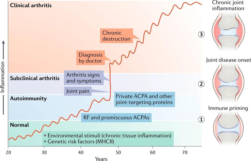
All previously published papers were reproduced with permission from the publisher.
Published by Karolinska Institutet.
Printed by Universitetsservice US-AB, 2021
© Erik Lönnblom, 2021
ISBN 978-91-8016-257-9
Cover illustration: Painting by Eugène Delacroix depicting the Sack of Constantinople in
1204. "It is tragic that the assailants, who set out to secure free access for Christians to the
Holy Land, turned against their brothers in the faith." - Pope John Paul II
Antibodies as Pathogenic Factors and Biomarkers in
Rheumatoid Arthritis
THESIS FOR DOCTORAL DEGREE (Ph.D.)

By

Erik Lönnblom

The thesis will be defended in public at Gustaf Retzius Lecture hall, Berzelius väg 3,
Karolinska Institutet, Solna, Friday the 11:th of June 2021, at 9:00 am

Principal Supervisor:                           Opponent:
Professor Rikard Holmdahl, MD, PhD              Professor Carl Turesson, MD, PhD
Karolinska Institutet                           Lund University
Department of Medical Biochemistry and          Department of Clinical Sciences
Biophysics                                      Division of Rheumatology
Division of Medical Inflammation Research
                                                Examination Board:
Co-supervisor(s):                               Professor Kristina Lejon, PhD
Professor Kutty Selva Nandakumar, PhD           Umeå University
Southern Medical University                     Department of Microbiology
School of Pharmaceutical Sciences               Division of Immunology/Immunochemistry
SMU-KI United Medical Inflammation Center
                                                Dr. Aikaterini Chatzidionysiou, MD, PhD
Dr. Chris Kessel, PhD                           Karolinska University Hospital
WWU Medical Center (UKM)                        Rheumatology Unit
Department of Pediatric Rheumatology &
Immunology                                      Assistant Professor Lina Marcela Diaz-Gallo, PhD
Division of Translation Inflammation Research   Karolinska Institutet
                                                Department of Medicine
                                                Division of Rheumatology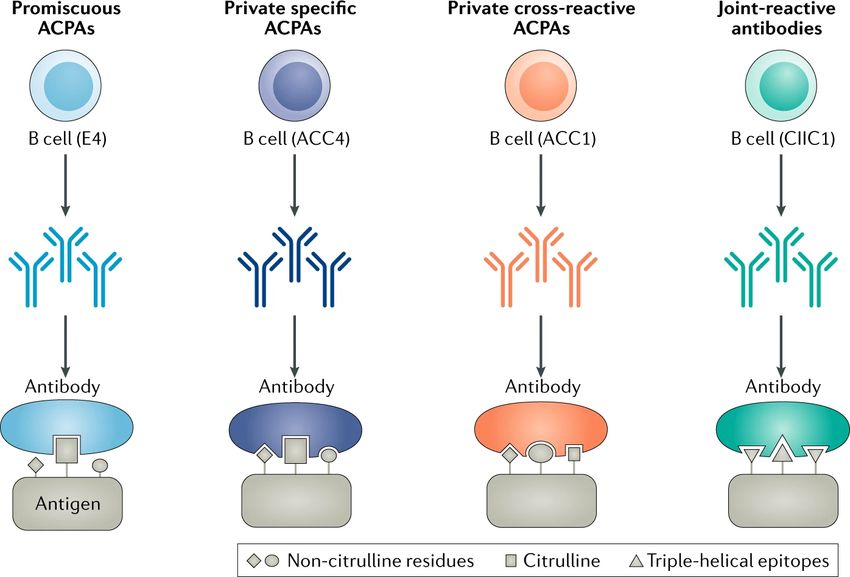
What we have done for ourselves alone dies with us; what we have done for others and the
world remains and is immortal.
– Albert Pike

I hate quotations. Tell me what you know.
– Ralph Waldo Emerson
POPULAR SCIENCE SUMMARY OF THE THESIS
The largest leap in the evolution of our immune system occurred around 400 million years ago
with the development of a new compartment known as the adaptive immune system. Instead
of relying on inherited immune receptors passed down over generations, a new set of tools
allowed the random generation of new immune receptors capable of binding almost any
antigen, even those never encountered before. While the new system greatly improved our
ability to quickly adapt to new pathogens, the random receptors also posed a new risk; some of
the new receptors might target antigens in our own body. This new weapon to protect the body
was the equivalent of a nuclear bomb in the evolutionary arms race between host and pathogen.
Although instead of fearing mutual destruction from a nuclear war, we were, from this point
forward, at risk of destroying our own body. Several control mechanisms developed to prevent
the immune system turning against us. Despite this, investigators today have identified over
100 different types of autoimmune diseases targeting almost every imaginable tissue, from
head to toe.

One of the most common autoimmune diseases is rheumatoid arthritis (RA) which affects
nearly 1% of the world population, predominantly women. In RA the joints are attacked by the
immune system causing joint inflammation and pain, this eventually leads to joint destruction
if not treated. The most common biomarkers used to diagnose RA in the clinics are
autoantibodies; the origin and function of these are however not known.

The work presented in this thesis is an attempt at discovering the origin and function of different
subclasses of autoantibodies. One of the subclasses are antibodies against type II collagen (CII),
the major protein in the joint cartilage. These antibodies have previously been shown to be
associated with the onset of RA, and cause arthritis when injected in animals. To better
understand them, we have synthesized a library of CII peptides and used them to screen the
serum of more than 4000 RA patients and 1500 healthy controls, confirming that several of
them are associated with RA.

If these antibodies could be identified in patients at an early stage, they may become the target
for new treatment strategies. Depleting or neutralizing them could reduce symptoms such as
antibody mediated pain or may even prevent RA from becoming established.

There is currently no diagnostic test available for these antibodies, although the hope is that the
new knowledge generated during this PhD project may help generate new biomarkers to aid in
the diagnosis and treatment of RA.
ABSTRACT
Ever since the evolution of an adaptive immune system capable of creating immune receptors
that may recognize self-antigens, we have been at risk of autoimmunity. There are over 100
different types of autoimmune diseases targeting almost every available tissue from head to
toe, with joints and connective tissues being a common target. In addition to autoimmune
diseases, infections and degenerative joint diseases can cause joint inflammation making
differential diagnosis between them difficult.

The most common autoimmune disease to afflict the joints is rheumatoid arthritis (RA),
affecting nearly 1% of the world population, predominantly women. The etiology of RA is not
known, although it involves interaction between multiple genes and environmental risk factors.
It is characterized by chronic inflammation of the joints, which without successful treatment
can lead to joint destruction. One of the hallmarks of RA is the presence of autoantibodies,
often observed in serum several years before any symptoms of disease. The two classes of
autoantibodies focused on today are rheumatoid factors (RF) and anti-citrullinated protein
antibodies (ACPA), the latter being a highly specific biomarker for a large subset of RA-
patients. The ACPA have greatly aided in diagnosing RA in many patients. Yet their function
and origin are still not known. Nevertheless, a subset of patients still lacks a specific biomarker.

All studies in this thesis have autoantibodies in arthritis as a common theme, and four of them
use a bead-based multiplex platform established during the PhD-project. In Study I, we
explored the hypothesized link between periodontitis induced by the oral pathogen
P.gingivalis, and its effect on arthritis progression and the production of ACPA. This study
revealed a citrulline specific antibody response against P.gingivalis peptidyl arginine
deiminase derived peptide, although the link to the arthritis development could not be
confirmed. In Study II, we synthesized a library of triple helical peptides (THP) as a tool to
characterize antibodies against type II collagen (CII). The peptides were tested in two cohorts
of RA patients, as well as on monoclonal antibodies (mAb), and in collagen induced arthritis.
The THPs were subsequently used in Study III to elucidate the specificity and function of
antibodies against type XI collagen (CXI), revealing a shared epitope between CXI and CII in
mice, rats and humans with arthritis. In addition, the THPs were also used in Study IV to
explore the cross-reactivity of a joint-reactive mouse ACPA, demonstrating a molecular
mechanism of how an ACPA can trigger arthritis. For Study V, the specificity of several human
ACPA were dissected with a bead based multiplex assay and compared to polyclonal responses
in two RA cohorts. Crystal structures of the ACPA revealed for the first time the structural
basis of how human ACPA bind citrulline residues on different peptides.

The data presented in this thesis provide further evidence that the major determinant of the
arthritogenicity of antibodies lies in their ability to cross-react to joint proteins. Dissecting these
specificities may lead to the establishment of new clinical biomarkers.
LIST OF SCIENTIFIC PAPERS

 I. Effects by periodontitis on pristane-induced arthritis in rats
    Eriksson K*, Lönnblom E*, Tour G, Kats A, Mydel P, Georgsson P,
    Hultgren C, Kharlamova N, Norin U, Jönsson J, L Lundmark A, Hellvard A,
    Lundberg K, Jansson L, Holmdahl R, Yucel-Lindberg T.
    J Transl Med. 2016 Nov 3;14(1):311

    * These authors contributed equally.

 II. Synthesis of an Array of Triple-Helical Peptides from Type II Collagen
     for Multiplex Analysis of Autoantibodies in Rheumatoid Arthritis
     Viljanen J, Lönnblom E, Ge C, Yang J, Cheng L, Aldi S, Cai W, Kastbom A,
     Sjöwall C, Gjertsson I, Holmdahl R, Kihlberg J.
     ACS Chem Biol. 2020 Sep 18;15(9):2605-2615

III. A Shared Epitope of Collagen Type XI and Type II Is Recognized by
     Pathogenic Antibodies in Mice and Humans with Arthritis
     Tong D, Lönnblom E, Yau ACY, Nandakumar KS, Liang B, Ge C, Viljanen
     J, Li L, Bãlan M, Klareskog L, Chagin AS, Gjertsson I, Kihlberg J, Zhao M,
     Holmdahl R.
     Front Immunol. 2018 Apr 12;9:451

IV. Anti-citrullinated protein antibodies cause arthritis by cross-reactivity to joint
    cartilage
    Ge C, Tong D, Liang B, Lönnblom E, Schneider N, Hagert C, Viljanen J,
    Ayoglu B, Stawikowska R, Nilsson P, Fields GB, Skogh T, Kastbom A,
    Kihlberg J, Burkhardt H, Dobritzsch D, Holmdahl R.
    JCI Insight. 2017 Jul 6;2(13):e93688

 V. Structural Basis of Cross-Reactivity of Anti-Citrullinated Protein Antibodies
    Ge C, Xu B, Liang B, Lönnblom E, Lundström SL, Zubarev RA, Ayoglu B,
    Nilsson P, Skogh T, Kastbom A, Malmström V, Klareskog L, Toes REM,
    Rispens T, Dobritzsch D, Holmdahl R.
    Arthritis Rheumatol. 2019 Feb;71(2):210-221
SCIENTIFIC PAPERS NOT INCLUDED IN THE THESIS
VI. Natural polymorphism of Ym1 regulates pneumonitis through
alternative activation of macrophages
Zhu W, Lönnblom E, Förster M, Johannesson M, Tao P, Meng L, Lu S,
Holmdahl R.
Sci Adv. 2020 Oct 21;6(43):eaba9337
VII. Cartilage-binding antibodies initiate joint inflammation and promote
chronic erosive arthritis
Li Y, Tong D, Liang P, Lönnblom E, Viljanen J, Xu B, Nandakumar KS,
Holmdahl R.
Arthritis Res Ther. 2020 May 24;22(1):120
VIII. The autoantibody response to cyclic citrullinated collagen type II
peptides in rheumatoid arthritis
Liang B, Ge C, Lönnblom E, Lin X, Feng H, Xiao L, Bai J, Ayoglu B,
Nilsson P, Nandakumar KS, Zhao M, Holmdahl R.
Rheumatology (Oxford). 2019 Sep 1;58(9):1623-1633
IX. The involvement of Toll-like receptor 9 in the pathogenesis of erosive
autoimmune arthritis
Fischer A, Abdollahi-Roodsaz S, Böhm C, Niederreiter B, Meyer B, Yau
ACY, Lönnblom E, Joosten LAB, Koenders M, Lehmann CHK, Dudziak D,
Krönke G, Holmdahl R, Steiner G.
J Cell Mol Med. 2018 Sep;22(9):4399-4409
X. Influence of hydrocarbon oil structure on adjuvanticity and
autoimmunity
Yau ACY, Lönnblom E, Zhong J, Holmdahl R.
Sci Rep. 2017 Nov 8;7(1):14998
XI. Regulation of autoantibody activity by the IL-23-TH17 axis determines
the onset of autoimmune disease
Pfeifle R, Rothe T, Ipseiz N, Scherer HU, Culemann S, Harre U, Ackermann
JA, Seefried M, Kleyer A, Uderhardt S, Haugg B, Hueber AJ, Daum P,
Heidkamp GF, Ge C, Böhm S, Lux A, Schuh W, Magorivska I, Nandakumar
KS, Lönnblom E, Becker C, Dudziak D, Wuhrer M, Rombouts Y, Koeleman
CA, Toes R, Winkler TH, Holmdahl R, Herrmann M, Blüml S, Nimmerjahn
F, Schett G, Krönke G.
Nat Immunol. 2017 Jan;18(1):104-113XII. Type II collagen antibody response is enriched in the synovial fluid of
rheumatoid joints and directed to the same major epitopes as in collagen
induced arthritis in primates and mice
Lindh I, Snir O, Lönnblom E, Uysal H, Andersson I, Nandakumar KS,
Vierboom M, 't Hart B, Malmström V, Holmdahl R.
Arthritis Res Ther. 2014 Jul 8;16(4):R143
XIII. Natural polymorphisms in Tap2 influence negative selection and
CD4∶CD8 lineage commitment in the rat
Tuncel J, Haag S, Yau AC, Norin U, Baud A, Lönnblom E, Maratou K,
Ytterberg AJ, Ekman D, Thordardottir S, Johannesson M, Gillett A;
EURATRANS Consortium, Stridh P, Jagodic M, Olsson T, Fernández-Teruel
A, Zubarev RA, Mott R, Aitman TJ, Flint J, Holmdahl R.
PLoS Genet. 2014 Feb 20;10(2):e1004151CONTENTS
1 INTRODUCTION........................................................................................................... 1
1.1 The immune system............................................................................................... 1
1.2 Innate immunity..................................................................................................... 1
1.3 Adaptive immunity ................................................................................................ 1
1.4 Rheumatoid arthritis .............................................................................................. 2
1.5 Classification of RA .............................................................................................. 2
1.6 Genetics of RA ...................................................................................................... 5
1.7 Environmental risk factors .................................................................................... 5
1.7.1 Cigarette smoking ..................................................................................... 5
1.7.2 Aerosols ..................................................................................................... 6
1.7.3 Periodontitis............................................................................................... 6
1.7.4 Porphyromonas gingivalis ........................................................................ 6
1.7.5 Aggregatibacter actinomycetemcomitans (Aa) ........................................ 7
1.7.6 Other infectious agents.............................................................................. 8
1.8 Structure of collagen.............................................................................................. 8
1.8.1 Type II collagen (CII) ............................................................................... 9
1.8.2 Type XI collagen (CXI) ............................................................................ 9
1.9 Animal models....................................................................................................... 9
1.9.1 Collagen induced arthritis (CIA) ............................................................10
1.9.2 Collagen type XI induced arthritis (CXIIA) ............................................10
1.9.3 Collagen antibody induced arthritis (CAIA) ..........................................10
1.9.4 Pristane induced arthritis (PIA) ..............................................................10
1.9.5 Other experimental arthritis models .......................................................11
1.10 Autoantibodies in RA ..........................................................................................11
1.10.1 Rheumatoid Factors (RF) ........................................................................11
1.10.2 Anti-citrullinated proteins antibodies (ACPA).......................................11
1.10.3 Anti-modified protein antibodies (AMPA) ............................................12
1.10.4 Anti-CII antibodies..................................................................................12
1.11 The pathogenicity of autoantibodies ...................................................................13
1.12 Pathogenesis of RA .............................................................................................14
2 AIMS .............................................................................................................................17
3 METHODOLOGICAL CONSIDERATIONS ............................................................19
3.1 Patient material ....................................................................................................19
3.1.1 TIRA-2 ....................................................................................................19
3.1.2 BARFOT .................................................................................................19
3.1.3 EIRA-1 ....................................................................................................19
3.1.4 Population controls..................................................................................19
3.2 Experimental animals ..........................................................................................20
3.3 Ligature-induced experimental periodontitis ......................................................20
3.4 Experimental arthritis ..........................................................................................20
3.4.1 Pristane Induced Arthritis (PIA) .............................................................203.4.2 Collagen Induced Arthritis (CIA) ........................................................... 20
3.4.3 Collagen Antibody Induced Arthritis (CAIA) ....................................... 20
3.4.4 Bead-based multiplex assay .................................................................... 21
3.5 Statistical Methods .............................................................................................. 21
3.6 Ethical considerations ......................................................................................... 21
4 PRESENT INVESTIGATIONS ................................................................................... 23
4.1 Study I: Effects by periodontitis on pristane-induced arthritis in rats ............... 23
4.2 Study II: Synthesis of an Array of Triple-Helical Peptides from Type II
Collagen for Multiplex Analysis of Autoantibodies in Rheumatoid
Arthritis ................................................................................................................ 25
4.3 Study III: A Shared Epitope of Collagen Type XI and Type II Is
Recognized by Pathogenic Antibodies in Mice and Humans with Arthritis ..... 27
4.4 Study IV: Anti-citrullinated protein antibodies cause arthritis by cross-
reactivity to joint cartilage................................................................................... 28
4.5 Study V: Structural Basis of Cross-Reactivity of Anti-Citrullinated
Protein Antibodies ............................................................................................... 30
5 CONCLUSIONS ........................................................................................................... 31
6 FUTURE PERSPECTIVES.......................................................................................... 33
7 ACKNOWLEDGEMENTS.......................................................................................... 35
8 REFERENCES .............................................................................................................. 41LIST OF ABBREVIATIONS ACPA Anti-citrullinated protein antibodies ACR American College of Rheumatology AMPA Anti-modified protein antibodies Anti-CarP Anti-carbamylated protein antibodies AKA Antikeratin antibodies APK Antiperinuclear Factor BCR B cell receptor CAIA Collagen antibody induced arthritis CCP Cyclic citrullinated peptides CCP2 Cyclic citrullinated peptides, version 2 CD Circular Dichroism CIA Collagen induced arthritis CII Type II collagen CEP-1 Citrullinated human alpha-enolase peptide 1 CFA Complete Freund’s adjuvant CLR C-type lectin receptors COMP Cartilage oligomeric matrix protein CPP3 Citrullinated P.PAD peptide 3 CTLA4 Cytotoxic T-lymphocyte-associated protein 4 CXI Type XI collagen DAMP Damage-associated molecular patterns DIP Distal interphalangeal EULAR The European League Against Rheumatism GPI Glucose-6-phospho-isomerase IgG Immunoglobulin G LPS Lipopolysaccharide mAb Monoclonal antibody MCP Metacarpophalangeal MHC Major histocompatibility complex NET Neutrophil extracellular trap
NLR            NOD-like receptors
PAD            Peptidyl Arginase Deiminase
PAMP           Pathogen-associated molecular patterns
P.gingivalis   Porphyromonas gingivalis
P.PAD          P.gingivalis PAD enzyme
PRRs           Pattern recognition receptors
PTM            Posttranslational modification
PTPN22         Protein tyrosine phosphatase, non-receptor type 22
RA             Rheumatoid arthritis
RF             Rheumatoid factor
RgpB           Arg‐gingipain B
PIA            Pristane induced arthritis
PIP            Proximal interphalangeal
ROS            Reactive oxygen species
RLR            RIG-I-like receptors
SE             Shared epitope
SLE            Systemic lupus erythematosus
TCR            T cell receptors
THP            Triple helical peptide
TLR            Toll-like receptors
TNF-α          Tumor necrosis factor-alpha
1 INTRODUCTION
1.1   THE IMMUNE SYSTEM
The role of the immune system is to protect the organism from diseases caused by foreign
organisms and materials such as bacteria, viruses, fungi, and toxins. Even though we lack
records from the earliest forms of life, it can be argued that the evolution of the immune system
started with the origin of life. At a first glance the complexity of the human immune system
involving a multitude of organs, cells and small molecules could be mistaken for an example
of intelligent design. Yet as we examine our evolutionary ancestors, we can follow the gradual
evolution of the immune system along the phylogenetic tree of life1.

The degree of complexity of the immune system varies between organisms of different
kingdoms and species, although it can be divided into different layers of protection. Surface
barriers are the first line of defence to keep pathogens outside. They can be either mechanical,
chemical, or biological. In humans they can be exemplified by the skin, low-pH environment
in the stomach and commensal flora.

1.2   INNATE IMMUNITY
If the surface barriers are breached, the immediate response to the threat is orchestrated by the
innate immune system, which is found in nearly all forms of life. Even bacteria have simple
heritable defense mechanisms2, while all multicellular organisms appear to have a complex
innate immune system3,4. The first function of the innate immune system is to identify the
pathogens. This is done through pattern recognition receptors (PRRs)5, which are mainly
expressed on cells of the innate immune system, such as macrophages, monocytes, dendritic
cells, neutrophils, and mast cells. PRRs recognize molecules of two different classes: pathogen-
associated molecular patterns (PAMP), which are conserved molecular motifs expressed on
pathogens and damage-associated molecular patterns (DAMP) released by host cells as a result
of cell damage or cell death. Examples of PRRs include Toll-like receptors (TLR) capable of
binding lipopolysaccharide (LPS) of Gram-negative bacteria6, and C-type lectin receptors
(CLR) binding carbohydrate structures such as mannan on fungi cell walls7. The two other
families of PRRs are Nod-like receptor (NLR) and RIG-I-like receptor (RLR)8. Once the PRRs
of the immune system have been activated, cells release inflammatory mediators to recruit
neutrophils and macrophages to the site of inflammation. Some of the effector mechanisms to
kill the pathogens include phagocytosis, complement system and release of reactive oxygen
species (ROS)9.

1.3   ADAPTIVE IMMUNITY

The final line of defense is the adaptive immune system. The principal components of our
adaptive immune system are T and B lymphocytes, which through somatic recombination can
assemble T and B cell receptors (TCR and BCR) specific for an immense range of antigens. In
contrast to the innate immunity, an adaptive immunity response is not immediate. However, it
is able to form immunological memory. These components are found in all jawed vertebrates

                                                                                               1
and marks a clear distinction in the evolution of the immune system, although jawless
vertebrates carry a precursor to our adaptive immune system with other recombinatorial
genes10. To put this into perspective, the earliest fossil records of jawed vertebrates are more
than 400 million years old11. The random generation of TCRs and BCRs does however have
the potential to generate receptors capable of recognizing self-antigens. To address this
problem the development of T and B cells undergo a stringent process of selection to ensure
self-tolerance; any cell not following the protocol may soon find itself on a path to
autoimmunity12.
1.4 RHEUMATOID ARTHRITIS
Rheumatoid arthritis (RA) is an autoimmune disease characterized by chronic inflammation of
peripheral joints causing cartilage destruction and bone erosion. The disease occurs in up to
1% of the population, with women more often being affected than men. While the peripheral
joints of the hands, feet and wrists are frequently affected, it is considered a systemic disease
since it can also affect cardiovascular, skeletal, and respiratory systems. The cause of RA is not
known, although the current dogma is that for a subset of patients, autoimmunity starts in a
mucosal tissue in response to a chronic inflammation. This subset of RA patients is
characterized by anti-citrullinated protein antibodies (ACPA) present in around 70% of
patients13, measured in clinics using the commercial CCP2-test (cyclic citrullinated peptides
kit, version 2). ACPA are a heterogenous subset of antibodies that can be detected in 1/5 of the
patients already 10 years before onset of arthritis14. The other prominent autoantibody is
rheumatoid factor (RF), often present together with ACPA15. The strongest genetic association
to RA is with the HLA-DRB1 allele which together with environmental risk factors such as
cigarette smoking show an epistatic interaction in the ACPA positive subset of RA patients16.
There is currently no cure for RA, although the introduction of biological drugs targeting pro-
inflammatory cytokines such as tumor necrosis factor-alpha (TNF-α) has significantly reduced
the burden of disease17.
1.5 CLASSIFICATION OF RA
In a disease such as rheumatoid arthritis in which the etiology is unknown and in which
there is no available proof of the diagnosis, a broad description of the disease usually
suffices for teaching and for diagnosis in individual cases. When such a method is used for
classifying patients for study, however, there is little uniformity in the cases included in
any series labelled rheumatoid arthritis. (Ropes MW et al, 1957)18
One of the first attempts to classify RA was made by the American Rheumatism Association
(today known as ACR, American College of Rheumatology) in 1957, in which patients needed
to fulfill 5 out of 11 criteria to be classified as “definite” RA18. There were gradual
modifications to the criteria over time, although it was not until a major overhaul leading to the
1987 ACR revised classification criteria for RA19, that this classification became widespread.
The revised 1987 guidelines were designed with the intention to be more specific creating a
stricter definition of RA. In addition, it also removed 3 earlier criteria which required invasive
procedures.
2Table 1. The 1987 ACR revised classification criteria for RA.
Criterion Description
Morning stiffness Morning stiffness in and around the joints, lasting at least one hour
before maximal improvement.
Arthritis of three or At least three joint areas (out of 14 possible areas; right or left PIP,
more joint areas MCP, wrist, elbow, knee, ankle, MTP joints) simultaneously have
had soft tissue swelling or fluid (not bony overgrowth alone) as
observed by a physician.
Arthritis of hand At least one area swollen (as defined above) in a wrist, MCP, or PIP
joints joint.
Symmetric arthritis Simultaneous involvement of the same joint areas (as defined
above) on both sides of the body (bilateral involvement of PIPs,
MCPs, or MTPs, without absolute symmetry is acceptable).
Rheumatoid Subcutaneous nodules over bony prominences or extensor surfaces,
nodules or in juxta-articular regions as observed by a physician.
Serum rheumatoid Demonstration of abnormal amounts of serum rheumatoid factor by
factor any method for which the result has been positive in less than 5
percent of normal control subjects.
Radiographic Radiographic changes typical of rheumatoid arthritis on
changes posteroanterior hand or wrist radiographs, which must include
erosions or unequivocal bony decalcification localized in, or most
marked adjacent to, the involved joints (osteoarthritis changes alone
do not qualify).
Note: For classification purposes, a patient has RA if at least four of these criteria are
satisfied (the first four must have been present for at least six weeks).
Table adapted from Arnett et. al, The American Rheumatism Association 1987 revised criteria for the classification
of rheumatoid arthritis, Arthritis & Rheumatism (1988) 31(3) 315-324
3Table 2. 2010 ACR/EULAR RA classification criteria
Domain Category Point score
A Joint involvement (0–5 points)
1 large joint 0
2–10 large joints 1
1–3 small joints (large joints not counted) 2
4–10 small joints (large joints not counted) 3
>10 joints including at least one small joint 5
B Serology (0–3 points)
Negative RF and negative ACPA 0
Low positive RF or low positive ACPA 2
High positive RF or high positive ACPA 3
C Acute-phase reactants (0–1 point)
Normal CRP and normal ESR 0
Abnormal CRP or abnormal ESR 1
D Duration of symptoms1.6 GENETICS OF RA
The heritability of RA is estimated to be around 60% based on twin studies, with a concordance
rate among monozygotic twins at 15-30%20. The major histocompatibility complex (MHC)
region contributes to more than half of the identified genetic heritability, with a set of HLA-
DRB1 alleles known as “shared epitope” having the largest contribution21. Shared epitope is a
five amino acid motif (70-74) in the DRβ1 chain with either QKRAA, QRRAA or RRRAA
shared between many DRB1 alleles. The most strongly associated allele in Caucasians is the
DRB1*0401 which in addition also encodes risk associated amino acids in position 11, 71 and
7422. Over 100 risk loci have been identified in RA23, although the disease association among
the individual non-MHC genes is much weaker. Many of these are involved in regulation of
the adaptive immune system (e.g., loci containing PTPN22, CD28 and CTLA4). Importantly,
the MHC class II region and several non-MHC genes are associated with a specific
autoantibody response to citrullinated proteins (anti-citrullinated protein antibodies, ACPA)24.
While the heritability is the same for ACPA positive and negative RA, there is no predominant
contribution of specific HLA-alleles in ACPA negative RA12. Another locus found in genetic
studies of Japanese and Korean cohorts contains PADI4 which encodes the enzyme Peptidyl
Arginine Deiminase 4 (PAD4) expressed in myeloid cells. PAD4 is proposed to be the PAD
enzyme responsible for generating citrullinated autoantigens during chronic inflammation in
mucosal tissues25. Surprisingly, this locus is associated to both ACPA positive and negative
RA16,26. The total contribution of the risk genes identified so far cannot explain the estimated
heritability in RA16, this gap is known as “the problem of missing heritability”, is seen in many
complex diseases and is hypothesized to be caused by rare genetic variants, gene-gene
interactions or gene-environment interactions not yet discovered27. Another explanation could
be that the heritability from early twin studies is overestimated. However, a recent study on
monozygotic twins shows that 15% have a substantial number of early developmental
mutations specific to only one of them, implicating we may have underestimated the
contribution of heritability in twin studies.28
1.7 ENVIRONMENTAL RISK FACTORS
1.7.1 Cigarette smoking
The strongest environmental risk factor to date is cigarette smoking that has been demonstrated
in a multitude of cohorts with RA patients29–33. The association with smoking also shows a
possible epistatic effect together with several genetic risk factors highlighting the complex
interactions between genes and environment in RA16,34,35. Adding to the complexity, cigarette
smoke is a complex mixture of more than 7000 chemicals. One major component of cigarette
smoke is nicotine which induces an anti-inflammatory effect through the cholinergic pathway
and is shown to be protective in several autoimmune diseases and animal models of RA36,37.
Nicotine is however also reported to induce formation of neutrophil extracellular traps (NETs)
which release intracellular components that may act as autoantigens38. There are in addition
other components of cigarette smoke shown to correlate with RA39,40.
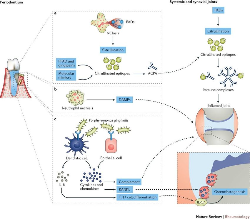
51.7.2 Aerosols Cigarette smoking is only one of the many aerosols that have been implicated in the etiology of RA as well as in several other autoimmune diseases; other examples are air pollution, silica dust, textile dust, pesticides, wood dust and mineral oils. Although they may act through different molecular pathways for the breach of self-tolerance, the common denominator is that these compounds may induce a chronic inflammation in the lung and in addition many of them show an epistatic effect together with cigarette smoking16,41. 1.7.3 Periodontitis Periodontitis is an inflammation of the periodontium, which in its severe chronic form affects 10% of the world population42. The association between oral health and inflammatory arthritis was described already by Hippocrates in ancient Greece who suggested pulling out teeth could cure arthritis. In addition to sharing pathophysiological similarities, they also have genetic (HLA-DRB1) and environmental (cigarette smoking) risk factors in common. 1.7.4 Porphyromonas gingivalis One of the bacteria causing periodontitis (PD) is Porphyromonas gingivalis (P.gingivalis) found in around 80% of patients with periodontal disease43. It expresses a PAD enzyme (P.PAD) capable of citrullination44 not found in any other prokaryote to date. The proposed mechanism is that P.PAD will generate citrullinated epitopes of bacterial origin, which through a process of molecular mimicry will lead to cross-reactivity to citrullinated self-antigens (Figure 1)45. Ever since a hypothesis that citrullination by P.PAD could be a causative link between PD and RA46 was presented, the scientific community has put considerable effort into investigating this with mixed results45,47,48. While most reported experimental models show an increased severity in the group treated with P.gingivalis, one major problem in characterizing the ACPA response is the lack of a non-citrullinated control; the CCP2-kit used in most studies does not include one. Moreover, other virulence factors in P.gingivalis such as LPS, collagenase and arginine gingipain B (RgpB) may also have a contribution to the link between PD and RA49. In summary, although epidemiological and experimental data support the role of P. gingivalis in the etiology of RA, there is yet no conclusive experimental evidence proving that citrullination by P.PAD is the causative link47. 6
Figure 1. Proposed mechanisms underlying the links between periodontal disease and the pathogenesis of
rheumatoid arthritis. Figure from Potempa, et. al. Nat Rev Rheumatol 13, 606–620 (2017). Reprinted with
permission from Nature.
1.7.5 Aggregatibacter actinomycetemcomitans (Aa)
While the scientific community studying the possible link between PD and RA has almost
exclusively focused on P.gingivalis, a study from 2016 by Konig et. al.50 proposed the
periodontal pathogen Aggregatibacter actinomycetemcomitans (Aa) as a candidate microbe
responsible for the break of tolerance in RA. The proposed mechanism is that a pore-forming
leukotoxin-A (LtxA) produced by Aa induces hypercitrullination in neutrophils, which through
a process similar to NET formation exposes citrullinated self-antigens in the gingival tissue.
Reports on experimental animal models exploring the link are so far scarce51, most certainly
due to earlier reports that LtxA is specific for leukocytes of human or Old World primate
origin52, although this narrow specificity is disputed53. The role of Aa in the development of
RA has so far yielded contradictory data47,54–57.
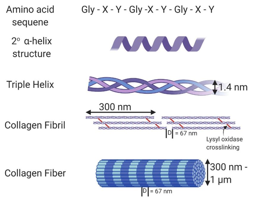
71.7.6 Other infectious agents The idea that a pathogen may cause RA is not new, early theories suggested that the disease originated in pre-Columbian America and was spread to the Old World by the Europeans58. One of the most well studied pathogens is Epstein-Barr virus59–61 which has been detected in the synovial fluid in RA patients62. Subsequent theories include an unknown microbe spreading through blood transfusions63 and subclinical urinary tract infections by Proteus mirablis64. 1.8 STRUCTURE OF COLLAGEN Collagens are the most abundant proteins; it is present in all animals with cells differentiated into tissues and makes up approximately 30% of the protein of mammals65. A total of 28 distinct collagen proteins have been described, with the main fibril-forming collagens (type I, II, III, V and XI) accounting for 80-90% of all collagens in humans. The structural motif that defines the collagen proteins is their ability to form a triple helix made up by three polypeptide chains (Figure 2). The trimers are made up of either three identical polypeptide chains to form homotrimers (type II), or different chains to form heterotrimer (type I, XI)66. Figure 2. Structure of collagen. The amino acid sequence of collagen consists of Gly-Xaa-Yaa repeats, with Xaa and Yaa commonly occupied by proline and hydroxyproline. This unique sequence allows collagen to form an α helix secondary structure. Fibrillar collagen is a triple helix containing crosslinks formed through the action of lysyl oxidase. In vivo, these collagen fibrils form fibers with varying thickness and a D-banding pattern of 67 nm. Figure from Walimbe et.al., Best of Both Hydrogel Worlds: Harnessing Bioactivity and Tunability by Incorporating Glycosaminoglycans in Collagen Hydrogels.Bioengineering. 2020; 7(4):156. 8
1.8.1 Type II collagen (CII)
CII is the major protein of the articular cartilage covering the joint surfaces and is made up of
three identical α1(II) chains translated from the Col2a1 gene. In addition, it is also found in the
cartilage of the nose, larynx, trachea and the vitreous. In similarity to other collagens, the
sequence is highly conserved between species and the major B-cell epitopes are shared between
humans, primates and rodents67.
1.8.2 Type XI collagen (CXI)
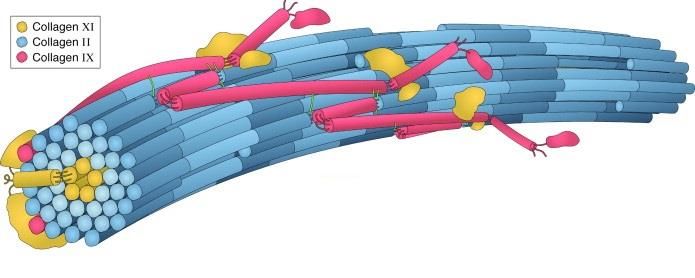
CXI is a heterotrimer composed of three distinct chains (α1(XI), α2(XI), α3(XI)). CXI forms
heterotypic fibrils together with CII and type IX collagen (CIX) and is often colocalized with
CII. The exact structure of the heterotypic fibrils is not determined, but it is hypothesized that
CXI constitutes the inner core of the fibrils and therefore is less exposed (Figure 3). The α3(XI)
is expressed by the Col2a1. As a result, it is identical to the α1(II) chain in CII apart from a
higher degree of hydroxylation and glycosylation68.
Figure 3. Three-dimensional model of the type II/IX/XI heterotypic fibril of developing cartilage matrix.
Reprinted from Methods, 45/1, Eyre et. al. Advances in collagen cross-link analysis, 65-74., Copyright (2008),
with permission from Elsevier
1.9 ANIMAL MODELS
The most commonly used animal for medical research is the mouse, and advances in recent
decades to create genetically modified mice has made it into the model organism of choice for
immunological studies69. Our latest common ancestor with the mouse is estimated at around
65 million years ago and our immune systems have since evolved not only to different
ecological niches, but also different body size and life span. Some of the major differences that
have emerged between the humans and mice immune system include the balance between
neutrophils and lymphocytes in peripheral blood, Toll receptors, Immunoglobulin subsets, Fc
receptors and regulation and development of different B- and T-cell subsets70. Despite the
lengthy period on diverging evolutionary paths using the mouse and other animals as a model
organism for the immune system has been instrumental for our current knowledge71 and
alternatives to animal models to study complex diseases are limited72.
91.9.1 Collagen induced arthritis (CIA) The current gold standard for preclinical experiments in arthritis is collagen induced arthritis (CIA) where CII is emulsified in complete Freund’s adjuvant73. Similar to RA, a major genetic risk factor for CIA is the MHC haplotype requiring a CII permissive MHC such as H-2r or H- 2q74. CIA can also induce arthritis in genetically susceptible rats and several species of monkeys, revealing that the major B-cell epitopes are conserved between species75. The disease development is dependent on both T and B-cells, leading to the production of anti-CII antibodies which are the major effector mechanism for disease induction. Finally, it was an important tool in development and testing of several biologically based therapeutics, such as IL-176, IL-677 anti-TNF-α78. 1.9.2 Collagen type XI induced arthritis (CXIIA) A variant of the traditional CIA model is to substitute CII with CXI in the experimental protocol79. CXI forms a heterotrimer from three different chains (α1(XI), α2(XI), α3(XI)), one of which (α3) is shared with CII. As a result, a subset of anti-CXI antibodies can cross-react to CII80. Arthritis induced with homologous CXI leads to a more pronounced chronic relapsing arthritis compared to CIA81, displaying increased titers of anti-CXI antibodies during the chronic phase. The model is also MHC-dependent, albeit in rats, the associated MHC haplotypes differs between CIA and CXIIA 82. 1.9.3 Collagen antibody induced arthritis (CAIA) The antibodies induced in CIA can be isolated and used to transfer disease in a model called collagen antibody induced arthritis (CAIA)83. Importantly, the dominant B-cell epitopes in CII are conserved between species67,84 and antibodies against different epitopes have been shown to be either pathogenic or protective85, yet all tested anti-collagen antibodies induce pain86. The standard CAIA cocktail uses 4 mAbs specific for C1, J1, U1 and the D3 epitope and is boosted with LPS87. The CAIA model is B- and T-cell independent and does not involve the priming phase in contrast to other models such as CIA. Consequently, it is used to study the disease onset phase and mechanisms for antibody mediated inflammation. 1.9.4 Pristane induced arthritis (PIA) Arthritis can also be induced in rats through the immunization of different mineral oils88, pristane induced arthritis (PIA) is the most studied model due to high incidence (99,6%) and excellent reproducibility89. PIA has a quick onset starting around 10 days with a peak at around 20 days. Thereafter, the inflammation steadily decreases before the start of a chronic relapsing disease with new inflammation. Isolated CD4+ T-cells can transfer the disease90, regulated by CLRs91. PIA induces the production of both RF, anti-CII92 and anti-CXI antibodies93. However, antibodies are not known to play a major role in the pathogenesis of PIA94. Interestingly, pristane induces chronic relapsing arthritis only in susceptible rat strains. In contrast, mice immunized with pristane display phenotypically different symptoms and instead develop a systemic lupus erythematosus (SLE) like disease in susceptible strains95. 10
1.9.5 Other experimental arthritis models
In addition to collagens, the immunization of other cartilage associated proteins such as
cartilage oligomeric matrix protein (COMP)96 and aggrecan97 can induce arthritis. The
ubiquitous protein glucose-6-phospho-isomerase (GPI) can also induce arthritis upon
immunization with an adjuvant98. The unlikely autoantigen was discovered in the K/BxN
model as a result of transgenic T-cells specific for GPI, leading to the production of anti-GPI
antibodies precipitating on the cartilage surface99 due to an interaction with proteoglycans100.
These antibodies induce arthritis in the K/BxN serum transfer model through similar molecular
mechanisms as CAIA101. Another similarity between GPI and CII as autoantigens is that
arthritis induction through protein immunization is MHC dependent with an association to the
H-2q haplotype98.

Many other proteins have also been implicated as autoantigens in RA due to the cross-reactivity
of ACPA including vimentin, fibrinogen, filaggrin, α-enolase and histones. There have been
attempts to use these proteins to establish new arthritis models102, yet it is debated if any of
them can induce experimental arthritis in a reproducible fashion103. The absence of data from
experimental animal models does not exclude them as potential autoantigens. None of the
animal models captures the full complexity of RA, instead each of them is an attempt to explain
a limited phase or pathway of the disease.

1.10 AUTOANTIBODIES IN RA

1.10.1 Rheumatoid Factors (RF)
Rheumatoid Factors (RF) are antibodies binding the Fc region of immunoglobulin G (IgG).
They were identified in patients with RA more than 70 years ago104. The name RF was given
due to the association with RA, although they are present both in other rheumatic diseases and
non-rheumatic conditions, as well as in healthy individuals, especially the elderly105. The high
prevalence of low-affinity RF during many infections points towards a key role in host
defense106 prior to the induction of the high affinity RF observed in autoimmune disorders. The
reported frequency in RA is between 60-90% and has a specificity of 85%. RF was the only
autoantibody to be included in the 1987 ACR RA classification criteria, and since the
introduction of standardized test for ACPA it is now interpreted together with ACPA status in
clinical settings as exemplified in 2010 ACR/EULAR classification criteria for RA107. The
presence of RF is correlated with a more erosive disease and studies exploring the interaction
between RF and ACPA show a significant additive effect in the double positive subset
(RF+/ACPA+) of patients108,109.

1.10.2 Anti-citrullinated proteins antibodies (ACPA)
The first observation of RA-specific antibodies was reported in 1964 and were named
Antiperinuclear Factor (APK)110. It was followed by antikeratin antibodies (AKA)111 which
were shown to bind the protein filaggrin112. Monoclonal antibodies against filaggrin showed
that APK and AKA were in fact the same RA-specific antibody113. Despite the high specificity

                                                                                             11
for RA, the inconvenient testing method required hindered their use as a serological marker for clinical use. It was not until the discovery that citrulline was the antigenic target114 that standardized assays could be developed and ACPAs were later included in the 2010 ACR/EULAR classification criteria for RA107. The standard assay used for detection of ACPA is the CCP2 ELISA test which contains a set of undisclosed synthetic cyclic citrullinated peptides with a reported sensitivity of over 70% and specificity above 98%115. In addition, there are several reports that the standard CCP2 assay doesn’t capture all ACPA, up to 20% of the CCP2 negative patients have antibodies binding other citrullinated antigens116. Even though ACPA have a high specificity for RA compared to RF105, they are also present in 1-2% of the general population without RA117. During a 3-year follow up of non-RA ACPA positive individuals in the Swedish twin registry only 8,5% developed RA, indicating that a positive ACPA status defined by the CCP2-kit has a limited predictive value for the onset of RA in the general healthy population118. For this reason, ACPA are currently only used as a biomarker for individuals which have symptoms of RA or other joint-related diseases. 1.10.3 Anti-modified protein antibodies (AMPA) In recent years, antibodies binding other posttranslational modifications (PTMs) such as homocitrulline and acetylation have also been found119,120. There has been a controversy if these antibodies are in fact a distinct class of autoantibodies in RA121. A recent report studying BCR sequences from B-cells using citrullinated or acetylated antigens revealed that most mAbs were highly cross-reactive between multiple PTMs120. The high cross-reactivity of antibodies between different PTMs has led to the proposed model that the term ACPA should be replaced by AMPA: anti-modified protein antibody. 1.10.4 Anti-CII antibodies The first report of collagen as a candidate autoantigen RA was made by Steffen in 1970122, although the initial studies in the field did not make clear distinction between different types of collagen making comparisons between studies confusing. The turning point came with the establishment of an experimental arthritis model comparing different types of collagen, identifying type II collagen (CII) as the major candidate123. While the link to the pathogenesis has been difficult to prove experimentally for other autoantibodies in RA, the arthritogenicity of anti-CII antibodies has been thoroughly investigated in experimental arthritis models such as CIA and CAIA67,73,83,85,124,125. In RA, anti-CII antibodies are associated with the acute onset and early stages of disease126,127. The prevalence of anti-CII antibodies in RA has yielded estimates between 3-88%128–130, highlighting the need for a standardized testing protocol with defined reagents. Despite a clear link to the pathogenesis of RA, the detection of anti-CII antibodies has never been established as a biomarker for clinical use. 12
1.11 THE PATHOGENICITY OF AUTOANTIBODIES
Epidemiological studies show that seropositive RA patients, in particular the double positive
RF+/ACPA+ subset, have a more severe disease outcome108,109. One proposed mechanistic link
for the additive effect is that RF forms immune complexes with ACPA which stimulate the
release of pro-inflammatory cytokines from macrophages to stimulate osteoclasts109. There are
reports indicating that ACPA in healthy individuals correlate with bone loss before clinical
onset, but the specificity of ACPAs is not known131. Since the citrullinated cyclic peptides used
in commercial kits are undisclosed and may not be expressed in vivo, one approach to find the
mechanistic link has been to study fine specificities of ACPA against citrullinated peptides
from different putative autoantigens. The proteins that have been studied most extensively
include fibrinogen, vimentin, α-enolase, filaggrin and CII, although with mixed results14,132–135.
Several animal models have reported arthritogenicity of ACPA, although low antibody titers
and lack of appropriate non-citrullinated control in most studies make the causative link
inconclusive136–138. The production of ACPA is however not the feature of any of the
established animal models for arthritis, and B-cells recognizing citrulline are negatively
selected in the mouse139. There are also reports of purified polyclonal and monoclonal human
ACPA with arthritogenic properties140–142.

Finally, the ACPA isolated from mice have been shown to induce (ACC1) or enhance (ACC4)
arthritis143,144. A recent publication of the first crystal structure of a human ACPA revealed how
it was able to cross-react between different citrullinated peptides145. In contrast, the crystal
structures of the two mouse ACPA revealed how they could either cross-react to native CII
(ACC1) or be specific to one citrullinated epitope on CII (ACC4)143,144. The different cross-
reactivity of ACPA in relation to antibodies known to induce arthritis in animal
models96,99,124,143, led to proposing a model of three different classes of ACPA where their
functional roles are determined by their specificity (Figure 4). In this proposed model the
antibodies which cross-react with joint proteins are arthritogenic and have an important role in
the onset of arthritis, while the ACPAs that are widely cross-reactive to citrullinated peptides
may have a role in the pathogenesis after the inflammation in the joint has been established.
However, they may also be non-functional or even regulatory146.

                                                                                               13
Figure 4. Examples of different types of ACPAs and joint-reactive autoantibodies in RA. Illustration of the binding of antigen by different types of autoantibodies in rheumatoid arthritis (RA), each exemplified by a crystallized antibody–peptide complex. Figure from Ge et. al. Nat Rev Rheumatol 15, 503–508 (2019). Reprinted with permission from Nature. 1.12 PATHOGENESIS OF RA To summarize the etiology of RA, a model for disease development in RA divides it into three separate stages; priming, disease onset and chronic joint inflammation147 (Figure 5). At stage one, autoimmunity emerges from exposure to environmental risk factors such as cigarette smoke and pathogens in genetically pre-disposed individuals. While epidemiological studies show a strong epistatic effect between genetic and environmental risk factors in the development of RA, a study of 12,590 twins suggests that induction of ACPA in the general population correlates more strongly with environmental exposures rather than genetic risk factors117. The exact location is not known, although it is postulated to occur in the mucosal membranes such as periodontium, gut, or the lung tissue148. The initial stage is characterized by the production of RF and ACPA which can be detected in the serum several years before the diagnosis in a subset of patients14,149. The antibody profile of these autoantibodies during the pre-symptomatic phase shows low titers and autoantibodies shown to induce arthritis are not present. In the second stage around the onset of RA, the autoantibody profile changes drastically with a rise in titer, affinity, epitope spreading and with antibodies targeting joint proteins14. The joint protein that has been most extensively studied is CII, which is the major protein in the joint cartilage and antibodies against CII in RA are associated with an early inflammatory and 14
destructive phenotype127. Why only a minority of individuals that are ACPA positive develop
arthritis is not known; the current dogma around this switch to joint reactivity argues for a
genetical predisposition, most notably HLA-DRB1 alleles and other T-cell activating genes
such as PTPN22.

The third and final stage is chronic inflammation of the joints. This phase leads to tissue
destruction and remodeling; the patients that have reached this stage represent the vast majority
seen by clinicians. This is also the focus for the current therapies. The most efficient treatments
at this stage target cytokines produced by macrophages such as TNF-α and interleukin-6 (IL-
6), although we currently lack biomarkers for predicting which patients will respond to the
treatment. For seropositive patients, therapies targeting T and B-cells have also shown to be
efficient, indicating that adaptive immunity also plays a role in chronic phase for this subset of
patients150. While anti-B-cell therapy works in a subset of RA patients, the functional role of
autoantibodies in the chronic phase is still not clarified. Once the disease is established, new
cartilage proteins/epitopes normally hidden to the immune system may be exposed. One
candidate autoantigen to illustrate this established phase is type XI collagen which is postulated
to be located inside the collagen fibers, and to which antibodies are seen at a late stage of
disease in experimental arthritis151. The exposure of neoantigens and cross-reactivity between
these and other joint proteins152 could further perpetuate the disease and have an impact on long
term remission.

Figure 5. The three stages of the development of RA. Figure from Ge et. al. Nat Rev Rheumatol 15, 503–508
(2019). Reprinted with permission from Nature.

                                                                                                       15
2 AIMS
The original goal of this PhD project was to investigate environmental risk factors of RA in
animal models of arthritis, with a focus on the origin and function of ACPA. Out of the 5
projects included in the original research plan, two of them are published in this thesis (Study
I and Study IV). The remaining original projects were not included in the thesis due to
limitations in reagents, genetically susceptible mice strains and access to facilities. Instead, the
PhD project took a new turn and led to establishing a platform for studying joint-reactive
autoantibodies and using it to analyze animal models of arthritis, monoclonal antibodies and
human patient cohorts with inflammatory joint diseases (Study II-V).
Study I examined the age-old theory of a link between periodontitis and arthritis. Our
investigation focused on the hypothesis by Rosenstein et. al. that the immune response to the
oral pathogen P.gingivalis may be involved in the etiology of RA46. While there are several
mechanisms by which a P.gingivalis induced periodontitis can influence arthritis development,
the cornerstone in this theory is that P.PAD enzyme could induce autoimmunity in RA through
molecular mimicry of citrullinated antigens.
In Study II, our goal was to address a major bottleneck for characterizing CII as an autoantigen
in RA: a library of high-quality triple helical peptides. Once established, the library would be
instrumental for investigating the role of anti-CII as biomarkers and pathogenic factors in
arthritis.
For Study III the aim was to examine CXI as an autoantigen in arthritis by immunization and
isolating anti-CXI clones to characterize their function, specificity and arthritogenicity.
An ACPA-clone of mouse origin that binds both cyclic citrullinated peptides and non-
citrullinated CII-peptides has been an enigma since its first report143. In Study IV the quest was
to solve this puzzle and determine the structural basis for the cross-reactivity and determine its
function.
Finally, in Study V, we searched to further explore the cross-reactivity of ACPA with a series
of human clones to elucidate their role in the pathogenesis of RA.
17You can also read