Swan Canning Riverpark dolphin population ecology & health investigations
←
→
Page content transcription
If your browser does not render page correctly, please read the page content below
Murdoch University
 School of Veterinary & Biomedical Sciences
School of Biological Sciences & Biotechnology

          Swan Canning Riverpark
      dolphin population ecology
             & health investigations

                       Final report to the Swan River Trust for
                                Project RSP10MUR03

                                       November 2012

                          D. Chabanne, L.M. Harrison, C. Holyoake,
                               H. Finn, N. Stephens, & L. Bejder

      Murdoch University
   Cetacean Research Unit
         http://mucru.org/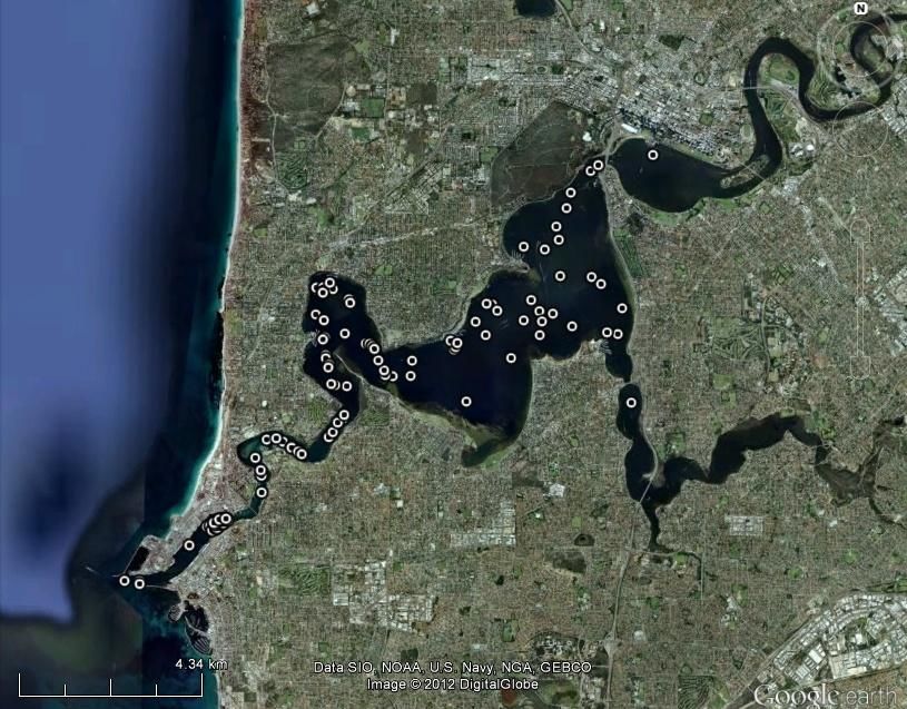
This document was prepared for the Swan River Trust as a final project report for Project RSP10MUR03: Swan
Canning Riverpark dolphin population health and ecology investigations.

Authors & Affiliations:

Delphine Chabanne1, Lisa-Marie Harrison1, Carly Holyoake2, Hugh Finn1*, Nahiid Stephens2, Lars Bejder1

1
    Murdoch University Cetacean Research Unit, School of Biological Sciences & Biotechnology, Murdoch University,
Murdoch, Western Australia 6150.
2
    School of Veterinary & Biomedical Sciences, Murdoch University, Murdoch, Western Australia 6150.

* Corresponding author: h.finn@murdoch.edu.au

                        School of Veterinary & Biomedical Sciences

                      School of Biological Sciences & Biotechnology

                                                                                                               2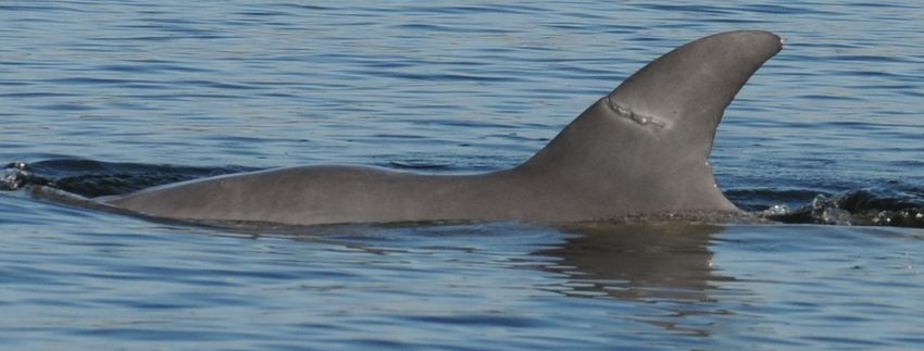
Project Report: Dolphin Health & Ecology Investigations

Summary

Introduction
Following the deaths of six Indo-Pacific bottlenose dolphins (Tursiops aduncus) in the Swan Canning
Riverpark in 2009 and recommendations of the Chief Scientist‘s Report to the Minister for
Environment (Beazley, 2010), the Swan River Trust commissioned this report in order to:

     1.   provide up to date information on the size and composition of the Riverpark dolphin
          population in context of historic datasets; and

     2.   review dolphin health investigations with up to date analyses of all findings from the 2009
          mortality event and an evaluation of the prevalence and incidence of epidermal disease in
          dolphins using the Swan Canning Riverpark.

Riverpark Dolphin Population
A total of 45 surveys (284 hours) were conducted over a set survey route within the Swan Canning
Riverpark between June 2011 and July 2012 resulting in 109 dolphin sightings. Thirty six dolphins,
including 27 adults/sub-adults and nine calves were photo-identified. Twenty of the 27 adult and sub-
adult individuals had been identified previously, either during intensive research from 2001-2003 or
during low-level monitoring from 2008-2011.

Of the nine mother-calf pairs identified, four of the females were known from the 2001-3 research
(Highnitch, Tworakes, Moon, Tupac). Three of these (Tworakes, Moon, and Tupac) had calves that
were 3-4 years old. Eden, a female identified in 2009, also had a calf of a similar age. Three females
had calves born after May 2011 (Pirulli, Highnitch, and Resource).

We classified 16 adult/sub-adult dolphins as ‗residents‘ within the Swan Canning Riverpark. These
dolphins had high seasonal sighting rates and were sighted in >10% of surveys between June 2011 and
May 2012. Four of the ‗resident‘ dolphins were females with dependent calves. Thus, if calves are
included, the total population size of resident dolphins in the Swan Canning Riverpark is 20 dolphins.
This is similar to the estimated number of resident dolphins present in 2001-3 (20-25 dolphins,
including calves).

Five adult/sub-adult dolphins were classified as ‗transients‘ because they were sighted only once or
twice within one month in the Swan Canning Riverpark and were mostly observed in adjacent coastal
areas (Cockburn Sound, Owen Anchorage, and Gage Roads). Two of the ‗transient‘ dolphins were
adult females with calves who were observed in consortship-type association with ‗resident‘ males.

                                                                                                     3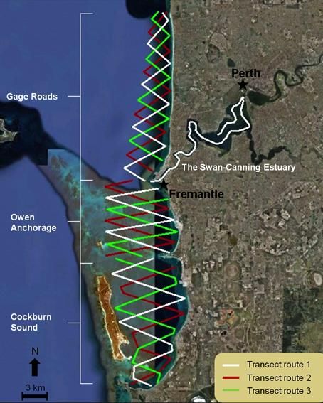
Project Report: Dolphin Health & Ecology Investigations

The other six adult/sub-adult dolphins sighted within the Swan Canning Riverpark were classified as
‗occasional visitors‘, although the sample sizes of sightings were small. Dolphins classified as
occasional visitors included three mother-calf pairs, a solitary male, and a male alliance from Cockburn
Sound that was first identified in 1993.

Analyses of sighting data indicate that the ranging patterns of adult/sub-adult dolphins occurring in the
Swan Canning Riverpark are best described by (but are not limited to) an emigration and re-
immigration model. This emigration and re-immigration model estimated that the population size was
17 adult/sub-adult individuals and that dolphins were spending an average of 142 days in the Swan
Canning Riverpark and 28 days in adjacent waters (e.g. Owen Anchorage). The difference between the
population sizes estimated by the model and by sighting information is minor and reflects the small
sample size of sightings for some dolphins. On-going study will improve the precision of these
estimates.

Analyses of association data indicate that adult males and females have stable associations and that
male associations are stronger than female associations. A model of constant companions explains the
adult male associations, while a model of casual acquaintances explains female associations (i.e.
individuals associate and disassociate after a certain time).

Further research is needed to confirm the composition of the current resident community and to assess
the genetic and demographic connectivity of the community to other dolphin populations in the Perth
area. We emphasise that a one-year study only provides a ‗snap-shot‘ of the current population –
further research is needed to better characterise the resident community and to confirm the residency
patterns observed to date.

Health Investigations

I. Review of the 2009 Mortality Event

In 2009, two clusters of mortality occurred in the Swan Canning Riverpark resulting in six deaths from
a resident community of c. 20 adult/sub-adult dolphins. By comparison, only six deaths were recorded
for the previous seven years. Thus, 2009 was an anomalous year. The first cluster occurred over three
weeks in June 2009 (winter), while the second occurred over five weeks in September-October 2009
(spring). The pathology of these deaths has been previously reported (Holyoake et al., 2010), but
results of subsequent tests to determine the role of pathogens are presented here and discussed in the
context of previous findings.

                                                                                                       4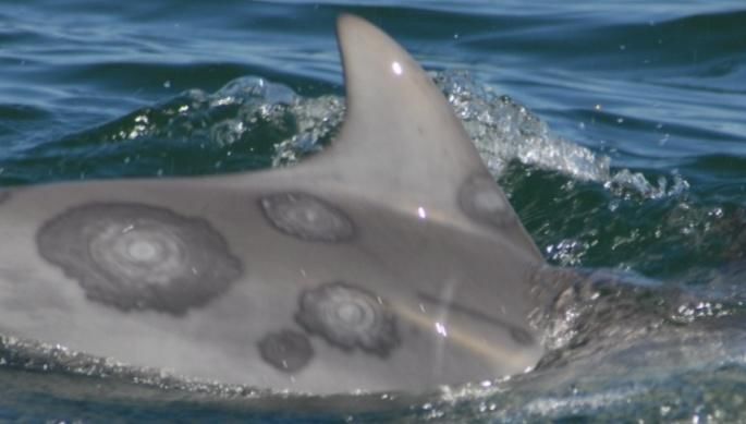
Project Report: Dolphin Health & Ecology Investigations

The dolphins in the first cluster (June 2009) were of mixed age classes. Two of these dolphins died
from opportunistic infections such as Aspergillus spp. bronchopneumonia or meningoencephalitis.
Characteristic dolphin morbillivirus lesions were not observed but cetacean morbillivirus (CeMV)
antigen was detected in multiple tissues from both animals using standard immunohistochemistry
methodology at an independent laboratory (Agri-Food and Biosciences Institute, Belfast – AFBI). PCR
on the same tissues at a second independent laboratory (Australian Animal Health Laboratory –
AAHL) confirmed the presence of CeMV nucleoprotein and phosphoprotein genes.

Dolphins from the second cluster (September-October 2009) were all adults. The most significant
pathology in the two dolphins examined post-mortem was severe extensive ulcerative dermatitis
associated with presumed poxvirus intracytoplasmic inclusion bodies and opportunistic bacterial and
mycotic infection. CeMV antigen was not detected in tissues from the two dolphins. Efforts to confirm
the presence of poxvirus by molecular methods and electron microscopy at AAHL were unsuccessful.
Analysis of the stranding records for the Swan-Canning Estuary found two further examples of severe
extensive dermatitis in October 2003 and November 2007, both affecting adult dolphins.

The June 2009 cluster of mortalities appears to have been associated with an outbreak of CeMV.
CeMV has been definitively identified as the primary aetiologic agent ultimately responsible for the
deaths of two of these mortalities, compounded by significant and severe secondary opportunistic
bacterial and fungal infections. The mixed age-class presentation of these two individuals suggests that
the Swan Canning Riverpark dolphins were a previously naïve population. Stranding data documenting
a 2009 mortality spike in cetaceans, including dolphins, on the coast of Western Australia suggests that
CeMV may also have affected other coastal and estuarine populations. This constitutes the first report
of CeMV-related dolphin mortality from the Indian Ocean and is the first time this pathogen has been
detected in marine mammals in Western Australia.

The September-October 2009 cluster followed the spring decline in water salinity and was associated
with an extreme presentation of what is presumed to be cetacean poxvirus (despite the lack of
definitive molecular diagnosis) causing extensive dermal ulceration exacerbated by osmotic stress.

The role of potentially immunosuppressive anthropogenic pollutants in either of these events is
unknown. Holyoake et al. (2010) previously identified that four of the deceased dolphins had high
dieldrin and PCB concentrations in blubber tissue.

                                                                                                      5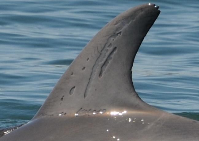
Project Report: Dolphin Health & Ecology Investigations

In conclusion, a multi-factorial model remains the best explanation of the 2009 mortality event in the
Swan-Canning Estuary. While we have identified some of the causal factors associated with the
pathology observed we cannot determine exactly how these factors combined to result in the deaths of
all of the dolphins involved. This conclusion is consistent with the findings of other studies of marine
mammal mortalities and illustrates the difficulties associated with wildlife disease and mortality
investigations, which often involve complex interactions between host, environment, and agent.

II. Epidermal Disease

We assessed the prevalence and incidence of epidermal disease in dolphins from the Swan Canning
Riverpark using photo-identification images of dolphins observed within the estuary between June
2011-June 2012. To support this assessment, we developed a classification scheme for dolphin skin
lesions within southwestern Australia.

The prevalence of skin lesion types varied. All dolphins had excoriations (e.g. tooth rake marks) that
occur during interactions with other dolphins. Of the lesion types having a potentially infectious
aetiology, hyperpigmented and hypopigmented lesions were present in more than 80% of individuals
and targetoid (rounded with rings of distinct colours creating a target-shaped mark) lesions were
present in more than 50% of dolphins. Estimating the actual prevalence of pathogenic lesions was
difficult because some lesions may represent scarring and different pathogens can manifest with similar
lesion presentations. Examinations of microscopic tissue sections and molecular analyses of lesion
biopsies are necessary to definitively determine the pathogen(s) present/responsible.

No ulcers, erosions, plaques, concentric rings, or abrasions were observed. No animals displayed multi-
nodular lesions consistent with descriptions of lacaziosis (lobomycosis). The genital area and oral
mucosa of dolphins were rarely visible, making it difficult to detect if herpesvirus-associated lesions
were present.

Tattoo-like lesions were present in five of 36 of individuals. Lesions in these individuals diminished in
size, number, and/or severity over the year. Tattoo-like lesions may indicate poxvirus (i.e.Tattoo Skin
Disease) infection, as it is likely that cetacean poxvirus is endemic to dolphin populations in this
region. Lesion prevalence appeared low and the absence of severe presentations of tattoo-like lesions
suggests that dolphins were generally able to control poxvirus infections. However, lesion prevalence
and severity may vary seasonally and annually in response to environmental conditions and to exposure
to immunosuppressive stressors (e.g. CeMV, contaminant burdens).

                                                                                                       6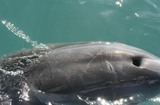
Project Report: Dolphin Health & Ecology Investigations

Table of Contents

Summary………………………………..……………………………………………………………………………………………...3

Table of Contents…..…...……………………………………………………………………………………………...…………..7

I. Introduction & Aims…………………………………………………………………………………………………………...8

II. Health Investigations: review of 2009 mortality event………….………………….…………….….……...12

II. Health Investigations: epidermal disease………………….....…………….….………….………………………28

IV. Ecology Investigations: 2001-3 research………..….……………………………….…………………………...47

V. Ecology Investigations: 2011-2 research...…………..……………………..………………...…………………..71

Acknowledgements….………………………………………………………………….…………………………...................86

References………………..……………………………………………………………………………………………...................87

                                                                                   7
Project Report: Dolphin Health & Ecology Investigations

I. Introduction& Aims

Project Background & Objectives
An investigation into the deaths of six Indo-Pacific bottlenose dolphins (Tursiops aduncus) within the
Swan Canning Riverpark between May-October 2009 found that a suite of factors likely contributed to
the mortalities (Beazley 2010; Holyoake et al. 2010). This investigation, along with research
undertaken for Swan Canning Research Innovation Program (SCRIP) Project: ―Toxicant exposure,
population genetics, and trophic associations of bottlenose dolphins (Tursiops sp.) in the Swan River‖
(Holyoake et al. 2011), emphasised the vulnerability of the resident community to natural and
anthropogenic stressors and the need to improve the scientific basis for the long-term conservation of
dolphins within the estuary.

To support this aim, the Swan River Trust and Murdoch University entered into a collaborative
agreement Project to undertake further health and ecology investigations into dolphins in the Swan
Canning Riverpark and to conclude analyses of existing data and samples. This report presents the
major findings for this project. The specific objectives of this project were to:

     (a) review the 2009 unusual mortality event in light of findings from further tissue analyses;
     (b) assess the prevalence and incidence of epidermal disease in dolphins from the Swan
         Canning Riverpark;
     (c) analyse historic datasets from 2001-3 to establish baseline information for dolphins within
         the Swan Canning Riverpark; and
     (d) characterise the size and composition of the current resident dolphin community in the Swan
         Canning Riverpark.

Structure of Report
This report contains a summary and five component sections:

1.   Section I: Introduction& Aims –project aims and background information;
2.   Section II: Health Investigations – review of the 2009 mortality event;
3.   Section III: Health Investigations – an assessment of epidermal disease in Riverpark dolphins;
4.   Section IV: Ecology Investigations – a review of research undertaken from 2001-3;
5.   Section V: Ecology Investigations – an assessment of the current Riverpark dolphin population;

                                                                                                       8
Project Report: Dolphin Health & Ecology Investigations

Scientific Basis for Report
This report draws on the scientific information obtained during field studies of dolphins conducted in
the Perth area since from the early 1990s onwards. Publications relevant to this report include:

Bejder, L., Samuels, A., Whitehead, H., Finn, H. and Allen, S. 2009. Impact assessment research: use and misuse
     of habituation, sensitisation and tolerance to describe wildlife responses to anthropogenic stimuli. Marine
     Ecology Progress Series 395: 177-185.
Cannell, B.L. 2004. Distributions of the major marine fauna found in the Perth metropolitan area (Yanchep to
     Mandurah). Technical Report: MMS/CWC, LNE/MMP, SEMP, SIMP-79/2004, Marine Conservation
     Branch, Department of Conservation and Land Management, Perth, Western Australia.
Donaldson, R., Finn, H., and Calver, M. 2011. Illegal feeding increases risk of boat-strike and entanglement in
     bottlenose dolphins. Pacific Conservation Biology 16: 157-161.
Donaldson, R., Finn, H., Bejder, L., Lusseau, D., Calver, M. (in press) The social side of human-wildlife
     interaction: wildlife can learn harmful behaviours from each other. Animal Conservationdoi:10.1111/j.1469-
     1795.2012.00548.x
Finn, H.C. 2005. Conservation biology of bottlenose dolphins (Tursiops sp.) in Perth metropolitan waters. PhD
      thesis. School of Biological Sciences & Biotechnology, Murdoch University, Perth, Western Australia.
Finn, H.C. and Calver, M.C. 2008. Feeding aggregations of bottlenose dolphins and seabirds in Cockburn Sound,
      Western Australia. The Western Australian Naturalist 26: 157-172.
Finn, H.C., Donaldson, R., and Calver, M.C. 2008. Feeding Flipper: a case study of a human-dolphin interaction.
      Pacific Conservation Biology 14: 215-225.
Gales, N and Waples, K. 1993. The rehabilitation and release of bottlenose dolphins from Atlantis Marine Park,
     Western Australia.Aquatic Mammals 19: 49-59.
Ham, G.S. 2009. Population biology of bottlenose dolphins (Tursiops sp.) in Cockburn Sound, Western Australia.
     Honours thesis. School of Biological Sciences & Biotechnology, Murdoch University, Perth, Western
     Australia.
Holyoake, C., Finn, H., Stephens, N., Duignan, P., Salgado, C., and others. 2010. Technical Report on the
     Bottlenose Dolphin (Tursiops aduncus) Unusual Mortality Event within the Swan Canning Riverpark, June-
     October 2009. Report to the Swan River Trust, May 2010. Murdoch University, Perth, Western Australia.
Holyoake, C., Finn, H., Stephens, N., Linke, T., Daniel, C., Allen, S., Smith,H., McElligott, D., Bejder, L. 2011.
     Toxicant exposure, population genetics, and trophic associations of bottlenose dolphins (Tursiopsaduncus) in
     the Swan River. SCRIP Project Report to the Swan River Trust, October 2011. Murdoch University, Perth,
     Western Australia.
Lo, H.N. 2009. Bottlenose Dolphins (Tursiops sp.) in the Swan River, Western Australia: community size and
     composition, residency patterns, and social structure. Unpublished Honours thesis.Department
     ofEnvironmental and Aquatic Science, Curtin University of Technology, Perth, Western Australia.
Moiler, K. 2008. Bottlenose Dolphins (Tursiops sp.) – a study of patterns in spatial and temporal use of the Swan
     River, Western Australia. Unpublished Honours thesis. Department of Environmental Biology, Curtin
     University of Technology, Perth, Western Australia.
Waples, K.A. 1997. The rehabilitation and release of bottlenose dolphins from Atlantis Marine Park, Western
    Australia. PhD thesis. Texas A & M University, Galveston, Texas.

Supporting Research Initiatives
This project was undertaken as part of two broader research initiatives: the Murdoch University Marine
Mammal Health Project and the Coastal and Estuarine Dolphin Project.

Murdoch University Marine Mammal Health Project

The Murdoch University Marine Mammal Health Project is a collaborative research program
investigating the health of marine mammals in Western Australia. Researchers from Murdoch

                                                                                                                9
Project Report: Dolphin Health & Ecology Investigations

University collaborate closely with personnel from the WA Department of Environment and
Conservation (DEC) and from other Australian and international research institutions. The general aims
of the Population Assessment project are to: (1) identify and characterise the factors associated with
cetacean strandings and (2) acquire baseline and epidemiological information on disease levels.

The specific research objectives of the Marine Mammal Health Project are to: (a) collect morphometric
and life history data (e.g. size, sex, age class); (b) undertake partial and where feasible full post-mortem
examinations to acquire information on causes of morbidity and mortality, and to collect tissues for
pathogen identification, histopathology and toxicology; (c) isolate and identify pathogenic viruses,
bacteria, protozoa, or fungi from tissue samples (where appropriate); (c) archive tissue samples for
long-term disease surveillance, toxicological monitoring, and retrospective study; and (d) establish
beneficial collaborations within WA, nationally and internationally in order to enhance the information
gained from the samples collected.

Coastal and Estuarine Dolphin Project

The Coastal & Estuarine Dolphin Project (CEDP) is a collaborative research program addressing the
health, ecology, and conservation of dolphins in the Perth region. CEDP partners include the Cockburn
Sound Management Council, Curtin University, Fremantle Ports, Murdoch University, and the Swan
River Trust.

In July 2011, we initiated field research for a four-year Population Assessment project to assess the
abundance, habitat use, and ranging and residency patterns of dolphins in the Perth region using photo-
identification, behavioural sampling, GIS, and line transect sampling. The design and methodology of
the project follows that of the South West Marine Research Program (SWMRP) in the Bunbury region
(Smith 2012). The study area for the project extends along the coast from Rockingham to Scarborough
and inwards to include the Swan Canning Riverpark; it comprises four sub-areas: Cockburn Sound,
Owen Anchorage, Gage Roads, and Swan Canning Riverpark (Figure 1.1).

The overall aim of the Population Assessment project is to characterise the population size, structure,
and connectivity of dolphins around Perth region. This aim addresses two information needs identified
following the deaths of six dolphins within the Swan Canning Riverpark in 2009: (a) the status of
populations and ‗communities‘ of dolphins within the Perth metropolitan area, particularly the resident
community associated with the Swan Canning Riverpark and (b) the connectivity of the different
populations and communities within the Perth metropolitan area.

The issue of population connectivity is important because bottlenose dolphins sometimes exhibit long-
term site fidelity to particular coastal and estuarine areas, suggesting that local extinction could occur.

                                                                                                         10
Project Report: Dolphin Health & Ecology Investigations
Genetic and ecological information is therefore needed to assess population structure and dispersal
rates and individual ranging patterns, and to describe the current size and composition of resident
communities (where they occur). Thus, although the resident community in the Swan Canning
Riverpark is a key focus of this research, we also need information on their connectivity to dolphins in
adjacent areas in order to better assess their vulnerability.
The general aims of the CEDP Population Assessment project are to: (1) determine how many dolphins
occur in this region; (2) investigate whether dolphins show long-term fidelity to particular locations;
(3) assess if discrete ‗communities‘ of dolphins occur within certain areas; and (4) evaluate what
environmental variables are associated with the distribution of dolphins. The specific research
objectives are to: (a) estimate dolphin abundance across the study area using photo-identification and
mark-recapture methods; (b) determine residency and ranging patterns for individuals in order to better
understand site fidelity and population structure; (c) determine habitat use patterns through habitat
modeling; and (d) collect behavioural, environmental, and epidemiological data related to interactions
with human activities and with dolphin health (e.g. entanglements, epidermal diseases).
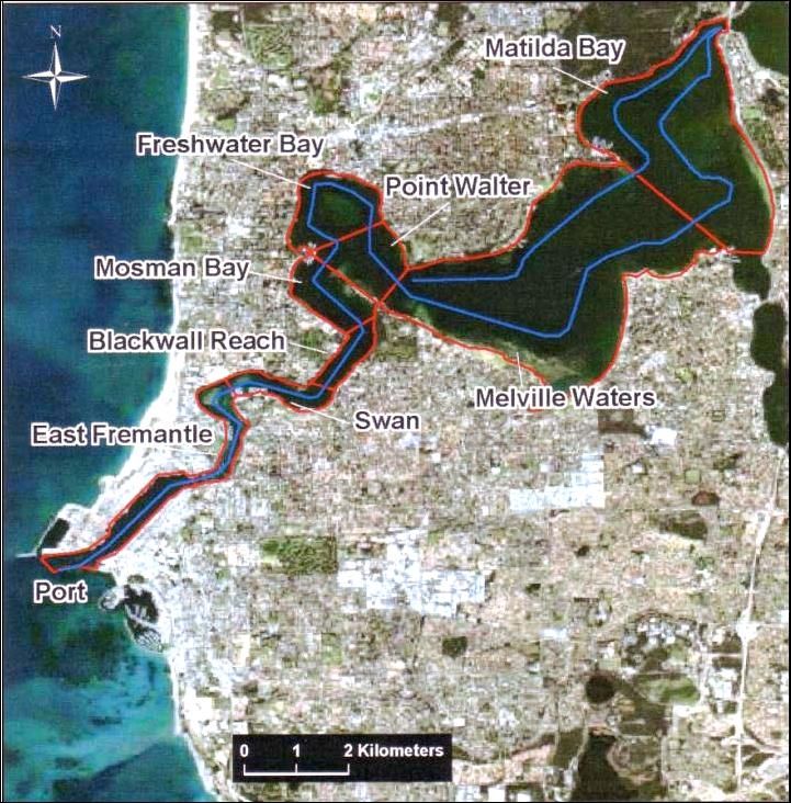
Swan Canning Riverpark
Figure 1.1: Overall study area for the Population Assessment project showing: (a) the location of
the four sampling areas (Cockburn Sound, Owen Anchorage, Gage Roads, Swan Canning
Riverpark) and (b) the lay-out of the line transect design within these areas. Sampling within these
four areas is balanced temporally with equal numbers of samples (n > 5 samples) per season (summer,
autumn, winter, spring). Dolphin observed during transect sampling are photo-identified and group size
and activity are recorded. Environmental data is also recorded on a systematic basis to allow for habitat
modelling (along with bathymetry and substrate databases).
11Project Report: Dolphin Health & Ecology Investigations
II. Health Investigations: Review of 2009 Unusual Mortality Event
Introduction
Factors Affecting Health & Mortality of Bottlenose Dolphins
The deaths of six Indo-Pacific bottlenose dolphins (Tursiops aduncus) in the Swan Canning Riverpark
over a five-month period in 2009 represented an ‗unusual mortality event‘ because of the small number
of dolphins thought to be resident within the estuary at the time (c. 20-25 individuals) and stranding
records indicating a typical stranding rate of about one carcass per year for the Riverpark (Holyoake et
al. 2010). The deaths occurred in two clusters, with the first cluster (comprising Cases 1-3 in Table 2.4)
occurring over a three-week period in June 2009 (winter) and involving mixed-age classes, and the
second cluster (comprising Cases 4-6 in Table 2.4) occurring over a five-week period from September
to October 2009 (spring) and involving solely adult animals.
An unusual mortality event of this kind implies that some factor or, more likely, suite of factors was
present and caused a perturbation from normal or ‗background‘ rates of mortality within the
population. Factors that cause or contribute to poor health and mortality within dolphin populations are
commonly described as stressors (Acevedo-Whitehouse and Duffus 2009) (Table 2.1). These stressors
can affect health or cause mortality in a range of ways (Table 2.2). Stressors may be natural or
anthropogenic, and may be further divided into persistent, acute, and lethal stressors (Table 2.3).
Table 2.1: Factors (or ‘stressors’) that may cause or contribute to poor health and mortality within dolphin
populations include:
infection by a virulent primary pathogen, e.g. cetacean morbillivirus (CeMV)
opportunistic (secondary) infection by bacterial and fungal pathogens
parasitism
natural predation
disturbance from human activities or interactions, e.g. with fishing operations
anthropogenic noise
adverse environmental conditions; e.g. exposure to very cold or very low salinity waters
epidermal disease, e.g. tattoo skin disease (TSD)
introduction of a novel or emerging disease, such as a more virulent variant of a pathogen;
low food availability, e.g. low abundance of prey species
human-induced injuries, e.g. entanglements; and
concentrations of contaminants sufficient to impair reproductive and immune function.
12Project Report: Dolphin Health & Ecology Investigations
Table 2.2: Stressors may affect health or cause mortality by causing:
physical trauma, e.g. wounds such as lacerations, avulsions, or amputations
reduced condition, e.g. emaciation, fat store depletion
systemic physiological stress, e.g. elevated production of stress hormones
physiological injury/impairment, e.g. acoustic damage
immune system impairment, e.g. lymphoid depletion resulting in immunosuppression1
reproductive impairment, e.g. reduced fertility, embryo toxicity, abortion/stillbirths
epidermal lesions, e.g. tattoo skin disease
neurological impairment, e.g. brevetoxicosis
internal lesions that impair normal physiologic function, e.g. pneumonia2
Table 2.3: Types of Stressors
(a) Persistent – Some stressors may cause chronic (i.e. repeated, sustained, long-term) and generalised (i.e.
affecting multiple bodily functions) stress affecting the physiology, body condition, immune function, and/or
reproductive viability of the animal. Specific effects may include: reduced immunological function; decreased
reproductive output; diminished blubber reserves and general body condition; increased susceptibility to
opportunistic pathogens and parasitism; and reduced regenerative/healing potential following cell/tissue
injury/damage.3
Examples include: toxicants, anthropogenic noise, vessel disturbance, adverse environmental conditions,4 low
prey availability, interactions with human activities (e.g. fishing or tourism operations), chronic infection
from a persistent but weakly virulent (i.e. non-lethal) pathogen, and an enduring entanglement injury.
(b) Acute – Certain stressors affect organisms over shorter time periods. For example, while a persistent stressor
may affect an organism over a period of months to years, an acute stressor will occur over a time-span of
weeks, days, and possibly even hours.
Examples include: infection by a primary pathogen, a severe entanglement injury, and a wound from a shark
attack or vessel strike.
(c) Lethal – In a few cases, stressors are sufficient to cause death by themselves (e.g. shark attack), or by
exacerbating pre-existing conditions in such a way that leads to the death of the animal in a short period of
time.5
1
Immunosuppression refers to the suppression (i.e. compromised) of normal immune function of an individual, and may involve
dysfunction of adaptive and/or innate immunity. Causes of immunosuppression include primary pathogens (e.g. morbillivirus),
contaminants, and environmental stress. These factors may cause lymphoid depletion, leading to immunosuppression.
2
The definition of lesion used in pathology differs from how the term is used in other contexts. Blood & Studdert (1988) define
the term as: ―any pathological or traumatic discontinuity of tissue or loss of function of a part. Lesion is a broad term, including
wounds, sores, ulcers, tumours, cataracts and any other tissue damage. They range from skin sores associated with eczema to the
changes in lung tissue that occur in tuberculosis‖ (p. 529). Thus, even a change that does not cause overt structural damage is
classified as a lesion if it adversely affects function.
3
Heavy parasitism is an indication of poor health rather than necessarily a cause. However, it will also adversely affect the
individual as a persistent stressor in its own right.
4
These would relate to: (a) prevailing long-term conditions; (b) conditions that persist for shorter periods of time; and (c)
variation in conditions, particularly those involving a rapid and substantial change.
5
In forensic pathology, the legal term cause of death refers to the injury, disease, or abnormality responsible for instigating the
sequence of functional disturbances culminating in mortality, either by itself or allied with other factors. The term mechanism of
death refers to the structural or functional change that makes organisms no longer able to sustain life without artificial
intervention. Finally, the term manner of death refers to the way in which the cause of death is classified, with particular
reference to human agency.
13Project Report: Dolphin Health & Ecology Investigations
A Conceptual Model of Health
An animal‘s state of health can be conceptualised as lying along a continuum running from very
healthy (i.e. absence of disease, good body condition, normal physiologic function) right through to
death, with a threshold for disease occurring somewhere in between (Figure 2.1). The presentation of
disease may vary from sub-clinical to severe. Some animals may lie near the threshold because their
immune system is challenged, e.g., they are affected by a suite of chronic stressors, have sustained a
traumatic injury, or carry a mild but recurrent infection. A small change in the stressors affecting these
individuals may be sufficient for disease to occur (or for pre-existing conditions to grow more severe).6
Most instances of disease are multi-factorial. A multi-factorial aetiology means that disease typically
occurs because two or more stressors interact (i.e. act synergistically) or exert a cumulative effect.
Figure 2.1: Continuum representing potential states of health for individual dolphins. Disease
refers to the finite abnormality of structure or function with an identifiable pathological basis.
Mortality Rates
Background rates of mortality for dolphins in the Perth area are not well-known, but mortality may
occur through: shark predation, infectious and non-infectious disease, senescence, and agonistic
interactions (e.g. male-male fighting). Anthropogenic factors add to natural background mortality rates
and are considered to have a ‗biologically significant‘ impact if their impact on demographic
parameters (birth and death rates) is sufficient to threaten population viability. This threshold of
biological significance provides part of the conceptual framework for statutory instruments designed to
identify and mitigate anthropogenic mortality on marine mammals.7
6
For example, infection by a primary pathogen (such as a virus) may cause immunosuppression leading to secondary infection
by bacterial, fungal, or protozoal pathogen(s), which may ultimately culminate in the animal‘s death. Diminished body condition
caused by one or more stressors may result in mobilisation of lipid reserves, thereby allowing lipophilic contaminants to enter the
bloodstream as these tissues are metabolised.
7
For example, the United States Marine Mammal Protection Act 1972 utilises a concept called Potential Biological Removal
(PBR) to provide guidelines for assessing the biological significance of fishing-related mortality for a marine mammal
population. The New Zealand Marine Mammal Protection Act 1977 uses a similar concept known as Maximum Allowable Level
of Fishing-Related Mortality (MALFIRM).
14Project Report: Dolphin Health & Ecology Investigations
Estuaries & Health
Coastal and estuarine ecosystems are challenging environments for dolphins (Reeves et al. 2003;
Balmer et al. 2011; Jefferson et al. 2011). Populations inhabiting these areas may experience: habitat
loss and degradation; exposure to environmental contaminants and biotoxins; incidental mortality from
interactions with fisheries and other activities; disturbance from vessel interactions and anthropogenic
noise; and greater risk of infectious disease (Van Bressem et al. 2009a; Jefferson et al. 2011; Schwacke
et al. 2012). These stressors can affect the behaviour, physiology, and health of dolphins and reduce
reproductive success and survivorship, particularly if stressors exert cumulative or synergistic impacts
(Gulland and Hall 2007; Balmer et al. 2008; Bejder et al. 2009; McHugh et al. 2011).
Estuaries are challenging environments for dolphins because of the diversity and severity of stressors
that may be present. Dolphins may experience rapid and substantial changes in environmental
conditions (e.g. salinity, temperature, tidal height) and the availability of food, as well as stress from
anthropogenic factors (e.g. debris, contaminants, harmful algal blooms, vessel disturbance, interactions
with fisheries). Estuarine dolphin populations are therefore at particular risk of suffering infectious
disease, physiological stress, human-induced injury, and exposure to algal biotoxins (Van Bressem et
al. 2009a). Along the eastern U.S. seaboard, for example, estuarine dolphins have been the most
affected by unusual mortality events (Hohn 2002).
Studying the Effects of Stressors
Pathology investigations may use a variety of methodologies. These include: post-mortem examination
through gross and histological approaches; ancillary laboratory analyses such as microbiological
culture/identification, serum or other body fluid chemistry, serology, immunohistochemistry, biotoxin
and contaminants analyses; collection of relevant environmental data (e.g. water quality, presence of
potentially harmful phytoplankton); and epidemiological analysis. Polymerase Chain Reaction (PCR)
or other molecular techniques are often used to screen for disease; these methods work by amplifying
the DNA/RNA belonging to infectious agents or pathogens present in tissues collected post-mortem or
by remote biopsy to a level where they can be detected. Ideally, pathology investigations are coupled
with ecological and demographic data so as to support assessments of the biological significance of
stressors (or suites of stressors), and determine whether identified health challenges are likely to
impinge upon demographic parameters and potentially threaten the viability of populations.
Cetacean Morbillivirus
Disease is often the cumulative result of interacting host, environment and agent factors. Complex
interactions between host, environment, and agent often result in difficulties in establishing clear cause
15Project Report: Dolphin Health & Ecology Investigations
and effect pathways in wildlife disease and mortality investigations. The lack of baseline and
epidemiological information for many marine mammal diseases is a further complication. The disease
outcome of cetacean infection with cetacean morbillivirus (CeMV) epitomises such complexity.
Cetacean morbillivirus has a worldwide distribution (Van Bressem et al. 2001) and has been implicated
as the cause of several epizootics resulting in mass mortalities in the NW Atlantic (Lipscomb et al.
1994; Krafft et al. 1995; Duignan et al. 1996); North Sea (McCullough et al. 1991; Visser et al.1993);
Black Sea (Birkun et al. 1999); and Mediterranean Sea (Domingo et al. 1990; Van Bressem et al. 1991;
1993; Fernàndez et al. 2008; Raga et al. 2008; Keck et al. 2010). The first confirmed case of fatal
CeMV in the southern hemisphere was recently described in an Australian offshore bottlenose dolphin
(Tursiops truncatus: Stone et al. 2011). Serological evidence of CeMV infection has since been
detected in five cetacean species from southeastern Queensland and northern New South Wales,
Australia (Stone et al. 2012).
Although cetacean morbillivirus is regarded as one of the most pathogenic infectious agents for
cetaceans (Van Bressem et al. 2001), much remains unknown about the pathogenesis and epidemiology
of disease. Evidence of subclinical infection has been detected in dolphins (Bossart et al. 2010), but the
factors that lead to clinical disease and the development of epizootics remain unknown (Bossart et al.
2010). Van Bressem et al. (2009a) suggested several factors such as fisheries interactions, inbreeding,
migration, high contaminant loads, higher sea-surface temperatures and limited prey availability may
have synergistically interacted to increase the severity of CeMV infection and favour the occurrence of
epizootics in striped dolphins (Stenella coeruleoalba) in the Mediterranean.
In 2009, over a period of five months (June-October 2009), there were six confirmed mortalities in a
small resident population of Indo-Pacific bottlenose dolphins (Tursiops aduncus) in the Swan Canning
Riverpark. The deaths were deemed an unusual mortality event as the population was thought to only
comprise about 20-25 individuals and previously on average only one or less stranded carcass per year
had ever been reported (Holyoake et al. 2010). The main aim of this study was to determine direct and
indirect causes of the mortalities, including consideration of potential interactions among proposed
environmental, host, and agent determinants of disease.
16Project Report: Dolphin Health & Ecology Investigations
Methods
Pathology & Disease Investigation
Four of the six individuals were necropsied (two were deemed too autolysed to examine) (Table 2.4). A
standard small cetacean necropsy protocol was followed (Duignan 2000; Pugliares et al. 2007). The
degree of post-mortem autolysis was determined and each carcass was assigned a condition code
according to Pugliares et al. (2007). Necropsies and histopathology were conducted at the Department
of Anatomic Pathology, School of Veterinary and Biomedical Sciences, Murdoch University;
Veterinary Department, Perth Zoo; and Animal Health, Department of Agriculture and Food Western
Australia (DAFWA).
Immunohistochemistry (IHC) for the detection of morbillivirus antigen was performed (on samples
from those individuals where the post-mortem/histopathological findings was indicative) at the Agri-
Food and Biosciences Institute, Belfast, Northern Ireland (AFBI), on paraffin-embedded formalin fixed
tissues from the four dolphins necropsied by using a monoclonal antibody specific for the
nucleoprotein of canine distemper virus (VMRD, Inc.) at a dilution of 1 in 100. The reagents used were
part of a commercially available streptavidin-biotin peroxidise technique kit and the methodology
followed that used by Kennedy et al. (1989). Tissues from healthy dolphins were used as negative
controls, while tissues from morbillivirus-infected striped dolphins (Domingo et al. 1992) and from a
harbour porpoise (Phocoena phocoena: Kennedy et al. 1991) were used as positive controls. Negative
controls in which the primary antibody was omitted were also included.
Reverse transcriptase polymerase chain reaction (RT-PCR) for detection of morbilliviral RNA was
performed on formalin-fixed paraffin embedded tissues found positive via IHC at the Commonwealth
Scientific and Industrial Research Organisation (CSIRO), Australian Animal Health Laboratory
(AAHL), Geelong, Victoria, using molecular techniqes as previously reported (Stone et al. 2011).Two
sets of primers (previously described in Stone et al. 2011) for the detection of morbillivirus
nucleoprotein (N) gene18 and phosphoprotein (P) gene19 were utilised. Amplified PCR products (~238
bp for N gene and ~429 bp for P gene) were gel purified and sequenced. BLASTn software
(http://blast.ncbi.nlm.nih.gov): a tool used by biologists world-wide to identify similarities between
nucleotide based biological genome sequences) was utilised to compare sequence similarity with
known dolphin morbillivirus sequences reported in the literature.
IHC for the detection of dolphin poxvirus antigen was performed by CSIRO / AAHL on samples from
Cases Four and Six (the only individuals where this was indicated by gross/histopathological findings).
A poxvirus antibody against capripox (recombinant LSDV P32 – envelope protein) was used (although
17Project Report: Dolphin Health & Ecology Investigations
it is somewhat divergent from cetacean poxvirus, it was the only one available at the time); according
to the literature (Bracht et al. 2006), the dolphin poxvirus DNA polymerase gene fragments are 74%
homologous with capripox.
Polymerase chain reaction (PCR), utilising several published and in-house methods for detection of
poxvirus, parapoxvirus and herpesvirus, was performed by CSIRO / AAHL on samples from Cases
Four and Six (the only individuals where this was indicated by gross / histopathological findings). Both
generic and specific primers for dolphin poxvirus were used.
Samples from the wet formalin fixed skin lesions from Case Six (the only individual where these
samples were available; Case Four was also indicated, however samples were not available) were
examined by thin section electron microscopy (EM) (in particular the transition zone where most
intracytoplasmic poxviral-like inclusions were seen within keratinocytes) at CSIRO/AAHL in an
attempt to identify the presence of intralesional pox virions. The same tissue was homogenised for
negative contrast EM. Targeted laser selection of the areas of interest (i.e. region of epidermis
containing keratinocytes affected by intracytoplasmic pox-viral like inclusion bodies) within formalin-
fixed, paraffin-embedded tissue from the same individual for thin section EM examination was also
performed.
Contaminants Analysis
From each dolphin necropsied approximately 100 grams of blubber was collected from a location
immediately anterior to the dorsal fin (Higgins and Noad 2006). Blubber samples were wrapped in
acetone rinsed and dried aluminum foil, placed in a ziplock bag and stored at -20°C prior to analysis.
Blubber samples were sent to the National Measurement Institute (NMI), Sydney, for analysis of
dichlorodiphenyltrichloroethane (DDT) and its metabolites, dieldrin and polychlorinated biphenyls
(PCBs). For each sample 4 g of blubber was mixedwith approximately 10 g anhydrous
sodium sulphate andsonicated for 2 hours using 50 mL hexane-acetone mix solvent before being
allowed to soak overnight (US EPA method 3540C). The next day the extract was purified using Gel
Permeation Chromatography (US EPA method 3640). The final extract was analysed by gas
chromatography with electron capture detection (GC-ECD) [Agilent 6890 Gas Chromatograph with
twin micro-ECDs, column 1: RTC-CL Pest (30 m x 0.32 mm x 0.25 µm) and column 2: DB-608 (15 m
x 0.32 mm x 0.25 µm)]. Samples were calibrated against a 0.01 ug/mL standard containing the analytes
of interest, while the surrogates TCMX, DF-DDE, DB-DDE, Terphenyl-d14 and Decachlorobiphenyl
were analysed along with the samples. Analytes found to be present at greater than the instrumental
linear dynamic range were diluted to within calibration range prior to re-analysis. Quality assurance
18Project Report: Dolphin Health & Ecology Investigations
consisted of a matrix spike, laboratory control spike, duplicate testing and analysis of one blank per 20
samples. The limit of reporting per wet weight of tissue was 0.1 µg/g for DDT, DDT metabolites and
dieldrin, and 2ng/g for PCBs.
Prior to analysis the fat content of each blubber sample was determined. Samples were blended to
obtain a homogeneous mass which was not free flowing, 6 g of this homogeneous mass was accurately
weighed into a thimble (height 100mm, diameter 25mm) and dried overnight at 102 (+/- 2) degrees
Celsius. After cooling, each thimble was placed in a Soxhlet apparatus and extracted with diethyl ether
for 16 hours. The ether extract was collected in a pre-weighed 250mL Erlenmeyer flask. After 16
hours extraction, the apparatus was disassembled and the diethyl ether extract was evaporated on a
water bath. The pre-weighed flask containing the fat was placed in an oven at 102 (+/- 2) degrees
Celsius for 1 hour. It was then placed in a desiccator containing dry silica gel to cool. After 1 hour it
was removed from the desiccator and weighed. This oven drying and weighing procedure was repeated
until successive measurements agreed within 0.005 g. The fat content of the blubber was calculated by
using the formula:
% fat = 100 x (Weight of flask - Weight of empty flask) / Weight of Sample in Thimble
Results
Pathology & Disease Investigation
Two of the four dolphins (Cases Two and Three) necropsied were immunopositive for morbillivirus
(CeMV) via IHC; infection of these two individuals by CeMV was confirmed by testing at a different
laboratory which confirmed the presence of the CeMV nucleoprotein and phosphoprotein genes by RT-
PCR (Table 2.4).
Two of the dolphins (Cases Four and Six) had numerous, extensive, multifocal to coalescing ulcerative
skin lesions covering up to 70% of their surface area with intracytoplasmic viral inclusion bodies
detected in numerous intralesional keratinocytes; these were consistent in their morphology with
dolphin poxvirus. However, efforts to determine the presence of poxvirus (as well as differentials such
as parapoxvirus and herpesvirus) via PCR and EM at CSIRO/AAHL were unsuccessful.
The blubber contaminants results are presented in Table 2.5. Case Three had the highest dieldrin,
∑DDT, and ∑PCB concentrations of the four dolphin blubber samples analysed.
19Project Report: Dolphin Health & Ecology Investigations
Discussion
Pathology & Disease Investigation
Prior to 2009 one or less dolphin deaths per year had been reported in the Swan Canning Riverpark
(Holyoake et al. 2010). In 2009 there were six reported deaths in a space of five months suggesting that
the deaths were possibly related. However, the necropsy results for the four dolphins examined did not
indicate one consistent finding that could be attributed as the cause of death for all. Lymphoid
depletion was noted histologically in all four dolphins necropsied, two animals were immunopositive
on IHC for morbillivirus (CeMV) antigen (Cases Two and Three) and the tissues from these same
animals were found to be RT-PCR positive for CeMV nucleoprotein (N) gene18 and phosphoprotein
(P) gene19, thus definitively identifying the presence of CeMV antigen in these two individuals.
Therefore, it would appear that the June cluster of mortality was associated with a CeMV outbreak.
Two animals (Cases Four and Six) had severe ulcerative skin lesions with intracytoplasmic inclusion
bodies detected in keratinocytes, the morphology of which was consistent for dolphin poxvirus (a
marine mammal specific orthopox virus). Attempts to positively identify the presence of poxvirus in
the lesioned tissue via PCR and EM were unsuccessful (additionally, testing for herpesvirus and
parapoxvirus as differentials was also negative). Thus, we have exhausted all means currently available
to confirm the aetiological diagnosis for the skin lesions. Unfortunately, it is unlikely we will learn any
more about their aetiopathogenesis unless they occur again in the population.
What follows is an attempt to discuss possible direct and indirect causes of the pathology and
mortalities, including consideration of potential interactions among proposed factors.
Cetacean Morbillivirus
This is the first time cetacean morbillivirus (CeMV) has been detected in marine mammals in Western
Australia; additionally, this is the first report of CeMV-related dolphin mortality from the Indian
Ocean. CeMV antigen was positively identified (using two different molecular techniques at two
different independent laboratories) in tissues from two of the three June cluster mortalities (Cases Two
and Three; a juvenile male and an adult female, respectively). Unfortunately, Case One (a male calf)
was too decomposed for necropsy and molecular investigation. Serological surveys indicate that
morbillivirus infection can be widespread in marine mammal populations without causing significant
acute mortality and/or mass strandings (Duignan et al. 1996). However, the presence of CeMV-related
mortality in mixed-age classes such as seen in the June cluster would suggest an outbreak in a
previously naïve population, or at least a scenario where the virus had not been circulating during the
lifetime of the oldest individual (Case Three, approximately 16 years of age based on dental aging and
20Project Report: Dolphin Health & Ecology Investigations
morphometric measurements). In populations in which CeMV infection is endemic, deaths tend to
occur solely in juveniles as immunity in individuals surviving exposure is typically life-long (Duignan
et al. 1996). The outbreak coincided with a significant increase in bottlenose dolphin strandings along
the WA coast in 2009 (affecting both coastal and estuarine dolphins) in comparison to relatively
constant stranding rates from 1981-2008 (Groom et al. 2012). This fact supports the hypothesis of
CeMV entering a naïve population, given the mortality spike in 2009 and the relative constancy of
stranding rates in the preceding 20 years.
Molecular characterisation of the CeMV N and P gene sequences identified and isolated from Cases
Two and Three using BLASTn software is ongoing at CSIRO/AAHL. Interestingly, preliminary partial
N and P gene sequencing carried out so far demonstrates that this particular virus is distinct from all
other hitherto reported cetacean morbilliviruses (i.e. it is a different strain of the virus), including the
virus identified previously in southeastern Queensland and northern New South Wales (Stone et al.
2011, 2012). This difference in viral strains between the east and west coast of Australia is probably
due to geographic isolation, given the distance between small cetaceans on either side of the continent.
This new information offers potentially significant insights into the long-term evolution of cetacean
morbilliviruses.
Neither of the two CeMV positive individuals exhibited the characteristic histopathological lesions of
acute morbilliviral infection, such as ‗Warthin-Finkeldey‘ type multinucleate giant syncytial cells in the
lungs, lymphohistiocytic encephalitis with lymphocytic perivascular cuffing and intranuclear
acidophilic viral inclusions within neurons, and oropharyngeal ulceration (Duignan et al. 1992; Stone et
al. 2011). However, most individuals do not die acutely of CeMV in isolation; instead, most animals
(of all age-classes) generally die of secondary infections such as fulminating interstitial pneumonia or
mycotic encephalitis. The 1987-1988 East Coast of USA Atlantic Bottlenose dolphin epizootic
demonstrated this point, as relatively few individuals had the classic acute CeMV lesions (Lipscomb et
al. 1994) and it was not until researchers in Belfast developed IHC during the phocine distemper
epidemic in Europe in 1988 (Kennedy et al. 1991) that retrospective investigation on the USA dolphin
deaths revealed CeMV to be the aetiologic agent, with RT-PCR subsequently confirming the diagnosis.
A characteristic histopathologic finding in these more chronic presentations was generalised lymphoid
depletion, leading to immunosuppression and hence opportunistic infections in those individuals
surviving the acute phase of the disease (Lipscomb et al. 1994).
Similar to these more chronic presentations described by Lipscomb et al. (1994), both Cases Two and
Three exhibited pathological changes indicative of immunosuppression (a characteristic of chronic
CeMV infection) including: secondary opportunistic infections by bacterial and/or fungal pathogens
21Project Report: Dolphin Health & Ecology Investigations
and generalised lymphoid depletion in the spleen and multiple lymph nodes. Both dolphins had
secondary mycotic infections associated with Aspergillus spp. In Case Two this had resulted in severe,
acute, focally extensive, suppurative and necrotising meningoencephalitis with extensive cerebral
vasculitis and malacia. In Case Three, infection had caused mycotic bronchopneumonia; secondary
bacterial infection with two opportunistic species was also present. Mycotic meningoencephalitis is a
common finding in dolphins with CeMV infection (Domingo et al. 1992; 1995; Schulman et al. 1997).
Mycotic pneumonia was also reported more frequently in bottlenose dolphins with CeMV infection
than in controls that died from other causes (Schulman et al. 1997). We hypothesise that the early,
more subtle and transient morphological changes typically associated with CeMV infection, which
were absent in the two CeMV IHC/RT-PCR positive animals, were present before the secondary
bacterial and mycotic infections, and may have been responsible for the tissue damage that would have
enabled (in conjunction with the immunosuppressive effects of the virus) the secondary infections to
occur and led eventually to the deaths of both individuals.
Skin Lesions
Within the September-October cluster, two dolphins (Cases Four and Six) exhibited acute, severe skin
lesions that were deeply ulcerated and infected with opportunistic microbial pathogens (mixed bacteria
and fungi), causing terminal debilitation. The presence of intracytoplasmic viral inclusion bodies in
numerous keratinocytes at the edges of these ulcers within the hyperplastic and spongiotic adjacent
epidermis suggests that the initial skin damage may have been caused by poxvirus infection.
Unfortunately, efforts to confirm the presence of poxvirus by PCR and EM were unsuccessful. PCR for
differentials such as herpesvirus and parapoxvirus was also negative.
Cetacean pox is generally a mild and self-limiting infection in juvenile dolphins that causes superficial
tattoo-like lesions (Van Bressem et al. 2009b). Infection is rare in adults and usually does not result in
epidermal ulceration as seen in the two adult cases reported here. In these dolphins the ulcers also
contained bacterial colonies and various fungal elements that may have exacerbated the presumed viral
lesions. One hypothesis to account for the atypical severity of the presumed poxvirus infection is that
the dolphins may have been previously infected with morbillivirus or had an undetected infection
which may have rendered them more susceptible to poxvirus infection and facilitated a more florid
manifestation of disease. Another hypothesis is that this event may represent a novel pathogen which
has yet to be identified. It should be noted that PCR using both generic and specific primers for dolphin
poxvirus (i.e. orthopox virus) as well as PCR for differentials such as parapoxvirus and herpesvirus has
been carried out; however, the specificity of these tests could potentially preclude diagnosis of viruses
with significantly divergent gene sequences. A primary viral aetiology of unspecified identity continues
22You can also read