Hedgehog Signaling: Implications in Cancers and Viral Infections - MDPI
←
→
Page content transcription
If your browser does not render page correctly, please read the page content below
Review

Hedgehog Signaling: Implications in Cancers and
Viral Infections
Sidney Iriana †, Kumari Asha, Miroslava Repak and Neelam Sharma-Walia *,†

                                          Center for Cancer Cell Biology, Immunology and Infection, Department of Microbiology and Immunology,
                                          Chicago Medical School, Rosalind Franklin University of Medicine and Science, North Chicago, IL 60064, USA;
                                          Sidney.iriana@my.rfums.org (S.I.); asha.kumari@rosalindfranklin.edu (K.A.);
                                          miroslava.repak@my.rfums.org (M.R.)
                                          * Correspondence: neelam.sharma-walia@rosalindfranklin.edu
                                          † These authors contributed equally to this work.

                                          Abstract: The hedgehog (SHH) signaling pathway is primarily involved in embryonic gut develop-
                                          ment, smooth muscle differentiation, cell proliferation, adult tissue homeostasis, tissue repair fol-
                                          lowing injury, and tissue polarity during the development of vertebrate and invertebrate organisms.
                                          GLIoma-associated oncogene homolog (GLI) family of zinc-finger transcription factors and smooth-
                                          ened (SMO) are the signal transducers of the SHH pathway. Both SHH ligand-dependent and inde-
                                          pendent mechanisms activate GLI proteins. Various transcriptional mechanisms, posttranslational
                                          modifications (phosphorylation, ubiquitination, proteolytic processing, SUMOylation, and acetyla-
                                          tion), and nuclear-cytoplasmic shuttling control the activity of SHH signaling pathway proteins.
                                          The dysregulated SHH pathway is associated with bone and soft tissue sarcomas, GLIomas, medul-
                                          loblastomas, leukemias, and tumors of breast, lung, skin, prostate, brain, gastric, and pancreas.
                                          While extensively studied in development and sarcomas, GLI family proteins play an essential role
                                          in many host-pathogen interactions, including bacterial and viral infections and their associated
                                          cancers. Viruses hijack host GLI family transcription factors and their downstream signaling cas-
                                          cades to enhance the viral gene transcription required for replication and pathogenesis. In this re-
                                          view, we discuss a distinct role(s) of GLI proteins in the process of tumorigenesis and host-pathogen
Citation: Iriana, S.; Asha, K.;
                                          interactions in the context of viral infection-associated malignancies and cancers due to other causes.
Repak, M.; Sharma-Walia, N.
Hedgehog Signaling: Implications in
                                          Here, we emphasize the potential of the Hedgehog (HH) pathway targeting as a potential anti-can-
Cancers and Viral Infections.             cer therapeutic approach, which in the future could also be tested in infection-associated fatalities.
Int. J. Mol. Sci. 2021, 22, 1042.
https://doi.org/10.3390/ijms22031042      Keywords: GLI transcription factors; cancer; virus; therapeutic

Received: 16 December 2020
Accepted: 14 January 2021
Published: 21 January 2021                1. Introduction
                                               Hedgehog (HH) signaling is a necessary, evolutionarily conserved developmental
Publisher’s Note: MDPI stays neu-
                                          process for human embryogenesis and organogenesis. First identified in Drosophila mela-
tral with regard to jurisdictional
                                          nogaster, HH signaling is extensively used to explain the complex topic of cell segment
claims in published maps and insti-
tutional affiliations.
                                          number patterning. Advanced research identified its important morphogenic properties
                                          for cell proliferation, polarity, and differentiation [1]. Mammals have three HH homologs,
                                          Desert (DHH), Indian (IHH), and Sonic (SHH), of which Sonic is the best-studied ligand
                                          of the vertebrate pathway [1]. SHH signaling promotes adult stem cells’ proliferation, in-
Copyright: © 2021 by the authors. Li-     cluding primitive hematopoietic [2], mammary, and neural stem cells [3]. Oncogenic
censee MDPI, Basel, Switzerland.          mechanisms, however, can alter their fates in adult tissues and cause aberrant upregula-
This article is an open access article    tion of the HH signaling pathway. Oncogenic mechanisms include cell proliferative ca-
distributed under the terms and con-      pacity, angiogenesis, epithelial to mesenchymal transition (EMT), and invasive migration
ditions of the Creative Commons At-       patterns typical of metastatic tissues [4–6]. With its first discovery in Gorlin syndrome,
tribution (CC BY) license (http://crea-   also known as nevoid basal cell carcinoma syndrome [7,8], HH signaling has been ex-
tivecommons.org/licenses/by/4.0/).        plored in gastrointestinal (GI), ovarian, brain, lung, skin, prostate, and breast cancers [9].

Int. J. Mol. Sci. 2021, 22, 1042. https://doi.org/10.3390/ijms22031042                                                   www.mdpi.com/journal/ijms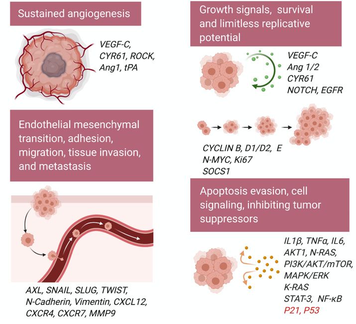
Int. J. Mol. Sci. 2021, 22, 1042                                                                                          2 of 30

                                   BCC and medulloblastoma tumors have constitutive activation of the HH pathway [10].
                                   Downstream HH signaling protein families such as GLI have become the spotlight as tar-
                                   gets for invasive epithelial cancers. HH pathway has been investigated for potential ther-
                                   apeutic antagonists as a strategy for cancer treatment, although the focus is limited to lig-
                                   and-dependent effectors such as Patched 1 (PTCH1) and Smoothened (SMO); Frizzled
                                   class receptor [11,12]. In this review, we discuss the prospect of HH/GLI signaling in can-
                                   cers due to viral infections and other causes.

                                   2. Various Types of GLI
                                         The GLI family includes three zinc finger proteins GLI1, GLI2, and GLI3 transcription
                                   factors identified as downstream targets of the HH signaling pathway—important in
                                   proper embryonic development, stem cell biology, and tissue homeostasis. Of the GLI
                                   proteins, GLI2 and GLI3 are the primary signal responders, with GLI2 mainly acting as a
                                   transcriptional activator and GLI3 predominantly acting as a repressor [13] (Figure 1).
                                   GLI1, a direct transcriptional target of HH signaling, is dispensable for mouse embryonic
                                   development [14,15]. HH signaling is GLI1 dependent in the spinal cord, and a combina-
                                   tion of GLI2 and GLI3 is required to regulate motor neuron and early ventral spinal cord
                                   development [16]. Structurally, they share similar features, including conserved tandem
                                   zinc fingers, conserved N- and C-terminal domains, and regions of protein kinase A (PKA)
                                   binding. There is an increased level of complexity in GLI transcription factors’ activation
                                   process as alternative splicing plays a significantly important role in controlling their ac-
                                   tivity. GLI1 alone functions as a transcriptional activator, whereas GLI2 and GLI3 have
                                   bifunctional transcription forms: full-length versions act as activators, and truncated N-
                                   terminal fragments act as repressors [13]. GLI2 and GLI3 display a repressor domain (RD),
                                   a processing determinant domain (PDD) [17], and a processing region (PR). N-terminal
                                   region of human GLI2 and GL13 contains a domain with a transcriptional repressor, and
                                   removal of this domain enhances the transcriptional activity of GLI2 [16,18,19]. The bal-
                                   ance between the GLI2/3 transcriptional activator and repressor actions dictates HH re-
                                   sponses that are both tissue-specific and developmental stage-specific. During limb and
                                   ureter development, SHH acts mainly by opposing GLI3 repression [13,20–22]. During
                                   neural tube and skeletal development, integrated regulation of GLI2 activation and GLI3
                                   de-repression has both overlapping and distinct functions. Both GLI2 and GLI3 constitu-
                                   tively traffic through primary cilia until activation of the HH pathway promotes the par-
                                   allel accumulation of GLI2 and GLI3 at the tip of primary cilia [23]. This region acts as an
                                   organizational center for activated GLI proteins, which then traffic to target gene promot-
                                   ers in the nucleus. The balance between these functional forms in the cell environment—
                                   which depend on post-transcriptional and post-translational processing—determines the
                                   HH transcription’s net direction and subsequent effects on tissue proliferation.
                                         Figure 1 shows GLI family functional domains, their similarities at the transcriptional
                                   level. GLI proteins belong to the GLI-Kruppel family of transcription factors and have
                                   Kruppel-type zinc-finger (ZF) motifs in their DNA binding domains [24–26]. Suppressor
                                   of fused (SUFU) binding site is highly conserved across all three mammalian GLI proteins
                                   [24–26]. Localization of GLI1 is influenced by the presence of a nuclear export signal
                                   (NES), and GLI1 becomes constitutively nuclear when this signal is mutated, or nuclear
                                   export is inhibited [27]. SUFU is a conserved negative regulator of GLI1 signaling that
                                   may affect the nuclear-cytoplasmic shuttling of GLI1 or the activity of GLI1 in the nucleus
                                   and thereby modulate cellular responses [27]. Recently, a PY-type nuclear localization sig-
                                   nal (PY-NLS) and the nuclear import factor karyopherin β2 (Kapβ2) were discovered to
                                   regulate GLI ciliary localization and HH pathway activity [28]. PY-NLS acts in conjunction
                                   with the canonical NLS (a bipartite NLS) localized in the Zn-finger domain [29]. The ca-
                                   nonical nuclear localization signal (NLS) in GLI plays a significant role, whereas the pro-
                                   line-tyrosine or PY-NLS has a minor role in targeting GLI to the nucleus. Interestingly,
                                   mutating the PY-NLS but not the canonical NLS impaired GLI ciliary localization and PY-
                                   NLSs interact with the Kapβ2, also known as transportin 1 (TRN1) or importin β2 and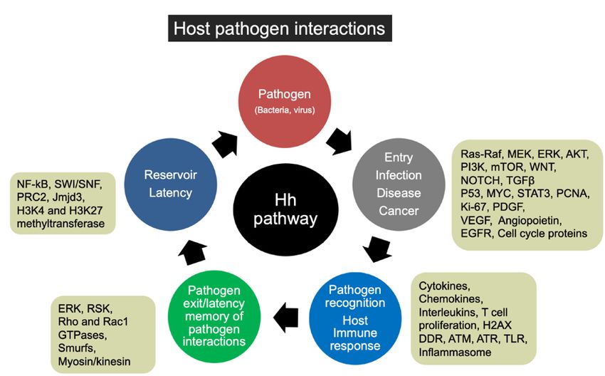
Int. J. Mol. Sci. 2021, 22, 1042                                                                                                3 of 30

                                   transport PY-NLS-containing proteins to the nucleus [28]. GLI activity is restrained by the
                                   phosphorylation of six conserved serine residues by protein kinase A (PKA) in the absence
                                   of HH ligands [30]. GLI protein functions are regulated via post-translational modifica-
                                   tions such as phosphorylation by PKA, glycogen synthase kinase 3 (GSK3), and casein
                                   kinase 1 (CK1) or ubiquitination, SUMOylation, acetylation, or methylation [24–26]. Two
                                   isoforms of GLI1 are termed GLI1ΔN and tGLI1 [31]. The full-length GLI1 is designated
                                   as GLI1FL [31]. However, GLI1ΔN lacks the SUFU-binding domain. It can activate and
                                   turn on target genes similarly to GLI1FL [19]. tGLI1 has a deletion of 41 amino acids, but
                                   it preserves all the functional domains present in GLI1FL. tGLI1 efficiently translocates
                                   into the nucleus to activate gene transcription and responds to HH ligand stimulation as
                                   well [6].

      Figure 1. Schematic shows known full-length GLIoma-associated oncogene homolog (GLI) family functional domains at
      the transcriptional level. Similarities shared among all three types include Suppressor of fused (SUFU) binding sites, zinc
      finger DNA-binding domain, and activation domains. GLI1 has an additional SUFU binding site at the C-terminus, while
      GLI2/3 has repressor domains; additionally, GLI2 has an extra activation domain that supports its main feature of activat-
      ing GLI-mediated transcription.

                                   3. GLI and Hedgehog Signaling
                                        The classical Hedgehog signaling pathway depends on the secretion of extracellular
                                   HH glycoproteins, Sonic HH (SHH), Indian HH (IHH), or Desert HH (DHH) [1]. SHH
                                   plays a critical role in maintaining normal embryogenesis, and abnormal fetal concentra-
                                   tions throughout its vast expression territory leading to the developmental defects, while
                                   its absence is lethal [32]. IHH, produced in hematopoietic cells, bone, and cartilage, has
                                   prominent HH signaling roles in the fetal liver [33]. DHH resides mostly in the peripheral
                                   nervous system and the testes, where its expression in pre-Sertoli cells serves as a marker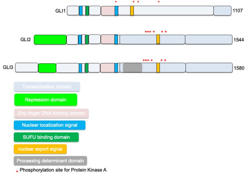
Int. J. Mol. Sci. 2021, 22, 1042 4 of 30
for male sexual differentiation [34]. Despite the macroscopic arrangement and responsi-
bilities of the HH family, canonical signaling remains conserved to the interaction of acti-
vated HH ligand binding and inactivating the cell-surface transmembrane protein,
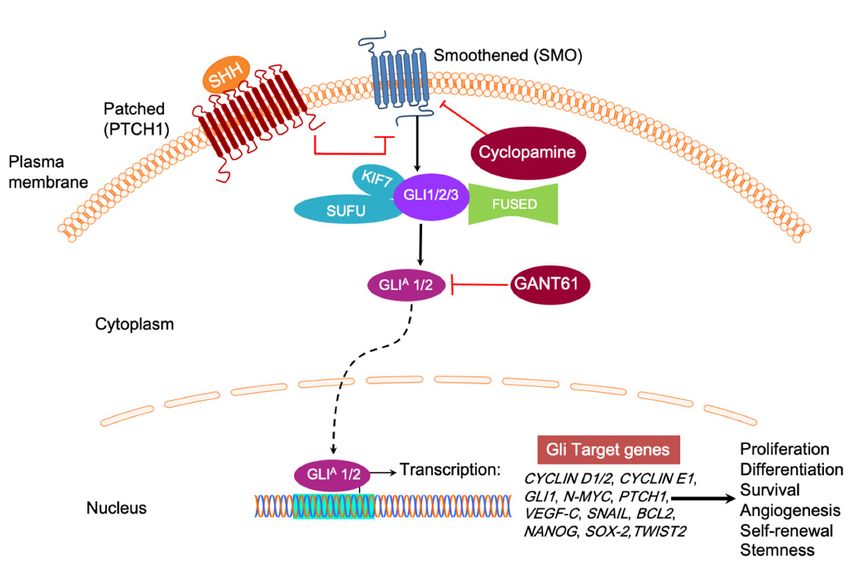
PTCH1 (Figure 2) [35]. In the inactive HH pathway, PTCH1 blocks the migration of SMO,
a G protein-coupled receptor (GPCR), to the cell membrane. When the SHH ligand binds
to PTCH1, inhibition upon SMO, a neighboring GPCR, is subsequently relieved, trigger-
ing downstream activation and nuclear localization of transcription factors like those in
the GLI family to regulate the expression of target HH genes, effectively inducing cell
proliferation, survival, differentiation, and angiogenesis. In the absence of HH ligand
binding, GLI proteins are typically sequestered in the cytoplasm by the negative regulator
called SUFU, which directly binds GLI to prevent activation of downstream pathway
genes (Figure 2) [36]. PKA initiates a phosphorylation cascade for PKA, GSK3, and CK1
phosphorylate GLI3 [37]. Phosphorylation of GLI3 targets it for ubiquitination [37]. Kine-
sin family member protein 7 (KIF7) is a conserved regulator of the HH signaling. KIF7
localizes to the cilium tip, the site of microtubule plus ends. KIF7 must relay the signals
from the membrane protein SMO to the GLI family transcription factors [38]. KIF7 affects
HH signaling, both positively and negatively (Figure 2). It functions as a negative regula-
tor of the SHH pathway by preventing GLI2 activation in the absence of ligand, and as a
positive regulator by preventing the processing of GLI3 into its repressor form. FUSED
(FU), the putative serine/threonine kinase, does not function in the mammalian HH sig-
naling but plays a role in motile cilia [39] (Figure 2). The balance between activated and
repressed forms of the GLI transcription factor determines the fate of target gene expres-
sion and cell phenotype.
Figure 2. In the absence of Hedgehog (HH) ligand (SHH, IHH, and DHH), the Patched 1 (PTCH) transmembrane receptor
at the base of the primary cilium maintains inhibition of Smoothened (SMO) G protein coupled receptor (GPCR) signaling.
Upon HH binding, SMO inhibition is relieved and activates GLI transcription factors, usually sequestered by SuppressorInt. J. Mol. Sci. 2021, 22, 1042 5 of 30
of fused (SUFU), KIF7, and FUSED. GLIA refers to the transcriptionally active form of GLI. SMO is a popular drug target
for cyclopamine, while GLI1 and GLI2-induced transcription can be inhibited by GANT61. It inhibits the HH signaling
pathway downstream of SMO and SUFU, causing GLI1 nuclear accumulation. Target gene expression includes CYCLIN
D1/2, CYCLIN E1, GLI1, N-MYC, Patched1, VEGF-C, and SNAIL to upregulate cell proliferation and tumor survival.
4. GLI Code
GLI expression is tightly regulated at the transcription level by multiple signaling
inputs—in the context of phase activity—that converge on the GLI code and direct its fate.
Whether cells undergo healthy development, homeostasis, or have a dynamic expression
of oncogenes and tumor suppressors during metastasis, GLI transcription responds ap-
propriately to the cells’ desired phenotype’s instructional cues. For example, tumor sup-
pressor p53 inhibits GLI1-induced neural stem cell self-renewal, proliferation, and tumor
growth through repressing nuclear localization and transcription activity of GLI1 [40]. In
contrast, the absence of p53, a hallmark event of most cancers, contributes to unregulated
GLI1 expression and tumor progression [41]. Similarly, a human orthologue of the mouse
tumor suppressor gene, RENKCTD11, also antagonizes GLI-mediated transactivation, and its
knock-down enhanced HH signaling and cell proliferation in medulloblastoma [42].
GLI transcription factors support tumorigenesis via cell signaling, cell proliferation,
angiogenesis, and metastasis [9]. Several studies suggest a crosstalk between RAS-RAF-
MEK, PI3K/AKT, and HH/GLI signaling pathways [43–46]. Not only does endogenous
GLI1 activity require intrinsic AKT and MEK signaling function, but AKT1, in combina-
tion with N-RAS, enhances nuclear localization and transcription of GLI1 (Figure 3) [43].
Activated K-RAS can also genetically cooperate with activated GLI2 to induce undiffer-
entiated pancreatic tumors [44,46]. A splice variant of p63, ΔNp63α, targets GLI2′s pro-
moter to increase expression and is an important event in osteosarcoma progression dur-
ing rare breast cancer forms [47]. Mediating this bidirectional transcription response af-
fecting GLI-level transcription determines the HH program’s status and phenotype of
cells in relevant tissues. HH pathway genes other than GLI1 include PTCH1, Wnt and
TGFβ superfamily proteins, cell cycle proteins (CYCLIN D), and stem-cell marker genes
(homeobox protein NANOG and SOX2) [48–50]. RAS/RAF/MEK/ERK, PI3K/AKT/mTOR,
epidermal growth factor receptor (EGFR), and NOTCH signaling pathways interact at the
level of the GLI transcription factors, except for NOTCH, which interferes with the ligand
SHH [51–53].
Constitutive canonical activation of the HH/GLI pathway is a classical representation
of aberrant signaling and is typically localized to mutations of PTCH1 (loss-of-function)
and SMO (gain-of-function) [54,55]. Inactive HH precursors undergo post-translational
modifications to form signal molecules consisting of cholesterol and palmitoyl residues,
which enhances ligand activity and diffusion capacity [56]. Non-canonical signaling is of-
ten recognized as a deviation from the typical motif of HH signaling, independent of GLI
activity, instead of acting through one of the multiple oncogenic pathways such as K-RAS,
TGFβ, PI3K-AKT, and PKC-α that target HH genes or are associated with a portion of the
HH pathway [54,55,57]. Not only does this provide more prospects for aberrant HH sig-
naling activity, but it also evades existing successful treatments for the canonical pathway
such as SMO inhibitor, cyclopamine. In vivo, there may be a combination of canonical and
non-canonical HH signaling that is regulated by crosstalk with other intracellular activity.
The HH pathway plays an essential role in cell proliferation, differentiation, apoptosis,
and migration, and it cross-talks with signaling pathways such as MAPK/ERK,
PI3K/AKT/mTOR, EGFR, and NOTCH (Figure 3) [52,58–60]. tGLI1 has been reported as a
stronger promoter of tumor migration and invasion as compared to GLI1 in glioblastoma
and breast cancer [61].Int. J. Mol. Sci. 2021, 22, 1042 6 of 30
Figure 3. Schematic shows the role of GLI1 and GLI2 in cancer cell proliferation, migration, invasion, cell cycle regulation,
angiogenesis, cell signaling, and survival kinases activation. Tumor suppressors downregulated by GLI are shown in red.
5. Involvement of GLI in Cancers
In recent years, the HH signaling pathway has shown significant contributions to
tumor initiation, progression to more advanced tumor stages, or low-grade to high-grade
tumors [62–65]. Inappropriate HH signaling plays a role in more than 30% of human can-
cers [66]. GLI1 overexpression in breast cancer serves as a significant marker of aberrant
activation of the SHH pathway driving the formation and progression of breast cancer
[67–69]. SHH pathway activation promotes mammary epithelial cell mesenchymal transi-
tion (EMT) [68,69], and regulates mammary cancer stem cell (CSC) self-renewal, and fa-
cilitates angiogenesis [70]. Additionally, inhibiting the GLI1 expression could efficiently
mitigate tumor growth and migration and showed its therapeutic potential in breast can-
cer management [71,72]. Studies reported no significant association between GLI1 expres-
sion and histological grade, T stage, clinical stage, and lymph node metastasis in breastInt. J. Mol. Sci. 2021, 22, 1042 7 of 30
cancer. A meta-analysis done in few studies explained GLI1 expression as one of the fac-
tors in aggressive biological behavior in breast cancer patients. Further, it elucidated the
link between GLI1 expression and prognosis of breast cancer [67,73,74]. GLI1 works
downstream of a protein lysine methyltransferase called SET7/9. The knockdown of
SET7/9 promotes the proliferation, migration, and invasion of breast cancer cells in vitro
and overexpression vice versa [75]. Investigation of the mechanism revealed that overex-
pression of SET7/9 inhibited GLI1 expression [75], suggesting that GLI1 expression in hu-
man breast cancer tissues negatively correlates with SET7/9 expression. Together, these
results establish that SET7/9 inhibits oncogenic activities by regulating GLI1 expression in
breast cancer [75]. High GLI1 expression in the claudin-low cells and tumors correlates
with EMT markers and breast CSCs [76]. GLI1 knockdown in claudin-low cells reduced
tumor growth of orthotopic xenografts, and treatment with nuclear factor κB (NF-κB)
pathway inhibitor decreases GLI1 expression and protein levels in breast cancer [76].
Inflammatory breast cancer (IBC), a rare (Int. J. Mol. Sci. 2021, 22, 1042 8 of 30
an initial genetic mutation acts as a marker for high HH signaling. It predicts poor prog-
nosis and suggests that the potential epigenetic reprogramming is an underlying natural
metastatic transition [92]. A novel variation in HH signaling that sustains the colon tumor
microenvironment inhibits the repressor form of GLI3 called GLI3R. SUFU, which typi-
cally sequesters GLI proteins in the absence of HH ligand binding, can also bind to GSK3β
to form the trimolecular complex GLI3/SUFU/GSK3β. This complex activates phosphory-
lation of GLI3 and subsequent processing/cleavage, which represses the HH pathway [93–
96]. High levels of GSK3β found in colon cancer tissues up-regulate non-canonical HH
signaling through maintaining the activator form of GLI3, thus effectively promoting can-
cer cell survival [93–96].
HH signaling downstream regulators are over-expressed in both squamous and ad-
enocarcinomatous esophageal cancers [97]. GLI1 binds to the caudal type homeobox 2
(CDX2), responsible for maintaining the intestinal phenotype promoter, activating the site
independently of SMO, which supports a non-canonical transition from squamous to co-
lumnar epithelium metaplasia in Barrett’s-associated adenocarcinoma cells [98]. A study
by Yang et al. found increased HH pathway activation in precancerous esophageal lesions
(Barrett’s esophagus and squamous dysplasia), suggesting HH signaling as an early event
esophageal cancer development [99]. The same study stated that high levels of SHH,
PTCH1, and GLI2 are focally expressed in the epithelium of carcinoma in situ, suggesting
potential early screening possibilities for unusual HH signaling activity [99]. Many
presentations of gastric cancer are epithelial-derived and manifest in the interstitium or
more diffusely as adenocarcinomas. El-Zaatari et al. demonstrated that GLI1 knock-down
in mice had reduced expression of a pre-neoplastic phenotype with low IL1β, TNFα, IL6,
phosphorylated signal transducer and activator of transcription 3 (STAT3), and prolifera-
tive marker Ki67 [100,101]. Studies that introduce GLI2 specific negative regulators in gas-
tric cancer cell lines informed a decrease in cell proliferation, migration, invasion, and an
increase in cell apoptosis [102,103].
Previous work in mouse models has shown that GLI2 over-expression in the skin
leads to the development of basal cell carcinoma (BCC), with sustained or aberrant upreg-
ulation of SHH/GLI cascade for cancer growth [104–106]. GLI2 acts as a direct upstream
activator of GLI1 via binding to its promoter site and initiates a positive feedback loop in
HH signaling.
HH signaling is also involved in ovarian cancer, bladder cancer, endometrial cancer,
rhabdomyosarcoma, pancreatic tumorigenesis, non-small-cell lung cancer (NSCLC), mel-
anoma, and hematological malignancies such as acute myeloid leukemia (AML), diffuse
large B-cell lymphoma (DLBCL), chronic lymphocytic leukemia (CLL), Hodgkin’s lym-
phoma (HL), ALK+ anaplastic large cell lymphoma (ALCL), mantle cell lymphoma
(MCL), multiple myeloma (MM) and chronic myeloid leukemia (CML) [66,107–115].
Dysregulation of SHH, the most potent and widely expressed HH ligand, is characteristic
of basal cell nevus syndrome, also known as Gorlin syndrome. [116,117]. Gorlin syndrome
is an autosomal dominant neurocutaneous disease mainly triggered by PTCH1 gene mu-
tations [118]. PTCH1 gene mutations permit SMO transposition and enhance the expres-
sion of GLI that drives cell proliferation and tumor growth [118]. SHH dysregulation in-
creases the risk of developing BCC, medulloblastoma, and meningioma. Activating or loss
of function mutations in the HH pathway genes SMO and PTCH1 occur in human gastric
tumors [119], sporadic BCC [120], medulloblastoma, nevoid basal cell carcinoma syndrome
(NBCCS), and colorectal cancer [121].
6. GLI and Hallmarks of Cancer
6.1. Angiogenesis
Initial stages in preparing the tumor microenvironment include a breakdown of the
vascular membrane and extracellular matrix (ECM), proliferation, and migration of endo-Int. J. Mol. Sci. 2021, 22, 1042 9 of 30
thelial cells (Figure 3). High expression of a common factor initiating angiogenesis, vas-
cular endothelial growth factor-C (VEGF-C), is often correlated to increased vascularity in
GLIoma formation [122]. There is a potential link between elevated HH signaling and in-
creased transcription of VEGF-C (Figure 3). Another factor, cysteine-rich angiogenic in-
ducer 61 called CYR61, is an ECM associated signaling molecule that promotes the adhe-
sion of endothelial cells through interaction via integrin. CYR61 contains pro-angiogenic
characteristics at the embryonic and wound healing properties at adult levels [123,124].
SHH-expressing breast cancer cells exhibit unregulated levels of CYR61 at the transcript
level. A GLI1 binding site exists upstream of the CYR61 transcription start site, and mu-
tation or deletion of this GLI1 binding site results in diminished activity of the CYR61
promoter in the presence of GLI1 [125]. SHH influences angiogenesis in endothelial cells
directly through the Rho/RhoA and Rho kinase (ROCK) signaling pathway [126]. SHH
induces the expression of matrix metalloproteinase 9 (MMP9) and osteopontin (OPN)
[126]. MMP9 is involved in the degradation of the ECM [126]. OPN, also known as bone
sialoprotein I (BSP1), is a highly chemotactic glycoprotein involved in bone remodeling,
bone mineralization, immune cell activation, and anti-apoptosis in various cancers and
viral infections [126]. Genetic ablation of the tissue plasminogen activator (tPA) in mouse
brain endothelial cells (MBECs) impaired tube formation and downregulated VEGF and
angiopoietin 1 (Ang1). Addition to rh SHH to tPA−/−, MBECs partially restored the tube
formation and upregulated Ang1, but not VEGF, although rh SHH increased VEGF and
Ang1 expression on wild-type MBECs. Complete restoration of tube formation in
tPA−/−MBECs was observed when both exogenous SHH and tPA were added, demonstrat-
ing the role of SHH-induced in vitro cerebral angiogenesis during the brain repair after
stroke [127].
6.2. Epithelial to Mesenchymal Transition (EMT)
Downstream effects of GLI family activation such as EMT transform adult tissues to
a mesenchymal-like state, losing polarity and adhesive properties while gaining migra-
tory characteristics that encourage the invasive nature of cancer cells (Figure 3) [128]. This
includes stem-cell features such as non-adherent growth, changes in the expression of cell-
surface glycoproteins, and surface marker expression of stem cells [129]. The net structural
transition of epithelial cells is from an attachment to the underlying ECM to migration
into the matrix [130,131]. HH signaling induces the trans-differentiation of epithelial cells
by decreasing E-Cadherin expression levels, increasing β-catenin and vimentin expres-
sion, tissue invasion, migration, and colony formation; it also transcribes necessary EMT
regulators such as SNAIL, SLUG, and TWIST via GLI1 in adult human placentas [132]. In
mouse models of gastric adenocarcinoma, EMT seems to be a particular phenotype of ac-
tivated GLI2 [133]. These metastatic cells undergo rapid proliferation and differentiation
typical of tumor heterogeneity and become locally invasive through basement tissues and
manifest as tumor islands in the gastric mucosa—features only present with activated
GLI2. Normal E-cadherin levels maintain cell attachment and layered phenotype of the
villous cytotrophoblast. In contrast, EMT-induced reduction of E-cadherin and redistri-
bution at cell junctional regions promotes loosened cell-to-cell connections and apicobasal
polarity [132].
Further evidence in human melanoma cells suggests that GLI2 is directly responsible
for turning off the E-cadherin gene (CDH1) expression during EMT in addition to enhanc-
ing the transcription of other EMT activators [134]. While this mesoderm expression pro-
cess is an essential precursor to the differentiation of multiple tissue types and generation
of organs in embryogenesis, it also enables a microenvironment of asymmetrical cell divi-
sion leading to macroscopic metastases in adult tissues. Various target molecules regulate
the proliferation pathway of HH signaling (Figure 3). Ectopic expression of Rab23 acts as
a negative regulator of HH signaling at the level of GLI1 and GLI2 mRNA expression
[135,136]. Interestingly, Rab23 upregulates the repressor form of GLI3, which endoge-
nously inhibits the HH signal cascade [137]. EMT cells increase breast cancer metastasisInt. J. Mol. Sci. 2021, 22, 1042 10 of 30
via paracrine GLI activation in neighboring tumor cells and triggering HH/GLI signaling
cascade [138]. GLI3 repressor (GLI3R) inhibits HH signaling, and GLI3R is essential for
response to SMO antagonist glasdegib in AML [139]. GLI3 is silenced in most AML pa-
tients [139]. GLI3R represses AML growth by downregulating AKT expression [140].
GLI3R plays an essential role in SMO-independent HH signaling in AML and suggests
that GLI3R could serve as a potential biomarker for patient selection in SMO antagonist
clinical trials. GLI3 inactivation results in additional digit formation in vertebrates [140].
GLI3 works as a negative modulator of the proliferative expansion of digit progenitors by
restricting the G1 to S cell-cycle transition by regulating CDK6 and constrains S phase
entry of digit progenitors [140].
6.3. Cell Cycle
Aberrant HH signaling triggers a series of vasculogenic and angiogenic processes
that endorse tumorigenesis and tumor growth in adult tissues. Several proposed that
feedback loops regulate HH pathway activity (Figure 3). IFN-γ/STAT1 signaling has tu-
mor suppressor function and is inactive in at least one-third of all melanoma and lung
adenocarcinoma cell lines in mice [141]. SOCS1, an IFN-γ/STAT1 inhibitor, is activated by
HH signaling pathway itself to create a negative feedback loop and a downstream target
of GLI1 and GLI2, which upregulates its transcriptional activity and subsequently relieve
the IFN-γ/STAT1 form obstructing tumor growth [142]. Common cell cycle genes turning
on via the HH pathway include CYCLIN D and E, which are necessary to induce G1-to-S
transition in the cell cycle, and CYCLIN B, which activates mitosis promoting factors (Fig-
ure 3). HH signaling opposes normal stimuli for epithelial cell cycle arrest such as P21 and
inhibits the P53 tumor suppressor gene [143,144].
6.4. Migration/Adhesion/Invasion/Metastasis
Mechanisms of HH induced metastasis are understood at a broad level, but specific
phases remain under deliberation. Upregulation of G-protein coupled receptors (GPCRs),
chemokines CXCR4 and CXCR7 enhance the directional migration of breast cancer cells
in lung metastasis by way of CXCL12 [145], which is a highly-secreted signaling protein
of metastatic organs [146]. HH pathway upregulation of GLI1 enhances the CXCL12 in-
duced migration of cancer cells [147]. Administering CXCR4 specific inhibitors or knock-
down treatments suppress cancer cell migration patterns in breast and pancreatic cancer
in mouse models and in vitro [145]. Part of the migration effectiveness depends on the
EMT-programmed loss of cell–cell adhesion that leads to motility. GLI1 activates EMT by
inducing SNAIL expression. SNAIL causes fibroblastic conversion, malignant transfor-
mation, and loss of a cell–cell adhesion molecule called E-cadherin [148]. Once the adhe-
sive properties of the membrane are compromised, metastatic cells invade the matrix. The
EMT program upregulates the expression of essential invasion factor AXL, which is re-
quired to maintain SNAIL, SLUG, and TWIST expression in pancreatic adenocarcinoma
cells [149]. Interestingly, these same factors potently induce AXL expression, contributing
to a positive feedback loop that continues the proliferation of malignant mesenchymal
tumor cells [149]. GLI2 knockdown studies supporting its role in migration and invasion
in osteosarcoma, prostate cancer, and hepatocarcinoma cell lines underscores the meta-
static potential of GLI [150,151].
GLI2 expression directly enhances tumorigenesis in a model of myofibroblastic cells
representing reactive stromal prostate cancer cells [152]. There is still some controversy
regarding the exact model of hormone signaling in prostate tissues: paracrine versus au-
tocrine. Paracrine signaling from the epithelium supports stromal differentiation during
prostate development and sustains the stroma in the adult prostate [153,154]. In xenograft
tumors, SHH is localized to the prostatic epithelium, while GLI1 mRNA is localized to the
stromal compartment suggesting paracrine HH signaling [155]. Other studies support the
idea of a shift to autocrine SHH signaling during pathogenesis and progression of prostateInt. J. Mol. Sci. 2021, 22, 1042 11 of 30
carcinoma [156]. There may be some interplay between these two forms of hormone ac-
tivity in vitro, at least during the introduction of tissue metastasis, if not during the tumor
progression.
7. HH Signaling Pathways during Infections and Viral Malignancies
Besides controlling processes involved in embryogenesis, there are broader implica-
tions of HH signaling. Recent studies revealed that aberrant activation of HH signaling
leads to pathological consequences [157]. Viruses enter the host by interacting via their
surface proteins, hijacking host replication machinery, targeting several signaling path-
ways concurrently, and overturning host immune mechanisms and evolutionary benefit
from the host cell machinery. The virus makes copies of itself and spreads those copies to
new hosts. Many viruses have evolved to stimulate host HH signaling to control their life
cycle and pathogenesis [157,158]. There is considerable speculation of pathogens to use
HH signaling to regulate their life cycle.
Here, we discuss influenza-A, hepatitis B virus (HBV), hepatitis C virus (HCV), hu-
man immunodeficiency virus (HIV), human papillomavirus (HPV), Merkel cell polyoma-
virus (MCPyV), human T-cell leukemia virus, type 1 (HTLV-1), Epstein–Barr virus (EBV)
and Kaposi’s sarcoma herpesvirus (KSHV) infection to highlight the plausible involve-
ment of HH pathway players in the viral life cycle. We discuss the possible role of HH
signaling in viral latency, virus reservoir maintenance, entry, replication, pathogenesis,
infected cell proliferation, infection progression, pathogen recognition in the host, and the
immune response of the host, exit of the pathogen and memory of the host immune me-
diators (Figure 4).
Figure 4. Schematic shows various stages and consequences of host-pathogen interactions. It also shows multiple signaling
cascades, and transcription factors are demonstrated to be regulated by the HH pathway. Signaling molecules and tran-
scription factors regulated by HH signaling could contribute to pathogen (virus or bacteria) entry, infection, pathogenesis
(angiogenesis, fibrosis, inflammation), and activation of host foreign recognition network, host immune response, patho-
gen exit/elimination, and pathogen latency in the host.Int. J. Mol. Sci. 2021, 22, 1042 12 of 30
HIV [159,160], HCV [161,162], HBV [163], EBV, and KSHV [164–166] utilize VEGF to
maintain the vascularity of the tumors and infected cell proliferation. HH signaling plays
an indispensable role in vascular development in mouse embryos via VEGF and NOTCH
signaling [167]. SHH regulates the angiogenic VEGF and angiopoietin (Ang 1 and Ang 2)
in astrocytes by activating the nuclear receptor subfamily 2 group F member 2 (NR2F2)
transcription factor [168] (Figure 3). HIV [169], HCV [170], HBV [171,172], EBV and KSHV
[173–176] infection promotes angiopoietin expression. Maturation of VEGF-induced new
vessels in the cornea involves a platelet derived growth factor (PDGF)-SHH axis that me-
diates PDGF-BB–mediated smooth muscle cell (SMC) migration by inducing ERK1/2 and
PI3Kγ activation [177]. Elegant studies described that KSHV utilizes host PDGF receptor-
α (PDGFRα) to drive its tumorigenesis and is prominently active in murine and human
AIDS-associated-Kaposi’s sarcoma (AIDS-KS) development [178]. Exclusively blocking
PDGFRα signaling could impede murine KS tumor formation [178]. There is a strong cor-
relation between the HH pathway and hypoxia and hypoxia-inducible factor 1 (HIF1).
Hypoxia induces upregulation and secretion of SHH in the human pulmonary arterial
smooth muscle cells (HPASMCs) [179]. SHH expression depends on HIF1 in the
HPASMCs, and hypoxia stimulates GLI1 nuclear translocation [179]. Overall, hypoxia in-
duces HPASMC proliferation, and the HH signaling pathway regulates apoptosis in the
HPASMCs subjected to hypoxia [179]. HIV [180], EBV [181], and KSHV [182–186], HCV
[187], and HBV [182] utilize the host HIF1 factor for redox signaling during their life cycle.
HH signaling is a potential target for manipulating viruses, as this pathway plays a
fundamental role in cell survival and proliferation [188–190]. The HH target gene PTCH1
is one of the critical host genes involved in influenza infection and favors viral dissemina-
tion [191,192]. Nonstructural protein 1 (NS1), one of the multifunctional proteins, limits
the lung’s injury to preserve the viral habitat in the host. HH signaling plays a significant
role in branching morphogenesis during pulmonary development [193]. Immunohisto-
chemical analyses have indicated increased TGFβ1 expression at the epithelial site of fi-
brosis, such as cryptogenic fibrosing alveolitis and bronchiectasis. SHH antagonizes
TGFβ1 by inducing epithelial repair [194]. Patients with scarring in distal lung secondary
to bronchiectasis show prominent perinuclear staining for SHH, thus relating the SHH
staining to the level of injury [195].
Moreover, the HH receptor, PTCH1, is expressed in infiltrating and circulating lym-
phocytes (CD4 and CD8) at both protein and mRNA levels. It is evident that immune cells
are equipped to respond to the HH ligand secreted from the inflamed area [196]. HH sig-
naling thus plays an essential role in repairing damaged lung tissue by remodeling the
epithelium directly or in concurrence with activated immune cells and regulates the com-
munication of immune cells. Therefore, controlling the HH response may help the host to
avoid the harmful consequences of viral infection. A similar study on larval Drosophila
melanogaster has shown that the effect of the NS1 on the HH pathway is conserved be-
tween the species. A point mutation (A122V) was identified in Drosophila, which could
reduce the HH-dependent activity of the NS1 in flies and transfected cells [191]. However,
the same mutation, when incorporated into a mouse-adapted influenza-A virus, intensi-
fied the expression of some HH targets in the mouse lung and significantly accelerated
lethality. No mutation at position 122 of NS1 has been recognized in any influenza strains,
and thus NS1 protects the host. This muting of HH signaling may be utilized to diminish
the harmful effects that could be caused by HH signaling to ensure optimal viral matura-
tion before dissemination.
HIV [197], HCV [198], and HBV [199], EBV [200], and KSHV [201–203] are well-
known to program their genes selectively to modulate the DNA damage response (DDR).
KSHV manipulates DDR either via activation of ataxia telangiectasia mutated (ATM)
pathway or by phosphorylating factors associated with the DDR, such as tumor suppres-
sor protein P53 [201–203]. Interestingly, canonical HH signaling or ectopic expression of
GLI1 causes genomic instability and cancer predisposition by faulting the S-phase check-Int. J. Mol. Sci. 2021, 22, 1042 13 of 30
point, DNA repair mechanisms, and inhibiting DNA double-strand breaks (DSBs)-medi-
ated DDR [204]. ATM and Ataxia telangiectasia and RAD3 related (ATR) protein serine-
threonine kinases involved in cell-cycle checkpoint signaling are regulated by HH signal-
ing [205]. GLI inhibitor GANT61 induces DNA damage. GANT61 treatment increases the
nuclear foci of γH2AX and activates ATM and Chk2 in human colon carcinoma cells [206].
HH signaling also contributes to the epigenetic regulations. HH target genes are
poised and are marked by an active (H3K4me3) and a repressive (H3K27me3) mark main-
tained by the H3K27 methyltransferase polycomb repressive complex 2 (PRC2) [207].
SHH induction recruits the Jumonji domain-containing protein D3 (Jmjd3), a histone
H3K27 demethylase, which dislodges PRC2, removes H3K27me3, and enlists the
Set1/MLL H3K4 methyltransferase complex to initiate gene expression [207]. DNA viruses
exploit epigenetic regulation in maintaining viral episomes through the generation of
chromatin, controlling viral gene transcription and replication or evading of the host in-
nate immune response [208]. Excitingly, HIV [209], HCV [210], HBV [211,212], EBV
[213,214] and KSHV [215,216] employ host methyltransferases, histone modification en-
zymes, histone acetylases, deacetylases, and demethylases, etc., for their chromatin re-
modeling, reactivation from latency, and viral life cycle propagation.
HBV and HCV are associated with liver cirrhosis and hepatocellular carcinoma
(HCC) worldwide [217]. Pereira et al. showed increased hepatocyte production of HH
ligands in patients with chronic HBV and HCV infection [218]. HH pathway activation
often occurs during fibrogenic repair of liver damage due to chronic viral hepatitis [218].
HH-responsive cells facilitate hepatocarcinogenesis and disease advancement in chronic
viral hepatitis [218]. Another study further confirmed the increased expression of HH tar-
gets in a GLI-dependent manner when liver cells were treated in vitro with the whole
HBV replicon or with serum from HCV-infected patients [219]. This caused pro-fibrotic
effects [219]. Hepatitis B virus x (HBx), one of the HBV viral proteins, increases GLI1 pro-
tein nuclear accumulation [220]. HBx protein stimulates the HH-GLI activation through
protein stabilization and nuclear localization of GLI1 in liver cancer cells while the exact
role of GLI1 protein translocation for these viral activities remains to be discovered [220].
Blocking HH signaling delayed hepatocarcinogenesis induced by HBx protein (Table 1)
[221]. HH signaling in liver cells is associated with increased permissiveness for HCV rep-
lication and viral production [222].
Table 1. Therapeutic strategies against viral diseases.
Hh Inhibitors Disease Mechanism of Action
Decreases liver fibrosis in human
Decreases tumor formation in a mouse model of fibrosis-associated HCC
Vismodegib (GDC-0449) HBV and HCV
Reduces the growth of HBV X-expressing tumor xenografts in nude mice
and HCC formation in transgenic mice expressing the HBV X protein
Reduces the pro-fibrotic effects
EBV and EBV linked
Inhibits autophagy in HCV-exposed fibroblasts
Nasopharyngeal cancer
GLI inhibitors: GANT-61 Reduces tumor-sphere formation in several EBV-infected cell lines
Decreases the proliferation of Human Papilloma Virus-derived cervical
HPV
cancer cells
GLI agonist Targeted against local uninfected environment
HIV Targeted against HIV infected Cells.
Smoothened agonist
Limits viral niche.
The HH pathway maintains and controls stem cells and policies of hematopoiesis
and lymphopoiesis to maintain immune cells [223–226]. Increased GLI1 expression in EBV
infection decreases the level of human leukocyte antigen (HLA) and helps the virus to
escape cytotoxic T cell recognition [227]. The HH pathway is activated in EBV derived
nasopharyngeal carcinoma (NPC) tissue, NPC-derived cell lines, and in EBV infected ep-
ithelial cells [228]. HIV-related nephropathy presented increased expression of GLI andInt. J. Mol. Sci. 2021, 22, 1042 14 of 30
associated proteins of the HH pathway [217]. EBV latent membrane protein 2A (LMP2A)
utilizes Gli1 to downregulate HLA in gastric cancer cells [229]. The same was observed in
a human podocyte cell line infected with HIV [217]. These aberrant pathway activities
decrease host defense by increasing proliferation and migration markers, loss of kidney
filtration barrier function, and increased permeability. All these changes observed could
seemingly boost viral dissemination and increase host infectivity [217]. HIV infection is
linked to the elevated level of immunoregulatory cytokine TGFβ1 that leads to the sup-
pression of host protective immune responses [230]. GLI2 regulates TGFβ1 at the tran-
scriptional level in human CD4+ T cells during HIV infection [230]. Human SMO inhibits
HIV-1 replication and disease [231]. The role of the HH pathway was tested in HIV-in-
duced EMT, which is critical for the progression of kidney injury [217]. The blockade of
the HH pathway with GANT 58 (GLI antagonist, a specific blocker for GLI1-induced tran-
scription) treatment could dramatically decrease HIV-induced podocyte EMT, permeabil-
ity, and fibrosis of the kidney (Table 1) [217].
HH regulates the expression of OPN in nonalcoholic steatohepatitis-related liver fi-
brosis as GLI directly interacts with the GLI-binding sites in the OPN promoter [232]. A
decrease in either HH signaling or OPN decreases fibrosis [232] as OPN is a direct tran-
scriptional target of the HH pathway [232]. Treatment with GLI1 inhibitor or cyclopamine
plays a protective role in a mouse model of renal fibrosis following injury and expressing
the activated HH signaling pathway [232]. Some viruses may promote HH signaling to
induce fibrotic damage to ensure viral spread [157,217,233]. Other pathogens, such as the
influenza virus, limit fibrotic tissue formation, thereby allowing more time for replication,
maturation, and dissemination of infection [157,191]. However, to maintain progeny,
these viruses make sure to support viable hosts available for reinfection.
Merkel cell polyomavirus (MCPyV) is detected in approximately 80% of Merkel cell
carcinoma (MCC), an aggressive neuroendocrine skin cancer mostly occurring in the el-
derly. Reactivation of HH signaling later in life can cause tumors. 29 MCPyV-positive and
21 MCPyV-negative MCCs were stained for SHH, IHH, PTCH1, SMO, GLI1, GLI2, and
GLI3 and expression of the HH signal pathway players associated with MCPyV infection
and prognosis of MCC [234].
HPV oncogenes (E6/E7) and estradiol, major etiologic factors associated with cervical
cancer, could induce GLI activity in the cervix and the skin in mice [235]. Treatment with
a putative novel HH inhibitor itraconazole, could not diminish HH signaling, but it re-
duced growth at an early stage of cervical carcinogenesis (Table 1) [235]. While these stud-
ies suggested the possible involvement of HH signaling in cervical carcinogenesis, the
mechanism is not known [235]. GLI1 and GLI2 overexpression serve as a prognostic factor
for overall and disease-free survival in patients with locally advanced HPV negative head
and neck cancer undergoing surgery and postoperative radiotherapy [236]. The human
poliovirus receptor CD155 gene acts as a transcriptional target of SHH and is activated by
SHH in neuroectodermal tumors [237].
Tumor cells of Hodgkin lymphoma (HL) are derived from mature B cells. The lineage
infidelity of Hodgkin/Reed-Sternberg cells (HRSs) often causes diagnostic problems as
HRS markers are also favorable for follicular dendritic cells (FDCs). Investigation of the
expression of FDC markers in HL and anaplastic large cell lymphoma (ALCL) revealed
GLI3, fascin (actin-bundling protein found in membrane ruffles), and TUBB3 (a member
of the beta-tubulin protein family) as the most sensitive markers, which were diffusely
positive in HL [238]. A recent study from our lab reported the increased expression of
GLI1 in KSHV infected primary effusion lymphoma (PEL) cells [239].
Anti-inflammatory lipoxin A4 treatment in PEL cells downregulated GLI1 expression
[239]. The decrease in GLI1 and PTCH1 expression was not dependent on SHH ligand
activity, as we did not observe any significant change in SHH expression in the solvent or
lipoxin A4 treated PEL cells. Interestingly, we found increased phosphorylation of GLI1
at Thr 1074 and decreased phosphorylation of AKT/mTOR proteins in lipoxin A4 treated
PEL cells. GLI1 phosphorylation at Thr 1074 led to the degradation of GLI1 through 5’Int. J. Mol. Sci. 2021, 22, 1042 15 of 30
adenosine monophosphate-activated protein kinase (AMPK) activation (through the
phosphorylation of AMPK at Thr 172) and reduced the oncogenic potency of GLI1 by
preventing the transcription of target genes GLI1 and PTCH1. We are testing the thera-
peutic potential of GLI inhibitors in KSHV related cancers, including KS and PEL.
HTLV1 expression is activated by the interaction of a viral transactivator protein,
TAX, and cellular transcription factor, CREB (cyclic AMP response element-binding pro-
tein), binds to the long terminal repeat (LTR). The human homolog of a member of the
GLI oncogene family, GLI2 (also termed hGLI2), helps HTLV1 infection progression as
the simultaneous binding of hGLI2 and CREB seems critical for TAX protein to activate
transcription [240]. HIV [241], HCV [242], HBV [243,244], EBV [245], and KSHV [246] uti-
lize MYC and cellular STAT3 for infected cell proliferation, the persistence of herpesvi-
ruses latency, and inhibition of viral reactivation. HIV [247], HCV [248], HBV [249], EBV
and KSHV [250,251] infected cells secrete cytokines and chemokines to regulate viral path-
ogenesis, evade host immune response (Toll-like receptors; TLRs and inflammasome), an-
giogenesis, and selectively chemoattracts T cells, activation and migration of immune
cells. HIV1 envelope protein R5 gp120 exposure to immature monocyte-derived DCs
(MDDCs) resulted in the CCR5-dependent production of interleukin-6 (IL6) cytokine via
mitogen-activated protein kinase (MAPK)/NF-κB pathways [252]. IL6 could activate
STAT3 by an autocrine loop, further contributing to IL6 secretion [252]. HH pathway has
been demonstrated to act synergistically with interleukin-6 to drive the growth of basal
cell carcinoma via STAT3 activation [253]. Mechanistically, IL6 and HH/GLI signaling in-
tegration occur at the level of cis-regulatory sequences by co-binding of GLI and STAT3
to common HH-IL6 target gene promoters and HH-IL6 pathway combinatorial blockade
could efficiently arrest cancer growth in BCC patients [253]. These transcription factors,
signaling pathways mediating IL6 induction have not been tested in the context of viral
infections, which depend significantly on the IL6 for survival and progression of infection
such as KSHV [254], H1N1 influenza A infection [255], Pneumovirus infection; closely
related to a respiratory syncytial virus [256], Hepatitis B Virus [257], and EBV [258].
HH signaling is also regulated by MAPK and NF-κB cascade and HH/GLI1, MAPK
(KRAS-MEK-ERK) cascade and NF-κB cooperate to regulate growth and cell proliferation
[259], and apoptosis resistance in many tumors [260]. These studies provide a potential
link that could be playing an important role in the lifecycle of many viruses, which acti-
vate MAPKs and NF-κB in host cells upon binding, entry, or during the stage of viral gene
expression. Similar to MAPK, ERK, PI3K, and NF-κB, HH/GLI pathway plays an im-
portant role in the induction and sustenance of Rho-GTPases and stimulates cell migration
[261,262]. HIV [263], HCV [264], HBV [265], EBV [266] and KSHV [267–269]. These viruses
utilize signaling pathways such as ERK, RSK, PI3K, Rho, and Rac1 GTPases for cell cycle
progression, viral entry, cellular transformation, expression of viral genes, and the estab-
lishment of infection. HIV [270], HCV [271,272], HBV [273–275], EBV, and KSHV [276–
279] exploit host AKT, PI3K, mTOR signaling pathways for the evasion of apoptosis, in-
fected cell survival, and proliferation, viral replication, production, vesicle formation, in-
tracellular motility and activation of transcription factors. HIV [280], HCV [281], HBV
[282], EBV [283], and KSHV [269] utilize myosin/kinesin for viral transmission, virion
transcytosis, virus entry, intracellular viral transport, and formation of highly metastatic
and invasive tumors with the leading edge.
Not only viruses, but bacterial infections such as Salmonella enteritidis ST183 [284]
Escherichia coli [285], and Helicobacter pylori [286] are also inclined to use the HH signaling
pathway to control the infection progression and the infected cell microenvironment
[284,285,287,288] (Figure 4). Helicobacter pylori infection mediated inflammation and repair
process involving macrophage recruitment activates transcription factor NF-κB and also
upregulates HH proteins [286].
8. GLI Inhibition and Implications as Anticancer TherapeuticsInt. J. Mol. Sci. 2021, 22, 1042 16 of 30
Currently, small molecule modulators such as SMO and GLI1 inhibitors of HH sig-
naling have been used in basic research to detect links between signaling and specific phe-
notypes of interest [289]. Few inhibitors are now in use to treat certain malignancies asso-
ciated with viral infections (Table 1), BCC and certain leukemia, whereas many others are
still in clinical trials [290]. Till now, vaccines and antivirals, which are specific to rapidly
mutating viral proteins, have been used, but now with the use of HH inhibitors, a broad
spectrum of strains can be targeted. Since the HH signaling controls many critical cellular
processes, simultaneously, there is a need to target a specific pathway component selec-
tively. This process may require combinatorial drug usage targeting different HH-de-
pendent processes, which may further regulate signaling in the infected cells. An in-depth
understanding of the precise mechanism by which viral factors would interact with the
HH pathway players would prove beneficial for targeted therapies.
Aberrant HH signaling has been found responsible for chemo-resistance in aggres-
sive cancers [291,292]. Much work has already shown the worth of the GLI family as
emerging targets for cancer therapy [293]. GLI1 inhibitors have demonstrated the broadest
therapeutic potential so far as a target in advanced and metastatic tumors [293]. GLI1 ex-
pression has been used as a potential prognostic factor for survival in bladder and colon
cancer [294]. Inhibition of upstream MEK1/2-ERK1/2 activity with U0126 inhibitor in hu-
man HT29 colon cancer cells suppress GLI transcriptional activity and subsequent protein
expression [96]. GLI1 also supports a correlation between low expression and more pro-
longed survival in patients with oral squamous cell carcinoma [295]. Breast tumor cells
upregulate GLI2 expression during bone metastasis, stimulating bone resorption, activat-
ing TGFβ, and subsequent tumor proliferation [296]. Aberrant HH signaling activation
promotes the growth of BCC, medulloblastoma, colorectal cancer, and small lung cell
cancer [289]. This has led to a large repertoire of small molecule inhibitors developed
for the treatment of cancers dependent on the HH pathway.
Among SMO inhibitors, GDC-0449 and LDE-225 are in the clinic for the treatment of
advanced BCC with aberrant HH activity due to loss of the functional allele of PTCH
[297,298] The steroidal alkaloid, cyclopamine, has progressively shown therapeutic po-
tential as an inhibitor for HH signaling. The mechanism of cyclopamine action suggests
an interaction with the SMO heptahelical bundle, promoting a protein conformation
through small endogenous molecules rather than direct protein-protein interaction
[292,299]. In GLIoma-derived neurospheres, cyclopamine blocked inhibited overall
growth rate by 30–70% [291,300]. Vismodegib (trade name Erivedge) is another SMO in-
hibitor that effectively terminates HH signaling. Vismodegib is also an FDA approved
treatment for basal cell carcinoma in adult patients [301]. There is less support for cyclo-
pamine and its analogs as therapies in cancers with bone metastases such as IBC, wherein
GLI2 needs to be inhibited further downstream from the HH receptors [302]. LDE-225
(Erismodegib/Sonidegib/Odomzo), HH pathway inhibitor, received FDA approval to
treat cancer patients [303,304]. Many phase I and phase II trials for Erismodegib as a mon-
otherapy and in combination are underway, treating malignancies including advanced
gastroesophageal adenocarcinoma, small cell lung cancer, myelofibrosis, advanced/meta-
static HCC, and relapsed medulloblastoma [305,306].
IPI-926 (Saridegib), CUR61414 binds to SMO and prevents its activation, BMS-
833923/XL139 (binds SMO), PF-04449913; Glasdegib (SMO inhibitor), PF-5274857 (SMO
antagonist), TAK-441 (SMO inhibitor), LY2940680; Taladegib (SMO antagonist), MRT-92
(anti-SMO activity by blocking several overlapping sites of the SMO transmembrane do-
main), Jervine (binds to SMO and preventing its conversion to an active state), RU-SKI 43
(SHH Inhibitor), and SHH Monoclonal Antibody 5E1 (SHH Inhibitor) is SMO or SHH
Inhibitor in clinical trials [305,306].
A well-known small molecule inhibitor, GANT61, reduces GLI1, GLI2, and PTCH1
mRNA expression in human colon carcinoma cell lines [96]. GANT61 appears to be a more
potent treatment in colon carcinoma cell lines than its upstream-acting counterpart, cyclo-You can also read