COVID-19: UNMASKING EMERGING SARS-COV-2 VARIANTS, VAC-CINES AND THERAPEUTIC STRATEGIES - PREPRINTS.ORG
←
→
Page content transcription
If your browser does not render page correctly, please read the page content below
Preprints (www.preprints.org) | NOT PEER-REVIEWED | Posted: 2 June 2021 doi:10.20944/preprints202106.0060.v1
Review
COVID-19: Unmasking emerging SARS-CoV-2 variants, Vac-
cines and Therapeutic Strategies
Renuka Raman 1,†, Krishna J. Patel 2,† and Kishu Ranjan 3,*
1 Department of Surgery, Weill Cornell Medical College, New York, NY 10065, USA;
rer2029@med.cornell.edu
2 Icahn School of Medicine at Mount Sinai, New York, NY 10029, USA; krishna.patel2@mssm.edu
3 School of Medicine, Yale University, New Haven, CT 06519, USA; kishu.ranjan@yale.edu
* Correspondence: kishu.ranjan@yale.edu; Tel.: +1- 203-785-3588
†These authors contributed equally to this work
Abstract: Severe acute respiratory syndrome coronavirus 2 (SARS-CoV-2) is the etiological agent of
the coronavirus disease 2019 (COVID-19) pandemic which has been a topic of major concern to
global human health. The challenge to restrain the COVID-19 pandemic is further compounded by
the emergence of several SARS-CoV-2 variants viz. B.1.1.7, B.1.351, P1 and, B.1.617., which show
increased transmissibility and resistance towards vaccines and therapies. Importantly, the likeli-
hood of susceptibility to SARS-CoV-2 infection among individuals with dysregulated immune re-
sponse or comorbidities needs greater attention. Herein, we provide a comprehensive perspective
regarding ongoing vaccine (mRNA, protein-based, viral vector based etc.) and therapeutic (mono-
clonal antibodies, small molecules, plasma therapy, etc.) modalities designed to curb the COVID-19
pandemic. We also discuss in detail the challenges posed by different SARS-CoV-2 variants of
concern (VOC) identified across the globe and their effects on therapeutic and prophylactic inter-
ventions.
Keywords: SARS-CoV-2; COVID-19; variants; vaccines; immune dysregulated; comorbidities; an-
tibody; Spike protein; biomolecules; coronavirus.
1. Introduction
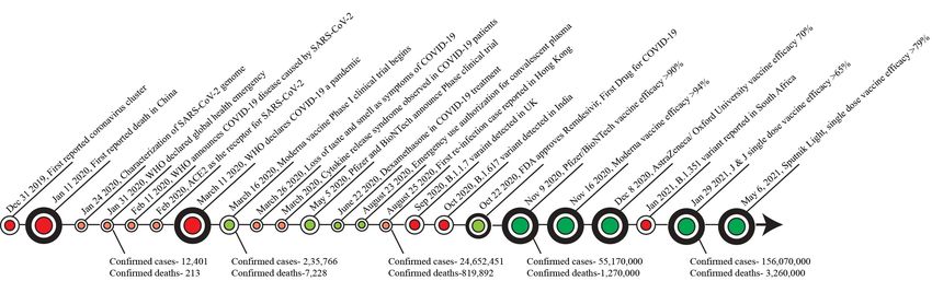
The catastrophic spread of coronavirus disease 2019 (COVID-19) has already claimed millions
of lives across the globe and has been declared as a public health emergency of international con-
cern by the World Health Organization (WHO) [1] (Figure 1). So far, there are 7 different types of
coronaviruses documented; among these, four common human coronaviruses- 229E, NL63, OC43
and HKU1, cause mild infections [2]. However, individuals infected with either of the other three
coronaviruses- severe acute respiratory syndrome coronavirus (SARS-CoV), Middle East respira-
tory syndrome coronavirus (MERS-CoV) and SARS-CoV-2, develop severe respiratory distress and
viral pneumonia and may ultimately succumb to the disease [3-5]. SARS-CoV-2, the causative
agent of the ongoing COVID-19 pandemic, is a newly identified, enveloped, highly diverse sin-
gle-stranded RNA virus [4-7]. It is noteworthy that the nucleotide sequence of SARS-CoV-2 nearly
matches (96% similarity) with a bat coronavirus RaTG13 (GenBank: MN996532.1), suggesting that
bats are the most likely progenitors of SARS-CoV-2 and the source for zoonotic spillover to human
[5,8]. The molecular characterization through an RNA-based metagenomic next-generation se-
quencing (mNGS) analysis revealed that the SARS-CoV-2 genome is 29,881 bp in length (GenBank
no. MN908947) and encodes 9860 amino acids [9].
SARS-CoV-2 genome encodes distinct structural and nonstructural proteins; genes encoding,
spike (S) glycoprotein, envelope (E) glycoprotein, membrane (M) glycoprotein, and nucleocapsid
(N) protein constitute the structural components, whereas 3-chymotrypsin-like protease, papa-
in-like protease, and RNA-dependent RNA polymerase in addition to several accessory proteins
constitute the nonstructural framework of SARS-CoV-2 [10]. (Figure 2A). Notably, the S glycopro-
tein is composed of 1273 amino acids constituted of N terminal signal peptide (amino acids 1–13),
the S1 subunit (14–685 residues), and the S2 subunit (686–1273 residues). Furthermore, the S1
subunit contains an N-terminal domain (NTD, 14–305 residues) and a receptor-binding domain
© 2021 by the author(s). Distributed under a Creative Commons CC BY license.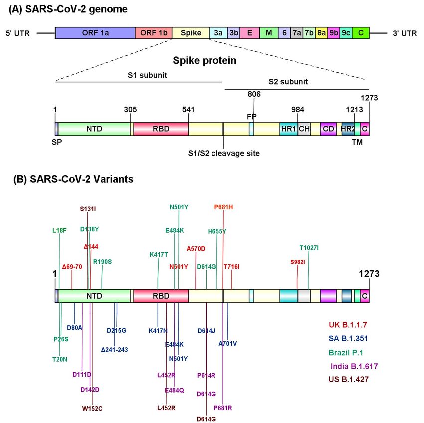
Preprints (www.preprints.org) | NOT PEER-REVIEWED | Posted: 2 June 2021                           doi:10.20944/preprints202106.0060.v1

                                                                                                                                        2 of 32

                                         (RBD, 319–541 residues), while the S2 subunit is composed of the fusion peptide (FP, 788–806 res-
                                         idues), heptapeptide repeat sequence 1 (HR1) (912–984 residues), HR2 (1163–1213 residues), TM
                                         domain (1213–1237 residues), and cytoplasm domain (1237–1273 residues) [11] (Figure 2A). The S1
                                         and S2 subunits are critical in assembly and surface projection of the S protein which interacts with
                                         cognate Angiotensin-Converting Enzyme 2 (ACE2) receptors expressed on the lower respiratory
                                         pneumocytes of host [5,12]. The S protein is cleaved by host proteases like (TMPRSS2), into the S1
                                         subunit and S2 subunit at the furin cleavage site, to facilitate viral fusion and entry. [13,14]. Post
                                         intracellular entry, SARS-CoV-2 hijacks the host cell machinery and rapidly synthesizes viral en-
                                         velope, nucleocapsid, and the replicase polyproteins, to assemble and release virus progenies
                                         [15,16]. Recent studies have identified several SARS-CoV-2 variants (B.1.1.7, B.1.351, P.1, B.1.617,
                                         CAL.20C) carrying deleterious mutations in the S protein that evade host immune recognition,
                                         which further exacerbate the pathogenicity and transmission of COVID-19 [4,5,8]. Molecular
                                         characterization of different SARS-CoV-2 variants is imperative to determine the transmission rate
                                         and further identify target sites to develop effective therapies for COVID-19.

          Figure 1. Timeline of major key events in the progression of COVID-19 pandemic and vaccine development

                                               The infection and pathogenicity of SARS-CoV-2 in humans was initially reported in lung [3],
                                         but further studies identified SARS-CoV-2 infection vulnerability to other organs, including liver,
                                         brain, kidneys and intestine [13,17-20]. Studies reported that an average incubation period of
                                         SARS-CoV-2 in the host is approximately 4–5 days [6,7,21,22] followed by onset of symptoms in
                                         11-12 days [23]. Notably, in some cases, the SARS-CoV-2-infected patients may remain completely
                                         asymptomatic, but could potentially transmit the virus [24,25]. The severely infected COVID-19
                                         patients develop acute respiratory distress syndrome (ARDS)- a common clinical complication as-
                                         sociated with viral pneumonia, and hypoxemia [26-30]. Given the fact that severe COVID-19 illness
                                         corresponds to altered immune response and exaggerated cytokine storm, it is important to un-
                                         derstand and design a better treatment approach for patients with pre-existing immunological
                                         comorbidities such as autoimmune diseases and cancer.
                                               Finally, the COVID-19 pandemic has led to the approval of novel vaccine candidates at an
                                         unprecedented pace. The pandemic has seen the emergence of nucleic acid vaccines as promising
                                         alternatives to conventional vaccine approaches, while the development of effective antiviral
                                         therapies for treating SARS-CoV-2 infection are in progress. In this review, we discuss the effect of
                                         SARS-CoV-2 infection in patients with altered immune responses, emergence of novel SARS_CoV2
                                         variants, as well as leading therapeutic approaches and vaccines in development to curb the
                                         COVID-19 pandemic.
Preprints (www.preprints.org) | NOT PEER-REVIEWED | Posted: 2 June 2021 doi:10.20944/preprints202106.0060.v1
3 of 32
Figure 2. Structural regions of SARS-CoV-2 involved in the pathogenicity. (A) SARS-CoV-2 ge-
nomic organization and structural components of spike (S) protein. (B) SARS-CoV-2 variants with
identified mutation sites in the structural region. UK, United Kingdom; SA, South Africa; US,
United states.
2. The pathophysiology of COVID-19 in immune-dysregulated patients
The severity of SARS-CoV-2 infection appears to be modulated by both viral infection and an
aberrant immune response in host. In critically ill COVID-19 patients a hyperactivation of proin-
flammatory cytokine (cytokine storm) phase, with subsequent tissue damage contributes to the
exacerbation of comorbidities [31]. An aberrant elevation of different inflammatory mediators in-
cluding IL-1β, IL-1RA, IL-8, IL-9, IL-7, IL-10, fibroblast growth factor (FGF), granulo-
cyte-macrophage colony-stimulating factor (GM-CSF), granulocyte-colony stimulating factor
(G-CSF), IFNγ, ,interferon-γ-inducible protein (IP10), monocyte chemoattractant protein (MCP1),
platelet-derived growth factor (PDGF), macrophage, inflammatory protein 1 alpha (MIP1A), vas-
cular endothelial growth factor (VEGF) and tumor necrosis factor (TNFα) contribute to the severity
of COVID-19 [27,32]. Most importantly, terminally ill patients with elevated IL-6 levels succumb to
COVID-19 more readily than survivors [33]. Therefore, an awry immune response may be a critical
risk factor in individuals with pre-existing morbidities. Some of the chronic illnesses associated
with inflammatory dysregulation will be further analyzed in this section.
2.1 Multiple Sclerosis (MS)
MS is the most predominant chronic inflammatory disease of the central nervous system
(CNS), affecting brain and spinal cord [34]. In a large cohort study, it was concluded that the prev-
alence of COVID-19 associated death was proportionally corelated with chronic neurological
manifestation including MS [35]. Furthermore, a small cohort study reported that there was a 2.5
times higher incidence measure of COVID-19 in patients with MS compared to the general popu-
lation [36]. A recent global cohort study (representing 13 countries and 4 continents) reported
neurological symptoms including headache and anosmia in COVID-19 patients, and those patients
with clinical neurological symptoms were more susceptible to in-hospital death [37]. In contrast,
two independent studies reported that patients with MS are less likely susceptible to COVID-19
infection [38,39]. Therefore, more comprehensive, and mechanistic studies are required to furtherPreprints (www.preprints.org) | NOT PEER-REVIEWED | Posted: 2 June 2021 doi:10.20944/preprints202106.0060.v1
4 of 32
evaluate the aggression of COVID-19 related immune dysregulation in MS patients, that will help
in designing therapeutic strategies.
2.2 Rheumatoid Arthritis (RA)
RA is a chronic inflammatory disease of the joints involving inflammation of the synovial
membrane, leading to damage of articular cartilage and juxta-articular bone [40]. Patients with RA
are more susceptible to respiratory illness, osteoporosis, infection, and cancer [41]. A retrospective
case–control study conducted at the national level found high susceptibility of SARS-CoV-2 infec-
tion in RA patients [42]. An interesting study suggested that the array of proinflammatory cyto-
kines elevated in COVID-19 patients are potential targets in the treatment of RA [43]. In contrast,
Monti S. et al. conducted a retrospective study on 320 patients treated with antirheumatic drugs,
and concluded that 4 clinically confirmed COVID-19 patients have no risk of respiratory compli-
cations from SARS-CoV-2 compared to the general population [44]. Given the relevance of immune
disorder in the pathogenesis of RA, further studies warrant reporting the clinical manifestation of
RA in COVID-19 patients, which will help in management and treatment of this disease.
2.3 Systemic Lupus Erythematosus (SLE)
SLE is a chronic multisystem autoimmune disorder characterized by generation of antibodies
to self-antigens, leading to altered immune tolerance, tissue, and organ damage [45]. Notably, vi-
ruses tend to mitigate anti-viral type I IFN response [46], an increased levels of type I IFN in SLE
patients therefore raises speculation of a protective phenotype in SLE patients during SARS-CoV-2
infection [47]. Some earlier studies aimed to investigate if SARS-CoV-2 infection modulates SLE
disease prevalence, however they have not found a substantial vulnerability of COVID-19 in in-
fected SLE patients [48,49]. A small cohort study by Fernandez-Ruiz et al. reported that COVID-19
confirmed SLE patients have a higher hospitalization rate compared to the general population,
independent of mortality rate [50]. The immunosuppressive treatment in SLE patients has not
shown a higher rate of SARS-CoV-2 infection or COVID-19 symptoms [51]. Still further studies
with large cohort size are needed to understand the impact of SARS-CoV-2 infection on SLE pa-
tients and design strategies to contain COVID-19 in SLE patients.
2.4 Cancer
The immunological manifestation of cancer is frequently associated with infiltration of im-
munosuppressive leukocytes, hyperactivation of immunosuppressive cytokines (e.g. TGFβ), sup-
pressive proinflammatory signals, and impaired dendritic cell maturation [52]. Studies reported
that cancer patients are more susceptible to risk of contracting the virus and developing COVID-19
[53,54]. Furthermore, a retrospective study analyzing data sets from the COVID-19 and Cancer
Consortium (CCC19) registry showed that, the all-cause mortality among cancer patients was
highly associated with COVID-19 progression [55]. Furthermore, cancer patients undergoing
chemotherapy and surgery develop an immunocompromised condition and are at a higher risk of
COVID-19 infection [56,57]. Immune checkpoint inhibitors (ICI) and CAR-T cell mediated cancer
immunotherapy usually lead to hyper activation of IL-6, IFN- γ and other cytokines, causing severe
illness and death [58], a similar condition was observed in severe COVID-19 patients [58]. In this
context, an elevated level of proinflammatory CCR6+ Th17 in CD4+T cells and hyperactivated cy-
totoxic CD8+T cells was observed in a confirmed COVID-19 patient, suggesting that pathologic
hyperactivation of the immune response contributes to severe immune injury [59]. A strong disease
management and precautionary treatment protocol, therefore, needs to be defined to protect cancer
patients from inflammatory injury.
2.5 Inflammatory Bowel Disease (IBD)
While SARS-CoV-2-infected patients exhibit severe respiratory distress, limited reports have
analyzed gastrointestinal (GI) complications in COVID-19 patients. The first case of SARS-CoV-2
colonization in the GI tract was reported in a 35-year-old man in the United States, when his res-
piratory and stool samples tested positive by real-time reverse-transcriptase polymerase chain re-
action (rRT-PCR) assay for SARS-CoV-2 infection [17]. Subsequent studies reported digestive stress
and frequent diarrhea in COVID-19 patients during the disease course [60,61], thereby demon-
strating that GI complications may aggravate disease severity. Furthermore, Lin et al. reported the
presence of SARS-CoV-2 RNA in the specimens of oesophagus, stomach, duodenum and rectum
from patients with severe COVID-19 disease [62], suggesting that SARS-CoV-2 penetration across
the GI organs may impose risks to mucosal integrity. IBD, comprising Crohn’s disease (CD) and
ulcerative colitis (UC), is a chronic inflammatory disease of the GI tract [31,63]. During early
COVID-19 outbreaks in Wuhan (China), no SARS-CoV-2 infection was reported in 318 registeredPreprints (www.preprints.org) | NOT PEER-REVIEWED | Posted: 2 June 2021 doi:10.20944/preprints202106.0060.v1
5 of 32
IBD patients (204 UC patients and 114 CD patients) [64] and there is no experimental evidence
showing exacerbation of IBD due to SARS-CoV-2 infection till date. However, this could be due to
less reporting of IBD cases vs non-IBD cases, or IBD patients not showing symptomatic COVID-19
infection. IBD patients usually require anti-inflammatory compounds in the course of treatment,
and it is possible that frequent use of immunomodulators to treat COVID-19 patients may interfere
with IBD clinical manifestations [65]. It should be noted that ACE2 receptor expression is detected
in the cells of terminal ileum and colon and these GI locations are more susceptible to
IBD-associated inflammation [66,67]. Although, limited reports of IBD pathogenesis in the context
of COVID-19 are available, the outcomes of these studies are indicating a possible GI and mucosal
alteration in COVID-19 patients, and further studies are needed to address various pressing ques-
tions to provide better treatment and care.
3. Vaccine Development Platforms
Spike (S) protein is a key antigenic target for COVID-19 vaccine development [68]. It is
noteworthy that, SARS-CoV-2 spike protein induces robust CD4 + T cell response and correlated
with the magnitude of the anti-SARS-CoV-2 IgG and IgA titers [69]. Furthermore, in mice models
and in human clinical trials it was observed that, vaccines encoding SARS-CoV S protein elicited
both humoral and cellular immune responses, required to neutralized infection [70].
Based on several different development platforms, multiple vaccine candidates (>280) have
been identified to target COVID-19. Currently, over 100 COVID-19 vaccines candidates are under
clinical development in an unprecedented expeditious development effort. Protein-based vaccines
constituted the largest category, accounting for 31% of all the vaccine candidates being developed
[71]. Other vaccine based on viral vectors, nucleic acid-based, inactivated virus, live attenuated
virus, and virus-like particle account for 21%, 26%,16%, 2%, and 5%, respectively [71].
3.1 Protein-based Vaccines (PV)
Protein-based vaccines are produced by recombinant approach and typically contain viral surface
protein, either the full-length S protein or its RBD domain to elicit antigenicity in host immune
system. These recombinant proteins can be expressed in different expression systems, including
Escherichia coli, yeasts, insect cells, and mammalian cells [72]. Noteworthy, vaccines based on this
technology including Hepatitis B, Influenza (FluBlok), Human Papilloma Virus (HPV) and Me-
ningococcal B are currently in market. Several COVID-19 proteins-based vaccines are in the ad-
vanced stage of clinical development (Table 1). A PV based vaccine developed by Novavax, the
NVX-CoV2373 (a recombinant pre-fusion S protein nanoparticle-based) is currently under Phase 3
clinical trial, and demonstrated an efficacy rate of 96% against SARS CoV-2 [73], [74]. Interest-
ingly, compared to other vaccine types (i.e. Viral Vector-based and mRNA Vaccines),
NVX-CoV2373 reportedly generates higher titers of total as well as neutralizing antibodies against
virus [73,75-77]. Another peptide-based COVID-19 vaccine UB-612 (developed by Vaxxinity) is the
first ‘multitope’ (derived from RBD, the S2 protein, as well as membrane and nucleoprotein regions
of the SARS-CoV-2 virus) vaccine that generates higher neutralizing antibody titers that exceed
those in human convalescent serum [78]. UB-612 induced neutralizing antibodies in 100% of par-
ticipants in Phase 1 Clinical Trial [79].
Recombinant Protein-based vaccines offer distinct advantages over other vaccine platforms
[80]. They induce a safe and robust immune response (along with adjuvants), are easy to generate
and require much less stringent storage and distribution requirements than mRNA vaccines. In
addition, unlike the viral vector-based vaccines, they do not carry the risk of pre-existing adeno-
viral immunity. However, such vaccines also have disadvantages. The membrane-bound spike
protein is relatively hard to express, which is likely to affect production yields [81]. In addition,
there are concerns over adverse immune reactions triggered by full-length spike protein [82]. Alt-
hough RBD is a relatively small protein and easier to purify, it lacks other neutralizing epitopes,
rendering RBD-based vaccines less effective than the full-length version[83]. In addition, pro-
tein-based vaccines are typically injected via intramuscular (IM) and are not expected to result in
robust mucosal immunity.
Virus-like particle (VLP) based vaccines are a subset of protein vaccines that constitute some
or all of the proteins derived from the viral capsid, which can then self-assemble into the virus-like
structure [84] . Since VLPs cannot replicate, they provide a safer alternative to Live-Attenuated
Vaccines (LAV). All four FDA-approved vaccines for hepatitis B and HPV, are based on highly
purified VLP [85]. Currently, three VLP candidates are in clinical development to target COVID-19
(Table 1).Preprints (www.preprints.org) | NOT PEER-REVIEWED | Posted: 2 June 2021 doi:10.20944/preprints202106.0060.v1
6 of 32
Table 1. Promising COVID-19 Protein Vaccine (PV) and Virus Like Particle (VLP) candidates in clinical development.
Type Manufacturer Name Phase RoA Trial registration
PV Novavax NVX-CoV2373 Phase 3 IM NCT04611802
PV Anhui Zhifei Longcom Biopharmaceutical SARS-CoV-2 vaccine Phase 3 IM NCT04466085
PV Center for Genetic Engineering and Biotechnology CIGB-66 Phase 3 IM RPCEC00000359
(CIGB)
PV Federal Budgetary Research Institution State Re- EpiVacCorona EUA (Russia) IM NCT04780035
search Center of Virology and Biotechnology
"Vector"
PV Instituto Finlay de Vacunas FINLAY-FR-2 Phase 3 IM RPCEC00000354
PV Sanofi Pasteur + GSK VAT00002 Phase 3 IM PACTR20201152310190
VLP VBI Vaccines Inc. VBI-2902a Phase 1/2 IM NCT04773665
VLP The Scientific and Technological Research Council SARS-CoV-2 VLP Phase 1 SC NCT04818281
of Turkey Vaccine
VLP Radboud University ABNCoV2 Phase 1 IM NCT04839146
RoA, route of administration; IM, Intramuscular; SC, subcutaneous.
3.2 Nucleic Acid Vaccines
Nucleic acid vaccines encode the genetic instruction to synthesize protein antigen using the
host cell translational machinery. Such a platform offers great flexibility in manipulating the coded
antigen and great potential for rapid production. Nucleic Acid Vaccines can be classified further
into mRNA and DNA-based vaccines.
3.2.1 mRNA Vaccines
mRNA vaccines comprise an RNA molecule, encapsulated in the lipid nanoparticles (LNPs).
Following intramuscular injection, LNP-mRNA is internalized in the host cells and serves as a
template to synthesize full-length spike protein antigen. mRNA vaccines have multiple advantages
over conventional approaches in terms of safety, cost effectiveness and induction of both cell and
antibody mediated immune response [86,87].
Two mRNA vaccines, from Pfizer/BioNTech (BNT162b2) and Moderna (mRNA-1273), have
already been authorized for emergency use (EUA) in multiple countries. Both BNT162b2 and
mRNA-1273 demonstrate a vaccine efficacy of 95% and 94. 1% percent respectively in preventing
COVID-19 illness [88,89]. Furthermore, mRNA-1273 had 100% efficacy against severe COVID-19
[90]. Interestingly, a clinical trial with more than 600 people who already received first dose of the
ChAdOx1 nCoV-19 (developed by Oxford–AstraZeneca, an adenovirus based vaccine) followed by
a booster (eight weeks later) of BNT162b2 showed robust humoral response compared to single
dose ChAdOx1 nCoV-19 vaccine. These antibodies were able to recognize and inactivate
SARS-CoV-2 in laboratory tests [91]. Another mRNA vaccine, CVnCoV (developed by CureVac
AG), effectively generates neutralizing antibodies against SARS-CoV-2 as observed in convalescent
patient sera [92]. Notably, CVnCoV vaccine is under Phase 3 clinical trial (Table 2) and stable for at
least three months when stored at 5°C [93].
3.2.2 DNA Vaccines
DNA vaccines are based on plasmid DNA constructs containing mammalian expression
promoters and a transgene encoding immunogenic Spike protein antigen. Compared with tradi-
tional approaches, DNA vaccines have several advantages, such as induction of broad immune
responses, thermal stability; possibility of encoding multiple antigens in a single vaccine; efficient
large-scale production in bacteria, and cost effective [94].
Inovio Pharma has designed a COVID-19 vaccine candidate (INO-4800) encoding a full-length S
protein with an N-terminal IgE leader to increase immunogenicity (Table 2). INO-4800 mode of
administration is intradermal and induces neutralizing antibodies that block SARS-CoV-2 S protein
binding to the host ACE2 receptor [95]. Furthermore, DNA vaccines ZyCov-D (Zydus Cadila, In-
dia) and AG0301 (Osaka University, Japan) are currently in late-stage clinical trials (Table 2).Preprints (www.preprints.org) | NOT PEER-REVIEWED | Posted: 2 June 2021 doi:10.20944/preprints202106.0060.v1
7 of 32
Table 2. Promising COVID-19 Nucleic Acid-based Vaccine candidates in clinical development.
Type Manufacturer Name Phase RoA Trial registration
RNA Pfizer-BioNTech + Fosun Pharma BNT162b2 (Comirnaty) Approved (US, EU, Canada, IM NCT04760132
Israel, etc.)
RNA Moderna mRNA -1273 Approved in Switzerland. EUA IM NCT04760132
(US, EU, UK, Canada, Israel)
RNA CureVac AG CVnCoV Vaccine Phase 3 IM NCT04674189
RNA Walvax Biotechnology ARCoV Phase 3 IM NCT04847102
DNA Zydus Cadila nCov vaccine Phase 3 ID CTRI/2020/07/026352
DNA Inovio Pharmaceuticals INO-4800 Phase 2/3 ID NCT04642638
DNA AnGes + Takara Bio + Osaka Univ AG0301 Phase 2/3 IM NCT04655625
RoA, route of administration; IM, Intramuscular; ID, Intradermal.
3.3 Viral Vector-based Vaccines.
Viral vector-based vaccines consist of a genetically modified virus (i.e, the vector) to express
foreign antigen(s) using the host translational machinery. Adenovirus, measles, lentivirus, and ve-
sicular stomatitis virus (VSV) vectors are commonly used vector designs for vaccine development.
Viral vector vaccines can be broadly classified into a non-replicating viral vector and replicating
vector vaccines
3.3.1 Non-replicating Viral Vector Vaccines
Adenoviral-based vector is the most widely used and advanced vector candidate for the
non-replicating viral vaccine design. They are typically rendered replication ineffective due to the
deletion of virus structural gene (E1 and E3); thus, no new virus particles are formed.
Non-replicating vectors have been engineered to produce the encoded antigen, i.e., spike/RBD
protein; hence both humoral and cellular immune responses are stimulated. One of the most sig-
nificant disadvantages is that some of these vectors are partially neutralized due to pre-existing
vector immunity, thus reducing the vaccine efficacy [96]
Multiple non-replicating vector vaccine candidates have progressed in clinical development
(Table 3). To avoid pre-existing immunity concerns, a modified version of a chimpanzee adenovi-
rus, known as ChAdOx1 was designed by the University of Oxford/AstraZeneca to develop and
test a ChAdOx1 nCoV-19 vaccine [97]. Overall, the vaccine offers strong protection, with an overall
efficacy of 76 % [98]. This vaccine is approved in Brazil and authorized for emergency use in mul-
tiple countries. JNJ-78436735, developed by Johnson & Johnson, demonstrated that a single dose of
the vaccine had an efficacy rate of 72% in the United States, and lower efficacy in countries where
more contagious variants are widespread [99]. The vaccine has been authorized for emergency use
by the European Union, the United States, and other countries. A recent study suggests that vac-
cine-induced immune thrombotic thrombocytopenia (VITT), a side effect linked to Ox-
ford/AstraZeneca and Johnson & Johnson, could be overcome by using re-optimized spike protein
open reading frame to avoid generating unintended splice protein variants and thereby increasing
the safety of adenoviral based COVID-19 vaccine [100]
Sputnik V or Gam-Covid-Vac developed by Gamaleya Research Institute (Russia) uses two
different Adenoviruses, Ad26 and Ad5 to overcome pre-existing adenoviral immunity. It is a
two-dose vaccine with an efficacy rate of 91.6% [101]. The vaccine is approved in Russia and is
authorized for emergency use in multiple countries. Recently, a single-dose version dubbed
“Sputnik Light” was authorized for emergency use in Russia with an efficacy of 79.4% [102]
3.3.2 Replicating Viral Vector Vaccines
Replicating viral vector vaccine has can propagate themselves in host cells such that a lower
dose might be able to induce robust immune response [83]. Beijing Wantai Biological Pharmacy is
developing a COVID vaccine based on intranasal flu-based-RBD is in Phase 2 trial (Table 3). Mul-
tiple vaccine candidates, including vectors based on lentivirus, vesicular stomatitis virus (VSV),
and Newcastle disease virus (NDV) are in clinical development [103,104]. NDV vector has several
advantages as it is safe in humans, can be amplified in embryonated chicken eggs, thereby allowing
for high yields, low costs per dose, and offers an intranasal route of administration [83].Preprints (www.preprints.org) | NOT PEER-REVIEWED | Posted: 2 June 2021 doi:10.20944/preprints202106.0060.v1
8 of 32
Table 3. Promising COVID-19 Viral Vector-based Vaccine candidates in clinical development.
Type Manufacturer Name Phase RoA Trial registration
VVnra AstraZeneca + University of Oxford ChAdOx1-S (Covishield) Approved (UK, India, Ar- IM NCT04760132
gentina, México, etc.)
VVnra CanSino Biological Recombinant coronavirus EUA (Mexico) IM NCT04526990
vaccine (Ad5 vector)
VVnra Gamaleya Research Institute Gam-COVID-Vac EUA (Russia, Argentina, IM NCT04530396
Bolivia, UAE, etc.)
VVnra Janssen Pharmaceutical Ad26.COV2.S EUA (US, Canada) IM NCT04505722
VVrb Beijing Wantai Biological Pharmacy DelNS1-2019-nCoV-RBD-OPT Phase 2 IN ChiCTR2000039715
1
VVrb Israel Institute for Biological Re- rVSV-SARS-CoV-2-S Vaccine Phase 1/2 IM NCT04608305
search
VVnra: Viral Vector non-replicating; VVrb: Viral Vector replicating RoA, route of administration; IM, Intramuscular; IN, Intranasal.
3.4 Inactivated Vaccines (IV)
IVs are chemical neutralized (typically by beta-propiolactone) SARS-CoV-2 virus, grown in
Vero cell lines in conditional medium [105]. IVs has been traditionally effective against polio, ra-
bies, and hepatitis A and showed promising antibody titers against SARS-CoV-2 outcomes com-
pared to other vaccine types [106]. Several IV candidates are under clinical trials, with CoronaVac
(developd by Sinovac) currently in the advanced stage of clinical development (Table 4). The vac-
cine is approved for use in China and has EUA in multiple countries [107]. In Brazil, researchers
found it had an efficacy of 50.65% [107]. In addition, COVAXIN (developed by Bharat Biotech, In-
dia) received authorization for emergency use in India, and trial results demonstrated an efficacy
rate of 78% against mild, moderate, and severe COVID-19 disease [108]. The advantage of this
approach is that the inactivated virus in these vaccines cannot undergo an active replication cycle
and usually are considered safer than live-attenuated vaccine constructs. Inactivated viruses are
likely to elicit an immune response not only against the S protein of SARS-CoV-2 but the entire
virus since the whole inactivated virus is presented to the immune system. However, manufac-
turing is time-consuming and requires a biosafety level 3 facility [83].
3.5 Live-Attenuated Vaccines (LAV)
Live-attenuated vaccines are produced by generating a weakened version of the virus with
limited replication capacity, but still retain the ability to induce an antiviral immune response
comparable to natural infection [109]. These vaccines typically induce both antibody and
cell-mediated immune responses [110](Li et al). A key advantage with LAVs are, they can be ad-
ministered intranasally to induce a mucosal immune response to protect the upper respiratory
tract, the primary entry site for SARS-CoV-2 virus replication. Currently, there are two candidates
in clinical development (COVI-VAC and MV-014-212), both of which use the codon
de-optimization approach for virus attenuation [111,112] (Table 4).Preprints (www.preprints.org) | NOT PEER-REVIEWED | Posted: 2 June 2021 doi:10.20944/preprints202106.0060.v1
9 of 32
Table 4. Promising COVID-19 Inactivated Vaccines (IV) and Live Attenuated Vaccine (LAV) candidates in clinical development.
Type Manufacturer Name Phase RoA Trial registration
IV Sinovac CoronaVac Approved (China, Indone- IM NCT04756830
sia)
IV Sinopharm SARS-CoV-2 vaccine Phase 3 IM ChiCTR2000034780
IV Sinopharm BBIBP-CorV Approved (China, Bahrain, IM NCT04863638
UAE)
IV Institute of Medical Biology + Chi- SARS-CoV-2 vaccine Phase 3 IM NCT04659239
nese Academy of Medical Sciences
IV Research Institute for Biological QazCovid-in® Phase 3 IM NCT04691908
Safety Problem (Kazakhstan)
IV Bharat Biotech COVAXIN® EUA (India) IM NCT04641481;
CTRI/2020/11/028976
IV Beijing Minhai Biotechnology Inactivated SARS-CoV-2 vac- Phase 3 IM NCT04852705
cine
IV Valneva, National Institute for VLA2001 Phase 3 IM NCT04864561
Health Research, United Kingdom
LAV Codagenix/Serum Institute of India COVI-VAC Phase 1 IN NCT04619628
LAV Meissa Vaccines MV-014-212 Phase 1 IN NCT04798001
RoA, route of administration; IM, Intramuscular; IN, Intranasal.
4. Pharmacological therapies
Currently, there is no effective antiviral therapy for treating SARS-CoV-2 infection. Pharmacolog-
ical therapies are recommended based on the understanding of the COVID-19 disease progression.
Typically, antiviral therapies (e.g. Remdesivir) would have the greatest effect early in the disease
course, primarily driven by SARS-CoV-2 virus replication. Later in the disease course when im-
mune/inflammatory response is amplified then immunosuppressive/anti-inflammatory therapies
(e.g. Dexamethasone, Baricitinib) are likely to be more beneficial to have a clinical impact.
4.1 Remdesivir
Remdesivir is an FDA-approved broad-spectrum antiviral drug made by Gilead Sciences for
COVID-19 treatment (Table 5). It is an intravenous nucleoside analog and inhibits the
RNA-dependent RNA polymerase (RdRp) of SARS-CoV-2, thus prematurely terminating viral
replication [113,114]. Multiple in vitro studies demonstrate nanomolar in vitro activity against
SARS-CoV-2 infection [115-117]. Interestingly, a recent study revealed that a combination of
remdesivir with repurposed hepatitis C virus (HCV) drugs was10 times more effective at inhibiting
SARS-CoV-2 in cell culture [118]. In addition, in nonhuman primate studies, remdesivir treatment
had a clinical benefit as reflected by reduced viral load and lung damage [119]
Currently, multiple clinical trials are ongoing to evaluate the safety and efficacy of remdesivir
in COVID-19 patients. A recent study demonstrates an 11-day median recovery time compared
with 15 days for hospitalized COVID patients receiving placebo, and while not statistically signif-
icant, remdesivir also helped to reduce mortality [120]. Yet many clinicians remain skeptical of
remdesivir's benefits. A recent meta-analysis study of randomized controlled trials demonstrates
no statistically significant evidence of reduced mortality in hospitalized COVID patients when
treated with Remdesivir [121]. On Nov19 2020, WHO recommended against using remdesivir.
Based on the Solidarity Trial Result in Feb 2021, it was concluded that remdesivir had little to no
effect on hospitalized COVID patients [122]
4.2 Dexamethasone
Dexamethasone is a glucocorticoid with potent anti-inflammatory properties. It's approved for the
treatment of multiple inflammatory diseases [123]. Due to their immunosuppressive and potent
anti-inflammatory effect, glucocorticoids have been widely used to treat COVID-19 related syn-
dromes, like SARS, Middle East respiratory syndrome (MERS), severe influenza, and Acute Res-
piratory Distress Syndrome (ARDS) [124-126]. The Randomised Evaluation of COVID-19 TherapyPreprints (www.preprints.org) | NOT PEER-REVIEWED | Posted: 2 June 2021 doi:10.20944/preprints202106.0060.v1
10 of 32
(RECOVERY) trial, a multicenter, open-label, randomized trial in hospitalized COVID patients,
indicated that the use of dexamethasone compared to standard of care reduced 28-days mortality
(endpoint) in patients requiring oxygen therapy and/or ventilation support [127]. In addition, a
prospective meta-analysis of seven randomized trials showed that 28-day all-cause mortality was
lower among patients who received corticosteroids compared with those who received usual care
or placebo [128]. Based on the current evidence, NIH COVID-19 treatment guideline recommends
using 6mg of dexamethasone per day up to 10 days or until hospital discharge in COVID patients
requiring ventilation and/or supplemental oxygen. [129]. It should be noted that corticosteroids
may be less likely to benefit COVID patients with more recent symptom onset.
4.3 Favipiravir
Favipiravir is an oral antiviral compound that is effective against a broad spectrum of RNA viruses
[130,131]. It has been approved to treat influenza in Japan under the brand name Avigan. Favipi-
ravir is a selective and potent inhibitor of viral RNA polymerase [132]. A report from Wang et al
showed Favipiravir inhibits (EC50: 61.88uM) SARS-CoV-2 infection in Vero cells [116]. In addition,
a recent clinical study (N=80) with mild to moderate COVID-19 patients revealed that treatment
with Favipiravir led to shorter viral clearance time (4 vs 11 days) and a significant improvement
rate in chest imaging (CT) (91.43% vs. 62.22%), were observed [133]. Furthermore, a meta-analysis
of nine clinical studies showed significant clinical improvement in the Favipiravir group versus the
control group as reflected by viral clearance rate, requirement for oxygen therapy, ICU transfer,
and reduced mortality [134]. Favipiravir is approved for COVID-19 in China, India, Japan, and
Russia (Table 5)
4.5 Opaganib
Opaganib is a novel, orally administered, sphingosine kinase-2 (SK2) selective inhibitor developed
by RedHill Biopharma for treating COVID-19 patients. Opaganib demonstrated potent
SARS-CoV-2 antiviral activity by completely inhibiting viral replication in vitro human lung tissue
model. Phase 2 proof-of-concept study (N=40) with opaganib (NCT04414618) showed a consistent
trend of more remarkable improvement in reducing oxygen requirement compared to the placebo
arm [135].
4.6 Tocilizumab
Tocilizumab is a humanized anti-IL-6 receptor monoclonal antibody that is FDA approved for
rheumatologic disorders, Giant cell arteritis, Castleman's disease, and cytokine release syndrome
(CRS) in chimeric antigen receptor T cell (CAR T-cell) cancer therapy [136-138]. Previous clinical
trials have so far shown mixed results for 28-day mortality: six trials reported no benefit while the
Randomized, Embedded, Multifactorial Adaptive Platform Trial for Community-Acquired Pneu-
monia (REMAP-CAP) trial reported improved outcomes, including survival in critically ill
Covid-19 patient requiring respiratory or cardiovascular organ support [139,140]. Based on the
current evidence, the FDA Panel recommends using tocilizumab (single intravenous dose of 8
mg/kg) in combination with dexamethasone (6 mg daily for up to 10 days) in hospitalized patients
exhibiting rapid respiratory decompensation due to COVID-19 [141].
4.7 Chloroquine (CQ) and Hydroxychloroquine (HCQ)
CQ and HCQ are antimalarial drugs and have also been approved to treat autoimmune diseases,
such as systemic lupus erythematosus (SLE) and rheumatoid arthritis [142]. Although the possible
mechanism of action is not yet fully understood, it is believed that both drugs elevate the pH of
intracellular organelles such as endosomes/ lysosomes, thereby impeding fusion and uncoating,
and ultimately viral replication [143]. Initial in vitro tests demonstrate inhibitory effects of CQ and
HCQ on SARS-CoV-2 replication and infection [144,145]. However, such in vitro effects are not
replicated in SARS-CoV-2 infection model studies in hamsters and non-human primates [146].
Furthermore, multiple reports from clinical trials investigating the potential therapeutic safety and
efficacy of CQ and HCQ demonstrate no significant difference in the rate of viral clearance, disease
progression, and 28-day all-cause mortality in mild to moderate COVID patients. [147-149]. Con-
sidering the lack of a benefit seen in the clinical trials and the potential for toxicity, FDA recom-
mends against using CQ/HCQ to treat hospitalized COVID-19 patients [150].Preprints (www.preprints.org) | NOT PEER-REVIEWED | Posted: 2 June 2021 doi:10.20944/preprints202106.0060.v1
11 of 32
4.8 Baricitinib
Baricitinib is an oral (Janus kinase) JAK inhibitor and is FDA approved for the treatment of rheu-
matoid arthritis [151]. Baricitinib is a selective JAK1 and JAK2 inhibitor and inhibits
JAK1/2-dependent cytokines (e.g., IL-6 and interferon [IFN]-γ), typically involved in COVID-19
inflammation [152]. The anti-inflammatory and antiviral activity of Baricitinib was demonstrated
by its ability to reduce viral infectivity in human primary liver spheroids and in COVID patients
exhibiting a rapid decline in viral load, inflammatory markers, and IL-6 levels [153]. Initial re-
ports from a multicenter, randomized, double-blind ACTT-2 trial showed that Baricitinib plus
remdesivir was superior to remdesivir alone in reducing recovery time by about a day with no
impact on mortality and accelerating clinical status improvement in hospitalized COVID-19 pa-
tients receiving high-flow oxygen or noninvasive ventilation [154]. Baricitinib and Dexamethasone
are the only two therapies that reduce inflammation and have demonstrated efficacy in clincal tri-
als to treat hospitalized COVID-19 patients.
4.9 SARS-CoV-2 Monoclonal Antibodies
Most of the antiviral monoclonal antibodies (mAbs) under development target the surface spike
protein of SARS-CoV-2 [155]. Currently, two combination products, Regeneron Pharmaceuticals
REGEN-COV (Casirivimab with Imdevimab); Eli Lilly (Bamlanivimab and Etesevimab) respec-
tively, and Vir Biotechnology/GlaxoSmithKline Sotrovimab, have been authorized for emergency
use by FDA to treat mild to moderate non-hospitalized COVID-19 patients.
REGEN-COV (Casirivimab with Imdevimab) contains two different monoclonal antibodies
that bind to unique epitopes of the spike protein RBD of SARS-CoV-2. In a randomized, dou-
ble-blinded, placebo-controlled phase 2 clinical trial (N=799), significant reductions were observed
in the level of the virus along with fewer medical visits within 28 days of receiving the RE-
GEN-COV treatment compared to placebo [156]
Bamlanivimab (also known as LY-CoV555 and LY3819253) is a neutralizing monoclonal antibody
that targets the RBD of the S protein of SARS-CoV-2. Etesevimab (also known as LY-CoV016 and
LY3832479) is another neutralizing monoclonal antibody that binds to a different but overlapping
epitopes in the RBD of the SARS-CoV-2 S protein. Initial reports from the randomized phase 2/3
BLAZE-1 clinical trial (N=577) showed that the treatment with a combination arm (Bamlanivimab +
eEesevimab) significantly decreased SARS-CoV-2 log viral load at day 11 compared with placebo
in mild to moderate COVID-19 patients [157].
Sotrovimab is a single dose monoclonal antibody targeting the spike protein of SARS-CoV-2,
thereby blocking virus attachment and entry into human cells. Interim analysis from Phase 3
COMET-ICE (COVID-19 Monoclonal antibody Efficacy Trial – Intent to Care Early) trial (N= 583)
demonstrated an 85% reduction in hospitalization or death in patients receiving sotrovimab
(monotherapy) compared to placebo in mild-to-moderate COVID-19 patients [158].
4.10 Convalescent Plasma Therapy (CPT)
CPT is a passive immunization approach to treat infectious diseases using plasma with high anti-
body titer which in principle, could stop the disease progression. CPT is considered standard of
care for the treatment of Argentine hemorrhagic fever [159]. In addition, multiple nonrandomized
trials have claimed efficacy in SARS, Middle East respiratory syndrome (MERS), H1N1 influenza,
and Ebola [160]. Given the lack of effective therapeutic strategy against SARS-CoV-2, CPT may be
an essential tool for treating COVID-19 patients. Multiple randomized trials of convalescent plasma
for the treatment of hospitalized patients with COVID-19 have been reported; however, none of
these trials have demonstrated a beneficial effect on mortality [161,162]. Recently, FDA revised to
limit the authorization to high-titer convalescent plasma only in early disease course COVID-19
hospitalized patients [163].
4.11 Additional Novel Therapies
4.11.1 Inhaled Nanobodies
Compared to mAbs, camelid single-domain antibody fragments called nanobodies are
cost-effective and exhibit unique biophysical properties, including small size and stability, allow-
ing efficient pulmonary administration via aerosolization [164]. A recent study demonstrates that
low doses (0.2 mg/kg) of an aerosolized nanobody named Pittsburgh inhalable Nanobody-21
(PiN-21) protected against moderate to severe COVID-19 infection in Syrian hamsters model from
infection-induced weight loss, decreases lung viral titers by a million-fold, prevents lung damagePreprints (www.preprints.org) | NOT PEER-REVIEWED | Posted: 2 June 2021 doi:10.20944/preprints202106.0060.v1
12 of 32
compared to placebo treatment that doesn’t neutralize the virus [165]. It remains to be seen
whether such therapeutic benefits can be translated in human clinical trials.
4.11.2 Mesenchymal Stem Cells Therapy
Mesenchymal Stem Cells (MSCs) are multipotent adult stem cells and have immunomodulatory
and immune-privileged potential. Furthermore, MSCs lack ACE2 receptors and TMPRSS2, making
them resistant to SARS-CoV-2 infection [166]. In a recent pilot study of seven patients with
COVID-19 pneumonia, intravenous administration of clinical-grade human MSC showed im-
proved functional outcomes and recovery compared to placebo arm [167]. At present, multiple
clinical trials (+70) are evaluating the efficacy of MSCs for Covid-19 treatment.
Table 5. Promising COVID-19 Therapeutic drugs in Clinical Development.
Manufacturer Name Target Mechanism of Action Phase RoA
Gilead Sciences Inc Veklury (Remdesivir) Viral RNA polymerase Inhibitor of viral replication Approved IV
None (generic) Dexamethasone Glucocorticoid receptor alters the body's normal im- Approved Oral
agonist mune system responses
Fujifilm Toyama Favipiravir Viral RNA polymerase Inhibitor of viral replication Approved (as Oral
Chemical
generics)
Eli Lilly Olumiant (Baricitinib) JAK1/2 inhibitor Decreases immune system acti- EUA Oral
vation
Regeneron/Sanofi Casirivimab and Im- Viral epitopes Binds to virus and neutralizes its EUA IV
devimab (REGN-COV2) ability for infection
Eli Lilly Bamlanivimab Viral epitopes Binds to virus and neutralizes its EUA IV
(LY-CoV555) and Etese- ability for infection
vimab (LY-CoV016)
GSK/ Vir Biotech Sotrovimab Viral epitopes Binds to virus and neutralizes its EUA IV
ability for infection
Roche/Chugai Tocilizumab (Actemra) IL-6 Decreases immune system acti- Phase 3 IV and SC
vation
Sanofi Chloro- Endosomal vesicles antiviral activity through pH Phase 3 Oral
quine/Hydroxychloroqui change
ne
Humanigen[168] Lenzilumab GM-CSF neutralizes circulating GM-CSF Phase 3 IV
RedHill[135] Opaganib Sphingosine kinase-2 SK2 inhibitor Phase 3 Oral
(SK2)
EUSA Pharma[169] Siltuximab IL-6 Decreases immune system acti- Phase 3 IV
vation
Merck[170] MK-4482 Viral RNA polymerase Inhibitor of viral replication Phase 3 Oral
Synairgen [171] SNG001 IFN-beta-1a Delivery of FN-beta inhibits Phase 3 IN
viral replication
GSK/Vir[158] GSK4182136 Viral epitopes Binds to virus and neutralizes its Phase 3 IV,IM
ability for infection
PharmaMar [172] Plitidepsin (Aplidin) eEF1A eEF1A inhibitor Phase 2 IV
Pfizer[173] PF-07321332 3CL protease 3CL protease inhibitor Phase 1 Oral
RoA, route of administration; IV, intravenous; IN, intranasal; SC, subcutaneous
5. SARS-CoV-2 variants
Genomes of coronaviruses such as SARS-CoV-2 can alter their genome sequence during replication
in host cells referred to as mutations. A population of coronaviruses that inherit the same distinc-Preprints (www.preprints.org) | NOT PEER-REVIEWED | Posted: 2 June 2021 doi:10.20944/preprints202106.0060.v1
13 of 32
tive mutations is called a variant. Several mutations and variants of SARS-CoV-2 have arisen
throughout the world over the course of the pandemic. From the evolutionary perspective, those
variants that confer a competitive advantage with respect to viral replication, viral transmission, or
escape from immunity are most likely to increase in frequency. However, chance events, chronic
infection in immunosuppressed individuals, and host shifts could also increase the frequency of a
particular strain. RNA viruses like SARS-CoV-2 mutate slowly than most RNA viruses due to
proofreading function during replication, which results in fewer mutations and higher accuracy in
virus replication [174]. SARS-CoV-2 variants first started emerging in early March of 2020 having a
single D614G mutation in the spike (S) glycoprotein, and variants having this mutation predomi-
nated since June of 2020 [175], possibly due to enhanced viral fitness and transmissibility
[176,177]. Although several vaccine constructs have shown promise, including two with ~95%
protective efficacy against COVID-19 [88,178], these interventions were directed toward the initial
SARS-CoV-2 virus that emerged in 2019. The recent emergence of new SARS-CoV-2 variants is a
matter of concern due to several mutations that have arisen in the spike protein. Such mutations
could impact the structure of the protein, thereby altering infection rates by modifying the interac-
tion of the spike protein with the human angiotensin-converting enzyme-2 (hACE2) receptor,
modifying immune response, or compromising the efficacy of treatments by monoclonal antibod-
ies. The World Health Organization has classified variants as ‘Variants of Interest’ and ‘Variants of
Concern (VOC)’. We will discuss the important SARS-CoV-2 VOCs in this section.
5.1 The B.1.1.7 lineage (a.ka. 20I/501Y.V1)
This variant is also known as 20I/501Y.V1, or B.1.1.7. It was first detected in the United Kingdom in
December 2020 and was named VOC 202012/01 since it quickly surged in other countries at an
exponential rate [179]. Coronaviruses from the B.1.1.7 lineage are between 40% to 83% more infec-
tious than the wild type B1 strain, result in higher nasopharyngeal viral loads, and cause more se-
rious disease [180-182]. The B.1.1.7 lineage has now been detected in over 50 countries, including
the United States.
The high infection rate of B.1.1.7 is attributed to several mutations in its Spike protein including 2
deletions namely H69-V70, and Y144/145, and 6 substitutions including N501Y, A570D, P681H/R,
T716I, S982A and D1118H. The key mutations of B.1.1.7 that highly affect transmissibility, disease
severity and infection rate are N501Y, H69-V70del, and P681H (Figure 2B). The H69/V70 deletion
results in a two-fold increase in S protein-mediated infectivity in vitro using pseudotyped lentivi-
rus [183]. This deletion is speculated to modify the immunodominant epitopes located at variable
loops within NTD, conferring resistance to neutralization by sera from both convalescent patients
and vaccinated individuals [184]. The Y144/145 deletion occurs on the edge of the spike tip and is
speculated to modify binding of the antibodies to the coronavirus. The N501Y mutation is in the
RBD of the spike protein and is thought to be critical in increasing virus transmission since it helps
the virus to increase binding to hACE2 receptors which are found on the membranes of the human
heart, kidney and lung cells [185,186]. The N501Y mutation has also been linked to increased in-
fectivity and virulence in mouse and ferret models [62]. Another key mutation is in the P681 resi-
due, which is adjacent to the furin cleavage site that separates S1 and S2 subunits of the S protein.
The P681H and P618R mutations facilitate easier access of the human proteases to the furin cleav-
age site, thus increasing SARS-CoV-2 transmission and infection [187,188].
Recent studies have demonstrated a reduced, but overall, largely preserved neutralizing titers us-
ing pseudoviruses with the complete set of mutations described for B.1.1.7 variant [189,190].
Correspondingly, modest reductions in the neutralizing activity of both plasma from convalescent
patients (2.7–3.8-fold) and sera from individuals that received Moderna or Pfizer vaccines
(1.8–2-fold) have been observed [191,192]. The Gam-Covid-Vac Sputnik V vaccine sera effectively
neutralized B.1.1.7. viruses, albeit with highly variable titers, while NVX-CoV2373 demonstrated
an efficacy rate of 86.3% against mild, moderate and severe COVID-19 caused by B.1.1.7 as com-
pared to 96% efficacy seen in the wild-type B1 strain [74,193]. Overall, although there are varia-
tions in study design, the efficacy of currently available vaccines is either similar or moderately
lower against the B.1.1.7 variant (Table 6). The B.1.1.7 variant also retains in vitro susceptibility to
the anti-SARS-CoV-2 monoclonal antibodies that are currently available through Emergency Use
Authorization (EUA) [194] (Table 6).Preprints (www.preprints.org) | NOT PEER-REVIEWED | Posted: 2 June 2021 doi:10.20944/preprints202106.0060.v1
14 of 32
5.2 The B.1.351 lineage (a.k.a 20H/501Y.V2)
This variant also known as 20H/501Y.V2, was first identified in South Africa in December 2020,
samples dating back to the beginning of October 2020 [195]. This variant has enhanced transmis-
sibility and is designated as a VOC since it has been detected outside of South Africa, including the
United States.
B.1.351 shares the N501Y mutation with B.1.1.7 in the RBD domain of the spike protein (Fig-
ure 2B). This variant also has two additional mutations in the same RBD domain (K417N and
E484K) that play a pivotal role in both the interaction with the receptor and immune evasion.
Compared to the Wuhan reference strain, the B.1.351 variant has 12 non-synonymous mutations
and one deletion. B.1.351 contains 9 mutations in the spike protein including L18F, D80A, D215G,
LAL 242–244 del, R246I, K417N, E484K, N501Y, D614G, and A701V, while the remaining ones are
located in ORF1a [K1655N], envelope (E) [P71L], and N [T205I] viral proteins. Out of the 9 spike
mutations, LAL 242-244 del & R246I mutations are in the NTD, while K417N, E484K, & N501Y are
in RBD, and A701V is located near the furin cleavage site [195]. Nelson et al., have demonstrated
using molecular dynamic simulation that the E484K mutation enhances spike RBD-ACE2 affinity
and the combination of E484K, K417N and N501Y mutations in the B.1.351 variant induce confor-
mational changes greater than N501Y mutant alone, resulting in an escape mutant [196].
VSV pseudoviruses with spike containing K417N-E484K-N501Y-D614G and full B.1.351
mutations resulted in 2.7 and 6.4-fold Geometric Mean Titer (GMT) reduction, respectively, as
compared to the D614G VSV original isolate. In addition, sera from the Moderna and Pfiz-
er-BioNTech vaccines show significantly reduced neutralization of B.1.351 (12.4 fold, Moderna; 10.3
fold, Pfizer) [192]. The Gam-Covid-Vac Sputnik V vaccine sera exhibited moderate and markedly
reduced neutralization titers, against E484K and B.1.351 variants respectively [193]. Serum samples
obtained after the second dose of the BBIBP-CorV vaccine (Sinopharm) or CoronaVac vaccine se-
rum samples showed complete or partial loss of neutralization against B.1.351 [197]. Finally, ran-
domized placebo-controlled clinical trials reported in press release by Novavax and Janssen com-
panies in South Africa indicate significant decrease in the efficacy of their vaccines in places where
the B.1.351 variant dominated [91,198]. Similarly, a clinical trial evaluating two-dose regimen of
AZD1222 (AstraZeneca/Oxford vaccine) in South Africa did not show protection against mild to
moderate COVID-19 due to B.1.351 variant [91,199]. In Qatar, mass immunization campaigns have
revealed that the estimated effectiveness of the Pfizer-BioNTech vaccine against the B.1.1.7 variant
was 89.5% at 14 or more days after the second dose, while the effectiveness against infection with
the B.1.351 variant was 75.0% [200]. Overall, the BNT162b2 (Pfizer-BioNTech) vaccine was effec-
tive against infection and disease in the population of Qatar, despite the B.1.1.7 and B.1.351 variants
being predominant within the country; however, vaccine effectiveness against the B.1.351 variant
was approximately 20 percentage points lower than the effectiveness (>90%) reported in the clinical
trial [88] and in real-world conditions in Israel [201] and the United States [202].
B.1.351 is resistant to a major group of potent monoclonal antibodies that target the RBM, in-
cluding three authorized for emergency use [191,203](Table 3). In vitro studies suggest that bam-
lanivimab plus etesevimab has markedly reduced activity against the B.1.351 variant.
Casirivimab activity is also significantly reduced in this variant possibly due to the K417N and
E484K mutation, although the combination of casirivimab and imdevimab appears to retain activ-
ity [194]. The US FDA has recently revoked the EUA for bamlanivimab, because of an increasing
number of reports of SARS-CoV-2 variants (having the E484K mutation) that are resistant to bam-
lanivimab alone, in addition to B.1.351.
5.3 P.1 variant (a.k.a. 20J/501Y.V3)
The P.1 variant is a branch of the B.1.1.28 lineage that was first detected in Brazil [204], and has
become a dominant variant in Brazil [205].
The P.1 variant has accumulated 12 mutations in the spike protein including the N501Y mu-
tation, which is also present in B.1.1.7 and B.1.351, while L18F, K417T, E484K, and D614G muta-
tions are shared with the B.1.351 variant (Figure 2B). Overall, P.1 contains 12 spike mutations in
addition to D614G, including K417T, E484K, and N501Y in the RBD, L18F, T20N, P26S, D138Y and
R190S in the NTD, and H655Y near the furin cleavage site [204-206].
Neutralizing activity for the P.1 variant among vaccinated persons was lower by a factor of 6.7 for
the BNT162b2 vaccine and by a factor of 4.5 for the mRNA-1273 vaccine [207]. A study using theYou can also read