Revised nomenclature and classification of inherited ichthyoses: Results of the First Ichthyosis Consensus Conference in Sore'ze 2009

Page created by Jeffrey Rice
 
CONTINUE READING
Revised nomenclature and classification of inherited ichthyoses: Results of the First Ichthyosis Consensus Conference in Sore'ze 2009
SPECIAL        REPORT

 Revised nomenclature and classification of inherited
 ichthyoses: Results of the First Ichthyosis Consensus
              Conference in Sorèze 2009
  Vinzenz Oji, MD,a Gianluca Tadini, MD,b Masashi Akiyama, MD, PhD,c Claudine Blanchet Bardon, MD,d
           Christine Bodemer, MD, PhD,e Emmanuelle Bourrat, MD,d Philippe Coudiere, PharmD,f
John J. DiGiovanna, MD,g Peter Elias, MD,h Judith Fischer, MD, PhD,i Philip Fleckman, MD,j Michal Gina, MD,k
            John Harper, MD, FCRCP, FRCPCH,l Takashi Hashimoto, MD,m Ingrid Hausser, PhD,n
            Hans Christian Hennies, PhD,o Daniel Hohl, MD, PhD,k Alain Hovnanian, MD, PhD,p,q
           Akemi Ishida-Yamamoto, MD, PhD,r Witold K. Jacyk, MD,s Sancy Leachman, MD, PhD,t
     Irene Leigh, MD, FRCP, FMedSci,u Juliette Mazereeuw-Hautier, MD, PhD,v Leonard Milstone, MD,w
             Fanny Morice-Picard, MD,x Amy S. Paller, MS, MD,y Gabriele Richard, MD, FACMG,z
              Matthias Schmuth, MD,aa,bb Hiroshi Shimizu, MD, PhD,c Eli Sprecher, MD, PhD,cc
       Maurice Van Steensel, MD, PhD,dd Alain Taı̈eb, MD,x Jorge R. Toro, MD,ee Pierre Vabres, MD,ff
                 Anders Vahlquist, MD, PhD,gg Mary Williams, MD,aa and Heiko Traupe, MDa
 Münster, Heidelberg, and Cologne, Germany; Milano, Italy; Sapporo, Fukuoka, and Asahikawa, Japan;
 Paris, Lavaur, Evry, Toulouse, Bordeaux, and Dijon, France; Providence, Rhode Island; San Francisco,
California; Seattle, Washington; Lausanne, Switzerland; London, United Kingdom; Pretoria, South Africa;
Salt Lake City, Utah; New Haven, Connecticut; Chicago, Illinois; Gaithersburg and Rockville, Maryland;
         Innsbruck, Austria; Tel Aviv, Israel; Maastricht, The Netherlands; and Uppsala, Sweden

     Background: Inherited ichthyoses belong to a large, clinically and etiologically heterogeneous group of
     mendelian disorders of cornification, typically involving the entire integument. Over the recent years, much

From the Department of Dermatology, University Hospital Mün-               Dermatology, National Reference Center for Rare Skin Diseases,
   stera; Centro Malattie Cutanee Ereditarie, Istituto di Scienze           Hôpital St André, Bordeauxx; Departments of Dermatology and
   Dermatologiche, Istituto Di Ricovero e Cura a Carattere                  Pediatrics, Northwestern University Feinberg School of Medi-
   Scientifico Ospedale Maggiore, Milanob; Department of Der-               cine, Chicagoy; GeneDx, Gaithersburgz; University of California
   matology, Hokkaido University Graduate School of Medicine,               San Franciscoaa; Department of Dermatology, Innsbruck Med-
   Sapporoc; Department of Dermatology, Saint-Louis Hospital,               ical Universitybb; Department of Dermatology, Tel Aviv Soura-
   Parisd; Department of Dermatology, Necker Enfants Malades                sky Medical Centercc; Department of Dermatology, Maastricht
   Hospital (Assistance Publique Hopitaux de Paris [APHP])eUni-             University Medical Center and GROW Research School for
   versity Paris V, National Reference Centre for Genodermatoseis           Oncology and Developmental Biology, University of Maas-
   Centre de reference sur les Maladies Génétiques à Expression          trichtdd; Genetics Epidemiology Branch, Division of Cancer
   Cutanéee; Pierre Fabre Dermatologie, Lavaurf; Division of               Epidemiology and Genetics, National Cancer Institute, Rock-
   Dermatopharmacology, Department of Dermatology, The War-                 villeee; Université de Bourgogne, Department of Dermatology,
   ren Alpert School of Medicine of Brown University, Providenceg;          Hôpital du Bocage, Dijonff; and Department of Medical Sci-
   Dermatology, Department of Veterans Affairs Medical Center,              ences, Dermatology and Venereology, Uppsala University.gg
   San Franciscoh; Centre National de Génotypage, Evryi; Division       The accommodation and travel costs of the participants and the
   of Dermatology, University of Washingtonj; Hospices Canto-               conference rooms of the Ichthyosis Consensus Conference
   nauxeCentre Hospitalier, Universitaire Vaudois, Service de               were sponsored by the Laboratories Pierre Fabre, Castres,
   Dermatologie des Hospices, Lausannek; Great Ormond Street                France. Moreover, our work is supported by the Network for
   Children’s Hospital, Londonl; Department of Dermatology,                 Ichthyoses and Related Keratinization Disorders (Bundesminis-
   Kurume University School of Medicine, Fukuokam; Department               terium für Bildung und Forschung, GFGM01143901), the Foun-
   of Dermatology, University Hospital Heidelbergn; Cologne                 dation for Ichthyosis and Related Skin Types (United States),
   Center for Genomics, Division of Dermatogenetics, University             and the Ichthyosis Patient Organization of Germany (Selbsthilfe
   of Cologneo; Departments of Genetics and Dermatology,                    Ichthyose e. V.).
   Necker Enfants Malades Hospital (APHP)eUniversity Paris Vp;           Conflicts of interest: None declared.
   Institut national de la santé et de la recherche médicale U781q;    Accepted for publication November 17, 2009.
   Department of Dermatology, Asahikawa Medical Colleger; De-            Reprint requests: Vinzenz Oji, MD, Department of Dermatology,
   partment of Dermatology, University of Pretorias; University of          University Hospital Münster, Von-Esmarch-Str. 58, 48149
   Utah Health Sciences Centert; Queen Mary and Westfield                   Münster, Germany. E-mail: ojiv@uni-muenster.de.
   College, Centre for Cutaneous Research, Institute of Cell and         Published online July 20, 2010.
   Molecular Science, Barts and the London Medical School Queen          0190-9622/$36.00
   Maryu; Reference Center for Rare Skin Diseases, Department            ª 2010 by the American Academy of Dermatology, Inc.
   of Dermatology, Purpan Hospital, Toulousev; Yale University,          doi:10.1016/j.jaad.2009.11.020
   New Havenw; Department of Dermatology and Pediatric

                                                                                                                                     607
Revised nomenclature and classification of inherited ichthyoses: Results of the First Ichthyosis Consensus Conference in Sore'ze 2009
608 Oji et al                                                                                       J AM ACAD DERMATOL
                                                                                                            OCTOBER 2010

    progress has been made defining their molecular causes. However, there is no internationally accepted
    classification and terminology.

    Objective: We sought to establish a consensus for the nomenclature and classification of inherited ichthyoses.

    Methods: The classification project started at the First World Conference on Ichthyosis in 2007. A large
    international network of expert clinicians, skin pathologists, and geneticists entertained an interactive
    dialogue over 2 years, eventually leading to the First Ichthyosis Consensus Conference held in Sorèze,
    France, on January 23 and 24, 2009, where subcommittees on different issues proposed terminology that
    was debated until consensus was reached.

    Results: It was agreed that currently the nosology should remain clinically based. ‘‘Syndromic’’ versus
    ‘‘nonsyndromic’’ forms provide a useful major subdivision. Several clinical terms and controversial disease names
    have been redefined: eg, the group caused by keratin mutations is referred to by the umbrella term,
    ‘‘keratinopathic ichthyosis’’eunder which are included epidermolytic ichthyosis, superficial epidermolytic
    ichthyosis, and ichthyosis Curth-Macklin. ‘‘Autosomal recessive congenital ichthyosis’’ is proposed as an umbrella
    term for the harlequin ichthyosis, lamellar ichthyosis, and the congenital ichthyosiform erythroderma group.

    Limitations: As more becomes known about these diseases in the future, modifications will be needed.

    Conclusion: We have achieved an international consensus for the classification of inherited ichthyosis that
    should be useful for all clinicians and can serve as reference point for future research. ( J Am Acad Dermatol
    2010;63:607-41.)

    Key words: autosomal recessive congenital ichthyosis; epidermolytic ichthyosis; genetics; histology;
    keratinopathic ichthyosis; mendelian disorders of cornification; superficial epidermolytic ichthyosis;
    ultrastructure.

    The ichthyoses form part of a large, clinically and
etiologically heterogeneous group of mendelian                     Abbreviations used:
disorders of cornification (MEDOC) and typically                   ARCI:  autosomal recessive congenital
involve all or most of the integument.1-3 During the                      ichthyosis
                                                                   CDPX2: chondrodysplasia punctata type 2
past few years, much progress has been made in                     CIE:   congenital ichthyosiform erythroderma
defining the molecular basis of these disorders, and               EI:    epidermolytic ichthyosis
in establishing genotype-phenotype correlations.4-11               EKV:   erythrokeratodermia variabilis
                                                                   EM:    electron microscopy
However, there is no universally accepted terminol-                HI:    harlequin ichthyosis
ogy and classification of the diseases considered                  IV:    ichthyosis vulgaris
under the umbrella term ‘‘ichthyosis.’’ Classification             KPI:   keratinopathic ichthyosis
                                                                   LB:    lamellar body
schemes and terminology continue to vary greatly                   LI:    lamellar ichthyosis
among European, North American, and Asian coun-                    MEDOC: mendelian disorders of cornification
tries. For example, the same entity may be referred to             NS:    Netherton syndrome
                                                                   PPK:   palmoplantar keratoderma
as epidermolytic hyperkeratosis, bullous congenital                RXLI:  recessive X-linked ichthyosis
ichthyosiform erythroderma (CIE), or bullous ich-                  SC:    stratum corneum
thyosis, depending on where it is diagnosed.9                      SG:    stratum granulosum
                                                                   TGase: transglutaminase
Therefore, a new consensus project was initiated at                TTD:   trichothiodystrophy
the First World Conference on Ichthyosis 2007 in
Münster, Germany (http://www.netzwerk-ichthyose.
de/fileadmin/nirk/uploads/Program.pdf). The subse-              Subcommittees were formed to address controver-
quent process of correspondence involved more than              sial issues including both terminology and nosology.
37 dermatologists, skin pathologists, biologists, and           The consensus achieved is presented in Tables I to
geneticists active in the field of ichthyoses. The              III. Tables IV to XII summarize the clinical and
discussions led to the 2009 Ichthyosis Consensus                morphologic findings of the inherited ichthyoses.
Conference on the terminology and classification of             Importantly, the clinical classification developed
inherited ichthyoses, held in Sorèze, France (http://          at the conference is consistent with current under-
www.netzwerk-ichthyose.de/index.php?id=28&L=1).                 standing of molecular causes and pathophysiology,
Revised nomenclature and classification of inherited ichthyoses: Results of the First Ichthyosis Consensus Conference in Sore'ze 2009
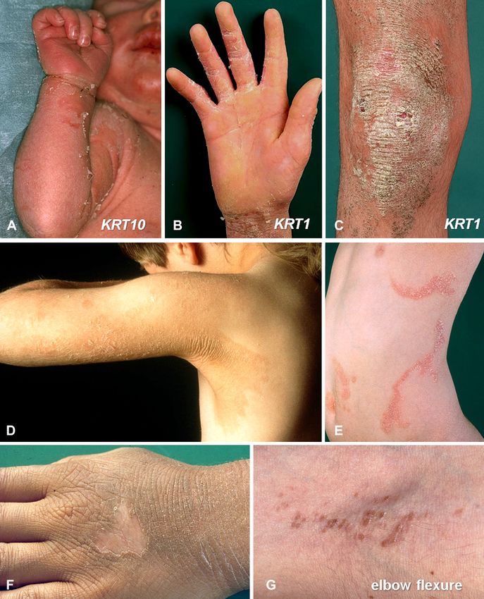
J AM ACAD DERMATOL                                                                                         Oji et al 609
VOLUME 63, NUMBER 4

as summarized in Table XIII, and should be amena-              diagnostics may be impeded by the high cost of
ble to modification as new information emerges.                analysis. Similarly, ultrastructural techniques are not
                                                               in common clinical use by pathologists and are not
AIMS AND LIMITATIONS OF THE                                    widely available to clinicians. Other laboratory tech-
CONSENSUS REPORT                                               niques, including light microscopy, narrow the dif-
   The overall goal of the revised classification is to        ferential diagnoses in some cases (see ‘‘Diagnostic
clarify the terminology of this heterogeneous group of         Aspects’’ section), but decisions regarding further
inherited skin diseases (Table I). The classification          testing, ie, molecular diagnostics, rest on an initial,
scheme and nosology should                                                               rigorous clinical evaluation.
be easily understandable for                                                             Therefore, the result of the
all clinicians, biologists, and      CAPSULE SUMMARY                                     consensus discussion process
students. It should guide cli-                                                           is a clinically based classifica-
nicians toward the correct           d Inherited ichthyoses belong to a large            tion, in which the diseases are
genotyping of their patients           and heterogeneous group of mendelian              referenced with the causative
and facilitate communication           disorders of cornification and involve the        gene or genes. Two principal
with investigators. The pro-           entire integument.                                groups are recognized: non-
posed classification (Tables II      d A conference of experts was convened              syndromic forms (Table II)
and III) will need to be               to reach a consensus on terminology               and syndromic forms (Table
modified or expanded as                and classification and to provide an              III). This algorithm is in the
new information accrues.               internationally accepted frame of                 tradition of previous con-
A pathophysiologic classifica-         reference.                                        cepts3,12-14 and based on the
tion of the ichthyoses and all       d The classification remains clinically based       following question:
MEDOC should be initiated in           and distinguishes between syndromic               d Is the phenotypic expres-

the future (Table XIII).               and nonsyndromic ichthyosis forms.                   sion of the disorder only
                                                                                            seen in the skin (proto-
RECOMMENDED
                                     d Bullous ichthyosis/epidermolytic                     types: lamellar ichthyosis
REVISION OF THE                        hyperkeratosis is redefined as                       (LI) and epidermolytic ich-
TERMINOLOGY AND                        keratinopathic ichthyosis. Autosomal                 thyosis [EI]), or is it seen in
CLASSIFICATION OF                      recessive congenital ichthyosis refers to            the skin and in other organs
INHERITED                              harlequin ichthyosis, lamellar ichthyosis,           (prototypes: Sjögren-Lars-
ICHTHYOSIS                             and congenital ichthyosiform                         son syndrome and tricho-
   The generic term ‘‘inher-           erythroderma.                                        thiodystrophy [TTD])?
ited ichthyosis’’ refers to
diseases that are MEDOC af-                                                                  Noteworthy,         recessive
fecting all or most of the integument. The skin changes        X-linked   ichthyosis (RXLI)   is regarded   as syndromic
are clinically characterized by hyperkeratosis, scaling,       when accompanied by associated manifestations
or both. Despite concern among some participants that          such as testicular maldescent, and nonsyndromic
                         2
the term ‘‘ichthyosis’’ is outmoded and sometimes              when ichthyosis occurs as an isolated type3 without
inaccurate, the consensus was to retain it, as it is too       extracutaneous signs. To facilitate the readability and
firmly entrenched in the literature and minds of clini-        understanding of the long list of autosomal ichthy-
cians to be abandoned. Inherited ichthyoses are                osis syndromes, subheadings have been introduced
regarded as one disease group within the greater               that point to the prominent associated signs, eg, hair
group of MEDOC. For greater clarity, we redefined              abnormalities or neurologic signs (Table III).
some important clinical and dermatologic terms that                Another question distinguishes between congen-
are in common usage (Table I). Specifically, the revised       ital ichthyosis and ichthyoses of delayed onset. This
classification is based on consent to a specific defini-       criterion is important for common ichthyoses (Table
tion of the term ‘‘autosomal recessive congenital ich-         IV), namely ichthyosis vulgaris (IV) and RXLI, which
thyosis’’ (ARCI), and a major change to nomenclature           often have a delayed onset (Fig 1). However, early
of the ichthyoses caused by keratin mutations (see             subtle skin changes may be overlooked, eg, RXLI
below).                                                        may present with fine superficial scaling shortly after
                                                               birth, which may fade within weeks and recur as a
General framework for the revised                              clear ichthyosis in later life. Therefore, considering
classification system                                          the high variability of the initial disease presentation
   At present, molecular diagnosis is not available            of some ichthyoses, eg, TTD, the age of onset has not
for all forms of ichthyosis, and access to genetic             been chosen as a major classification criterion.
Revised nomenclature and classification of inherited ichthyoses: Results of the First Ichthyosis Consensus Conference in Sore'ze 2009
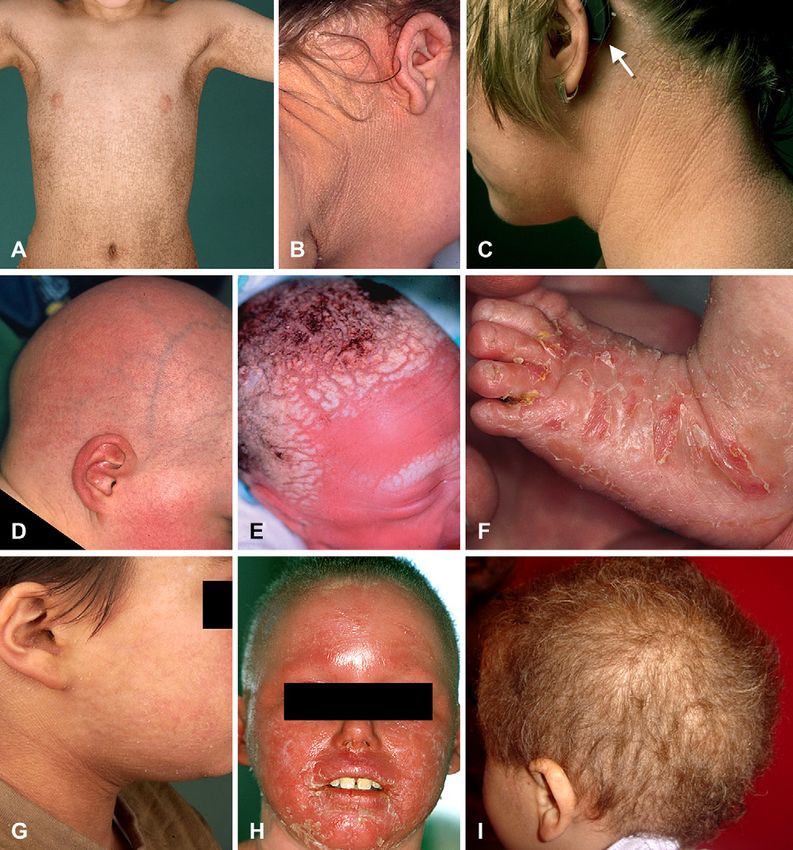
610 Oji et al                                                                                                  J AM ACAD DERMATOL
                                                                                                                        OCTOBER 2010

Table I. Main definitions, and recommended new terms and disease names
Recommended terms                                                                       Definition
General terminology
  Disorder of cornification (DOC)                  Disease with abnormal terminal keratinocytic differentiation
  MEDOC                                            Mendelian disorders of cornification
Inherited ichthyosis                               MEDOC affecting all or most of integument characterized by hyperkeratosis
                                                      and/or scaling
  Common ichthyoses                                Ichthyoses with high prevalence: IV (1:250-1000) and RXLI (1:2000-6000)
  Acquired ichthyosis                              Noninherited ichthyosis associated with malignancy; autoimmune,
                                                      inflammatory, nutritional, metabolic, infectious, and neurologic diseases;
                                                      or medications
Autosomal recessive congenital ichthyosis          Modified umbrella term for nonsyndromic congenital ichthyoses referring to
  (ARCI)*                                             HI and spectrum of LI and CIE (Tables II and V)
Keratinopathic ichthyosis (KPI)y                   New umbrella term for ichthyoses caused by keratin mutations, namely EI,
                                                      SEI, and other minor variants (Tables II and VI)
  Epidermolytic ichthyosis (EI)                    New disease name for bullous ichthyosis, bullous CIE, epidermolytic
                                                      hyperkeratosis, ichthyosis exfoliativa
  Superficial epidermolytic ichthyosis (SEI)       New disease name for ichthyosis bullosa Siemens
Diagnostic main criteria for classification
  Nonsyndromic ichthyosis                          Phenotypic expression of underlying genetic defect is only seen in skin
  Syndromic ichthyosis                             Phenotypic expression of underlying genetic defect is seen in skin and other
                                                     organs
Clinical and dermatologic terms
   Collodion membrane                              Tight shiny cast encasing newborn that cracks after some time, resulting in
                                                      irregularly branched fissures
  Congenital                                       Disorder is evident at birth or soon after birth (\1 wk)
  Delayed onset                                    Disorder becomes evident after weeks, months, or years
  Hyperkeratosis                                   Histopathological: increased thickness of SC
                                                   Clinical descriptive: thick and horny skin; it is not necessarily accompanied by
                                                      visible scaling
  Hystrix                                          Massive hyperkeratosis, cobblestone-like or spiky
  Keratoderma                                      Localized form of hyperkeratosis
  Lamellar scaling                                 Phenotype in which scales tend to be coarse and large (platelike scales)
  Scaling                                          Visible flakes of SC of variable size, color, and thickness

CIE, Congenital ichthyosiform erythroderma; HI, harlequin ichthyosis; IV, ichthyosis vulgaris; LI, lamellar ichthyosis; MEDOC, mendelian
disorders of cornification; RXLI, recessive X-linked ichthyosis; SC, stratum corneum.
*Previously termed LI/nonbullous ichthyosiform erythroderma.
y
  Previously used umbrella term: bullous ichthyosis, epidermolytic hyperkeratosis, or exfoliative ichthyosis.

Classification of ARCI                                                    One difficulty of the ARCI classification is the
    The acronym ‘‘ARCI’’ has been used as an um-                       limited genotype-phenotype correlation within the
brella term for nonsyndromic disorders, eg, LI and                     LI/CIE spectrum. Mutations in 6 genes have been
CIE, and for syndromic types of ichthyosis, such as                    described in non-HI ARCI to date, including TGM,
Netherton syndrome (NS). We propose that ‘‘ARCI’’                      the gene encoding transglutaminase (TGase)-1,22,23
should be used to refer to harlequin ichthyosis (HI)                   the genes ABCA12,17 NIPAL4 (also known as
and disorders of the LI/CIE phenotypic spectrum                        ICHTHYIN ),24 CYP4F22,25 and the lipoxygenase
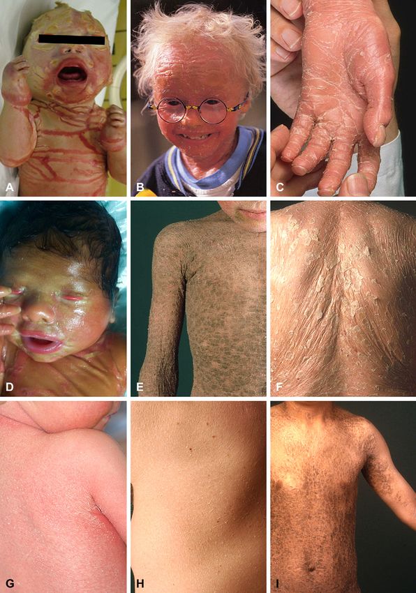
(Table V) exclusively. HI (Fig 2, A) was included,                     genes ALOX12B and ALOXE3.26 A large cohort of
because functional null mutations in the ABCA12                        520 affected families showed a mutation distribution
gene cause the disease,15,16 whereas missense mu-                      of 32% for TGM1, 16% for NIPAL4, 12% for ALOX12B,
tations in the same gene may result in a milder                        8% for CYP4F22, 5% for ALOXE3, and 5% for
phenotype that shows collodion membrane at birth                       ABCA12,27 which approximately correlated with a
and develops into LI17,18 or CIE,19,20 often with                      recent report of 250 patients.28 At least 22% of these
palmoplantar keratoderma (PPK). Those infants                          cases did not exhibit mutations in any of the known
with HI who survive the perinatal period go on to                      ARCI genes,27 implying that further loci must exist,
express a severe and very scaling erythroderma21                       such as two loci on chromosome 12p11.2-q13.29,30
(Fig 2, B and C ).                                                     A preliminary clinicogenetic correlation based on the
Revised nomenclature and classification of inherited ichthyoses: Results of the First Ichthyosis Consensus Conference in Sore'ze 2009
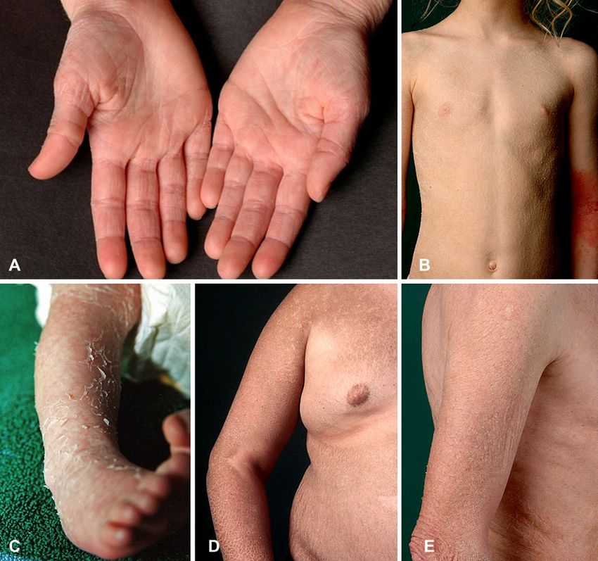
J AM ACAD DERMATOL                                                                                                       Oji et al 611
VOLUME 63, NUMBER 4

Table II. Clinicogenetic classification of inherited ichthyoses, part A: nonsyndromic forms
                                                           Inherited ichthyoses
                                                       Part A: nonsyndromic forms
Disease                                      Mode of inheritance                                        Gene(s)
Common ichthyoses*
  IV                                     Autosomal                           FLG
                                           semidominant
  RXLI
  Nonsyndromic                           X-linked recessive                  STS
    presentation
ARCI
  Major types
     HI                                  Autosomal recessive                 ABCA12
     LIy                                            ‘‘                       TGM1/NIPAL4z/ALOX12B/ABCA12/loci on 12p11.2-q13
     CIE                                            ‘‘                       ALOXE3/ALOX12B/ABCA12/CYP4F22/NIPAL4z/TGM1/loci
                                                                               on 12p11.2-q13
  Minor variants
    SHCB                                 Autosomal recessive                 TGM1, ALOX12B, ALOXE3
    Acral SHCB                                      ‘‘                       TGM1
    BSI                                             ‘‘                       TGM1
Keratinopathic ichthyosis (KPI)
  Major types
    EI§                                  Autosomal dominant                  KRT1/KRT10
    SEI                                             ‘‘                       KRT2
  Minor variants
    AEI§                                 Autosomal dominant                  KRT1/KRT10
    ICM                                             ‘‘                       KRT1
    AREI                                 Autosomal recessive                 KRT10
    Epidermolytic nevi//                 Somatic mutations                   KRT1/KRT10)
Other forms
  LK                                     Autosomal dominant                  LOR
  EKV{                                                ‘‘                     GJB3/GJB4
  PSD                                    Autosomal recessive                 Locus unknown
  CRIE                                   Autosomal dominant (?)              Locus unknown
                                           (isolated cases)
  KLICK                                  Autosomal recessive                 POMP

AEI, Annular epidermolytic ichthyosis; ARCI, autosomal recessive congenital ichthyosis; AREI, autosomal recessive epidermolytic ichthyosis;
BSI, bathing suit ichthyosis; CIE, congenital ichthyosiform erythroderma; CRIE, congenital reticular ichthyosiform erythroderma; EI,
epidermolytic ichthyosis; EKV, erythrokeratodermia variabilis; HI, harlequin ichthyosis; ICM, ichthyosis Curth-Macklin; IV, ichthyosis
vulgaris; KLICK, keratosis lineariseichthyosis congenitaekeratoderma; LI, lamellar ichthyosis; LK, loricrin keratoderma; PSD, peeling skin
disease; RXLI, recessive X-linked ichthyosis; SEI, superficial epidermolytic ichthyosis; SHCB, self-healing collodion baby.
*Often delayed onset (in RXLI mild scaling and erythroderma may be present already at birth).
y
  Few cases of autosomal dominant LI described in literature (locus unknown).
z
  Also known as ICHTHYIN gene.
§
  KRT1 mutations are often associated with palmoplantar involvement.
//
  May indicate gonadal mosaicism, which can cause generalized EI in offspring generation.
{
  Whether progressive symmetric erythrokeratodermia represents distinct mendelian disorders of cornification form is debated.

recent literature17-20,22-45 and our discussions at the                  scaling and pronounced erythroderma.31,45 The phe-
consensus conference is given in Tables II and III.                      notypes can change over time and in response to
   LI is characterized by coarse and brown/dark                          treatment, eg, LI treated with oral retinoids can
scaling (Fig 2, E and F ). Affected individuals are                      evolve into an erythrodermic ichthyosis with a finer
often born with collodion membrane and pro-                              scale pattern.46 In a recent North American study of
nounced ectropion (Fig 2, D). CIE is characterized                       104 patients with non-HI ARCI, mutations in TGM1
by fine, white scaling with varying degrees of                           were significantly associated with collodion mem-
erythema (Fig 2, G and H ). Individuals with CIE                         brane, ectropion, platelike scales, and alopecia.
may also be born with collodion membrane (often                          Patients who had at least one mutation predicted to
less severe), and then transit to generalized fine                       truncate TGase-1 were more likely to have severe
612 Oji et al                                                                                                J AM ACAD DERMATOL
                                                                                                                        OCTOBER 2010

Table III. Clinicogenetic classification of inherited ichthyoses, part B: syndromic forms
                                                         Inherited ichthyoses
                                                       Part B: syndromic forms
Disease                                                           Mode of inheritance                         Gene(s)
X-linked ichthyosis syndromes
   RXLI*
   - Syndromic presentation                                     X-linked recessive            STS (and othersy)
   IFAP syndrome                                                          ‘‘                  MBTPS2
   Conradi-Hünermann-Happle syndrome (CDPX2)                   X-linked dominant             EBP
Autosomal ichthyosis syndromes (with)
   Prominent hair abnormalities
      NS                                                        Autosomal recessive           SPINK5
      IHSz                                                              ‘‘                    ST14
      IHSC syndrome§                                                    ‘‘                    CLDN1
      TTD                                                               ‘‘                    ERCC2/XPD ERCC3/XPB GTF2H5/TTDA
      *TTD (not associated with congenital ichthyosis)                  ‘‘                    C7Orf11/TTDN1
   Prominent neurologic signs
      SLS                                                                  ‘‘                 ALDH3A2
      *Refsum syndrome (HMSN4)                                             ‘‘                 PHYH/PEX7
      MEDNIK syndrome                                                      ‘‘                 AP1S1
   Fatal diseases course
      Gaucher syndrome type 2                                              ‘‘                 GBA
      MSD                                                                  ‘‘                 SUMF1
      CEDNIK syndrome                                                      ‘‘                 SNAP29
      ARC syndrome                                                         ‘‘                 VPS33B
   Other associated signs
      KID syndrome                                              Autosomal dominant            GJB2 (GJB6)
      Neutral lipid storage disease with ichthyosis             Autosomal recessive           ABHD5
      IPS                                                               ‘‘                    SLC27A4

ARC,      Arthrogryposiserenal      dysfunctionecholestasis;     CDPX2,     chondrodysplasia    punctata   type 2; CEDNIK,  cerebral
dysgenesiseneuropathyeichthyosisepalmoplantar keratoderma; HMSN4, hereditary motor and sensory neuropathy type 4; IFAP, ichthyosis
folliculariseatrichiaephotophobia; IHS, ichthyosis hypotrichosis syndrome; IHSC, ichthyosisehypotrichosisesclerosing cholangitis; IPS,
ichthyosis prematurity syndrome; MEDNIK, mental retardationeenteropathyedeafnesseneuropathyeichthyosisekeratodermia; MSD,
multiple sulfatase deficiency; NS, Netherton syndrome; RXLI, recessive X-linked ichthyosis; SLS, Sjögren-Larsson syndrome; TTD,
trichothiodystrophy.
*Often delayed onset (in RXLI mild scaling and erythroderma may be present already at birth).
y
  In context of contiguous gene syndrome.
z
  Clinical variant: congenital ichthyosis, follicular atrophoderma, hypotrichosis, and hypohidrosis syndrome.
§
  Also known as neonatal ichthyosis sclerosing cholangitis syndrome.

hypohidrosis and overheating than those with TGM1                     of intracellular vacuolization, clumping of tonofila-
missense mutations only.35                                            ments, and formation of small intraepidermal blisters,
    Clinically other minor ARCI variants/subtypes can                 as commonly seen in ichthyoses as a result of keratin
be distinguished: bathing suit ichthyosis47 has been                  mutations. Therefore the term ‘‘epidermolytic hyper-
attributed to particular TGM1 mutations that render                   keratosis’’ is used (by some) as synonymous with
the enzyme sensitive to ambient temperature                           bullous ichthyosis, ichthyosis exfoliativa, bullous CIE
(Fig 2, I ).32,42,43,48 The self-healing collodion baby               (of Brocq), or ichthyosis bullosa of Siemens.50-55
representing approximately 10% of all ARCI cases36,49                 However, the light microscopic features of the
has so far been associated with TGM1 or ALOX12B                       cytoskeletal abnormalities as a result of keratin
mutations.37,44 The recently described acral self-                    mutations may not be observed in all instances.56-59
healing collodion baby, ie, at birth the collodion                    To replace the long list of names, which have been
membrane is strictly localized to the extremities and                 used for these ichthyosesethose that are all a result of
then resolves, can also be a result of TGM1 mutations.41              keratin mutationsewe propose the novel umbrella
                                                                      term and definition ‘‘keratinopathic ichthyosis’’ (KPI)
Classification of the keratinopathic ichthyoses                       (Table I). In analogy to the prevalent morphologic
   The term ‘‘epidermolytic hyperkeratosis’’ derives                  key features, we suggest the term ‘‘epidermolytic
from the characteristic light microscopic observation                 ichthyosis’’ as a novel name for the specific disease
J AM ACAD DERMATOL                                                                                                         Oji et al 613
VOLUME 63, NUMBER 4

Table IV. Common forms of ichthyosis: summary of clinical and morphologic findings
                                              IV (prevalence: 1:250-1000)                         RXLI (prevalence: 1:2000-6000)
Mode of inheritance                    Autosomal semidominant                          XR
Onset                                  After ;2-6 mo                                   Exaggerated scaling and/or erythroderma in
                                                                                         newborn period or late onset after ;2-6 mo,
                                                                                         mild collodion-like skin at birth may be
                                                                                         possible
Initial clinical presentation          Xerosis, scaling, pruritus, eczema              Scaling
Disease course                         Stable, often better in summer                  Stable, often better in summer
Cutaneous findings
   Distribution of scaling             Generalized, antecubital or popliteal           Generalized, sparing of body folds, neck is often
                                          fossae often spared                            more severely involved
  Scaling type                         Fine or light                                   Large rhomboid scales or fine scaling
  Scaling color                        White-gray                                      Dark brown or light gray
  Erythema                             Absent                                          Absent
  Palmoplantar involvement             Accentuated palmoplantar markings               No accentuated markings
  Hypohidrosis                         Possible                                        Possible
  Scalp abnormalities                  Absent                                          Absent
  Others                               Eczema                                          -
Extracutaneous                         Strong association with atopic                  Incidence of cryptorchidism/testicular
  involvement                             manifestations                                 maldescent seems to be increased
                                                                                         (estimated numbers range from 5%-20%),
                                                                                         subclinical corneal opacities in ;50%;
                                                                                         insufficient cervical dilatation in female
                                                                                         carriers
                                                                                         *Contiguous gene syndromes have to be
                                                                                         ruled out
Ultrastructure                         Small or only rudimental KG                     Retained corneodesmosomes within SC
Special analyses                       Reduced or absent SG, reduced or                Absent steroid sulfatase (arylsulfatase-C)
                                         negative filaggrin staining by                  activity (leukocytes or fibroblasts), FISH test
                                         antigen mapping                                 for STS deletion; elevated blood cholesterol
                                                                                         sulfate levels
                                                                                         (Fetal steroid sulfatase deficiency leads to
                                                                                         low maternal serum/urinary estriol levels;
                                                                                         therefore, RXLI may be detected in utero,
                                                                                         when prenatal screening for Down syndrome
                                                                                         and other disorders includes measurement of
                                                                                         maternal estriol levels, as in triple-screen
                                                                                         blood test)

FISH, Fluorescent in situ hybridization; IV, ichthyosis vulgaris; KG, keratohyaline granules; RXLI, recessive X-linked ichthyosis; SC, stratum
corneum; SG, stratum granulosum; XR, X-linked recessive.
*RXLI within context of contiguous gene syndrome (Table III), eg, in Kallmann syndrome, chondrodysplasia punctata (brachytelephalangic
type), or ocular albinism type 1.

spectrum that is accompanied by epidermolytic                               (Fig 3, A), not seen thereafter except for focal blisters.
hyperkeratosis at the ultrastructural level. The term                       The blistering phenotype present at birth, which is a
‘‘epidermolytic hyperkeratosis’’ should be used ex-                         result of loss of mechanical resilience in the upper
clusively as an ultrastructural or histopathological                        epidermis, evolves into a hyperkeratotic one (phe-
descriptor. We propose the novel disease name                               notypic shift) (Fig 3, C ); this is suggested to be
‘‘superficial epidermolytic ichthyosis’’ for the well-                      influenced primarily by abnormal lamellar body (LB)
defined entity ichthyosis bullosa Siemens, which in                         secretion, rather than corneocyte fragility.60
contrast to EI shows a more superficial pattern of                          Superficial EI (Fig 3, D) has a milder phenotype
epidermolysis and is caused by mutations in keratin                         than EI and can be distinguished by the lack of
2, rather than in keratins 1 or 10.                                         erythroderma and by a characteristic ‘‘moulting’’
    Clinically, KPI show a broad spectrum of skin                           phenomenon (Fig 3, F ). Here, light microscopy
manifestations and severity (Table VI). Widespread                          and ultrastructure reveal cytolysis that correlates
skin blistering is characteristic of neonates with EI                       with the distinctive expression pattern of keratin 2
614 Oji et al                                                                                                        J AM ACAD DERMATOL
                                                                                                                               OCTOBER 2010

Table V. Autosomal recessive congenital ichthyoses: summary of clinical and morphologic findings
                                           HI                                  LI                                        CIE
Mode of inheritance           AR                        AR                                    AR
Onset                         At birth, often           At birth                              At birth
                                preterm babies
Initial clinical              Severe collodion          Collodion membrane with               CIE or less frequently mild collodion
   presentation                 membrane with             ectropion and eclabium;               membrane
                                armorlike membrane,       less frequently CIE
                                extreme ectropion and
                                eclabium, and
                                contractures,
                                broadened nose,
                                synechiae of auricles,
                                sometimes toes
Disease course                Development of            Ranging from very mild                Ranging from very mild to severe
                                exfoliative/very scaling to severe (probably
                                erythroderma similar      never completely heals)
                                to severe CIE with fine
                                or large scales
                                                        Minor variants
                                                          - SHCB: nearly complete resolution of scaling within
                                                          first 3 mo of life (in ;10% of cases)
                                                          - Acral SHCB: at birth only acral collodion membranes
                                                          are observed that later on heal
                                                          - BSI: collodion membrane at birth and development of LI or CIE
                                                          Then, within first months of life, skin predominantly of extremities
                                                          heals, but warmer skin areas, eg, axillary region, scalp, (mid-) trunk,
                                                          remain involved and show localized form of LI
Cutaneous findings
  Distribution of scaling Generalized                       Generalized; focally pronounced           Generalized; focally pronounced
                                                              scaling possible                           scaling possible
  Scaling type                Coarse and large              Coarse and large (platelike)              Fine
                                (platelike)
  Scaling color               Gray or yellowish             Brownish or dark                   White or gray
  Erythema                    Severe                        Variable, less pronounced          Variable, often pronounced
  Palmoplantar                Yes, possibly with            *NIPAL4: pronounced keratoderma; ALOX12B and CYP4F22:
    involvement                 synechiae of digits         pronounced lichenification and mild keratoderma; ALOXE3:
                                                            IV-like; TGM1: frequent palmoplantar involvement
  Hypohidrosis                Severe temperature            Moderate to severe                 Moderate to severe
                                dysregulation
  Scalp abnormalities         Scarring alopecia        Scarring alopecia possible (often Scarring alopecia possible
                                                          with TGM1)
   Other skin findings        Prone to skin infections -                                    -
Extracutaneous                Contractures; failure to Short stature (if severe)            Failure to thrive, short stature
   involvement                  thrive; short stature                                         (if severe)
Risk of death                 Very high during         Elevated during neonatal period Present during neonatal period
                                neonatal period
  Skin ultrastructure         Vesicular LB ghosts;     ABCA12 = absence of LB content; *NIPAL4 = weak correlation with
                                paucity of secreted    vesicular complexes, defective LB, perinuclear membranes within
                                lamellar structures in SG in glutaraldehyde fixation; TGM1: thin CE and disorganization
                                SC                     of lamellar bilayers (with glutaraldehyde fixation: polygonal clefts
                                                       within corneocytes)
  Other analyses              None                     In situ monitoring of TGase-1 activity in cryostat sections, SDS heating
                                                       test of scales

AR, Autosomal recessive; BSI, bathing suit ichthyosis; CE, cornified cell envelope; CIE, congenital ichthyosiform erythroderma; HI, harlequin
ichthyosis; IV, ichthyosis vulgaris; LB, lamellar body; LI, lamellar ichthyosis; SC, stratum corneum; SG, stratum granulosum; SHCB, self-healing
collodion baby; TGase, transglutaminase.
*NIPAL4 also known as ICHTHYIN.
VOLUME 63, NUMBER 4
                                                                                                                                                                                                      J AM ACAD DERMATOL
Table VI. Keratinopathic ichthyoses and congenital reticular ichthyosiform erythroderma: summary of clinical and morphologic findings
                                               EI                                   SEI                              ICM                                 CRIE*
Mode of inheritance             AD or rarely AR (KRT10)            AD                                 AD                                   AD (?) (isolated cases)
                                Annular type: AD
Onset                           At birth                           At birth                           Early childhood                      At birth
Initial clinical presentation   Large erosions, mild scaling,      Erythroderma, widespread           Striate or diffuse PPK               Exfoliative CIE, larger areas
                                  erythroderma at birth              blistering                                                              forming reticular pattern
                                                                                                                                             predominantly on
                                                                                                                                             extremities
Disease course                  Resolution of erosions replaced    Within weeks development of        Progressive worsening of PPK         During childhood and puberty
                                  by hyperkeratosis in first         hyperkeratosis particularly        and development of                   characteristic patchy pattern
                                  months                             over extensor sides of joints      hyperkeratotic plaques over          starts to evolve
                                Annular type: development of                                            joints and/or hyperkeratotic
                                  numerous annular,                                                     papules on trunk and
                                  polycyclic, erythematous,                                             extremities
                                  scaly plaques on trunk and
                                  extremities that enlarge
                                  slowly, and then resolve
                                  (intermittent presentations
                                  of EI)
Cutaneous findings
  Distribution of scaling       Generalized, or predilection for   Friction areas                     Palms and soles, large joints,       Generalized, later reticular
                                  friction areas, over joints                                           rarely extremities and/or            ichthyosiform pattern
                                                                                                        trunk
  Scaling type                  Adherent, moderate                 Adherent, fine to moderate         Thick, spiky hyperkeratosis          Fine
  Scaling color                 White-brown                        Brown (mauserung/moulting)         Yellow-brown hyperkeratoses          Yellow-brown
  Erythema                      Frequent                           Initially, fades                   Erythroderma possible                Pronounced
  Palmoplantar involvement      KRT1: epidermolytic PPK            Usually no                         Massive PPK leading to deep,         Yes
                                KRT10: palms and soles are                                              bleeding, and painful
                                  spared (exceptions possible)                                          fissures; flexural contractures;
                                                                                                        constriction bands
  Hypohidrosis                  Possible                           Possible                           None                                 -
  Scalp abnormalities           Scaling                            -                                  None                                 Scaling
  Other skin findings           Pruritus, blisters after minor     Pruritus, bullae may occur after   -                                    -
                                   trauma, prone to skin             minor mechanical trauma
                                   infections/impetigo               (often in summer)

                                                                                                                                                                                                   Oji et al 615
Extracutaneous involvement      Growth failure with some                                              Gangrene and loss of digits          Growth failure with some
                                   severe phenotypes                                                                                          severe phenotypes
Risk of death                   Elevated during neonatal           -                                  -                                    Elevated during neonatal
                                   period                                                                                                     period
                                                                                                                                                                 Continued
616 Oji et al                                                                                                                                                                                                                                                                                                J AM ACAD DERMATOL
                                                                                                                                                                                                                                                                                                                     OCTOBER 2010

                                                                                                                                                                                                                                                                          in the stratum granulosum (SG) or upper stratum

                                                                                 AD, Autosomal dominant; AR, autosomal recessive; CIE, congenital ichthyosiform erythroderma; CRIE, congenital reticular ichthyosiform erythroderma; EHK, epidermolytic hyperkeratosis;
                             granular cells and (often?) so
                             far unidentified filamentous
                             material in vacuolated cells
                           Vacuolization of superficial                                                                                                                                                                                                                   spinosum.61 Different features such as distribution,
                                                                                                                                                                                                                                                                          erythema, or blistering were used for separating
                                                                                                                                                                                                                                                                          patients with EI into 6 clinical groups, with the most
                                                                                                                                                                                                                                                                          distinctive characteristic being involvement of palms
                   CRIE*

                                                                                                                                                                                                                                                                          and soles (1-3 vs non-palms and soles 1-3).62 PPK is
                                                                                                                                                                                                                                                                          usually predictive of a KRT1 mutation (Fig 3, E ). One
                                                                                                                                                                                                                                                                          explanation is that keratin 9, which is expressed in
                                                                                                                                                                                                                                                                          palms and soles, may compensate for a keratin 10
                                                                                                                                                                                                                                                                          defect, whereas keratin 1 is the only type II keratin
                                                              -

                                                                                                                                                                                                                                                                          expressed in palmoplantar skin.63-65 However, PPK
                                                                                                                                                                                                                                                                          may occur with KRT10 mutations as well.66
                             aberranteputativelyekeratin

                                                                                                                                                                                                                                                                              Similar to pachyonychia congenita or the epider-
                                                                                 EI, epidermolytic ichthyosis; ICM, ichthyosis Curth-Macklin; LB, lamellar body; PPK, palmoplantar keratoderma; SEI, superficial epidermolytic ichthyosis.

                                                                                                                                                                                                                                                                          molysis bullosa simplex group, the vast majority of
                           Binuclear cells, particular
                             concentric perinuclear

                                                                                                                                                                                                                                                                          the KPI arise from autosomal dominant mutations.
                                                                                                                                                                                                                                                                          The resulting mutant keratin is normally expressed
                   ICM

                                                                                                                                                                                                                                                                          but interferes with the assembly and/or function of
                                                                                                                                                                                                                                                                          keratin intermediate filaments, often leading to ker-
                             ‘‘shells’’ of

                                                                                                                                                                                                                                                                          atin intermediate filament aggregation and cytolysis.
                             material

                                                                                                                                                                                                                                                                          However, KRT10 nonsense mutations have been
                                                                                                                                                                                                                                                                          observed that do not lead to the usual dominant
                                                                                                                                                                                                                                                                          negative effect and cause an autosomal recessive
                           -

                                                                                                                                                                                                                                                                          KPI form.67 Therefore, autosomal recessive EI is
                                                                                                                                                                                                                                                                          listed as a new separate KPI. For ichthyosis Curth-
                                                                                                                                                                                                                                                                          Macklin,57-59,68 which represents a very rare form of
                             granular cells of affected
                           Superficial EHK, cytolysis in

                             body areas; no keratin

                                                                                                                                                                                                                                                                          KPI and shows a characteristic ultrastructure (Table
                                                                                                                                                                                                                                                                          VI), we propose to omit the adjective ‘‘hystrix’’ and
                                                                                                                                                                                                                                                                          retain the eponym Curth-Macklin. Hystrix skin
                   SEI

                                                                                                                                                                                                                                                                          changes can be observed in other ichthyoses, eg,
                                                                                                                                                                                                                                                                          KID syndrome (Table XII), or in particular types of
                             clumping

                                                                                                                                                                                                                                                                          ectodermal dysplasia.69 The annular EI (Fig 3, E ),
                                                                                                                                                                                                                                                                          which is a result of KRT1 or KRT10 mutations,70,71 is
                                                                                                                                                                                                                                                                          classified as a clinical variant of EI.
                                                              -

                                                                                                                                                                                                                                                                              Importantly, linear epidermolytic nevi, ie, those
                                                                                                                                                                                                                                                                          epidermal nevi exhibiting the histopathology of
                             clumping of keratin filaments

                                                                                                                                                                                                                                                                          epidermolytic hyperkeratosis, may indicate a so-
                             cytolysis, LB accumulation
                             in suprabasal cells; partly

                                                                                                                                                                                                                                                                          matic type 1 mosaicism for mutations in KRT1 or
                                                                                 *Also known as ichthyosis variegata and ichthyosis en confettis.
                           EHK, aggregations and

                                                                                                                                                                                                                                                                          KRT10, which, if also gonadal, can result in gener-
                                                                                                                                                                                                                                                                          alized EI in the patient’s offspring (Fig 3, A and
                   EI

                                                                                                                                                                                                                                                                          G).72-74 Because recognition of this risk is important
                                                                                                                                                                                                                                                                          for genetic counseling, epidermolytic nevi have
                                                                                                                                                                                                                                                                          been included (in brackets) in the classification of
                                                                                                                                                                                                                                                                          KPI (Table II).
                                                              -

                                                                                                                                                                                                                                                                          Other diseases considered in the classification
                                                                                                                                                                                                                                                                          of inherited ichthyoses
                                                                                                                                                                                                                                                                             The inclusion of disease entities into this classifi-
                                                                                                                                                                                                                                                                          cation of inherited ichthyosis rests on an appropriate
                                                                                                                                                                                                                                                                          clinical disease description and our definition of
                           Skin ultrastructure
Table VI. Cont’d

                                                              Special analyses

                                                                                                                                                                                                                                                                          inherited ichthyosis (Table I). A detailed overview of
                                                                                                                                                                                                                                                                          the disease onset, initial clinical presentation, disease
                                                                                                                                                                                                                                                                          course, cutaneous and extracutaneous findings, and
                                                                                                                                                                                                                                                                          of the skin ultrastructure is given for each entity: (1)
                                                                                                                                                                                                                                                                          common forms of ichthyosis (Table IV); (2) ARCI
                                                                                                                                                                                                                                                                          (Table V); (3) KPI and congenital reticular
Table VII. Other nonsyndromic ichthyosis forms: summary of clinical and morphologic findings

                                                                                                                                                                                    VOLUME 63, NUMBER 4
                                                                                                                                                                                                             J AM ACAD DERMATOL
                                               LK                                  EKV                                    KLICK                               PSD*
Mode of inheritance             AD                                 AD                                      AR                                  AR
Onset                           At birth                           At birth or within first year of life   At birth                            At birth (or first weeks of life)
Initial clinical presentation   CIE or collodion baby              Co-occurrence of transient,             Congenital                          IE, atopic dermatitis-like lesions
                                                                     migratory erythematous                  ichthyosis
                                                                     patches and hyperkeratosis
                                                                     limited to geographic outlined
                                                                     plaques or generalized
Disease course                  Improvement and development        Relapsing-remitting, erythema           Mild                                Mild to moderate, spontaneous
                                  of PPK                             are fleeting (hours-days),                                                  remissions, and relapses
                                                                     hyperkeratosis more stable
                                                                     (months to years)
Cutaneous findings
  Skin distribution             Generalized mild scaling with      Generalized or focally accented         Generalized, accentuated            Generalized (to be differentiated
                                   accentuated hyperkeratosis        hyperkeratosis, predominantly           linear keratoses in skin folds,     from acral PSS)
                                   over joints, flexural areas       on extremities, buttocks                (sclerosing) PPK
  Scaling type                  Fine                               Rough, thickened skin, possibly                                             Large peeling scales
                                                                     hystrix skin; occasionally
                                                                     peeling
  Scaling color                 White                              White to gray, yellow                   White-brown                         White
                                                                     or brown
  Erythema                      Uncommon                           Focal migratory                         Uncommon                            Varying from mild to moderate,
                                                                                                                                                 may improve with age
  Palmoplantar involvement      Noninflammatory diffuse PPK        Diffuse PPK present                     —                                   Yes
                                  with honeycomb pattern, mild       in about 50% of patients
                                  digital constriction, brown
                                  hyperkeratosis, knuckle pads
                                  over back aspects
  Hypohidrosis                  -                                  No                                      Yes                                 No
  Scalp abnormalities           No                                 No                                      No                                  No hair abnormalities
  Other skin findings           Keratoderma (thickening),          No                                      Linear keratosis                    Pruritus
                                  pseudo-ainhum or (mild) linear
                                  constrictions
Extracutaneous involvement      -                                  None                                    None                                Associated atopic diathesis, short
                                                                                                                                                  stature (single cases)
Risk of death                   Normal                             Normal                                  Normal                              Elevated during neonatal period

                                                                                                                                                                                                          Oji et al 617
                                                                                                                                                                       Continued
618 Oji et al                                                                                                                                                                                                                                                                                                                              J AM ACAD DERMATOL
                                                                                                                                                                                                                                                                                                                                                   OCTOBER 2010

                                                                                                                                                                                                                                                                                                        ichthyosiform erythroderma (Table VI); (4) other

                                                                                                   Acral PSS, Acral peeling skin syndrome; AD, autosomal dominant; AR, autosomal recessive; CE, cornified cell envelope; CIE, congenital ichthyosiform erythroderma; EKV, erythrokeratodermia
                            Superficial exfoliation, separation

                                                                                                   variabilis; IE, ichthyosiform erythroderma; KG, keratohyaline granules; KLICK, keratosis lineariseichthyosis congenitaekeratoderma; LB, lamellar body; LK, loricrin keratoderma; PPK, palmoplantar
                              directly above SG or within SC;

                                                                  Immunohistochemistry: LEKTI is
                              between, adjacent, or within
                                                                                                                                                                                                                                                                                                        nonsyndromic ichthyosis forms (Table VII); (5) X-

                                                                    normal or even elevated
                                                                                                                                                                                                                                                                                                        linked ichthyosis syndromes (Table VIII); and (6)
                                                                                                                                                                                                                                                                                                        autosomal ichthyosis syndromes with prominent
                                                                                                                                                                                                                                                                                                        hair abnormalities (Table IX), prominent neurologic
                    PSD*

                                                                                                                                                                                                                                                                                                        signs (Table X), fatal disease course (Table XI), and
                              corneocytes

                                                                                                                                                                                                                                                                                                        other associated signs (Table XII).
                                                                                                                                                                                                                                                                                                           Diseases that are classically regarded as ichthyosis
                                                                                                                                                                                                                                                                                                        in the previously published scientific literature and
                                                                                                                                                                                                                                                                                                        that will continue to be included are shown in Figs 4
                                                                                                                                                                                                                                                                                                        and 5. They include Sjögren-Larsson syndrome75,76
                                                                                                                                                                                                                                                                                                        (Fig 5, B), Refsum syndrome,77,78 neutral lipid storage
                                                                                                                                                                                                                                                                                                        disease with ichthyosis (also referred to as Chanarin-
                                                                                                                                                                                                                                                                                                        Dorfman syndrome) (Fig 5, G),40,79,80 ichthyosis
                                                                                                   *We propose to classify disorder as nonsyndromic form and therefore modified name ‘‘peeling skin syndrome (PSS)’’ into ‘‘peeling skin disease.’’

                                                                                                                                                                                                                                                                                                        folliculariseatrichiaephotophobia syndrome (Fig 5,
                                                                                                                                                                                                                                                                                                        D),81,82 Conradi-Hünermann-Happle syndrome
                              abnormally big KG
                            Hypergranulosis and
                    KLICK

                                                                                                                                                                                                                                                                                                        (CDPX2) (Fig 5, F ),83,84 multiple sulfatase defi-
                                                                                                                                                                                                                                                                                                        ciency,85,86 congenital reticular ichthyosiform eryth-
                                                                                                                                                                                                                                                                                                        roderma also referred to as ichthyosis variegata87 (or
                                                                                                                                                                                                                                                                                                        ichthyosis en confettis88) (Fig 4, E ), and ichthyosis
                                                                                                                                                                                                                                                                                                        prematurity syndrome89,90 (Fig 5, E ). In ichthyosis
                                                                                                                                                                                                                                                                                                        prematurity syndrome, affected pregnancies exhibit
                                                                  -

                                                                                                                                                                                                                                                                                                        abnormal amniotic fluid both on ultrasound imaging
                              keratinization and reduction of

                                                                                                                                                                                                                                                                                                        and clinically.91 It must be distinguished from the self-
                            Mostly nonspecific changes with
                              various degrees of deviations

                                                                                                                                                                                                                                                                                                        healing collodion baby, because in both diseases the
                                                                                                                                                                                                                                                                                                        skin heals almost completely soon after birth.89 Many
                                                                                                                                                                                                                                                                                                        advances in the heterogeneous field of the TTDs (Fig
                                                                                                                                                                                                                                                                                                        5, A) have been made.92,93 Recent studies on
                              or suppression of
                    EKV

                                                                                                                                                                                                                                                                                                        genotype-phenotype correlation distinguish the
                                                                                                                                                                                                                                                                                                        TTD syndromes associated with ichthyosis of de-
                              LB in SG

                                                                                                   keratoderma; PSD, peeling skin disease; SC, stratum corneum; SG, stratum granulosum.

                                                                                                                                                                                                                                                                                                        layed onset or accompanied with collodion mem-
                                                                                                                                                                                                                                                                                                        brane from other forms of TTD.94
                                                                                                                                                                                                                                                                                                           Diseases relatively new in the list of ichthyoses are
                                                                                                                                                                                                                                                                                                        loricrin keratoderma, also referred to as Camisa variant
                            -

                                                                                                                                                                                                                                                                                                        of Vohwinkel keratoderma (Fig 4, C ),95-97 the cerebral
                               granules in granular cells, thin

                                                                                                                                                                                                                                                                                                        dysgenesiseneuropathyeichthyosisePPK syndrome,98
                                                                  Histology: parakeratosis, and
                               CE in lower SC, abnormal
                            Electron dense intranuclear

                                                                                                                                                                                                                                                                                                        the arthrogryposiserenal dysfunctionecholestasis syn-
                                                                                                                                                                                                                                                                                                        drome,99-101 the mental retardationeenteropathye
                               extracellular lamellae

                                                                                                                                                                                                                                                                                                        deafnesseneuropathyeichthyosisekeratodermia syn-
                                                                                                                                                                                                                                                                                                        drome,102 the ichthyosisehypotrichosisesclerosing
                                                                    hypergranulosis
                    LK

                                                                                                                                                                                                                                                                                                        cholangitis syndrome (also known as neonatal ichthy-
                                                                                                                                                                                                                                                                                                        osis sclerosing cholangitis syndrome),103-105 the ichthy-
                                                                                                                                                                                                                                                                                                        osis hypotrichosis syndrome (Fig 5, I )106 and its allelic
                                                                                                                                                                                                                                                                                                        variant congenital ichthyosisefollicular atrophoder-
                                                                                                                                                                                                                                                                                                        maehypotrichosisehypohidrosis syndrome,107,108 and
                                                                                                                                                                                                                                                                                                        keratosis lineariseichthyosisecongenital sclerosing
                                                                                                                                                                                                                                                                                                        keratoderma (Fig 4, F ).109,110
                                                                                                                                                                                                                                                                                                           Erythrokeratodermia variabilis (EKV),111-113
                                                                                                                                                                                                                                                                                                        which is characterized by migratory erythematous
                            Skin ultrastructure
Table VII. Cont’d

                                                                                                                                                                                                                                                                                                        patches and more fixed, symmetric hyperkeratotic
                                                                  Other analyses

                                                                                                                                                                                                                                                                                                        plaques often with palmoplantar involvement (Fig 4,
                                                                                                                                                                                                                                                                                                        B), is genetically heterogeneous and can in 50% to
                                                                                                                                                                                                                                                                                                        65% of cases114 be caused by mutations in GJB3
                                                                                                                                                                                                                                                                                                        coding for the gap junction protein connexin 31,115
                                                                                                                                                                                                                                                                                                        or GJB4 coding for connexin 30.3.116 Whether
J AM ACAD DERMATOL                                                                                                  Oji et al 619
VOLUME 63, NUMBER 4

Table VIII. X-linked ichthyosis syndromes (for recessive X-linked ichthyosis see Table IV): summary of clinical
and morphologic findings
                                                   IFAP syndrome                      Conradi-Hünermann-Happle syndrome (CDPX2)
Mode of inheritance               XR*                                                XD
Onset                             At birth                                           At birth
Initial clinical presentation     Mild collodion skin, congenital atrichia           Ichthyosiform erythroderma may be severe
Disease course                    Development of generalized follicular              CIE clears up after few months, lifelong
                                    keratosis that can be severe or improves            hyperkeratosis distributed in linear,
                                    during first year of life                           blotchy pattern, follicular atrophoderma
Cutaneous findings
  Distribution of scaling         Generalized (mosaic in carriers)                   Generalized or mosaic pattern of skin lesions
  Scaling type                    Mild to moderate                                   Discrete IV-like scaling
  Scaling color                   Whitish                                            Variable
  Erythema                        Mild                                               Resolving after birth
  Palmoplantar involvement        Inflammatory focal to diffuse (also possible       Unusual
                                     in carriers)
  Hypohidrosis                    Mild                                               No
  Scalp abnormalities             Follicular keratoses, atrichia, occasionally       Patchy areas of cicatricial alopecia
                                     some sparse and thin hair may be present
  Other skin findings             Disturbed nail growth (possible), prone to         Sparse eyelashes and eyebrows, nail
                                     infections                                         anomalies
Extracutaneous involvement        Severe photophobia (vascularizing keratitis        Stippled calcifications of enchondral bone
                                     or anomalies in Bowman membrane),                  formation, chondrodysplasia punctata,
                                     retarded psychomotor development, in               short stature, asymmetric shortening of
                                     some cases: cerebral atrophy, temporal             legs, kyphoscoliosis, dysplasia of hip
                                     lobe malformation, hypoplasia of corpus            joints, sectorial cataracts, asymmetric
                                     callosum, failure to thrive, atopic                facial appearance as result of unilateral
                                     manifestations, inguinal hernia,                   hypoplasia, flattened nose bridge
                                     aganglionic megacolon, testicular or renal
                                     anomalies
Risk of death                     Present during neonatal period                     Present during neonatal period
   Skin ultrastructure            Nonepidermolytic hyperkeratosis                    Cytoplasmic vacuoles of keratinocytes in SG
   Other analyses                 Histology: numerous atrophic hair follicles        Histology: calcification in follicular keratoses
                                     and absence of sebaceous glands                   (in neonates); roentgenographic
                                                                                       examination; serum GC-MS for high
                                                                                       8-DHC and cholesterol level

DHC, Dehydrocholesterol; CDPX2, chondrodysplasia punctata type 2; CIE, congenital ichthyosiform erythroderma; GC-MS, gas
chromatography-mass spectrometry; IFAP, ichthyosis folliculariseatrichiaephotophobia; IV, ichthyosis vulgaris; SG, stratum granulosum;
XD, X-linked dominant; XR, X-linked recessive.
*Female carriers may present with linear pattern of mild follicular ichthyosis, mild atrophoderma, hypotrichosis, and hypohidrosis
(X-chromosomal lyonization effect).

progressive symmetric erythrokeratodermia,111,112                     syndrome118,119 (Fig 5, C ), which is identical to
which has a considerable clinical overlap with                        ichthyosis hystrix type Rheydt120 or hystrixlike ich-
EKV,113 represents a distinct MEDOC form is debated                   thyosis deafness syndrome.3 KID syndrome is caused
and depends on future genetic data. At present, it is                 by heterozygous mutations in GJB2 (connexin 26)121
known that progressive symmetric erythrokeratoder-                    and patients with congenital presentation in partic-
mia is heterogeneous and patients of two families                     ular have generalized skin involvement. In some
given the diagnosis of progressive symmetric eryth-                   cases, it may overlap with Clouston syndrome, which
rokeratodermia were found to have the same GJB4                       is caused by mutations in GJB6 (connexin 30).69,122
mutation as others with EKV.114,117 Previously,                           One could argue that NS123 (Fig 5, H ) should not
erythrokeratodermia was differentiated from the                       be classified with the ichthyoses, because it is char-
ichthyosis group as it is not generalized in most                     acterized by premature desquamation and a thinner
cases. However, the majority of the participants                      rather than thicker stratum corneum (SC). However,
thought that the inclusion of EKV into this classifica-               the clinical features often overlap with the CIE
tion is appropriate and useful and in accordance with                 phenotype, and scaling is a common clinical feature.
the inclusion of KID (keratitiseichthyosisedeafness)                  The consensus was to retain the disorder in the
620 Oji et al                                                                                                      J AM ACAD DERMATOL
                                                                                                                            OCTOBER 2010

Table IX. Autosomal ichthyosis syndromes with prominent hair abnormalities: summary of clinical and
morphologic findings
                                           NS                                    IHS                              IHSC syndrome*
Mode of inheritance       AR                                     AR                                     AR
Onset                     At birth (or later)                    At birth                               At birth (or shortly after)
Initial clinical          CIE in most of cases, collodion        LI, severe hypotrichosis, absent       Mild scaling, neonatal jaundice
   presentation             membrane rare, ILC, atopic              eyebrows and eyelashes                with hepatomegaly, frontal
                            dermatitis-like lesions                                                       alopecia in early childhood
Disease course            Mild to severe, spontaneous            Over time, scalp hair growth           Mild ichthyosis, liver
                            remissions, and relapses               and appearance/color may               involvement variable
                                                                   improve
Cutaneous findings
  Skin distribution       Localized (ILC type) or                Generalized, including scalp,          Predominant on trunk
                             generalized (CIE type)                face may be unaffected
  Scaling type            Fine or large, double-edged            Coarse, platelike, adherent            Fine to polygonal, thin
                             scales (ILC)
  Scaling color           White                                  Brown to dark                          Normal
  Erythema                Frequent, varying from                 Unusual                                Unusual
                             moderate to severe, may
                             improve with age
  Palmoplantar            Possible                               No                                     No
    involvement
  Hypohidrosis         No                                        Yes                                    No
  Scalp                Short, fragile, and brittle hair;         Hypotrichosis in youth, sparse,        Major criterion: coarse thick hair,
    abnormalities         alopecia (hair, lashes, and              unruly hair in adolescence,            frontotemporal scarring
                          eyebrows); spontaneous                   recessing frontal hairline in          alopecia; hypotrichosis,
                          remissions and relapses                  adults                                 curly/woolly hair
   Other skin findings Severe pruritus, prone to                 Follicular atrophoderma
                          bacterial and viral (HPV) skin
                          infections
Extracutaneous         HS abnormalities, failure to              Sparse and curly eyebrows,             Major criterion: sclerosing
   involvement            thrive, severe atopic diathesis,         occasionally photophobia              cholangitis or congenital
                          increased IgE level and                  and pingueculum                       paucity of bile ductsy
                          eosinophilia, frequent skin
                          infections (Staphylococcus
                          aureus or HPV)
Risk of death          Life-threatening neonatal                 Normal                       Not observed, but theoretically
                          hypernatremic dehydration,                                            possible from liver
                          and sepsis                                                            involvement
   Skin ultrastructure Suppressed keratinization, thin           High presence of intact      Splitting of desmosomal
                          or absent SC and SG,                     corneodesmosomes in upper    anchoring plaques in SG
                          reduction of                             SC, residues of membranous
                          corneodesmosomes,                        structures in SC
                          intercorneal clefts
   Other analyses      Trichorrhexis invaginata: highly          Hair microscopy may reveal             Liver function tests,
                          diagnostic (usually after 1 y),          dysplastic hair, pili torti, or pili    cholangiography, liver biopsy
                          but inconsistent; skin                   bifurcate
                          immunochemistry: absent or
                          reduced expression of LEKTI

AR, Autosomal recessive; CIE, congenital ichthyosiforme erythroderma; HPV, human papillomavirus; HS, hair shaft; IHS, ichthyosis
hypotrichosis syndrome; IHSC, ichthyosisehypotrichosisesclerosing cholangitis; ILC, ichthyosis linearis circumflexa; LI, lamellar ichthyosis;
NS, Netherton syndrome; SC, stratum corneum; SG, stratum granulosum.
*Also known as neonatal ichthyosis sclerosing cholangitis or ichthyosis, leukocyte vacuoles, alopecia and sclerosing cholangitis (ILVASC)
syndrome.
y
  Previously described leukocyte vacuoles are probably artifact and no longer diagnostic criteria.
Table X. Autosomal ichthyosis syndromes with prominent hair abnormalities and/or neurologic signs: summary of clinical and morphologic findings

                                                                                                                                                                                    VOLUME 63, NUMBER 4
                                                                                                                                                                                                             J AM ACAD DERMATOL
                                        TTD              TTD (not associated with CI)                SLS                 Refsum syndrome (HMSN4)         MEDNIK syndrome
Mode of inheritance      AR                            AR                               AR                           AR                            AR
Onset                    At birth                      Childhood or late                At birth                     Childhood or late             At birth or within first weeks
                                                         adulthood                                                     adulthood                     of life
Initial clinical         Collodion baby, CIE           Xerosis, scaling,                CI of mild type, focal       Xerosis, scaling              Erythematous rashes, similar
   presentation                                          IV-like                           accentuation of                                           to EKV
                                                                                           hyperkeratosis on scalp
                                                                                           and neck
Disease course           Postneonatal improvement Progressive                           Mild to moderate             Progressive                   Progressive
                           in most cases, mild
                           LI possible
Cutaneous findings
  Distribution of scaling Generalized                  Generalized                      Generalized but more         Generalized                   Generalized,
                                                                                          severe on trunk and
                                                                                          neck
  Scaling type           Fine, rarely lamellar         Fine or light                    Velvetlike, fine scaling     Fine or light                 EKV-like
  Scaling color          White, gray                   White-gray                       Grayish                      White-gray                    ‘‘
  Erythema               Caused by photosensitivity    Absent                           Yes                          Absent                        ‘‘
  Palmoplantar           Possible PPK                  Accentuated palmoplantar         Yes                          Accentuated palmoplantar      Not specifically
    involvement                                           markings                                                      markings
  Hypohidrosis           No                            No                               Yes                          Unusual                       ?
  Scalp abnormalities    Hair fragility,               Hair fragility,                  -                            Absent                        Not specifically
                           variable                       variable
  Other skin findings    Photosensitivity,             -                                Pruritus                     -                        Nail thickening, mucous
                           atopic dermatitis                                                                                                     membrane affected
Extracutaneous           Growth and developmental delay,                                Spastic                     Development of night      Congenital sensorineural
  involvement            short stature, recurrent infections, cataracts                   paraplegia, mental          blindness (retinitis       deafness, peripheral
                                                                                          retardation, ocular         pigmentosa), anosmia,      neuropathy, psychomotor
                                                                                          involvement                 progressive deafness,      and growth retardation,
                                                                                                                      peripheral neuropathy,     chronic diarrhea, mental
                                                                                                                      cerebellar ataxia          retardation
Risk of death            High risk of death in childhood because of infection           Increased                   Without                   Life-threatening congenital
                                                                                                                      treatment present          diarrhea
  Skin ultrastructure    Limited studies: perinuclear vacuoles in                       Not specific: abnormal LB, Mostly nonspecific: lipid  Histology: hyperkeratosis
                         cytoplasm of keratinocytes, irregularly                          cytoplasmic lipid vacuoles vacuoles in melanocytes,    with hypergranulosis
                         arranged bundles of tonofilaments (?)                            and lamellar/nonlamellar    basal keratinocytes and

                                                                                                                                                                                                          Oji et al 621
                                                                                          phase separations layers    dermal cells
                                                                                                                                                                      Continued
622 Oji et al                                                                                                                                                                                                                                                                                                                                                                                 J AM ACAD DERMATOL
                                                                                                                                                                                                                                                                                                                                                                                                      OCTOBER 2010

                                                                                                                                                                                                                                                                                                                                                           classification. Peeling skin disease (Fig 4, D)124 has to

                                                                                                                                                             AR, Autosomal recessive; CI, congential ichthyosis; CIE, congenital ichthyosiform erythroderma; EKV, erythrokeratodermia variabilis; HMSN4, hereditary motor and sensory neuropathy type 4;
                                                                                                                                                             IV, ichthyosis vulgaris; LB, lamellar body; LI, lamellar ichthyosis; MEDNIK, mental retardationeenteropathyedeafnesseneuropathyeichthyosisekeratodermia (;EKV 3, Kamouraska type); NAD,
                                                                                                                                                                                                                                                                                                                                                           be differentiated from NS. Unlike NS, peeling skin
                  MEDNIK syndrome

                                                                                                                                                                                                                                                                                                                                                           disease does not show hair anomalies, is not caused
                                                                                                                                                                                                                                                                                                                                                           by SPINK5 mutations,125 and has different immuno-
                                                 Elevation of VLCFAs

                                                                                                                                                                                                                                                                                                                                                           chemical features,126 but may also be accompanied
                                                                                                                                                                                                                                                                                                                                                           by atopic diathesis.3,124
                                                    (blood)

                                                                                                                                                                                                                                                                                                                                                           Diseases related to inherited ichthyoses
                                                                                                                                                                                                                                                                                                                                                               A certain number of MEDOC forms can be
                                                                                                                                                                                                                                                                                                                                                           regarded as phenotypically and/or etiologically re-
                  Refsum syndrome (HMSN4)

                                                                                                                                                             nicotinamid-adenin-dinucleotid; PPK, palmoplantar keratoderma; SLS, Sjögren-Larsson syndrome; TTD, trichothiodystrophy; VLCFA, very long chain fatty acids.                                  lated to ichthyosis, or have to be considered as
                                                                                                                                                                                                                                                                                                                                                           differential diagnoses. Examples are the PPKs, which
                                                                                                                                      phytanic acid levels

                                                                                                                                                                                                                                                                                                                                                           sometimes show nonacral involvement, eg,
                                                                                                                                                                                                                                                                                                                                                           Vohwinkel keratoderma127 caused by a particular
                                                                                                                                                                                                                                                                                                                                                           dominant GJB2 mutation (connexin 26),128 Mal de
                                                                                                                                                                                                                                                                                                                                                           Meleda129 caused by recessive SLURP1 mutations,130
                                                                                                                                      (blood)
                                                 Hair shafts with alternating light and dark bands under Eye examination; increased Increased

                                                                                                                                                                                                                                                                                                                                                           and Papillon-Lefèvre syndrome131 caused by reces-
                                                                                                                                                                                                                                                                                                                                                           sive CTSC mutations encoding cathepsin C.132
                                                                                                                                                                                                                                                                                                                                                           Mutations in keratin 5 or 14 cause epidermolysis
                                                                                                                                                                                                                                                                                                                                                           bullosa simplex,133,134 which can present with
                                                                                                           dehydrogenase or fatty

                                                                                                                                                                                                                                                                                                                                                           severe neonatal blistering clinically indistinguishable
                                                                                                           fatty alcohols (blood);

                                                                                                                                                                                                                                                                                                                                                           from EI.62,65,135 Importantly, hypohidrosisea
                                                                                                           reduced aldehyde

                                                                                                                                                                                                                                                                                                                                                           common symptom in ichthyoses, especially
                                                                                                           oxidoreductase
                  SLS

                                                                                                                                                                                                                                                                                                                                                           ARCI136erepresents one main criterion for the het-
                                                                                                           alcohol NAD

                                                                                                           (leukocytes)

                                                                                                                                                                                                                                                                                                                                                           erogeneous group of the ectodermal dysplasia.137,138
                                                                                                                                                                                                                                                                                                                                                           Generalized erythroderma with scaling, and even
                                                                                                                                                                                                                                                                                                                                                           collodion membranes, have been described in single
                                                                                                                                                                                                                                                                                                                                                           cases of hypohidrotic ectodermal dysplasia.139,140
                                                                                                                                                                                                                                                                                                                                                           One important differential diagnosis of HI (or severe
                                                 polarizing microscopy and structural abnormalities such
                  TTD (not associated with CI)

                                                                                                                                                                                                                                                                                                                                                           collodion babies) is lethal restrictive dermopa-
                                                                                                                                                                                                                                                                                                                                                           thy,141-143 which is associated with intrauterine
                                                                                                                                                                                                                                                                                                                                                           growth retardation, congenital contractures, tight
                                                                                                                                                                                                                                                                                                                                                           skin, and ectropion, but does not develop hyperker-
                                                                                                                                                                                                                                                                                                                                                           atosis and scaling. Another perinatal lethal syn-
                                                 as trichoschisis, low-sulfur hair content

                                                                                                                                                                                                                                                                                                                                                           drome, the Neu-Laxova syndrome, should be
                                                                                                                                                                                                                                                                                                                                                           considered in neonates with ichthyosis and multiple
                                                                                                                                                                                                                                                                                                                                                           anomalies, including tight translucent skin similar
                                                                                                                                                                                                                                                                                                                                                           to that in restrictive dermopathy, abnormal facies
                                                                                                                                                                                                                                                                                                                                                           with exophthalmos, marked intrauterine growth
                                                                                                                                                                                                                                                                                                                                                           retardation, limb deformities, and central nervous
                                                                                                                                                                                                                                                                                                                                                           system anomalies.144 CHILD (congenital hemidys-
                  TTD

                                                                                                                                                                                                                                                                                                                                                           plasiaeichthyosiform nevuselimb defect) syn-
                                                                                                                                                                                                                                                                                                                                                           drome145 is strictly limited to one half of the body
                                                                                                                                                                                                                                                                                                                                                           and does not fulfill the ichthyosis criterion of a
                                                                                                                                                                                                                                                                                                                                                           generalized cornification disorder; it is here consid-
                                                                                                                                                                                                                                                                                                                                                           ered ichthyosis related. Conradi-Hünermann-
                                                                                                                                                                                                                                                                                                                                                           Happle (CDPX2) and CHILD syndrome are both
                                                                                                                                                                                                                                                                                                                                                           caused by an enzyme defect within the distal cho-
                                                                                                                                                                                                                                                                                                                                                           lesterol biosynthetic pathway as a result of X-linked
Table X. Cont’d

                                                                                                                                                                                                                                                                                                                                                           dominant mutations in the EBP (CDPX2) and NSDHL
                                                                                                                                                                                                                                                                                                                                                           (CHILD) genes, respectively.84,146 However, CDPX2
                                                   analyses

                                                                                                                                                                                                                                                                                                                                                           may present with severe CIE or collodion membrane
                                                                                                                                                                                                                                                                                                                                                           and is therefore regarded as an ichthyosis (Fig 4,
                                                 Other

                                                                                                                                                                                                                                                                                                                                                           F ).147 Darier disease148,149 and HaileyeHailey dis-
                                                                                                                                                                                                                                                                                                                                                           ease150 are autosomal dominant genodermatoses
You can also read