Treacher Collins syndrome: a case report
←
→
Page content transcription
If your browser does not render page correctly, please read the page content below
Netherlands Journal of Critical Care
Submitted July 2020; Accepted August 2020

CASE REPORT

Treacher Collins syndrome: a case report

R. Ibrahim, D. Hejazi Albasha, H. Daood
Pediatric Intensive Care Unit, Children’s Damascus University Hospital, Syria

Correspondence
R. Ibrahim - dr.r.e345@gmail.com

Keywords - Treacher Collins syndrome, mandibulofacial dysostosis, Berry's syndrome, Franceschetti-Zwahlen-Klein syndrome

Abstract                                                                      in intubation, and many techniques aiming at successful airway
Treacher Collins syndrome is an inherited and rare, autosomal                 management have been reported, such as intubation under
dominant condition that presents several craniofacial                         fibre-optic bronchoscopy, the use of a laryngeal mask airway,
deformities at different levels. The disorder is characterised                fibre-optic intubation through a laryngeal mask airway, and
by abnormalities of the auricular pinna, hypoplasia of facial                 even tracheostomy as a last resort.[4]
bones, antimongoloid slanting palpebral fissures with coloboma                The care of individuals affected by Treacher Collins syndrome
of the lower eyelids and cleft palate. This condition affects                 requires a multidisciplinary approach and may involve
an estimated 1 in 50,000 people. Upper airway obstruction                     intervention from a number of healthcare professionals both
and difficult tracheal intubation are often encountered in                    preoperatively and postoperatively.[5,6]
patients with this syndrome. Neonates and small infants with                  We describe the case of a 40-day-old baby girl with Treacher
craniofacial abnormalities may represent great challenges                     Collins syndrome. By presenting this case, we intend to show
regarding the management of the airway. We present a 40-day-                  that in small infants with this syndrome, in whom difficulties
old baby girl with Treacher Collins syndrome. The infant was                  in ventilation and intubation are expected, thoughtful airway
seen in our hospital for sudden-onset respiratory distress that               management planning is very important. A multidisciplinary
progressed rapidly to respiratory failure. Chest X-ray was                    team should be formed to coordinate the available expertise to
consistent with pneumonia. The patient developed worsening                    manage these complex situations. An appropriate algorithm of
respiratory distress and was transferred to the intensive care                airway management should be clearly envisaged.
unit and mechanically ventilated. She proved difficult to
intubate and difficult to wean. Tracheostomy and gastrostomy                  Case report
were necessary in the management of this patient. We discuss                  A 40-day-old baby girl, weight 2 kg, was seen in our emergency
the importance of a multidisciplinary planned approach in the                 room with a three-day history of sudden-onset shortness of
management of this rare syndrome.                                             breath, poor feeding and lethargy. In the previous 12 hours, she
                                                                              had developed respiratory distress and cyanosis. Her mother
Introduction                                                                  denied having a history of fever or chills. The infant was started
Treacher Collins syndrome, otherwise known as mandibulofacial                 on antibiotic therapy without any improvement.
dysostosis, is a congenital disorder of craniofacial development,[1]
mainly characterised by maxillary, zygomatic and mandibular                   History
hypoplasia, a high arched palate and temporomandibular joint                  The baby was born vaginally at full term; weight at birth was
abnormalities. Patients with this syndrome are particularly                   2 kg and there was no delay in crying. This patient was the
difficult or even impossible to mask, ventilate or intubate.[2]               youngest of the three children born to parents with no family
Treacher Collins syndrome can present one of the most                         history of any syndrome. She had two normal siblings. Her
difficult airway management problems encountered by an                        parent’s marriage was not consanguineous; there was no
anaesthesiologist, because children with this condition can                   history of exposure to known teratogenic agents or of maternal
have many craniofacial abnormalities and they often require a                 diseases. The mother also had no history of alcohol, smoking
variety of corrective surgical procedures, thus confronting the               and drug abuse. The baby was having difficulty in feeding and
anaesthesiologist with a possibly difficult intubation.[3]                    not gaining weight. She developed pneumonia at the age of 6
Treacher Collins syndrome is well known to present difficulties               days and aspirated several times.

36          NETH J CRIT CARE - VOLUME 29 - NO 1 - JANUARY 2021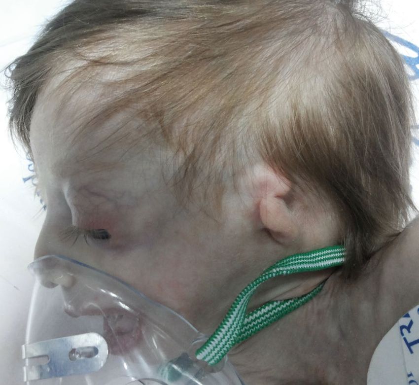
Netherlands Journal of Critical Care
                                                                                                                                                 Treacher Collins syndrome

Physical examination                                                                  atresia of the auditory meatus, microcephaly, ‘beak-like’ nose,
On her arrival to the emergency room, the infant looked ill, with                     macrostomia ‘fishlike mouth’ and thin hair (figures 1 and 2).
a temperature of 38.5°C, a respiratory rate of 55 breaths/min,                        Intraoral examination revealed a high arched cleft palate
and a pulse rate of 150 beats/min. The blood pressure was 85/60                       without cleft lip (figure 3).
mmHg and the oxygen saturation was 78% by pulse oximetry                              A detailed examination of this patient revealed clinical features
while breathing room air. Examination of her chest revealed                           of mandibulofacial dysostosis, bilaterally symmetrical but
crackles at both lungs, cardiac examination was normal.                               abnormal face characteristics. Computed tomography showed
The infant had a characteristic appearance with antimongoloid                         bilateral choanal obstruction and bilateral atresia of the external
slanting of the palpebral fissures, bilateral coloboma of the lower                   auditory canals. Echocardiograph and abdominal ultrasound
eyelids with absence of cilia of the lower eyelids, hypertelorism                     revealed normal findings.
of the eyes, proptosis, normal-sized pupils with a normal                             Based on the phenotypic and radiographic findings, the
reaction to light, malar hypoplasia of the zygomatic arch with                        diagnosis of Treacher Collins syndrome was made.
a ‘sunk-in’ appearance, sad facial expression, a ‘bird-like’ face
with trigonocephaly, mandibular hypoplasia, micrognathia,                             Hospital course
retrognathia, low-set ears, malformations of the auricular pinna,                     During the first day, the patient’s condition deteriorated, her

Figure 1. The clinical view of the 40-day-old baby girl with                          Figure 2. The clinical manifestations of Treacher Collins syndrome
Treacher Collins syndrome. Consent of the patients family was obtained for the publication of these images.

                                                                                                NETH J CRIT CARE - VOLUME 29 - NO 1 - JANUARY 2021                    37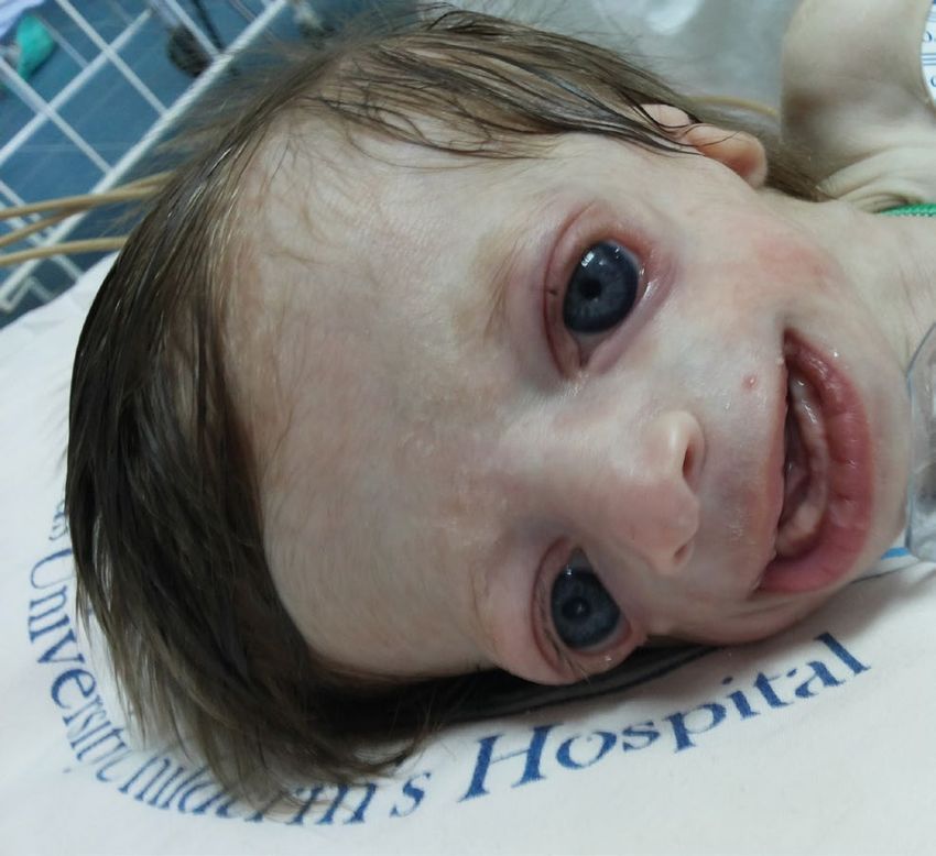
Netherlands Journal of Critical Care
Treacher Collins syndrome
stabilised enough for the ventilator device to be removed.
The patient was extubated; however, this led to shortness of
breath, a rapid heart rate and elevated blood pressure with no
improvement after high flow oxygen inhalation and noninvasive
ventilator-assisted respiration. Therefore, she was intubated
again.
During the period she was connected to the ventilator, she
proved difficult to wean. Weaning was attempted 24 hours
after using a T-piece and 4 l/min of oxygen was administered.
She had a good cough reflex. Three attempts were made to
remove the endotracheal tube but all of them failed due to
immediate respiratory distress with documented low saturation
requiring reintubation. Therefore, after consulting the
paediatric otolaryngologist, the decision was made to perform
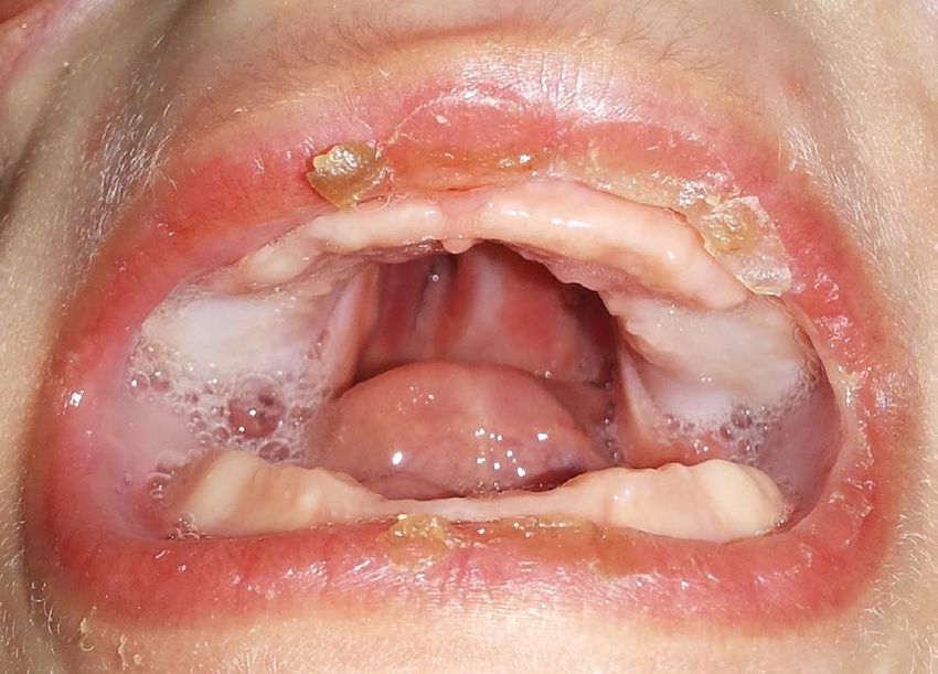
Figure 3. A high arched cleft palate a tracheostomy.
Because of feeding problems, the patient was commenced
dyspnoea worsened, and she experienced respiratory distress. on central total parenteral nutrition. After a discussion with
Two hours after admission to the hospital, she was transferred the paediatric gastroenterologist, the decision was made to
to the paediatric intensive care unit (PICU). Examination of perform a gastrostomy to aid in growth and development, and
the chest X-ray indicated bilateral infiltrates consistent with for prevention of aspiration.
pneumonia. The patient was treated with a combination of The treatment of this baby girl required the coordinated
antibiotics. Bronchoalveolar samples showed Streptococcus efforts of a team of specialists, including a paediatrician,
pneumoniae. However, the blood culture which was collected paediatric otolaryngologist, paediatric nurse, plastic surgeon,
for the bacteriological diagnosis was negative. audiologist, ophthalmologist, geneticist and other specialists to
During the next day, she underwent intubation for worsening systematically and comprehensively plan the child’s treatment.
hypoxia and respiratory distress, and a three-way central Multiple surgeries would have been needed to treat the various
venous catheter (CVC) was inserted into the right internal craniofacial abnormalities associated with Treacher Collins
jugular vein. As great difficulty was anticipated in securing the syndrome.
airway, the otorhinolaryngologist and anaesthesiologist teams Unfortunately, her clinical condition deteriorated; 36 days after
were urgently requested to be present at the procedure. These admission, she developed septic shock. Cultures of blood from
teams were waiting in the adjacent room with all the necessary the CVC and peripheral vein, and culture of urine were obtained.
equipment laid out and ready, including a paediatric intubation Because ventilator-associated pneumonia was suspected,
set and a tracheostomy set. diagnostic bronchoalveolar lavage culture was performed
The intubation of the infant was very difficult with two to evaluate for pneumonia as a sepsis source. The antibiotic
unsuccessful attempts at orotracheal intubation. One treatment was empirically changed to wide-spectrum antibiotic
intubation attempt was with a video laryngoscopy and one therapy (linezolid and colistin). She required high inotropic
was through the laryngeal mask airway. Finally, we decided to support and high ventilator settings. Inotropes and vasopressors
perform tracheal intubation via a fibreoptic bronchoscope. The were used to maintain her mean arterial pressure within the
fibreoptic bronchoscope was checked and loaded with a 3.5 mm normal range. Chest X-ray revealed bilateral infiltrates. A
endotracheal tube. We were ready to perform an emergency nosocomial, ventilator-associated pneumonia was diagnosed.
tracheostomy if required with the otorhinolaryngologist and The results of blood cultures from the CVC and peripheral vein,
anaesthesiologist on hand. The fibrescope was inserted into the and bronchoalveolar samples yielded Pseudomonas aeruginosa.
mouth. The trachea was entered and the tube was passed into Urine culture was negative.
the trachea. The CVC was removed and a new catheter was inserted into the
left internal jugular vein. She was switched to high-frequency
After seven days, the patient’s condition had improved. The oscillatory ventilation but the patient never responded
chest X-ray showed no infiltrates. Computed tomography of adequately to any therapy.
the rhinopharynx showed bilateral choanal obstruction. The Despite appropriate treatment, she remained critically ill with
diagnosis of choanal atresia was confirmed by nasofibroscopy. unstable systemic blood pressure.
The patient underwent successful correction of bilateral Forty days after admission, she went into cardiac arrest.
choanal atresia. Although cardiopulmonary resuscitation was performed for 30
Three days after the operation, the pulmonary situation had minutes, the patient succumbed to her illness.
38 NETH J CRIT CARE - VOLUME 29 - NO 1 - JANUARY 2021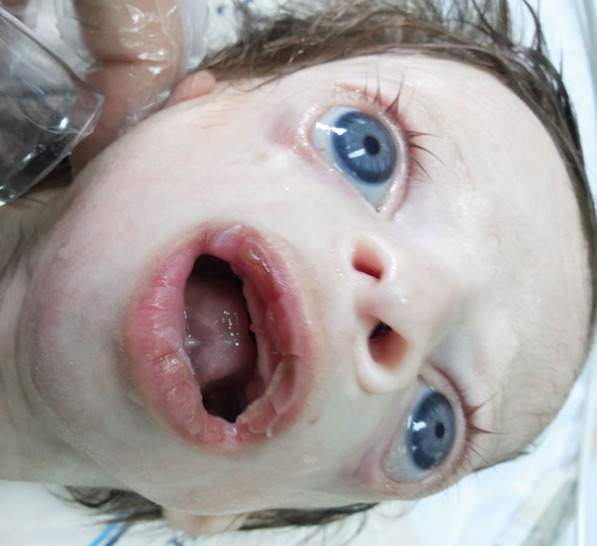
Netherlands Journal of Critical Care
                                                                                                                        Treacher Collins syndrome

Discussion                                                             the most relevant and challenging tasks for anaesthetists.
Treacher Collins syndrome is a rare congenital disorder                Many airway management techniques had been described
of craniofacial dysmorphism characterised by numerous                  in paediatric patients with craniofacial malformations.[2] In
developmental anomalies restricted to the head and neck.[7]            our case we used different devices: intubation with video
Early descriptions were attributed to Berry (1889), Treacher           laryngoscopy was attempted once but failed, then we used
Collins (1900) and Franceschetti and Klein (1949) and hence            the laryngeal mask airway once which also failed and, finally,
the names Berry's syndrome and Franceschetti-Zwahlen-Klein             intubation was achieved by a fibreoptic bronchoscope. Each
syndrome.[1,9,11]                                                      device was only used once in order to prevent the risk of airway
The frequency of Treacher Collins syndrome is 1 in 50,000              trauma which is associated with multiple attempts.
live births.[5,7,8] The most frequent clinical manifestations           We formed a multidisciplinary team consisting of paediatric
are antimongoloid palpebral fissures, malar and mandibular             critical care doctor, anaesthesiologist and otorhinolaryngologist
hypoplasia, malformation of auricular pinna, coloboma of               to manage intubation difficulties.
the lower eyelids, conductive deafness, cleft palate and dental        Nagamine et al. reported a 13-year-old girl with Treacher
anomalies.[9]                                                          Collins syndrome who had a history of difficult intubation and
Additional abnormal structures that are occasionally found in          was scheduled for plastic surgery. They took three-dimensional
Treacher Collins syndrome include absent parotid glands, cervical      computed tomography images to better evaluate the anatomical
spine malformation, cryptorchidism, extremity malformation,            features of the upper airway. The patient’s anaesthetic airway
renal anomalies, and congenital heart disease.[8,10]                   management was influenced successfully by the findings of
Treacher Collins syndrome is caused by mutations in the TCOF1,         the images.[15] In our case, airway management was urgent and
POLR1C or POLR1D genes that affect facial development                  there was not enough time for additional investigations.
before birth.[9,11,12] Mutations in the TCOF1 gene ac¬count for        Grohskopf et al. carried out a study to determine the prevalence
81% to 93% of all cases. POLR1C and POLR1D gene mutations              of ICU-acquired infections, which is a major cause of morbidity
are responsible for 2% of Treacher Collins syndrome cases.[9]          in PICU patients. They concluded that age-adjusted risk factors
The three genes have been involved with a dominant (TCOF1,             for infection included central intravenous catheters, arterial
POLR1D) or a recessive (POLR1D, POLR1C) autosomal mode                 catheters, total parenteral nutrition, or mechanical ventilation.[16]
of inheritance.[13]                                                    In our case, the patient had multiple risk factors for infection
Forty percent of the cases are associated with previous family         such as central intravenous catheters, parenteral nutrition, and
history and those affected have a 50% chance of passing it on          mechanical ventilation
to their next generation. The remaining 60% of the cases are           Çelik et al. performed a study to determine the prevalence of
thought to arise because of new mutation.[6,9,13] Our case did         infections and the predominant organisms. They concluded
not report a familial history of the syndrome and the cause was        that the rate of nosocomial infection is high in ICU patients,
likely a new mutation.                                                 especially for respiratory infections. The most frequently
The diagnostic features of Treacher Collins syndrome include           reported infection was ventilator-associated pneumonia. The
abnormalities in eyes, ears, nose, mouth and facial bone.              predominant bacteria were P. aeruginosa and S. aureus.[17] In
The vast majority of these features were present in our case.          our case, a nosocomial, ventilator-associated pneumonia was
Based on these clinical features five clinical forms of Treacher       diagnosed. The results of blood cultures and bronchoalveolar
Collins syndrome were identified by Franceshetti and Klein:            samples yielded P. aeruginosa.
the complete form presenting with all known features, an               Vincent et al. provided an up-to-date, international picture of the
incomplete form presenting with less severe ear, eye, zygoma,          extent and patterns of infection in ICUs. They concluded that
and mandibular abnormalities, the abortive form with only              infections are common in patients in contemporary ICUs, and
lower lid pseudo coloboma and zygoma hypoplasia, the                   the risk of infection increases with the duration of ICU stay.[18] In
unilateral form with anomalies limited to only one side of the         our case, the baby girl stayed in the PICU for 40 days.
face and the atypical form presenting with other abnormalities         Treacher Collins syndrome can be detected using prenatal
not usually part of this syndrome.[1,7,9] In our case, the patient     screening ultrasound. Three-dimensional sonographic imaging
presented with the complete form of this syndrome.                     has been shown to detect these subtle features including
Treacher Collins syndrome has been recognised to be associated         downslanting palpebral fissures, micrognathia, and lowset ears/
with upper airway obstruction and difficult tracheal intubation.[14]   microtia. Polyhydramnios is seen as well.[10] In our case, the
In neonates and other paediatric patients with predicted difficult     syndrome was not detected before birth. For a good outcome
ventilation and intubation, thoughtful airway management               it is mandatory to anticipate problems in securing the airway
planning is mandatory. Usually, difficult intubation in these          and have a plan ready on how to proceed. Ideally appropriate
patients is anticipated, which gives us some time to prepare.[2]       prenatal planning is needed and prompt intervention after birth
Management of difficult airway in children remains one of              by a team of experienced medical and surgical personnel.

                                                                               NETH J CRIT CARE - VOLUME 29 - NO 1 - JANUARY 2021            39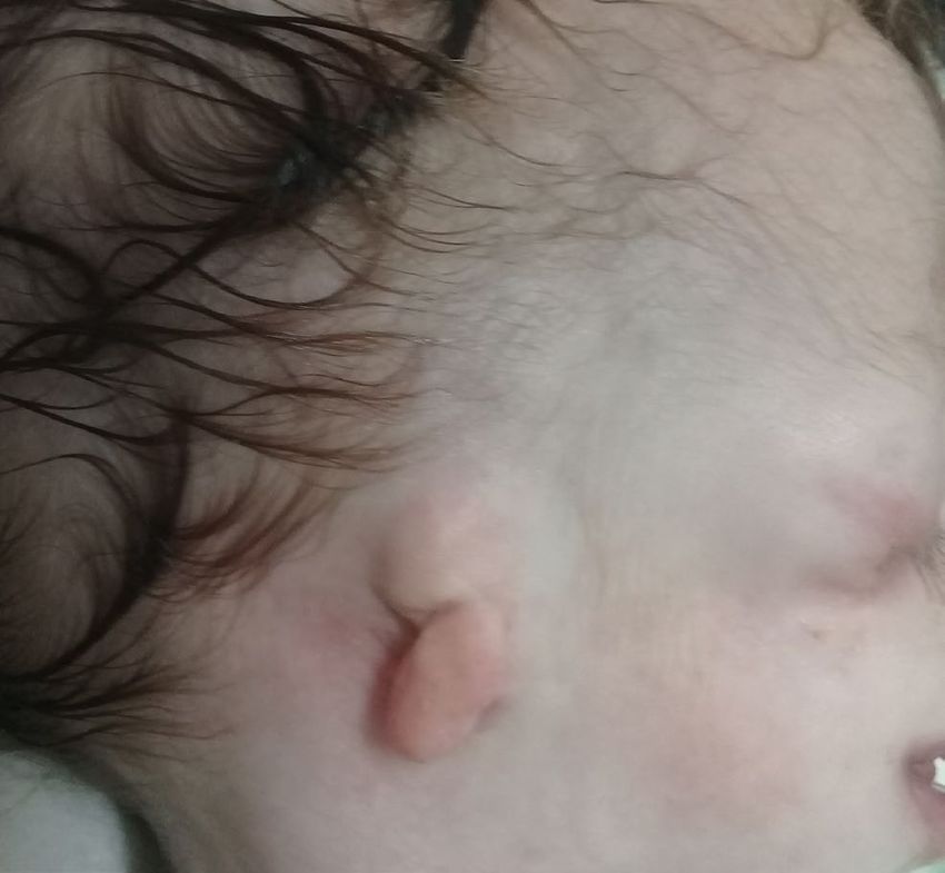
Netherlands Journal of Critical Care
Treacher Collins syndrome

There is no cure for Treacher Collins syndrome. Treatment is       Disclosures
aimed at the specific needs of each individual. Many children      All authors declare no conflict of interest. No funding or
require a multidisciplinary approach involving a craniofacial      financial support was received.
team, including a paediatric otolaryngologist, audiologist,
plastic surgeon, geneticist, psychologist, dental surgeons and     Informed consent was obtained from the patient’s family for the
other healthcare professionals.[1]                                 publication of this case report (and the accompanying images).
Of primary concern are breathing and feeding problems that
present at birth as a consequence of micrognathia and tongue       References
obstruction of the hypopharynx.[5,9]
                                                                   1. 	Renju R, Varma BR, Kumar SJ, Kumaran P. Mandibulofacial dysostosis (Treacher
A tracheostomy may even be necessary in some cases to                    Collins syndrome): A case report and review of literature. Contemp Clin Dent.
maintain an adequate airway. Furthermore, a gastrostomy                  2014;5:532.
                                                                   2. 	Marques-Pires R, Trindade H. The airway approach to a neonate with Treacher
could be necessary to ensure an adequate caloric intake while            Collins syndrome–Case report. Rev Esp Anestesiol Reanim. 2017;64:233-6.
protecting the airway. Surgery to restore a normal structure of    3. 	Muraika L, Heyman JS, Shevchenko Y. Fiberoptic tracheal intubation through a
                                                                         laryngeal mask airway in a child with Treacher Collins syndrome. Anesth Analg.
the face is generally performed at defined ages, depending on            2003;97:1298-9.
the developmental stage.[9] Tracheostomy was required at some      4.	Lin TC, Soo LY, Chen TI, et al. Perioperative airway management in a child with
                                                                         Treacher Collins syndrome. Acta Anaesthesiol Taiwan. 2009;47:44-7.
stage in childhood in 41% of children with TCS, and the ratio      5. 	Trainor PA. Craniofacial birth defects: The role of neural crest cells in the etiology
increased to 84% in TCS patients who also had choanal atresia            and pathogenesis of Treacher Collins syndrome and the potential for prevention.
                                                                         Am J Med Genet. 2010;152A:2984-94.
or stenosis.[4]                                                    6. 	Trainor PA, Dixon J, Dixon MJ. Treacher Collins syndrome: etiology, pathogenesis
                                                                         and prevention. Eur J Hum Genet. 2009;17:275-83.
In our case, tracheostomy, gastrostomy and surgical repair of      7. 	Thomas P, Krishnapillai R, Ramakrishnan BP. Treacher Collins Syndrome: A Case
bilateral choanal atresia were essential in the management of            Report and Review of Literature. Oral Maxillofac Pathol J. 2019;10:90-4.
                                                                   8. 	Martelli H, Coletta RD, Miranda RT, de Barros LM, Swerts MS, Bonan PR. Orofacial
the baby girl. We made a plan with our team of specialists to            features of Treacher Collins syndrome. Med Oral Patol Oral Cir Bucal. 2009;14
systematically and comprehensively treat the patient.                    :E344-8.
                                                                   9. 	Khodadadi E, Dehghan Z. Treacher Collins Syndrome: A Case Report and Review
                                                                         of Literature. J Pediatr Rev. 2019;7:45-54.
Conclusion                                                         10. 	Chang CC, Steinbacher DM. Treacher Collins syndrome. Semin Plast Surg.
                                                                         2012;26:83-90.
Each case of Treacher Collins syndrome is unique and needs to      11. 	Papageorgiou E, Papoulidis I, Zavlanos A, Papanikolaou E, Manolakos E, Fidani S.
be managed individually. Many manifestations of the disease can          A novel familial mutation associated with
                                                                   12. 	Vincent M, Geneviève D, Ostertag, et al. Treacher Collins syndrome: a clinical and
be improved by surgery and other supportive treatments. Early            molecular study based on a large series of patients. Genet Med. 2016;18:49-56.
detection and well-planned treatment, tailored to the individual   13. 	Shete, P, Tupkari, JV, Benjamin T, Singh A. Treacher Collins syndrome. J Oral
                                                                         Maxillofac Pathol. 2011;15:348.
patient, can reduce disease symptoms and produce excellent         14. 	Bucx MJ, Grolman W, Kruisinga FH, Lindeboom JA, Van Kempen AA. The
                                                                         prolonged use of the laryngeal mask airway in a neonate with airway obstruction
results for complete restoration of the form and function of the         and Treacher Collins syndrome. Pediatr Anesth. 2003;13:530-3.
patient. Airway management should also be specific for each        15. 	Nagamine Y, Kurahashi K. The use of three-dimensional computed tomography
                                                                         images for anticipated difficult intubation airway evaluation of a patient with
individual patient. A multidisciplinary approach and careful             Treacher Collins syndrome. Anesth Analg. 2007;105:626-8.
planning is mandatory for the appropriate management of            16. 	Grohskopf LA, Sinkowitz-Cochran RL, Garrett DO, et al. A national point-
                                                                         prevalence survey of pediatric intensive care unit-acquired infections in the
these patients. The members of the team are determined by the            United States. J Pediatr. 2002;140:432-8.
complexity of the case. Preparing for potential difficulties and   17. 	Çelik İ, İnci N, Denk A, Sevim E, Yaşar D, Yaşar MA. Prevalence of hospital acquired
                                                                         infections in anesthesiology intensive care unit. Fırat Tıp Dergisi. 2005;10:132-5.
communicating effectively can lead to successful results and       18. 	Vincent JL, Rello J, Marshall J, et al. International study of the prevalence and
avoid catastrophic outcomes.                                             outcomes of infection in intensive care units. JAMA. 2009;302:2323-9.

40          NETH J CRIT CARE - VOLUME 29 - NO 1 - JANUARY 2021
You can also read