RHABDOMYOLYSIS ACCOMPANYING THE STAPHYLOCOCCAL SEPSIS
←
→
Page content transcription
If your browser does not render page correctly, please read the page content below
Archives of the Balkan Medical Union                                                      vol. 55, no. 3, pp. 518-522
Copyright © 2020 Balkan Medical Union                                                                September 2020

                                                                                           CASE REPORT

RHABDOMYOLYSIS ACCOMPANYING
THE STAPHYLOCOCCAL SEPSIS
Leonid I. DVORETSKY1, Maria A. KARNAUSHKINA1 , Georgy O. ZAYRATIANTS2,
Elend V. SERGEEVA1, Svetlana S. BOBKOVA2
1
  Department of Hospital Therapy No. 2, „I.M. Sechenov“ First Moscow State Medical University, Moscow,
Russian Federation
2
  Municipal Hospital „S.S. Yudin“ of the Moscow Healthcare Department, Moscow, Russian Federation
                                                              Received 31 May 2020, Accepted 27 July 2020
                                                                           https://doi.org/10.31688/ABMU.2020.55.3.20

ABSTRACT                                                   RÉSUMÉ

Introduction. Rhabdomyolysis (RM) is a clinical and        Rhabdomyolyse accompagnant la septicémie staphy-
laboratory syndrome resulting from skeletal muscle         lococcique
damage, with the release of a big number of intracel-
lular substances of myocytes into the systemic circu-      Introduction. La rhabdomyolyse est un syndrome cli-
lation and endotoxemia. The entry of the muscle de-        nique et de laboratoire résultant de lésions musculaires
struction products into the systemic circulation leads     squelettiques avec libération d’un grand nombre de
to the development of systemic manifestations, serious     substances intracellulaires de myocytes dans la circula-
disorders in homeostasis, multiple organ failure syn-      tion systémique et endo-toxémie. L’entrée des produits
drome, including acute renal failure, often threatening    de destruction musculaire dans la circulation systé-
the patient’s life.                                        mique entraîne le développement de manifestations
Case report. A rare case of RM in a patient with           systémiques, de troubles graves de l’homéostasie, du
staphylococcal sepsis (SS) and development of multiple     syndrome de défaillance multiviscérale, y compris une
organ failure is described. The diagnosis of RM was        insuffisance rénale aiguë, menaçant souvent la vie du
confirmed by the results of pathological and anatomi-      patient.
cal studies. Possible causes of RM in staphylococcal       Rapport de cas. Un cas rare de rhabdomyolyse chez
sepsis are discussed here.                                 un patient souffrant de septicémie staphylococcique et
Conclusions. Only a few cases of RM upon SS were           de développement d’une défaillance d’organes multi-
found in the literature. The development of RM in          ples est décrit. La présence de la rhabdomyolyse est
combination with the systemic inflammatory reaction        confirmée par les résultats d’études pathologiques et
syndrome in SS exacerbates the course of the disease       anatomiques. Les causes possibles de la rhabdomyolyse
and worsens its prognosis. Therefore, the timely diag-     dans la septicémie staphylococcique sont discutées ici.
nosis of RM and appropriate treatment are extremely        Conclusions. Seuls quelques cas de rhabdomyolyse à
important. The mechanism of RM development upon            fond de septicémie staphylococcique ont été trouvés

      Address for correspondence:       Maria A. KARNAUSHKINA
                                        Department of Hospital Therapy No. 2, „I.M. Sechenov“ First Moscow State
                                        Medical University, Moscow, Russian Federation
                                        Address: Trubezkaja street no. 8, Moscow, Russia
                                        Email:kar3745@yandex.ru
Archives of the Balkan Medical Union
SS is complex. After staphylococcus was isolated from dans la littérature, et au deuxième millénaire, seules
the affected muscles in a patient with SS, one of the trois observations ont été publiées. Le développement
first descriptions of RM upon SS suggested the role of de la rhabdomyolyse en combinaison avec le syndrome
direct invasion of the infectious agent into the muscles, de réaction inflammatoire systémique dans les septicé-
followed by the development of „pyomyositis.“ Another mies staphylococcique exacerbe le cours de la maladie
mechanism for RM occurrence of PM may be the pres- et aggrave son pronostic, et donc le diagnostic rapide
ence of toxin upon the staphylococcal toxic shock syn- de la rhabdomyolyse et le traitement approprié sont
drome in patients with SS. extrêmement importants. Le mécanisme de dévelop-
pement de la rhabdomyolyse sur septicémie staphy-
Keywords: rhabdomyolysis, staphylococcal sepsis, in- lococcique est complexe. Après que le staphylocoque
fectious endocarditis, acute renal failure. ait été isolé à partir de muscles affectés chez un patient
atteint de septicémie staphylococcique, l’une des pre-
List of abbreviations: mières descriptions de la rhabdomyolyse sur septicé-
RM – rhabdomyolysis mie staphylococcique a suggéré le rôle d’une invasion
SS – staphylococcal sepsis directe de l’agent infectieux dans les muscles, suivie
ARN – acute renal failure du développement d’une «pyomyosite». Un autre mé-
IE – infectious endocarditis canisme pour l’apparition de la rhabdomyolyse peut
CPK – creatine phosphokinase être la présence de toxines sur le syndrome de choc
ALT – alanine aminotransferase toxique staphylococcique chez les patients atteints de
AST – aspartate aminotransferase septicémie staphylococcique.
Mots-clés: rhabdomyolyse, septicémie staphylococ-
cique, endocardite infectieuse, insuffisance rénale ai-
guë.
INTRODUCTION with septic shock, ARN development and resulting
in death 5. The following case of RM upon SS (the
Rhabdomyolysis (RM) is a clinical and labora- presence of staphylococcus in the blood and urine)
tory syndrome resulting from skeletal muscle dam- without signs of shock and ARN, with a positive
age, with the release of a large number of intracellular treatment outcome, was described two years later in a
substances of myocytes into the systemic circulation 20-year-old patient with infectious endocarditis (IE)6,
(myoglobin, lysosomal and mitochondrial enzymes, and two years later two more cases of RM upon SS
histamine, serotonin, oligo- and polypeptides, etc.) were reported7,8. Recent publications of the last cen-
and endotoxemia. The entry of the muscle destruc- tury cover two cases of RM upon SS: in one patient
tion products into the systemic circulation leads to with acquired immunodeficiency syndrome9, and in
the development of systemic manifestations, serious another – with staphylococcal pneumonia10. It is note-
disorders in homeostasis, multiple organ failure syn- worthy that in almost 30 years after the first descrip-
drome, including acute renal failure (ARN), often tions of RS upon SS, there were no publications on
threatening the patient’s life. this topic, and only in the second millennium did
The various causes of RM (traumatic injuries, individual information reappear. Thus, a report was
metabolic disorders, epilepsy, excessive muscle stress, published on the development of RM in a 12-year-old
drugs, etc.) specifically include various infections1,2. child with staphylococcal IE, manifested by myalgia,
A search in the Medline system for the publication of fever, and secretion of dark urine11. In adults, two
RM cases with infections over 30 years (1966-1996) cases of RM upon IE caused by methicillin-sensitive
returned information on viral and bacterial infec- staphylococcus have been described. One patient
tions as the cause of RM in 59 and 60 cases, respec- previously operated for a congenital malformation
tively3. developed IE complicated by severe RM with high
RM is relatively rare against a background of CPK indices (up to 49068 IU), successfully treated
staphylococcal infections, particularly, staphylococ- with antibiotics followed by surgical intervention12.
cal sepsis (SS). The first reports of RM upon SS in In another patient with IE, RM was diagnosed in
children and adults appeared in the 1980s. The pedi- combination with purpura, purulent pericarditis and
atric literature described a case of RM upon SS in intracerebral hematoma. Despite intensive therapy
a 15-year-old boy4. The earliest description of RM (antibiotics, drainage of the pericardial cavity and
upon SS in adults is related to a 70-year-old patient cerebral hematoma), a fatal outcome occurred13.
September 2020 / 519Rhabdomyolysis accompanying the staphylococcal sepsis – DVORETSKY et al
In this article, we present a patient with severe flutter, arrested by cordarone. The values of urea
RM that developed against the background of SS. (45.3 mmol/L), creatinine (332 μmol/L), CPK (4809
U/L), and lactate dehydrogenase (729 U/L) were
CASE PRESENTATION increasing. The microbiological examination of the
blood and urine revealed Staphylococcus aureus sen-
A 46-year-old man was admitted to the neuro- sitive to methicillin. Antibacterial therapy (oxacillin,
logical department of the State Clinical Hospital linezolid) was prescribed. Despite intensive therapy,
„S.S. Yudin“ (Moscow, Russian Federation) on June the condition deteriorated ,progressively and the pa-
28, 2019, with complaints of pain in the lower spine, tient died on July 11, 2019.
and weakness in the legs. The medical history includ- The positive diagnosis was: Sepsis from undiag-
ed type 2 diabetes mellitus, for which he received nosed origin. Systemic inflammatory reaction syn-
insulin preparations of short and prolonged action, drome. Rhabdomyolysis. Multiple organ failure. Poly
nephrectomy for urolithiasis, polytrauma (fracture of segmental pneumonia. Diabetes mellitus type 2.
ribs, craniocerebral trauma, fractures in the vertebra, The pathological diagnosis after necropsy was:
contusion of the liver and spleen) at the age of 20 Sepsis. Septic shock: shocked kidneys, liquid blood
years. in the cavities of the heart and large vessels. Bilateral
The condition at admission is satisfactory. The focal-confluent pneumonia in 6-9 segments. Multiple
consciousness is clear. Forced position on the back. organ failure syndrome: parenchymal dystrophy of
Vesicular breathing, without wheezing, breath rate the liver, myocardium, kidneys. Myocardial infarc-
16/min. The heart sounds are rhythmic, heart rate tion in the posterior wall of the left ventricle, with
80/min, blood pressure 120/80 mm Hg. The abdo- dimensions of 2.5x3.0 cm about one day old. Focal
men is soft and painless on palpation. The liver and necrosis of the ileum. Acute erosion of the stomach
spleen are not enlarged. No acute neurological and and duodenum. Pulmonary edema. Cerebral edema.
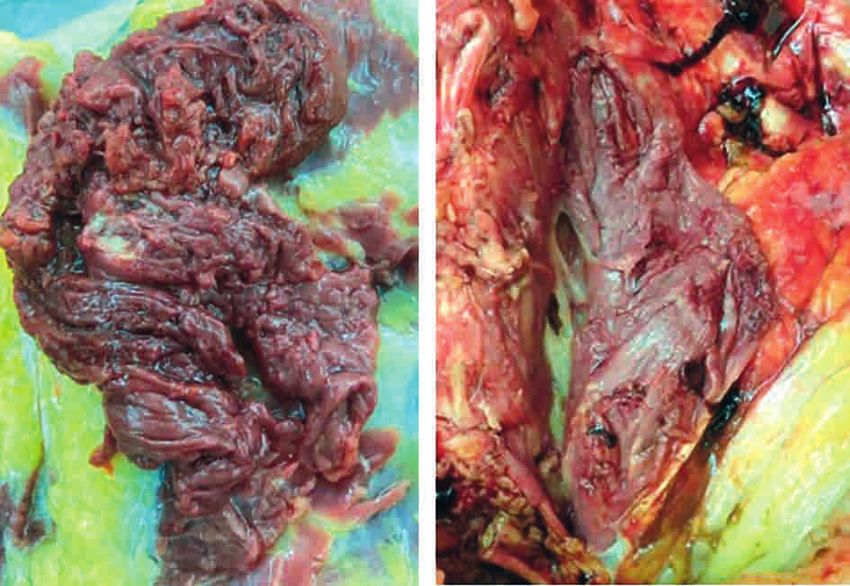
surgical pathology, or foci of infection were found. Attention was drawn to the change in the appearance
We noted the changes of laboratory tests in dynam- of striated muscles, especially the femoral and iliac
ics. Peripheral blood showed leukocytosis (32-35 x muscles, which were mottled, with uneven blood sup-
109/L) with a sharp shift to the left (17-37% of stab ply and flabby (“boiled meat“ appearance) (Fig. 1 a,b).
neutrophils) to the appearance of young forms in the
blood (myelocytes, metamyelocytes). Hemoglobin DISCUSSION
and platelet counts were within the normal range.
Biochemical blood tests revealed an increase in the The presence of a systemic inflammatory reac-
level of C-reactive protein (CRP) (302-440 mg/L), tion syndrome (marked leukocytosis with a shift to
procalcitonin (5.3-17.02 ng/mL), AST and ALT ac- myelocytes, a high-level of CRP), the results of the
tivity (118 IU/L and 101 IU/L, respectively), cre- microbiological study (Staphylococcus aureus in the
atine phosphokinase (2200-2700 IU/L), urea (16-23 blood and urine), and a high-level of procalcitonin in-
mmol/L), creatinine (153-234 mmol/L), potassium dicated the development of SS, although the primary
(5.2-5.5 mmol/L), glucose (20 mmol/L). During his site of infection could not be defined. Peculiarities of
stay in the neurological department, the patient’s con- SS progression in the form of myalgia syndrome in
dition deteriorated, with sharp pain in the muscles of combination with elevated CPK, ALT, AST, the pres-
the limbs, pain on palpation of the muscles, disorders ence of myoglobin in the blood, brown urine, and ul-
of the central nervous system (lethargy, confusion), trasound data on soft tissues indicated the diagnosis
a decrease in the blood pressure to 90/70 mm Hg. of RM, as a rare complication of SS. This clinical and
Myoglobin (14350 ng/mL) was detected in the blood. laboratory syndrome raised the suspicion of polymy-
Brown urine secretion was marked. The patient was ositis, which had to be differentiated from RM in
transferred to the intensive care unit on July 01, 2019, various pathological processes. Similar difficulties
where intubation of the trachea, and catheterization in the differential diagnosis were reported in one of
of the internal jugular vein were performed, followed the RM case descriptions upon SS in a patient with
by artificial ventilation, hemodiafiltration, and no- IE, successfully treated with antibiotics, followed by
radrenaline infusion. the disappearance of RM signs14. Muscle lesion was
Ultrasound of the soft tissues of the medial confirmed by autopsy and subsequent histological
surface of the upper third of the thigh revealed an examination.
increase in echogenicity, without clear delineations, Only a few cases of RM upon SS were found
with single an-echogenic inclusions (edema?, infil- in the literature, and in the second millennium,
trate?). No fluid accumulation detected in soft tis- only three observations were published12-14. The de-
sues. Electrocardiogram showed episodes of atrial velopment of RM in combination with the systemic
520 / vol. 55, no. 3Archives of the Balkan Medical Union
A. B.
Fig. 1 a,b. Iliac muscles of the mottled flabby appearance with uneven blood supply.
inflammatory reaction syndrome in SS exacerbates development of RM upon SS (statins, alcohol con-
the course of the disease and worsens its prognosis. sumption, etc)18. In our patient, one additional factor
Therefore, the timely diagnosis of RM and appropri- may be considered the presence of diabetes mellitus.
ate treatment are extremely important.
The mechanism of RM development upon SS is CONCLUSIONS
complex. After staphylococcus was isolated from the
affected muscles in a patient with SS, one of the first One of the RM complications is renal damage,
descriptions of RM upon SS suggested the role of with the development of ARF19. The incidence of
direct invasion of the infectious agent into the mus- ARF reaches 40% among all cases of RM20. RM is the
cles, followed by the development of „pyomyositis.“ cause of 15% of ARF cases21. The main pathogenetic
Another mechanism may be the presence of toxins mechanism of renal damage upon RM is the appear-
upon the staphylococcal toxic shock syndrome in pa- ance of myoglobin, having a nephrotoxic effect, in
tients with SS15. the blood. Furthermore, the fluid redistribution into
Among other toxic mechanisms not related to the muscles results in hypovolemia, leading to renal
the toxic shock syndrome, are the staphylococcal en- hypoperfusion. Attempts to predict the risk of devel-
terotoxins associated with the development of RM oping ARF in patients with RM based on the values
in a patient with SS, who was found to have the of CPK, creatinine, potassium, calcium, and the con-
genes encoding staphylococcal enterotoxins C, G tent of myoglobin in urine were inconclusive20. An
and I. One of the mechanisms of myocyte damage additional factor for the development of ARF in our
upon infections may be the release of interleukin-1 patient could be the nephrectomy in the past.
from cells, which is, moreover, a pyrogen during fe- The treatment of SS patients complicated by RM
ver development16. The role of the increased content includes, along with adequate antibacterial therapy,
of intracellular calcium entering the cells under the the correction of hypovolemia, electrolyte imbalance
action of prostaglandins and leading to cell destruc- (acidosis, hyperkalemia, hypercalcemia), and the
tion activated by neutral proteases is not excluded17. hemostatic system, alkalization of urine in order to
Various additional risk factors may contribute to the prevent the nephrotoxic effect of myoglobin, as well
September 2020 / 521Rhabdomyolysis accompanying the staphylococcal sepsis – DVORETSKY et al
as the methods of replacement therapy for ARF (he- 7. Kawamoto R, Fujii Y, Tao S. An autopsy case of rhabdomy-
modialysis)22. olysis associated with staphylococcal septicemia. J Jpn Soc
Intern Med 1988;77:1278.
8. Tai l l a n B, Vi nt i H, Fu zibet JG, Four nier JP,
Montagne N, Dujardin P. Non-traumatic rhabdomy-
Author Contributions: olysis in Staphylococcus aureus septicemia. Presse Med.
L.I.D., E.V.S., and S.S.B. were responsible for the 1988;17(36):1860-1.
diagnostic procedures, clinical diagnosis, and treatment 9. Wu AW, Benirschke K, McCutchan JA. Rhabdomyolysis
and Staphylococcus aureus septicemia in a man with the
decisions. G.O.Z. made the histopathological diagnosis. acquired immunodeficiency syndrome. West J Med 1990;
L.I.D. and M.A.K. analyzed the literature and wrote the 152:716-719.
manuscript. All authors have read the text and reached an 10. Bando T, Fujimura M, Noda Y, Ohta G, Hirose J, Matsuda
agreement for the manuscript text. T. Rhabdomyolysis associated with bacteremic pneumonia
due to Staphylococcus aureus. Intern Med 1994;33:454-5.
11. Bandi S, Chikermane A. Rhabdomyolysis in a child second-
Compliance with Ethics Requirements:
ary to Staphylococcus aureus endocarditis. J Global Infect Dis
„The authors declare no conflict of interest regarding 2009;1(2):146-8.
this article“ 12. Ravry C, Fedou AL, Dubos M, et al. Severe rhabdomyoly-
„The authors declare that all the procedures and ex- sis associated with Staphylococcus aureus acute endocarditis
periments of this study respect the ethical standards in the requiring surgery. Surg Infect (Larchmt). 2015;16(6):840-2.
Helsinki Declaration of 1975, as revised in 2008(5), as 13. Georgescu AM, Azamfirei L, Szalman K, Szekely E. Fatal
endocarditis with methicilin-sensible Staphylococcus aureus
well as the national law. Informed consent was obtained and major complications: rhabdomyolysis, pericarditis, and
from the patient included in the study“ intracerebral hematoma. A case report and review of the
„No funding for this study“ literature. Medicine 2016;95 (41): e5125.
14. Ojeda J, López-López L, González A, Vilá LM. Infective en-
Acknowledgements: docarditis initially presenting with a dermatomyositis-like
syndrome. BMJ Case Rep 2014;2014:bcr2013200865.
None 15. Scobie BA. Staphylococcal toxic shock: Two fulminant cases
with recovery. NZ Med J 1982;95:145-7.
16. Baracos V, Rodemann P. Dinarello CA, et al. Stimulation
REFERENCES of muscle protein degradation and prostaglandin E2 release
by leukocytic pyrogen (interleukin-1): a mechanism for the
1. Blanco JR, Zabalza M, Salcedo J, Echeverria L, García A, increased degradation of muscle proteins during fever. N
Vallejo M. Rhabdomyolysis of infectious and noninfectious Eng J Med 1983;308: 553.
causes. South Med J. 2002;95(5):542-4. 17. Clowes GH, George BC, Villee CA, et al. Muscle proteolys
2. Lionte C, Sorodoc L, Petris O, Sorodoc V, Bologa C, Anton is induced by a circulating peptide in patients with sepsis or
G. Non-traumatic rhabdomyolysis in medical practice. Rev trauma. N Engl J Med 1983;308:545.
Med Chir Soc Med Nat Iasi. 2009;113(4):1025-3. 18. Gheorghe G, Toth PP, Bungau S, et al. Cardiovascular risk
3. Singh U, ScheId WM. Infectious etiologies of rhabdomyoly- and statin therapy considerations in women. Diagnostics
sis: three case reports and review. Clinical Infectious Diseases 2020;10:483.
1996;22:642-9. 19. Stoicescu M, Csepento C, Mutiu G, Bungau S. The role
4. Adamski GB, Garin EH, Ballinger WE, Shulman ST. of increased plasmatic renin level in the pathogenesis of
Generalized nonsuppurative myositis with staphylococcal arterial hypertension in young adults. Romanian Journal of
septicemia. J Pediatr 1980;96:694-7. Morphology and Embriology 2011;52(Suppl. 1):419-423.
5. Lannigan R, Austin TW, Vestrup J. Myositis and rhabdomy- 20. Khan FY. Rhabdomyolysis: a review of the literature. Neth J
olysis due to Staphylococcus aureus septicemia. Infect Dis. Med. 2009;67(9):272-283.
1984;150(5):784. 21. Kasaoka S, Todani M, Kaneko T, et al. Peak value of blood
6. Saito H, Yoshii F, Ishihara T, Shinohara Y. Rhabdomyolysis myoglobin predicts acute renal failure induced by rhabdomy-
associated with staphylococcal septicemia. Rinsho Shinkeigaku olysis. J Crit Care. 2010;25(4):601-604.
(Clin Neurol) 1986;26:693-7. 22. Better OS, Abassi ZA. Early fluid resuscitation in patients
with rhabdomyolysis. Nat Rev Nephrol. 2017;7(7):416-422.
522 / vol. 55, no. 3You can also read