HHS Public Access Author manuscript J Am Acad Dermatol. Author manuscript; available in PMC 2018 March 08.
←
→
Page content transcription
If your browser does not render page correctly, please read the page content below
HHS Public Access
                                Author manuscript
                                J Am Acad Dermatol. Author manuscript; available in PMC 2018 March 08.
Author Manuscript

                    Published in final edited form as:
                     J Am Acad Dermatol. 2013 November ; 69(5): 736–741. doi:10.1016/j.jaad.2013.07.024.

                    Atypical Hand-Foot-Mouth Disease Associated with
                    Coxsackievirus A6 Infection
                    Jason P. Lott, MD MSHP*,†, Kristina Liu, BA*, Marie-Louise Landry, MD‡, W. Allan Nix, BS§,
                    M. Steven Oberste, PhD§, Jean Bolognia, MD*, and Brett King, MD PhD*
                    *Department      of Dermatology, Yale University School of Medicine (SOM)
                    †Robert    Wood Johnson Foundation Clinical Scholars Program, Yale University SOM
Author Manuscript

                    ‡Departments       of Laboratory Medicine and Internal Medicine, Yale University SOM
                    §Divisionof Viral Diseases, National Center for Immunization and Respiratory Diseases, Centers
                    for Disease Control and Prevention

                    Abstract
                         Background—Hand, foot, and mouth disease (HFMD) is an acute viral illness commonly
                         caused by coxsackievirus A16 (CV-A16) and enterovirus 71 infections. Recently, atypical HFMD
                         has been reported in association with CV-A6, an uncommon enterovirus strain.

                         Objective—To describe the clinical features of atypical HFMD associated with CV-A6 infection
                         and its diagnostic laboratory evaluation.
Author Manuscript

                         Methods—Patients presenting to our institution with history and examination suggestive of
                         atypical HFMD from January 2012 to July 2012 were identified. Morphology and distribution of
                         mucocutaneous lesions were recorded. Enterovirus infection was assessed by reverse transcriptase
                         polymerase chain reaction of biologic specimens. Enterovirus type was determined by viral capsid
                         protein 1 gene sequencing.

                         Results—Two adults and 3 children with atypical HFMD were identified. Four of 5 patients
                         exhibited widespread cutaneous lesions. In 2 patients with a prior history of atopic dermatitis,
                         accentuation in areas of dermatitis was noted. Associated systemic symptoms prompted 4 of 5
                         patients to seek emergency care, and both adults were hospitalized for diagnostic evaluation.
                         Infection with CV-A6 was confirmed in all patients.

                         Limitations—This study is a case series from a single institution.
Author Manuscript

                         Conclusion—Consideration of the expanded range of cutaneous findings in atypical HFMD
                         caused by CV-A6 infection may assist clinicians in diagnosis and management.

                    Corresponding Author (send reprint requests here): Brett King, MD PhD, 333 Cedar St, LCI 501, PO Box 208059, New Haven,
                    CT 06520-8059, Phone: 203-785-4091, Fax: 203-785-7637.
                    Conflicts of Interest: The authors have no conflicts of interest to declare.
                    Prior Presentation: This material has not been presented previously
Lott et al. Page 2
Keywords
Author Manuscript
Hand-foot-mouth disease; coxsackievirus A6; enterovirus; exanthem; atypical; diagnosis;
evaluation
Introduction
Hand-foot-mouth disease (HFMD) is an acute viral illness characterized by fever, intraoral
vesicles and erosions, and papulovesicles that favor the palms and soles.1 Typically, the most
common causative agents are coxsackievirus A16 (CV-A16) and enterovirus type 71
(EV-71). HFMD ordinarily occurs from spring to fall, usually affecting children 5 years of
age and younger.2
Recently, the Centers for Disease Control and Prevention (CDC) reported HFMD with
Author Manuscript
atypical features in several states, including Connecticut, caused by the uncommon strain
coxsackievirus A6 (CV-A6).3 We report 5 patients with HFMD secondary to CV-A6 with
atypical, widespread cutaneous manifestations. Given the increasing incidence of this
distinct form of HFMD, clinicians should become familiar with its clinical presentation and
laboratory confirmation.
Patient 1
A 43-year-old male was hospitalized in January 2012 with a 2-day history of headache,
fever, chills, night sweats, odynophagia, myalgias, and a painful palmoplantar eruption
impairing his ability to grasp and ambulate.3 His wife and son had been ill with fever,
nausea, and vomiting the week before, and several children at the son’s daycare center had
Author Manuscript
been recently diagnosed with HFMD.
Physical examination revealed 2–9 mm pink-red papules and papulovesicles on the left
forearm, dorsal hands, palms, fingers, and on one toe (Fig. 1A). Several 1–2 mm vesicles
and erosions were present on the hard and soft palates. One 4 mm painful subungual macule
was present on the left thumb.
An initial evaluation including a complete blood cell count was normal and polymerase
chain reaction (PCR) of serum for parvovirus B19 was negative. Reverse transcriptase-
polymerase chain reaction (RT-PCR) 4 of plasma for enterovirus was positive. Histologic
examination of a papule on the right hand showed scattered necrotic keratinocytes,
exocytosis of lymphocytes, minimal interface change, and a perivascular lymphocytic
infiltrate within the dermis. Viral typing performed by the CDC confirmed CV-A6 infection
Author Manuscript
via viral capsid protein (VP) 1 gene sequencing.5
He received supportive care only and had a full recovery.
Patient 2
A 21-year-old male was hospitalized in March 2012 with fever and shaking chills after
having been recently diagnosed with Graves’ disease. The day after admission he
J Am Acad Dermatol. Author manuscript; available in PMC 2018 March 08.Lott et al. Page 3
complained of odynophagia, and the following day he developed a “rash” on his hands,
Author Manuscript
prompting a dermatologic consultation.
Physical examination revealed 2 mm vesicles in the posterior pharynx and numerous 1–2
mm pink-red papules and papulovesicles on the lips, ears, upper extremities (antecubital
fossae, dorsal hands, palms, fingers), and lower extremities (thighs, shins, dorsal feet, soles)
(Fig. 1B–C).
Histologic examination of a papule on the right forearm showed orthokeratosis, mild
spongiosis, focal vacuolar degeneration, and a perivascular lymphocytic infiltrate with rare
neutrophils, suggestive of a viral exanthem. Enterovirus RT-PCR of plasma, stool, and
vesicular fluid was positive, and the virus in vesicular fluid was identified as CV-A6 by the
CDC.
Author Manuscript
The eruption began to resolve spontaneously, and the patient was discharged from the
hospital.
Patient 3
In February 2012, a 22-month-old girl with a history of atopic dermatitis (AD) and recurrent
otitis media was referred to the dermatology clinic for a tender cutaneous eruption of two
days’ duration. One day before the onset of the rash, she had presented to the ED because of
a fever and was given oral amoxicillin for presumed otitis media.
Physical examination revealed numerous, 3–5 mm, pink-red papulovesicles involving the
lips, anterior axillary line, upper extremities (antecubital fossae, forearms, dorsal hands,
palms), gluteal cleft, and lower extremities (thighs, popliteal fossae, shins and soles). There
Author Manuscript
was an obvious predominance of lesions in her antecubital fossae (Fig. 1D), a site where she
commonly manifested AD. A stool sample was positive for enterovirus by RT-PCR and was
typed as CV-A6 by the CDC.
The child’s illness resolved within one week without treatment, although she subsequently
developed onychomadesis (also described in other CV-A6 HFMD outbreaks).6,7 The
patient’s older sister and several children in the patient’s daycare center were subsequently
reported to have developed HFMD.
Patient 4
A 4-year-old boy with a history of asthma and an ileal resection for intussusception
presented to the ED in March 2012 because of fever, diarrhea, and a tender cutaneous
Author Manuscript
eruption. He had been hospitalized overnight seven days prior for a fever, cough, and
abdominal pain and had been discharged home on oral amoxicillin for presumed pneumonia.
Physical examination revealed 2 mm erosions on the soft palate and 2–4 mm pink-red
papulovesicles on the mucosal and cutaneous lips (Fig. 1E). Similar lesions were present on
the ears, back, flexor wrists and palms, buttocks, flexor lower extremities, and soles (Fig.
J Am Acad Dermatol. Author manuscript; available in PMC 2018 March 08.Lott et al. Page 4
1F–G). Enterovirus RT- PCR of stool and vesicular fluid was positive. Subsequently, virus in
Author Manuscript
the vesicular fluid was typed as CV-A6 by the CDC.
The patient was discharged from the ED with supportive care only, and his illness resolved
without sequelae.
Patient 5
A 19-month-old girl with a history of atopic dermatitis presented to the ED in July 2012 for
a 3-day history of a fever and a “rash.” Her mother stated she had had contact with other
children diagnosed with HFMD several weeks prior.
Physical examination revealed 4–5 mm erosions on the anterior and lateral aspects of the
tongue, erythema of the posterior oropharynx, and edema of the tonsillar pillars. There were
Author Manuscript
1–5 mm pink-red papulovesicles on the cheeks, perioral area, upper and lower extremities,
back, buttocks, and mons pubis. At sites of previous atopic dermatitis, especially the dorsal
hands and fingers, the papulovesicles coalesced into plaques. The palms and soles were
spared.
Scrapings from a lesion on the left foot were negative for herpes simplex virus (HSV) and
varicella-zoster virus (VZV) by direct fluorescent antibody assay and cell culture. Oral and
rectal swabs were positive for enterovirus by RT-PCR. Subsequently, the enterovirus in the
oral sample was typed as CV-A6 by the CDC.
The patient was discharged from the ED with supportive care only, and her illness resolved
without sequelae.
Author Manuscript
Discussion
Hand, foot, and mouth disease is an acute vesicular exanthem and enanthem caused by
enteroviruses and seen primarily in young children.8,9 Cutaneous lesions vary from
erythematous papules to oval-shaped vesicles; they may be asymptomatic or tender and
favor the palms and soles.3,10 The dorsal aspects of the hands and feet as well as the
buttocks may also be involved.10 A vesicular enanthem involving the hard and soft palates,
tongue, gingiva, and buccal mucosa is present in nearly 100% of patients.11,12 Typically,
HFMD resolves spontaneously within one week without therapeutic intervention.10 Most
epidemics of HFMD have been caused by serotypes CV-A16 and EV-71. Less commonly
associated serotypes include coxsackieviruses A5, A7, A9, A10, B1, B3, and B5.10,13–15
Whereas CV-A16 infection is almost always mild, EV-71-associated HFMD may be more
severe with encephalitis, acute flaccid paralysis, myocarditis, pulmonary edema, and even
Author Manuscript
death, though the more severe manifestations are rare.
CV-A6 infection usually presents as herpangina, not HFMD.16 Since 2008, however,
outbreaks of CV-A6-associated HFMD have occurred in Singapore,17 Japan,18 Finland,6,19
and Taiwan.20,21 A recent report highlighted 63 cases of atypical and severe HFMD that
occurred in the U.S. from November 2011 to February 2012,3 and infection with CV-A6 – a
strain not commonly identified in the U.S. – was confirmed in 25 of 34 patients whose
J Am Acad Dermatol. Author manuscript; available in PMC 2018 March 08.Lott et al. Page 5
clinical specimens were tested.3 Although all 25 CV-A6 strains were closely related
Author Manuscript
genetically (based on partial VP1 gene sequences) to CV-A6 strains circulating
internationally, no epidemiologic evidence (e.g. travel history) directly linked any of the
U.S. cases to importation. Among the features that made CV-A6 HFMD atypical and more
severe were the disease occurrence in late fall and winter22 and a greater than expected
proportion of affected adults (24%) and hospitalizations (19%).3
The patients described here highlight the atypical clinical presentation of CV-A6 HFMD
(Table 1). Three of the five patients presented during winter months, two of the patients were
adults, and the eruption in four of the five patients was widespread. Prominent involvement
of sites of dermatitis – the antecubital and popliteal fossae in Patient 3 and the dorsal hands
and fingers in Patient 5 - was seen in two patients with atopic dermatitis. Notably, another
patient (Patient 2) with no personal or family history of atopy had involvement of the
antecubital fossae. Systemic symptoms, e.g. fever, chills, diarrhea, and myalgias, together
Author Manuscript
with tender mucocutaneous lesions, led 4 of 5 patients to seek emergency hospital care; both
adults were hospitalized for diagnostic evaluation.
The predilection of lesions of CV-A6 HFMD for areas of atopic dermatitis resembles
eczema herpeticum (Kaposi varicelliform eruption). Honing of other viral exanthems to sites
of inflammation or cutaneous injury has also been described, e.g. localization of varicella to
an area of sunburn23 or diaper dermatitis,24 and accentuation of CV-A16 HFMD in an area
of diaper dermatitis.24 Viremia in the setting of increased vascular permeability at sites of
inflammation may underlie this phenomenon.
The differential diagnosis for typical HFMD varies depending on the presence or absence of
cutaneous involvement. When only the oral mucosa is involved, the differential diagnosis
Author Manuscript
includes primarily herpangina, orolabial herpes simplex infection, and aphthous stomatitis.
However, the differential diagnosis includes eczema herpeticum, varicella, disseminated
zoster, and erythema multiforme major if there is widespread cutaneous involvement.
Whereas the diagnosis of typical HFMD relies primarily on physical examination and
history, confirmation of atypical cases may require additional laboratory evaluation (Table
2). Molecular amplification methods such as RT-PCR are the most sensitive for detecting
enteroviruses. RT-PCR assays used in diagnostic laboratories are “type-common”, detecting
all enterovirus serotypes, which number over 100, without differentiation. Viral typing
requires nucleic acid sequencing of the viral capsid protein 1 and is only performed at
specialized research or public health laboratories such as the CDC.
Detecting virus by RT-PCR from vesicular fluid is preferred for HFMD, but throat swabs
Author Manuscript
and stool samples are also acceptable. Testing blood samples is not recommended since
enterovirus viremia occurs early, seeds the skin, then recedes and, with the exception of
young infants, is often negative when patients present with disease.25 The real-time RT-PCR
assay used in our laboratory is very sensitive, allowing for the detection of very low level
viremia, as in Patients 1 and 2 described above.4
Virus isolation is suboptimal for HFMD since group A coxsackieviruses and EV-71 often
require specialized cell systems such as rhabdomyosarcoma cell lines or inoculation of
J Am Acad Dermatol. Author manuscript; available in PMC 2018 March 08.Lott et al. Page 6
suckling mice,25 neither of which are available in most laboratories. Routine cultures of skin
Author Manuscript
lesion samples from three of our patients were negative (Patients 1, 2, and 4), yet two of
these were tested by enterovirus RT-PCR and were strongly positive (Patients 2 and 4).
Commercially available enterovirus antibody assays utilize outdated complement fixation
methodology, require acute and convalescent serum samples for diagnosis, are limited to 6 to
12 serotypes, and do not include enterovirus types commonly associated with HFMD. For
these reasons, they are not recommended for use.
The emergence of CV-A6 has led to atypical presentations of HFMD. Clinicians need to be
aware of the expanded range of cutaneous findings in CV-A6 HFMD in order to consider the
diagnosis, initiate appropriate laboratory evaluation (Table 2), and recommend supportive
care.
Author Manuscript
Acknowledgments
Funding Sources: None.
The findings and conclusions in this report are those of the author(s) and do not necessarily represent the views of
the Centers for Disease Control and Prevention
References
1. Bolognia, J.Jorizzo, JL., Schaffer, JV., editors. Dermatology. 3. Philadelphia: Saunders Elsevier;
2012.
2. Krugman, S.Gershon, AA.Hotez, PJ., Katz, SL., editors. Krugman's infectious diseases of children.
11. Philadelphia Pa: Mosby; 2004.
3. McIntyre MG, Stevens KM, Davidson S, et al. Notes from the field: severe hand, foot, and mouth
disease associated with coxsackievirus A6 - Alabama, Connecticut, California, and Nevada,
Author Manuscript
November 2011–February 2012. MMWR Morb Mortal Wkly Rep. 2012; 61:213–4. [PubMed:
22456122]
4. Nijhuis M, van Maarseveen N, Schuurman R, et al. Rapid and sensitive routine detection of all
members of the genus enterovirus in different clinical specimens by real-time PCR. J Clin
Microbiol. 2002; 40:3666–70. [PubMed: 12354863]
5. Nix WA, Oberste MS, Pallansch MA. Sensitive, seminested PCR amplification of VP1 sequences
for direct identification of all enterovirus serotypes from original clinical specimens. J Clin
Microbiol. 2006; 44:2698–704. [PubMed: 16891480]
6. Osterback R, Vuorinen T, Linna M, Susi P, Hyypia T, Waris M. Coxsackievirus A6 and hand, foot,
and mouth disease, Finland. Emerging Infect Dis. 2009; 15:1485–8. [PubMed: 19788821]
7. Wei SH, Huang YP, Liu MC, et al. An outbreak of coxsackievirus A6 hand, foot, and mouth disease
associated with onychomadesis in Taiwan, 2010. BMC Infect Dis. 2011; 11:346. [PubMed:
22168544]
8. Fauquet, CM., Mayo, M., Maniloff, J., Desselberger, U., Ball, LA. Virus taxonomy: VIIIth report of
Author Manuscript
the International Committee on Taxonomy of Viruses. London: Elsevier Academic Press; 2005.
9. McMinn PC. An overview of the evolution of enterovirus 71 and its clinical and public health
significance. FEMS Microbiol Rev. 2002; 26:91–107. [PubMed: 12007645]
10. James, WD.Berger, T., Elston, D., editors. Andrew's diseases of the skin: clinical dermatology. 11.
Philadelphia: Saunders Elsevier; 2011.
11. Yu JG, Liu YD, Qiao LY, Wang CJ. Epidemiological study and clinical analysis of 931 children
with hand foot and mouth disease in Yantai. Zhonghua Shi Yan He Lin Chuang Bing Du Xue Za
Zhi. 2011; 25:374–6. [PubMed: 22338229]
J Am Acad Dermatol. Author manuscript; available in PMC 2018 March 08.Lott et al. Page 7
12. Zhou H, Guo SZ, Zhou H, Zhu YF, Zhang LJ, Zhang W. Clinical characteristics of hand, foot and
mouth disease in Harbin and the prediction of severe cases. Chinese Medical J. 2012; 125:1261–5.
Author Manuscript
13. Tseng FC, Huang HC, Chi CY, et al. Epidemiological survey of enterovirus infections occurring in
Taiwan between 2000 and 2005: analysis of sentinel physician surveillance data. J Med Virol.
2007; 79:1850–60. [PubMed: 17935170]
14. Podin Y, Gias EL, Ong F, et al. Sentinel surveillance for human enterovirus 71 in Sarawak,
Malaysia: lessons from the first 7 years. BMC Public Health. 2006; 6:180. [PubMed: 16827926]
15. Tu PV, Thao NT, Perera D, et al. Epidemiologic and virologic investigation of hand, foot, and
mouth disease, southern Vietnam, 2005. Emerg Infect Dis. 2007; 13:1733–41. [PubMed:
18217559]
16. Grist NR, Bell EJ, Assaad F. Enteroviruses in human disease. Prog Med Virol. 1978; 24:114–57.
[PubMed: 360295]
17. Wu Y, Yeo A, Phoon MC, et al. The largest outbreak of hand; foot and mouth disease in Singapore
in 2008: the role of enterovirus 71 and coxsackievirus A strains. Int J Infect Dis. 2010; 14:e1076–
81. [PubMed: 20952237]
18. Fujimoto T, Iizuka S, Enomoto M, et al. Hand, foot, and mouth disease caused by coxsackievirus
Author Manuscript
A6, Japan, 2011. Emerg Infect Dis. 2012; 18:337–9. [PubMed: 22304983]
19. Ooi MH, Wong SC, Lewthwaite P, Cardosa MJ, Solomon T. Clinical features, diagnosis, and
management of enterovirus 71. Lancet Neurol. 2010; 9:1097–105. [PubMed: 20965438]
20. Chen KT, Chang HL, Wang ST, Cheng YT, Yang JY. Epidemiologic features of hand-foot-mouth
disease and herpangina caused by enterovirus 71 in Taiwan, 1998–2005. Pediatrics. 2007;
120:e244–52. [PubMed: 17671037]
21. Lo SH, Huang YC, Huang CG, et al. Clinical and epidemiologic features of Coxsackievirus A6
infection in children in northern Taiwan between 2004 and 2009. J Microbol Immunol Infect.
2011; 44:252–7.
22. Flett K, Youngster I, Huang J, et al. Hand, foot, and mouth disease caused by coxsackievirus a6.
Emerg Infect Dis. 2012; 18:1702–4. [PubMed: 23017893]
23. Castrow FF 2nd, Wolf JE Jr. Photolocalized varicella. Arch Dermatol. 1973; 107:628.
24. Messner J, Miller JJ, James WD, Honig PJ. Accentuated viral exanthems in areas of inflammation.
J Am Acad Dermatol. 1999; 40:345–6. [PubMed: 10025865]
Author Manuscript
25. Tsao KC, Huang CG, Huang YL, et al. Epidemiologic features and virus isolation of enteroviruses
in Northern Taiwan during 2000–2008. J Virol Methods. 2010; 165:330–2. [PubMed: 20214926]
26. de Crom SC, Obihara CC, van Loon AM, et al. Detection of enterovirus RNA in cerebrospinal
fluid: comparison of two molecular assays. J Virol Methods. 2012; 179:104–7. [PubMed:
22024398]
Author Manuscript
J Am Acad Dermatol. Author manuscript; available in PMC 2018 March 08.Lott et al. Page 8
Author Manuscript
Author Manuscript
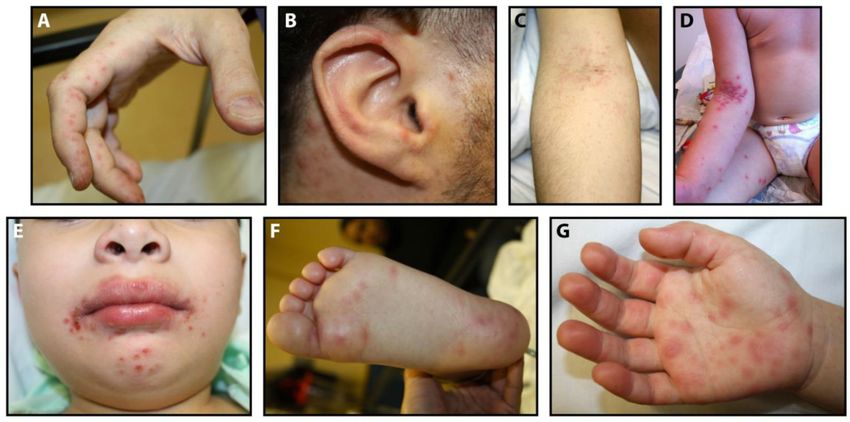
Figures 1.
Cutaneous lesions of atypical HFMD. Erythematous papulovesicles involving the fingers of
Patient 1 (A). Erythematous papules involving the ear and antecubital fossa of Patient 2 (B,
C). Significant involvement of the antecubital fossa, a common site of involvement of AD in
Patient 3, in addition to scattered papules on the forearm and thigh (D). Papulovesicles
involving the perioral area and lips (E), sole (F), and palm (G) in Patient 4.
Author Manuscript
Author Manuscript
J Am Acad Dermatol. Author manuscript; available in PMC 2018 March 08.Author Manuscript Author Manuscript Author Manuscript Author Manuscript
Table 1
Patient demographics, clinical presentations and laboratory results. RT-PCR, reverse transcriptase-polymerase chain reaction.
Patient Age Enterovirus Body Sites Involved Clinical History of Presented to the
Lott et al.
(years) Detection by Features Atopic Emergency
RT-PCR Dermatitis Department
1 43 *Plasma + • Oropharynx • Fever No Yes, subsequently hospitalized
• Left forearm • Chills
• Dorsal hands, palms, fingers • Night sweats
(including subungual thumb)
• Headache
• Toe
• Odynophagia
• Myalgias
2 21 Stool + • Oropharynx, lips • Fever No Yes (for fever), subsequently
hospitalized
Plasma + • Ears • Chills
*Fluid from a • Antecubital fossae • Odynophagia
cutaneous • Dorsal hands, palms, fingers
vesicle +
• Thighs, shins
• Dorsal feet, soles
3 2 *Stool + • Lips • Fever Yes Yes
• Peri-axillary chest, antecubital • Onychomadesis (subsequently)
fossae (AD site), forearms
• Dorsal hands, palms
• Gluteal cleft
• Thighs, popliteal fossae (AD site),
shins
J Am Acad Dermatol. Author manuscript; available in PMC 2018 March 08.
• Soles
4 4 Stool + • Oropharynx, lips • Fever No Yes
*Fluid from a • Ears • Diarrhea
cutaneous • Flexor wrists
vesicle +
• Palms
• Back
• Buttocks
Page 9Author Manuscript Author Manuscript Author Manuscript Author Manuscript
Patient Age Enterovirus Body Sites Involved Clinical History of Presented to the
(years) Detection by Features Atopic Emergency
RT-PCR Dermatitis Department
• Flexor aspect of the lower
extremities
Lott et al.
• Soles
5 4 Stool + • Oropharynx, tongue • Fever Yes Yes
*Oral + • Perioral face, cheeks
• Upper extremities
• Dorsal hands and fingers (AD
sites)
• Back, buttocks, mons pubis
• Lower extremities
• Dorsal feet
*
Virus in sample sequenced as CV-A6 at the Centers for Disease Control and Prevention (CDC).
J Am Acad Dermatol. Author manuscript; available in PMC 2018 March 08.
Page 10Lott et al. Page 11
Table 2
Laboratory evaluation of suspected atypical hand-foot-mouth disease (HFMD). CDC, Centers for Disease
Author Manuscript
Control and Prevention; DFA, direct fluorescent antibody assay; HSV, herpes simplex virus; PCR, polymerase
chain reaction; RT, reverse transcriptase; VZV, varicella-zoster virus.
Atypical HFMD should be considered when either papulovesicles extend beyond the typical distribution pattern, lesions favor sites of atopic
dermatitis as in eczema herpeticum, the disease presents in the winter, or adults are affected. In such cases, the following diagnostic laboratory
evaluation is recommended:
• DFA or PCR of keratinocytes from the floor of an intact vesicle to exclude HSV or VZV infection
• RT-PCR of vesicle fluid* to confirm enterovirus infection. If vesicle fluid is unavailable, then RT-PCR of oropharyngeal or stool
specimen*. In the latter case, a stool specimen rather than a swab of the perianal area should be obtained.
• If an epidemic is suspected, contact a specialized research or public health laboratory such as the CDC for additional testing, i.e.
viral capsid protein (VP) 1 gene sequencing.
Differential diagnosis
• Eczema herpeticum, varicella, disseminated zoster, erythema multiforme major
Author Manuscript
*
Culture and serology are not recommended (see text)
Author Manuscript
Author Manuscript
J Am Acad Dermatol. Author manuscript; available in PMC 2018 March 08.You can also read