Multiple endo bronchial lipoma: a rare case report - BMC ...
←
→
Page content transcription
If your browser does not render page correctly, please read the page content below
Zhao et al. BMC Pulmonary Medicine      (2020) 20:251
https://doi.org/10.1186/s12890-020-01287-4

 CASE REPORT                                                                                                                                        Open Access

Multiple endo bronchial lipoma: a rare case
report
Shunjin Zhao* , Yuexiang Shui and Zhong Dai

  Abstract
  Background: Endobronchial lipoma is an extremely rare benign tumor, which is generally located in the first three
  subdivisions of the tracheobronchial tree. According to the existing literature, all endobronchial lipomas are single
  (one per patient). Here, we report a rare case in which the patient presented with two endobronchial lipomas in
  the same patient, and underwent a bronchoscopic tumor resection in the left main bronchus and the left lower
  bronchus. Both tumors were pathologically confirmed as endobronchial lipoma.
  Case presentation: A 52-year-old Chinese man presented at the clinic reporting a mild cough with yellow color
  sputum and exertional dyspnea for 2 weeks. He was a heavy smoker (45 pack-years). Chest auscultation
  demonstrated faint wheezing in left lower lobe. Computed tomography (CT) revealed two low-density
  endobronchial masses located in the middle segment of the left main bronchus and the posterior basilar
  segmental bronchus of the left lower lobe. The neoplasms measured a CT-attenuation value of -70HU, −98HU in
  density with air trapping and atelectasis in the segmental bronchus of the left lower lobe. The patient underwent
  interventional bronchoscopic management to remove the neoplasms by using an electrosurgical snare,
  cryotherapy, and electrocautery. The locations of the neoplasms were confirmed at the left main bronchus and the
  superior segment of the left lower lobe during bronchoscopic intervention. Histopathological examination
  confirmed that both tissues were consistent with lipomas. After 18 months of follow-up, the patient was free of
  symptoms and CT revealed that bronchiectasia remained in the superior segment of the left lower lobe; however,
  no mass lesion was present in the left bronchus.
  Conclusions: This case suggests that an endobronchial lipoma can present as multiple lesions, and both proximal
  and distal types can simultaneously occur in the same patient. Thus, these findings help us further understand the
  biology of endobronchial lipomas.
  Keywords: Bronchoscopy, Case report, Lipoma, Endobronchial lipoma

Background                                                                             damage if undiagnosed early. Endobronchial lipomas are
Endobronchial lipoma is an extremely rare benign                                       more common in men and in the right lung [3]. Histo-
tumor, comprising approximately 0.1–0.5% of all bron-                                  logically, endobronchial lipomas have been shown to
chial tumors [1]. In most cases, tumors are located in                                 contain numerous uniform adipocytes [4]. According to
the first three subdivisions of the tracheobronchial tree                              the existing literature, all endobronchial lipomas are sin-
[2]. When they become large enough, these tumors can                                   gle (one per patient). Here, we report a rare case in
lead to endobronchial obstruction, thereby causing atel-                               which a patient presented with two lesions, and under-
ectasis and recurrent pneumonia, even irreversible lung                                went bronchoscopic tumor resection in the left main
                                                                                       bronchus and the left lower bronchus. Both tumors were
* Correspondence: 20969971@qq.com                                                      pathologically confirmed as endobronchial lipoma.
Department of Respiratory Medicine, Lanxi People’s Hospital, No. 1359,
Xishan Road, Lanxi, Jinhua 321100, Zhejiang Province, China

                                        © The Author(s). 2020 Open Access This article is licensed under a Creative Commons Attribution 4.0 International License,
                                        which permits use, sharing, adaptation, distribution and reproduction in any medium or format, as long as you give
                                        appropriate credit to the original author(s) and the source, provide a link to the Creative Commons licence, and indicate if
                                        changes were made. The images or other third party material in this article are included in the article's Creative Commons
                                        licence, unless indicated otherwise in a credit line to the material. If material is not included in the article's Creative Commons
                                        licence and your intended use is not permitted by statutory regulation or exceeds the permitted use, you will need to obtain
                                        permission directly from the copyright holder. To view a copy of this licence, visit http://creativecommons.org/licenses/by/4.0/.
                                        The Creative Commons Public Domain Dedication waiver (http://creativecommons.org/publicdomain/zero/1.0/) applies to the
                                        data made available in this article, unless otherwise stated in a credit line to the data.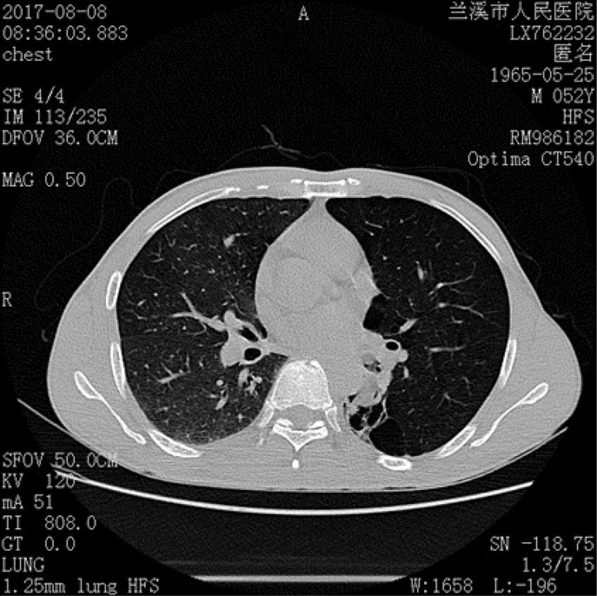
Zhao et al. BMC Pulmonary Medicine (2020) 20:251 Page 2 of 7
Case presentation
A 52-year-old Chinese man presented at the clinic
reporting a mild cough with yellow color sputum and
exertional dyspnea for 2 weeks. He denied any fever,
chest pain, night sweats, or weight loss. He was a heavy
smoker (45 pack-years) and had a history of a splenec-
tomy for abdominal injury 23 years ago. After a failed 3
day-course of antibiotic treatment at the clinic, he was
referred to our hospital for further examination and
treatment. Upon physical examination, the body mass
index (BMI) was 17.97, SpO2 was 98% when breathing
ambient air, the respiratory rate was 20/min, and the
blood pressure was 119/77 mmHg. Chest auscultation
demonstrated faint wheezing in left lower lobe. A com-
puted tomography (CT) scan was performed, which re-
vealed two low-density endobronchial masses located in
the middle segment of the left main bronchus and the
posterior basilar segmental bronchus of the left lower
lobe. The neoplasms measured the CT-attenuation value
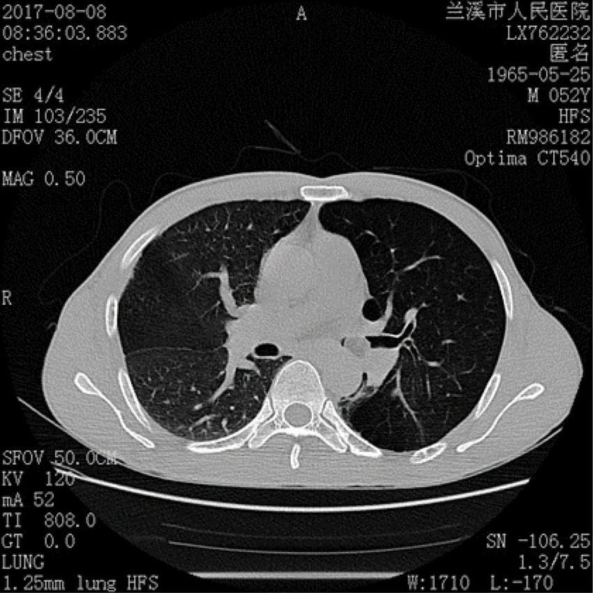
-70HU, −98HU (Figs. 1 and 2) in density with air trap- Fig. 2 CT suggesting low-density mass obstructing the left lower
lobe, the CT value is -98HU
ping and atelectasis in the segmental bronchus of the left
lower lobe. The complete blood count showed that the
white blood cell count increased to 16 * 10^9/L, the Subsequently, the patient underwent a flexible bronchos-
platelet count increased to 549 * 10^9/L, whereas other copy, which confirmed an exophytic spherical lesion that
blood cell counts were within the normal range. No ab- caused almost complete occlusion of the middle of the
normalities were observed in liver function, renal func- left main bronchus (Fig. 3), however, the cytological
tion, blood lipids, blood glucose, C-reactive protein, and diagnosis failed by using bronchoscopic brushing cells.
erythrocyte sedimentation rate. The tumor markers In view of the above, interventional bronchoscopic
alpha fetoprotein (AFP), carcinoembryonic antigen management was undertaken to remove the neoplasms.
(CEA), carbohydrate antigen 125 (CA125) and carbohy- First, we therapeutically resected the neoplasm, of which
drate antigen 19–9 (CA19–9) were in the normal range, the base was located in the lateral wall of the left main
and no acid-fast bacilli were found in sputum smears.
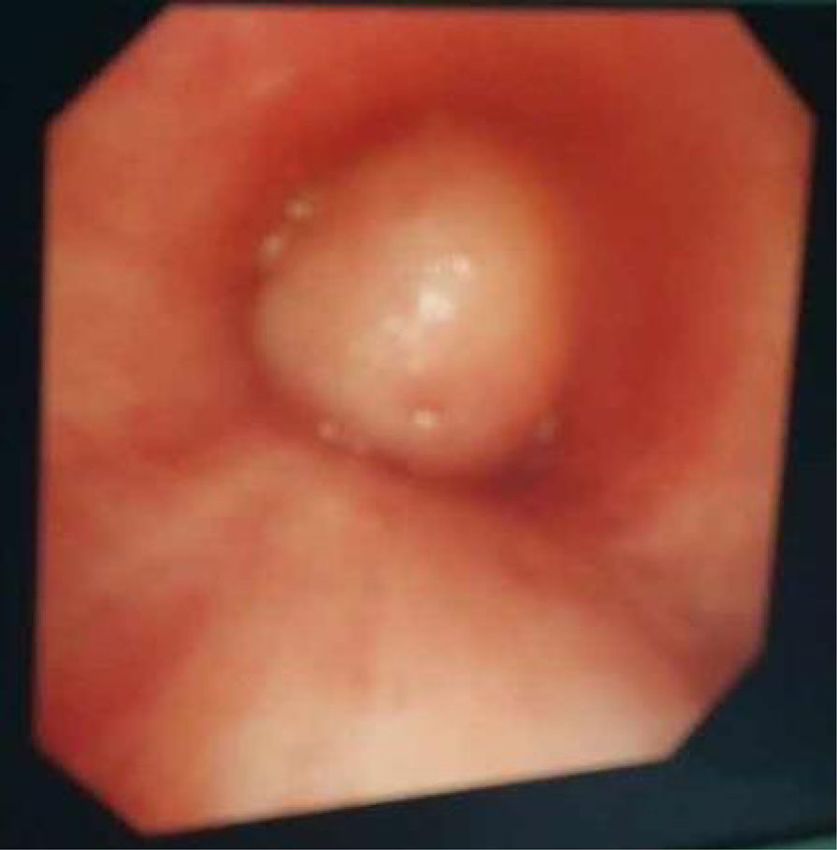
Fig. 1 CT showing a low-density mass obstructing the left main Fig. 3 A spherical neoplasm growing in the left main bronchus with
bronchus, the CT value is -70HU a smooth, soft, and red appearance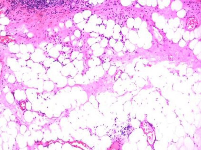
Zhao et al. BMC Pulmonary Medicine (2020) 20:251 Page 3 of 7
bronchus using electrosurgical snare under flexible
bronchoscope (Fig. 3). Then, the spherical and pink neo-
plasms were taken out by cryotherapy (Fig. 4). Subse-
quently, another yellowish dumb-bell neoplasm was
found in the superior segment of the left lower lobe
(Fig. 5) and the main part of the neoplasm was removed
(Figs. 6 and 7) by electrosurgical snare, electrocautery,
and cryotherapy. Only the C branch of the superior seg-
ment of the left lower lobe could not be completely
resected or ablated (Fig. 8) because the neoplasm origi-
nated distally. Histopathological examination confirmed
that both tissues from the left main bronchus and the
superior segment of the left lower lobe were consistent
with lipoma (Figs. 9 and 10).
After bronchoscopic intervention, the patient recov-
ered and was discharged. After 18 months of follow-up,
the patient was free of symptoms and CT revealed that
bronchiectasia remained in the superior segment of the
left lower lobe (Figs. 11 and 12), however, no mass lesion
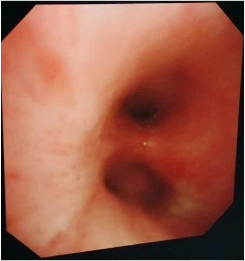
was present in the left main bronchus. Fig. 5 The left lower lobe bronchus is blocked by a neoplasm. Its
surface is smooth, soft, has a light-yellow appearance, and is
dumbbell shaped
Discussion and conclusion
Endobronchial lipoma is a type of rare benign tumor,
which originates from fat cells located in peribronchial lipomas with a clear pathological diagnosis that pre-
and submucosal tissues of the bronchi [5]. Although lip- sented as a single lesion. The case reported here was
omas in connective tissue, such as subcutaneous tissue, unique in that two endobronchial lipomas were present
can present as multiple lesions, lipomas in the bronchi in the same patient. According to the location, endo-
are almost always present as a single lesion. The authors bronchial lipomas are divided into two types [3]: 1. Prox-
used ‘lipoma’ and ‘bronchus’ as keywords to search the imal type: often occurs in the large bronchi, and is rich
PubMed Database, thereby retrieving all endobronchial in normal adipose tissue, including trachea, carina, main
bronchus, and bronchus intermedius [6]. 2. Distal type:
Fig. 4 Neoplasm located in left main bronchus, about 2.0 × 1.3 × 0.9
cm in size, with an oval polypoid shape Fig. 6 Image of left main bronchial tumor after resection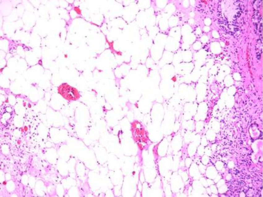
Zhao et al. BMC Pulmonary Medicine        (2020) 20:251                                                                           Page 4 of 7

 Fig. 7 Five pieces of neoplasia tissues that have been removed from    Fig. 9 Histology of the neoplasm at low magnification
 the left main bronchus and the lower left bronchus                     demonstrating the pseudo-stratified ciliated columnar epithelium
                                                                        overlying the mature adipocytes and little fibrous tissue. The
                                                                        neoplasm in the left main bronchus is consistent with lipoma.
located below the segmental bronchus, and originates                    Hematoxylin and eosin (HE) staining
from the surrounding bronchial wall adipose tissue, in-
cluding segmental and subsegmental [7]. The case pre-                    For most cases, lipomatous neoplasms arise in the set-
sented here was unique in that two endobronchial                       ting of lipomatosis and rarely involve the tracheobron-
lipomas were present in one patient, and were located in               chial tree [1, 5]. Endobronchial lipomas arise from
the left main bronchus, and the superior segment of the                submucosal fat in airways that contain cartilage and
left lower lobe, respectively. These findings suggested                bronchial glands, and are usually covered by normal re-
that an endobronchial lipoma can also present as mul-                  spiratory epithelium [8]. Most patients are men (80%)
tiple lesions, and both proximal and distal types can sim-             with a mean age of 60 years, and a significant smoking
ultaneously occur in the same patient. It is necessary to              history [2]. These benign tumors typically occur in the
compare the characteristics of this endobronchial lipoma               large bronchi (first 3 subdivisions), and may lead to ob-
case with previous lipomas because of its uniqueness.                  structive complications [9]. The patient suffering from
                                                                       endobronchial lipomas was a 52-year-old man who was
                                                                       a heavy smoker, which was consistent with the risk fac-
                                                                       tors of lipomas for age, sex, and smoking history [9].
                                                                       However, the patient’s BMI was only 17.96, which was

                                                                        Fig. 10 Histology of the neoplasm at low magnification
                                                                        demonstrating the pseudo-stratified ciliated columnar epithelium
                                                                        overlying mature adipocytes. The neoplasm in the left lower lobe is
 Fig. 8 Image of the left lower lobe bronchial after tumor resection    consistent with lipoma. HE staining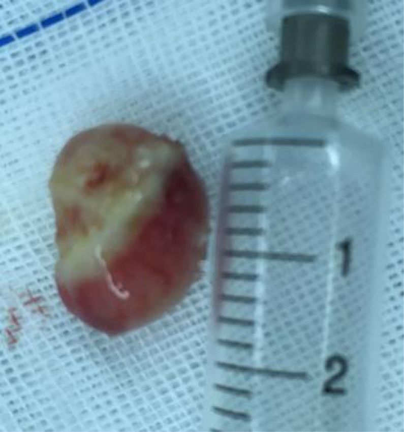
Zhao et al. BMC Pulmonary Medicine       (2020) 20:251                                                                 Page 5 of 7

                                                                       the lumen, it can lead to cough, sputum, fever, dyspnea,
                                                                       and other symptoms [11]. Nassiri et al. reported that
                                                                       atelectasis was present in 30% of cases, and that the ma-
                                                                       jority of patients was symptomatic (63.2%) [12]. How-
                                                                       ever, the patient in this case did not have clinical
                                                                       symptoms in the early stage until he developed post-
                                                                       obstructive pneumonia. Patients are often misdiagnosed
                                                                       as having asthma or chronic bronchitis and endobron-
                                                                       chial lipomas can remain undetected for months or years
                                                                       [13]. Thus, to shorten the diagnostic time of endobron-
                                                                       chial lipomas, CT and bronchoscopy should be per-
                                                                       formed early when a patient is actively undergoing
                                                                       unexplained cough, dyspnea, fever, or other symptoms.
                                                                          At present, a chest CT and bronchoscopy are main ap-
                                                                       proaches to diagnose endobronchial lipoma as the sensi-
                                                                       tivity of traditional X-ray examination is lower (66%)
                                                                       [14, 15]. The diagnosis can be suggested with fat attenu-
                                                                       ation on a chest CT and the lack of enhancement after
                                                                       contrast administration [16, 17], because the density of
 Fig. 11 CT revealing that the left main bronchus and the left lower   the lipoma is similar to that of normal adipose tissue,
 lobe were unobstructed, and that the left lower lobe                  and the CT-attenuation value is between − 120 Hu and
 showed bronchiectasis
                                                                       − 40 Hu. It is helpful to distinguish lipoma from other
                                                                       tumors by accurately measuring the CT-attenuation
not consistent with previous findings that obesity was a               value of the tumor. In our case, we measured the CT-
risk factor for endobronchial lipomas [8].                             attenuation value of the neoplasms, which was within
  Endobronchial lipomas grow slowly and have different                 the CT-attenuation value range of the lipoma, and
clinical manifestations [10]. The clinical symptoms are                helped us make the diagnosis of endobronchial lipoma.
related to the location of the tumor and the severity of               In addition, a chest CT can accurately display the shape,
endoluminal obstruction [11]. Symptoms can remain                      size, location, degree of lumen stenosis, the relationship
clinically silent for a prolonged period when the tumor                with the bronchial wall, and indirect obstructive signs of
is small. As the tumor grows and significantly obstructs               a tracheobronchial lipoma [8]. However, it is a challenge
                                                                       to distinguish endobronchial lipoma from bronchial
                                                                       hamartoma by CT, because a bronchial hamartoma is
                                                                       predominantly composed of adipose tissue. Therefore, a
                                                                       bronchoscopy is another important tool for the diagnosis
                                                                       of endobronchial lipoma, can visually show the charac-
                                                                       teristics of lipoma and biopsy, and can accurately deter-
                                                                       mine the tumor’s location, shape, and degree of blockage
                                                                       of the tumors. In previous reports, it has been suggested
                                                                       that the diagnostic value of a bronchoscopy is very low
                                                                       because of the solid cystic tissue covering the surface of
                                                                       the bronchial lipoma [15]. Only one third of patients can
                                                                       be diagnosed by bronchoscopic biopsy [2]. However, in
                                                                       some studies it was reported that using a cryoprobe,
                                                                       large pieces of the tumor can be extracted, which can
                                                                       help overcome the limitation of a low diagnostic yield
                                                                       [18]. In addition, surgical excision can be performed to
                                                                       obtain pathological diagnosis when neither CT nor
                                                                       bronchoscopy is confirmed. In this case, the chest CT
                                                                       had accurately judged the quantity, size and CT-
                                                                       attenuation value of the tumors, but made an error in
 Fig. 12 CT revealing that the left main bronchus and the left lower   judging the location (CT indicated one of the lesions
 lobe were unobstructed, and that the left lower lobe
                                                                       was in the posterior basilar segment of left lower lobe,
 showed bronchiectasis
                                                                       however the bronchoscopic resection confirmed that the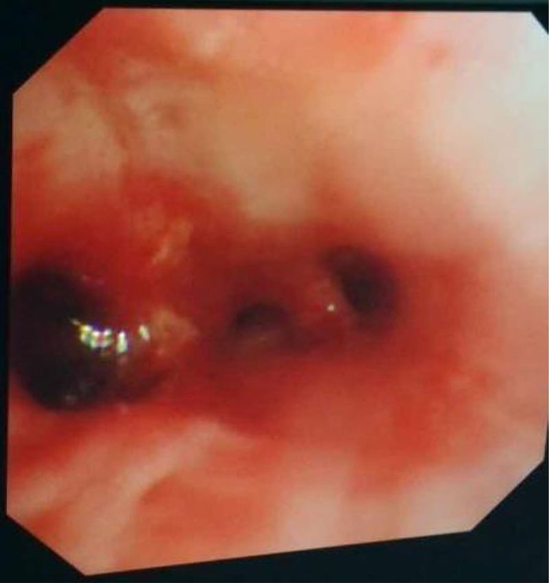
Zhao et al. BMC Pulmonary Medicine   (2020) 20:251                                                                                   Page 6 of 7

location was in the superior segment of the left lower         tumor in the left lower lobe, because it did not cause
lobe). These findings display that CT is a reliable            serious damage in the distal bronchus and the lung. It
method for the diagnosis of endobronchial lipoma even          was a slowly growing tumor without excess risk of ma-
if there are minor faulty judgements. In this case, we         lignant potential [2]. During a prolonged follow-up (18
failed in diagnosing the tumors by bronchoscopic brush-        months), no recurrence and new obstruction had oc-
ing cytology, however, bronchoscopy was very important         curred, and no aggravation of distal bronchiectasis and
to differentiating between benign and malignant tumors         lung damage was observed. This demonstrated that
since the shape, activity, surface condition, and obstruc-     endobronchial resection was effective and safe and pre-
tion degree of the tumors could clearly be observed            served lung tissue in this subgroup of benign pulmonary
under the bronchoscope, which provided a basis for the         tumors [12].
formulation of treatment.                                         In summary, in this case we reported multiple endo-
   Endoscopic features of tracheobronchial lipomas have        bronchial lipomas that coexisted proximal and distal, in-
been classified as follows. These tumors always appear         dicating a novel discovery of endobronchial lipoma, as
with a smooth surface and are oval-shaped, only a few          all cases previously reported were single. We demon-
could be lobulated, and they are poorly vascularized with      strated that the clinical features, radiological features,
a yellow to rose appearance. Furthermore, the mobility         endoscopic features, and histopathological features sup-
of the tumors is in general mobile, it is rarely to see        ported previous observations. Taken together, this case
fixed one. Finally, the proportions of proximal and distal     helps us further understand the biology of endobronchial
types are similar when it comes to the location of the tu-     lipomas.
mors [8, 12]. The two endobronchial lipomas in our case
                                                               Acknowledgements
were different in shape, color, and mobility, thereby sug-     We gratefully thank Dr. Saibin Wang (Department of Pulmonary Medicine,
gesting that the characteristics of multiple endobronchial     Jinhua Municipal Central Hospital, China) for his constructive guidance.
lipomas can be diverse, and more attention should be
                                                               Authors’ contributions
paid on it to avoid misdiagnosis.                              SZ was responsible for the literature review, design, and writing of the
   The treatment of endobronchial lipomas includes two         manuscript. YS and ZD participated in the diagnosis and treatment, and
methods: bronchoscopic resection and surgery [19].             revised the manuscript. All authors read and approved the final manuscript.
With the development of bronchoscopic interventional
                                                               Funding
technology, bronchoscopic interventional therapy for be-       Not applicable.
nign central airway stenosis has been widely used [2, 4,
12, 15]. When compared with surgery, bronchoscopic             Availability of data and materials
                                                               The data are available from the corresponding author on reasonable request.
resection can completely relieve the symptoms of pa-
tients, has a low risk, fewer complications, better patient    Ethics approval and consent to participate
tolerance, and would preserve lung tissue and lung func-       Written informed consent was obtained from the patient.
tion [2, 20]. Most cases of proximal endobronchial lip-        Consent for publication
oma can be removed by bronchoscopy. There is                   Written informed consent was obtained for the publication of this case
variability in strategy, including both rigid/flexible bron-   report and the accompanying images from the relative of the patient. A
                                                               copy of the written consent is available for review by the Editor-in-Chief of
choscopy, and use of electrocautery, cryotherapy, argon        this journal.
plasma coagulation, laser, and/or mechanical debulking
[4]. A rigid bronchoscope is preferred over a flexible         Competing interests
                                                               The authors declare that they have no competing interests.
bronchoscope because of the wider internal diameter
and the higher airway safety [14]. Surgery is another          Received: 12 March 2020 Accepted: 14 September 2020
method of treatment, and the most effective method,
however, surgery has a greater risk for more complica-
                                                               References
tions, has a higher cost, and requires a patient to be fit     1. Huisman C, van Kralingen KW, Postmus PE, et al. Endobronchial lipoma:a
enough to undergo surgical resection. However, surgical            series of three cases and the role of electrocautery. Respiration. 2000;67(6):
procedures should be reserved for patients with a pos-             689–92.
                                                               2. Muraoka M, Oka T, Akamine S, et al. Endobronehial lipoma:review of 64
sible coexistent malignant tumor, severe irreversible              cases reported in Japan. Chest. 2003;123:293–6.
damage of the distal bronchus and lung tissue, distal          3. Moran AM, Jian B, Min H, et al. Peripheral intrapulmonary lipoma in a 26-
type tumors growing around the bronchus, or technical              year-old woman-a case report. P0l J Pathol. 2011;62(2):113–5.
                                                               4. Ryan G, Elamin E. Two cases of endobronchial lipoma. Chest. 2017;152(4):
difficulties during bronchoscopic procedure, such as               A726.
multidirectional tumor growth [19, 21]. In this case, we       5. Sivapalan P, Gottlieb M, Christensen M, Clementsen PF. An obstructing
removed both the proximal type and distal type lipoma              endobronchial lipoma simulating COPD. Eur Clin Respir J. 2014;1:25664.
                                                                   https://doi.org/10.3402/ecrj.v1.25664.
using a flexible bronchoscope without any complica-            6. Eren F, Candan T, Eren B, et al. Endobronchial lipoma. JPak Med Assoc.
tions. Surgery had not been performed for the rest of              2013;63(6):784–5.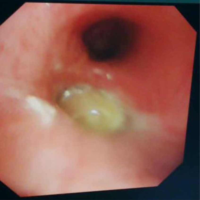
Zhao et al. BMC Pulmonary Medicine               (2020) 20:251                        Page 7 of 7

7.    Kim NR, Kim HJ, Kim JK, et al. Intrapulmonary lipomas:report of four cases.
      Histopathology. 2003;42(3):305–6.
8.    Yang YM, Pu C, Li Y, Ke HX, Xu XM, Fang BM. Endobronchial lipoma: report
      of 2 cases and review of the Chinese literature. Zhonghua Jie He He Hu Xi
      Za Zhi. 2012;35(3):176–9.
9.    Boland JM, Fritchie KJ, Erickson-Johnson MR, Oliveira AM, Colby TV, Folpe
      AL. Endobronchial lipomatous tumors: clinicopathologic analysis of 12 cases
      with molecular cytogenetic evidence supporting classification as "lipoma".
      Am J Surg Pathol. 2013;37(11):1715–21.
10.   Yun SC, Na MJ, Choi E, Kwon SJ, Lee SJ, Oh SH, Cha EJ, Son JW. Successful
      removal of endobronchial lipoma by flexible bronchoscopy using
      electrosurgical snare. Tuberc Respir Dis. 2013;74(2):82–5.
11.   Wang H, Du Z, Li A, Song J. Surgical treatment of an endobronchial lipoma
      obstructing the right upper bronchus: Imaging features with pathological
      correlation. Pak J Med Sci. 2013;29(6):1447–9.
12.   Nassiri AH, Dutau H, Breen D, Colchen A, Quiot JJ, Nguyen B, Vergnon JM. A
      multicenter retrospective study investigating the role of interventional
      bronchoscopic techniques in the management of endobronchial lipomas.
      Respiration. 2008;75(1):79–84.
13.   Irani F, Kumar B, Reddy P, Narwal-Chadha R, Kasmani R, Tita J. An
      endobronchial lipoma mimicking asthma and malignancy. Prim Care Respir
      J. 2010;19:281–3.
14.   Alazemi S, Majid A, Ruiz AI, et al. An elderly woman with chronic dyspnea
      and endobronchial lesion. Chest. 2010;137(2):460–6.
15.   Madan K, Agarwal R, Bal A, et al. Bronchoscopic management of a rare
      benign endobronchial tumor. Rev Port Pneumol. 2012;18(5):251–4.
16.   On R, Kushima H, Ishii H, Watanabe K. Endobronchial Lipoma: The
      Diagnostic Benefit of Computed Tomography Findings. Intern Med. 2018;
      57(2):285–6.
17.   Kassem H, Dhillon S, Huang M, Kumar A, Qiu J. Endobronchial lipoma:
      bronchoscopy, imaging and pathology. Ther Adv Respir Dis. 2014;8(5):162–
      4.
18.   Lamprecht B, Hutarew G, Porsch P, Wegleitner B, Studnicka M. Successful
      bronchoscopic cryorecanalization in a case of endobronchial lipoma. Diagn
      Ther Endosc. 2011;2011:845686.
19.   Dy RV, Patel S, Harris K, Mador MJ. Endobronchial lipoma causing
      progressive dyspnea. Respir Med Case Rep. 2017;22:95–7.
20.   Liew CJ, Tham KY, Poh AC, Tee A. Endobronchial lipoma. Singap Med J.
      2017;58(8):510–1.
21.   Carlos Galvez ,Julio Sesma, Sergio Bolufer, Francisco Lirio, Jose Navarro-
      Martinez, Maria Galiana, Benno Baschwitz, Maria Jesus Rivera. Single-incision
      video-assisted anatomical segmentectomy with handsewn bronchial closure
      for endobronchial lipoma. Ann Transl Med, 2016,4(15):284.

Publisher’s Note
Springer Nature remains neutral with regard to jurisdictional claims in
published maps and institutional affiliations.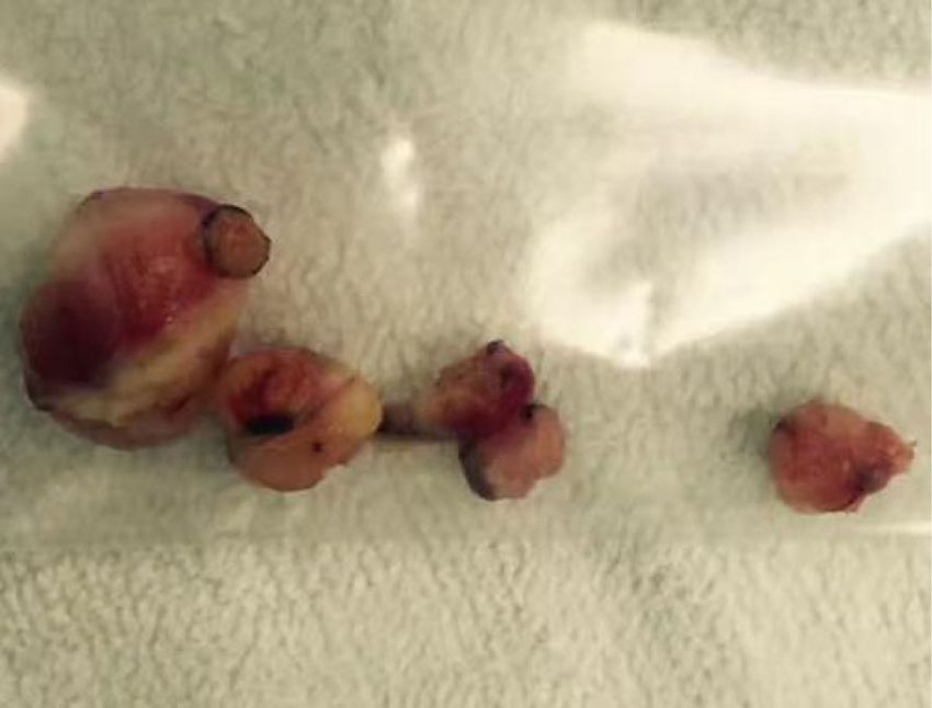
You can also read


