Disseminated herpes simplex virus: a case of eczema herpeticum causing viral encephalitis - Royal College of Physicians of ...
←
→
Page content transcription
If your browser does not render page correctly, please read the page content below
J R Coll Physicians Edinb 2018; 48: 36–9 | doi: 10.4997/JRCPE.2018.108 CASE OF THE QUARTER
Clinical
Disseminated herpes simplex virus: a case of
eczema herpeticum causing viral encephalitis
C Finlow1, J Thomas2
Eczema herpeticum is a dermatological emergency causing a mortality Correspondence to:
of up to 10% if untreated. It frequently presents in a localised form and C Finlow
Noble’s Hospital
Abstract rarely disseminates via haematogenous spread with pulmonary, hepatic,
ocular and neurological manifestations. Although it commonly appears on a Strang
background of atopic dermatitis, many other dermatological conditions have Douglas IM4 4RJ
been described preceding this disease. Eczema herpeticum can be easily Isle of Man
mistaken for folliculitis and is often treated accordingly with antibacterial drugs; therefore
patients will often deteriorate before a diagnosis of eczema herpeticum has been considered. Email:
c.finlow@gmail.com
Keywords: eczema herpeticum, herpes simplex, Kaposi’s vericelliform eruption, rash, toxic
confusional state, viral encephelitis
Patient consent: obtained
Declaration of interests: No conflict of interests declared
Background top of the chest and back and eventually to all four limbs. In
places the rash produced serous and yellow fluids.
Eczema herpeticum (EH) was initially described by Moriz
Kaposi in 1887 and is also known as Kaposi varicelliform On admission the patient was being treated with oral
eruption.1 It can be a dermatological emergency manifesting fl ucloxacillin and amoxicillin for folliculitis, after initial
as a generalised vesicular eruption in a toxic patient with presentation in the community. After being admitted this
high morbidity and mortality. It is often associated with a pre- was changed to intravenous fl ucloxacillin, intravenous
existing eczema diagnosis and for this reason it has a higher benzylpenicillin and topical fusidic acid for presumed
incidence rate in children; however, it is also common in the folliculitis unresponsive to oral antibiotics. The patient also
second and third decades of life.2–4 It affects all ages with an complained of a stiff neck, dry ‘gritty’ eyes, nausea, fever
equal incidence in males and females.5 Some predisposing and a headache. The headache was initially a stabbing pain
factors associated between atopic eczema and EH include in the occipital region and progressed to be a holocephalic,
an earlier onset of atopic eczema, a low level of natural constant ache. There was no photophobia or vomiting,
killer cells, high level of immunoglobulin E and Malassezia no change in pain with position or time of day. Due to
sympodialis antibodies.5 the ocular symptoms, topical chloramphenicol was also
commenced.
Prompt treatment with antiviral medication is needed due
Examination on admission
to dissemination internally. If this occurs, and depending on
the site of infection, the patient may present with pulmonary, The patient looked generally unwell, but was alert and
hepatic, ocular and neurological symptoms. With the process orientated with a GCS of 15/15. Temperature was 38°C,
of dissemination comes viraemia and scepticaemia.1 respiratory rate 15 breaths/min, and oxygen saturation was
100% on room air. His chest was clear to auscultation with
Case presentation no changes on percussion. Heart rate was 67 bpm, blood
pressure 100/65 mmHg, and heart sounds were normal.
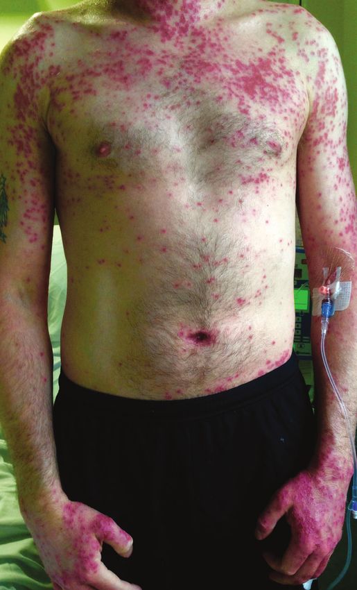
A 36-year-old male presented to accident and emergency There were eruptions of vesico-pustules on a pink base, with
with a 4-day history of rash, fever, headaches and generally some punched out lesions that were well demarcated and
feeling unwell. The rash was painful, with areas of blisters approximately 0.5 cm in diameter. The rash was widespread
and punched out erythematous lesions approximately 0.5 cm on the face, arms, trunk, back and backs of upper leg (Figures
in diameter, which were non-blanching in nature. The lesions 1–3). The patient had a normal range of movement of the
started on the left side of the neck and progressed over the neck in all directions.
mandible area onto the face, the right side of the neck, the
FY2, 2Consultant Geriatrician, Noble’s Hospital, Isle of Man
1
36 JOURNAL OF THE ROYAL COLLEGE OF PHYSICIANS OF EDINBURGH VOLUME 48 ISSUE 1 MARCH 2018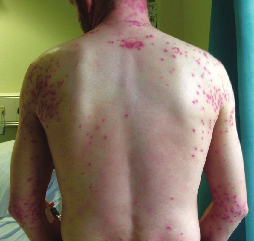
Eczema herpeticum causing viral encephalitis

Figures 1–3 A 36-year-old male with widespread eczema herpeticum.

Vesicles were present on the right eyelid. Cranial nerve            A differential diagnosis of EH was considered when, on
examination was normal. There was mild reduction in                 further history taking, it was noted that the patient’s wife
power in all four limbs but sensation, tone and coordination        had suffered from oral herpes 2 weeks prior to the patient’s
were normal. His abdomen was soft, non-tender, with no              admission. Dermatology input on the same night confirmed
organomegaly and no masses.                                         the diagnosis of EH and the patient was advised to continue
                                                                    antiviral treatment: intravenous aciclovir 10 mg/kg, every 8
Past medical history
                                                                    h for 10 days to 2 weeks. The patient started to clinically
The patient had been diagnosed with eczema in 2002; it was          improve within 24 h of treatment.
usually well controlled with regular emollients (Diprobase)
and occasional Eumovate for any flares. He had previously            On day 3 of admission the patient presented feeling ‘spaced
been treated with tacrolimus 0.1% ointment between 2005             out’ and at times confused. He described blurring of his
and 2010 and Oilatum cream and Plus Bath between 2002               vision that had started in the past 12 h. His headache and
and 2008. He had received no blood transfusions, no history         neck stiffness had been persistent since admission with no
of intravenous drug use and no known drug allergies. He             improvement. The patient felt feverish; his temperature at this
was a non-smoker, and consumed 24 units of alcohol at               time was 37°C while on regular paracetamol. Neurological
the weekend.                                                        examination showed a decreased GCS of 14/15 (E4 V4 M6),
                                                                    diplopia and decreased visual acuity. All other cranial nerves
Clinical progress                                                   remained intact and gross ocular examination still showed
                                                                    the vesicles on the right eyelid, which had been present on
After admission, a lumbar puncture was undertaken due to            admission. Upper and lower motor examination showed no
the possibility of meningitis. This was considered because of       changes from admission. Neck examination showed slightly
the headache, stiff neck and rash. Ceftriaxone and aciclovir        decreased range of movements in both flexion and lateral
were administered to cover for bacterial and viral infection        flexion. A clinical diagnosis of viral encephalitis secondary
until the cerebrospinal fluid (CSF) results were returned.           to the EH was made.

                                      MARCH 2018 VOLUME 48 ISSUE 1 JOURNAL OF THE ROYAL COLLEGE OF PHYSICIANS OF EDINBURGH     37
C Finlow, J Thomas

Table 1 Blood test results
                                                                     Computed tomography of the head showed no changes in the
                             On admission           Day 3            parenchyma, ventricles and skull. There was no evidence of
 WCC                            10.59                8.46            space occupying lesions or acute haemorrhage.
 Neutrophils                     9.14                7.02
 Haemoglobin                     146                 132
                                                                     Treatment
 MCV                             92.4                91.5
                                                                     While in hospital, this patient received intravenous 10 mg/kg
 Platelets                       212                 192             aciclovir 8 hourly for 14 days, 4 mg chlorphenamine 4 times
 Sodium                          134                 133             a day to aid pruritus, a 5 day tapering course of prednisolone
 Potassium                        3.7                 3.8            starting at 30 mg, 3.75 mg zopiclone once daily in the
 Creatinine                       82                  72             evening to support sleep, 300 mg gabapentin twice daily to
                                                                     settle the ulnar nerve involvement and 1 g paracetamol 4
 Urea                             2.9                 3.1
                                                                     times a day to aid with pain. Long term medications prior to
 Bilirubin                        20                  15             admissions were continued. These included Balneum Plus
 AST                              26                  23             oil used once a morning in the bath, Diprobase applied twice
 ALT                              24                  21             per day and Eumovate used in acute flare-ups of eczema.
 ALP                              73                  64
 GGT                              36                  33             Medication that had been discontinued during inpatient
                                                                     admission included chloramphenicol 4 times a day for 5 days
 CRP                              9.1                17.1
                                                                     and antibiotic medication (1 g intravenous flucloxacillin 4
                                                                     times a day and 1.2 g benzylpenicillin 4 times a day).
Ophthalmologic review confirmed right upper lid involvement,
with no evidence of corneal infection and no changes to the          Discussion
optic discs. These findings were thought to be consistent
with herpes zoster ophthalmicus; aciclovir eye drops were            EH is caused by a viral infection usually on a background
advised for 10 days. Visual changes improved after day 4             of a previous dermatological condition. Mortality from EH
of admission.                                                        can be 6–10% if untreated1 and is therefore regarded as
                                                                     an emergency. HSV1 and 2 are the common pathogens but
The patient described a tingling of his left little finger and half   others include coxsackie virus A16, smallpox and vaccinia.1
of the ring finger, which persisted intermittently over 48 h. On
examination of the upper limbs there was no change in power,         The majority of cases are in patients with pre-existing
no deficit in sensation and no change in tone, coordination           atopic eczema, but it can occur with other conditions such
or reflexes. The patient was treated with gabapentin for ulnar        as pemphigus, seborrheic dermatitis, lupus erythematous,
nerve involvement, which settled the symptoms.                       psoriasis, Darier’s disease and after thermal burns.2–4 EH can
                                                                     occur in all cases of atopic eczema but is often worse in more
On day 7 the patient started to feel systemically improved.          severe cases.5 There is no seasonal variation.
The headache took 10 days to resolve. He received 2 weeks
of intravenous aciclovir, and during this time the number of         Atopic eczema is due to underlying genetic and inflammatory
vesicles decreased, others crusted over and became less              changes as well as filaggrin gene mutations. Compromised
erythematous. There were remaining patches of vesicles               innate immunity leads to an increased vulnerability to
over the wrists that had coalesced but showed signs of               infections via a decrease in antimicrobial peptides such
healing. The patient was discharged with ophthalmology and           as cathelicidin LL-37, β-defensin 2 and β-defensin 3.5 The
dermatology outpatient follow up.                                    most common bacterial infection is due to Staphylococcus
                                                                     aureus and most common viral infections include HSV1 and
Investigations                                                       2, coxsackievirus and pox virus.5

Investigations undertaken included bloods tests (Table 1) that       Many mechanisms in the pathogenesis of EH have been
showed neutrophilia and raised C-reactive protein, with these        identified. It is thought that pre-existing dermatological
settling on day 3 of admission. HIV testing, hepatitis screen        conditions decrease the efficacy of both humeral and
and blood cultures on admission were negative. Facial vesicle        cell-mediated immunity, especially in immune pathways
swabs and a throat swab revealed normal flora. No group               intertwined with the skin.3 Beck et al. found that patients
A/C/G haemolytic streptococci were seen on throat swab.              with atopic dermatitis and a history of EH had an increased
                                                                     rate of skin infections, which is thought to be related to
The CSF fluid was clear and colourless. Gram staining showed          these patients having a higher level of Th-2 cytokine. 6
no organisms. White cell count was < 5/cu mm; red cell               Excoriation and scrubbing of the epithelium by patients with
count 16/cu mm. CSF protein was 0.36 g/L (normal), CSF               underlying dermatological conditions is thought to cause a
glucose 3.5 mmol/L with serum glucose being 5.3 mmol/L.              disruption of the stratum corneum which can also play a role
On day 4 of admission, the polymerase chain reaction                 in the pathological mechanism.1,7 Low levels of LL-37, an
(PCR) result was positive for herpes simplex virus (HSV)1.           antimicrobial peptide cathelicidin, is linked to a decrease in

38     JOURNAL OF THE ROYAL COLLEGE OF PHYSICIANS OF EDINBURGH VOLUME 48 ISSUE 1 MARCH 2018
Eczema herpeticum causing viral encephalitis
dermal innate immunity.5 Dissemination of EH in more severe
Viral encephalitis
cases is thought to be via haematogenous spread.5
Since the introduction of aciclovir, the mortality rate of HSV
The rash, which can be described as clusters of itchy encephalitis has dropped from 70% to 20%; this may be lower
blisters or punched-out erosions, usually arises during the if treatment is started within 48 h of admission.8
primary infection of herpes, 5–12 days after contact with A large number of viruses have been implicated in viral
an infected individual. It affects both areas with previous encephalitis, although in many cases a definite diagnosis in
involvement with another pathology, and skin not formerly not attained. The most common agents belong to the herpes
involved. It is commonly a localised rash, but, rarely, virus family: HSV1 and 2, varicella zoster virus and Epstein-
continues to disseminate over 7–10 days to distant dermal Barr virus.9 HSV1 is thought to be the causative organism
sites and internal structures.2 Each lesion appears akin to for 42% of all encephalitis cases.10,11
the next and may be described as ‘monomorphic’.1,2 The
blisters themselves may be filled with clear/yellow serous, A wide variety of neurological symptoms have been described,
purulent or blood-stained fluid. New blisters often have a including confusion, behavioural change, agitation, and focal
central umbilication, discharging fluid and go on to crust over, or generalised seizures. Many more symptoms based on the
forming erosions that can take between 2–6 weeks to heal.1,2 site of infection can be produced, including upper and lower
A secondary bacterial infection can occur with Staphylococcus motor neurone symptoms and cranial nerve deficits.9
or Streptococcus2 and should be treated accordingly.
CSF PCR has become the primary investigation in suspected
The patient may feel generally unwell and have signs and HSV encephalitis; it has a sensitivity of 96% and specificity
symptoms such as fever and lymphadenopathy. In the case of 99%.9 However, an initial CSF PCR may be negative and if
of disseminated disease involving organs such as the eyes, there is a high index of suspicion then antiviral therapy should
brain, lungs and liver, 1,2 each gives rise to its own set of signs not be delayed, and a second lumbar puncture carried out to
and symptoms. Investigations into the cause can include confirm the diagnosis at the earliest opportunity.8 CSF may
scrapings of the blister for viral culture, direct fluorescent show a lymphocytosis with a small rise in protein (< 150
antibody stain, PCR sequencing or Tankz smear.2 Bacterial g/dL); typically glucose is within normal limits.9,12 HSV can
swabs should also be taken to differentiate the cause of cause haemorrhagic encephalitis and in these case red cells
the rash.2 may be present in an atraumatic lumbar puncture.9
Antivirals such as oral or intravenous aciclovir should be MRI reveals focal temporal lobe changes in 80% of HSV
started immediately on considering the diagnosis.2 Topical encephalitis, 10% in extra-temporal regions and 10% will have
steroids are only indicated to treat flares of eczema. An a normal MRI in those with CSF PCR positive for HSV.9 EEG
ophthalmologist should be involved if there is indication findings consistent with HSV encephalitis include general
of eye or eyelid involvement.2 EH can recur and therefore low amplitude with occasional temporal lobe spikes.9 Other
patients with a history of EH should avoid contact with active investigations include culture of the CSF to exclude bacterial
HSV.5 In the case described here, it was close contact with or fungal causes, and, rarely, brain biopsy.13 Treatment with
active HSV which was the triggering event. 10 mg/kg aciclovir 3 times a day for 14–21 days is the
current recommendation.10,14
References
1 Khan A, Shaw L, Bernatoniene J. Fifteen-minute consultation: eczema 9 Jameson, J. Larry et al. Harrison’s Principles of Internal Medicine.
herpeticum in a child. Arch Dis Child Educ Pract Ed 2015; 100: 64–8. 18th ed. McGraw Hill, 2012.
2 Yin C-Y. Eczema herpeticum. DermNet New Zealand 2016. http:// 10 Bale JF Jr, Du Pasquier R. Relapse in herpes simplex virus
www.dermnetnz.org/topics/eczema-herpeticum (accessed 3/10/16). encephalitis: It’s not just about the virus. Neurology 2015; 85:
3 Robles DT, Habashy J. Kaposi Varicelliform Eruption. Medscape 1730–1.
2015. https://emedicine.medscape.com/article/1132622-overview 11 Mailles A, Stahl JP et al. Infectious encephalitis in France in 2007:
(accessed 3/10/16). a national prospective study. Clin Infect Dis 2009; 49: 1838–47.
4 Ferrari B, Taliercio V, Luna P et al. Kaposi’s varicelliform eruption: A 12 Hjalmarsson A, Blomqvist P, Sköldenberg B. Herpes simplex
case series. Indian Dermatol Online J 2015; 6: 399–402. encephalitis in Sweden 1990-1991: incidence, morbidity, and
5 Burns T, Breathenach S, Cox N et al., editors. Rook’s Textbook of mortality. Clin Infect Dis 2007; 45: 875–80.
Dermatology. 8th ed. Oxford: Blackwell Publishing; 2010. Chapter 13 Gluckman S, Hirsch M, Mitty J. Viral encephalitis in adults.
33.35, p. 1523–24. UpToDate. 2016. https://www.uptodate.com/contents/viral-
6 Beck L, Boguniewicz M, Hata T et al. Phenotype of atopic dermatitis encephalitis-in-adults?source=search_result&search=viral%20
subjects with a history of eczema herpeticum. J Allergy Clin Immunol encephalitis&selectedTitle=1~75 (accessed 3/10/16).
2009; 124: 260-9, 269.e1-7. 14 Benson P, Swadron S. Empiric acyclovir is infrequently initiated in
7 Kramer SC, Thomas CJ, Tyler WB et al. Kaposi’s varicelliform the emergency department to patients ultimately diagnosed with
eruption: a case report and review of the literature. Cutis 2004; 73: encephalitis. Ann Emerg Med 2006; 47: 100–5.
115–22.
8 Bell D, Suckling R, Rothburn MM et al. Management of suspected
herpes simplex virus encephalitis in adults in a UK teaching
hospital. Clin Med 2009; 9: 231–5.
MARCH 2018 VOLUME 48 ISSUE 1 JOURNAL OF THE ROYAL COLLEGE OF PHYSICIANS OF EDINBURGH 39You can also read