Clinical, histological and therapeutical aspects in the management of uterine and extrauterine stromal sarcomas: Case reports
←
→
Page content transcription
If your browser does not render page correctly, please read the page content below
EXPERIMENTAL AND THERAPEUTIC MEDICINE 22: 1456, 2021

Clinical, histological and therapeutical aspects in the management
   of uterine and extrauterine stromal sarcomas: Case reports
                 LAURA FLORENTINA REBEGEA1‑3, DOREL FIRESCU4,5, RODICA MARCELA ANGHEL6,7,
                      LAURENTIA GALES6,7*, ANA MARIA ILIE1, MIHAELA EMILIA DUMITRU1,
                       MIHAELA CRAESCU1,8, ELENA NICULET8*, ALIN LAURENTIU TATU2,3,9,
                   MARIANA STUPARU CRETU3,10, MIHAELA LUNGU2,11 and ANCA IULIA NEAGU8

                 1
                  Department of Radiotherapy, ‘Sfantul Apostol Andrei’ Emergency Clinical Hospital, 800578 Galati;
                   2
                    Medical Clinical Department, Faculty of Medicine and Pharmacy, ‘Dunarea de Jos’ University;
        3
          Research Center in The Field of Medical and Pharmaceutical Sciences, ReFORM‑UDJ, ‘Dunarea de Jos’ University,
       800010 Galati; 4IInd Clinic of Surgery, ‘Sfantul Apostol Andrei’ Emergency Clinical Hospital of Galati, 800578 Galati;
           5
             Surgical Clinical Department, Faculty of Medicine and Pharmacy, ‘Dunarea de Jos’ University, 800010 Galati;
              6
               Radiology, Oncology and Hematology Department, ‘Carol Davila’ University of Medicine and Pharmacy,
          050474 Bucharest; 7Clinical Department of Radiotherapy II, ‘Prof. Dr. Alexandru Trestioreanu’ Oncology Institute,
           022328 Bucharest; 8Department of Morphological and Functional Sciences, Faculty of Medicine and Pharmacy,
                ‘Dunarea de Jos’ University, 800010 Galati; 9Dermatology Department, ‘Sfanta Cuvioasa Parascheva’
                Clinical Hospital of Infectious Diseases, 800179 Galati; 10Family Planning Department, ‘Buna Vestire’
                      Clinical Hospital of Obstetrics and Gynecology, 800151 Galati; 11Department of Neurology,
                       ‘Sfantul Apostol Andrei’ Emergency Clinical Hospital of Galati, 800578 Galati, Romania

                                      Received August 6, 2021; Accepted September 7, 2021

                                                   DOI: 10.3892/etm.2021.10891

                                                                     Abstract. Endometrial stromal sarcoma (ESS) is a rare tumor,
                                                                     predominantly occurring as a primary tumor of the uterus. Rare
Correspondence to: Dr Mariana Stuparu Cretu, Research Center in      cases of primary extrauterine ESS (EESS) have been reported.
The Field of Medical and Pharmaceutical Sciences, ReFORM‑UDJ,
                                                                     Low‑grade ESS (LG-ESS) is more common than high‑grade
‘Dunarea de Jos’ University, 35 Al. I. Cuza Street, 800010 Galati,
Romania                                                              ESS (HG‑ESS). We present five cases of ESS and one case of
E‑mail: marianascretu@yahoo.com                                      EESS. All cases received external radiotherapy (EBRT) at the
                                                                     Radiotherapy Department of the Emergency Clinical Hospital
Dr Mihaela Lungu, Department of Neurology, ‘Sfantul Apostol
                                                                     ‘Sfantul Apostol Andrei’ Galati, during 2004‑2020. Five cases
Andrei’ Emergency Clinical Hospital of Galati, 177 Brailei Street,
                                                                     underwent EBRT in two‑dimensional (2D) technique and
800578 Galati, Romania
E‑mail: mihaelalungu17@yahoo.com                                     only one patient received EBRT with three-dimensinal
                                                                     conformational radiotherapy (3DCRT) technique with a linear
*
    Contributed equally                                              accelerator, Elekta Synergy. Five patients were referred to
                                                                     postoperative radiotherapy after hysterectomy. The median
Abbreviations:       ESS,      endometrial    stromal     sarcoma;   age of the patients was 57.4 years. One patient was referred
EESS, extrauterine endometrial stromal sarcoma; LG‑ESS,              to radiotherapy with palliative intent. EESS localized in the
low‑grade endometrial stromal sarcoma; HG‑ESS, high‑grade            retroperitoneum, in the para‑aortic region, was identified in one
endometrial stromal sarcoma; EBRT, external radiotherapy; 2D,        64‑year‑old patient with a personal history of hysterectomy and
two‑dimensional; 3DCRT, three‑dimensional conformational             bilateral salpingo‑oophorectomy in 1997; EESS was compli‑
radiotherapy; IHC, immunohistochemistry; CT, computer
                                                                     cated with vertebral extension at the L1‑L2 level and spinal
tomography; H&E, hematoxylin and eosin; HHF35, anti‑muscle
                                                                     cord compression syndrome. ESS represents a rare diagnosis
actin antibody; MRI, magnetic resonance imaging; DFS,
disease‑free survival; PFFS, pelvic failure‑free survival; LRFFS,    and a high‑ or low‑grade tumor profile is distinguished by
local‑regional failure‑free survival; HPF, high‑power field; HPE,    immunohistochemistry (IHC) tests. Up to 30% of patients have
histopathological examination; GIST, gastrointestinal stromal        EESS at presentation. The treatment of ESS is multimodal,
tumor; PEComa, perivascular epithelioid cell tumor                   its management requiring a multidisciplinary team, and it is
                                                                     different according to the primary tumor location and tumor
Key words: endometrial stromal sarcoma, surgery, radiotherapy,       staging. The role of adjuvant radiotherapy remains controversial
immunohistochemistry, extrauterine                                   in high‑grade EESS and due to the rarity of these cases there
                                                                     are limited data concerning the efficacy of adjuvant EBRT
                                                                     available from prospective randomized control clinical trials.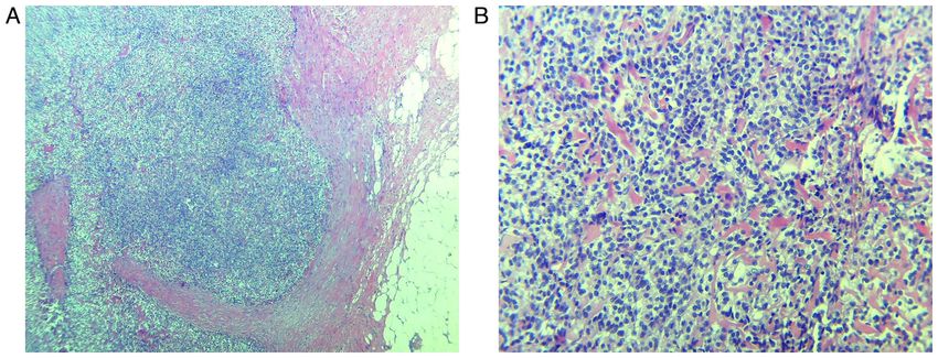
2                     REBEGEA et al: MANAGEMENT OF UTERINE AND EXTRAUTERINE STROMAL SARCOMAS

Introduction                                                        the long survival and death of breast cancer complications,
                                                                    we consider that the histological aspects for this patient would
Endometrial stromal sarcoma (ESS) is a rare tumor, predomi‑         have been LG‑ESS.
nantly occurring as a primary tumor of the uterus. Rare cases          Patient no. 3 presented for post‑therapeutic controls, up
of primary extrauterine ESS (EESS) have been reported.              until 2009, evading medical care since then. Patient no. 5
Low‑grade ESS (LG‑ESS) is more common than high‑grade               had a 23‑month survival. We could not calculate a median
ESS (HG‑ESS). Heterogeneous mixtures of morphologic                 survival due to the lack of data regarding all of the analyzed
and genetic features of LG‑ESS and HG‑ESS are distinct              cases.
histopathological entities. The diagnosis is often made after
hysterectomy (1).                                                   Patient no. 6. EESS was diagnosed in a 64‑year‑old patient
    ESS represents a rare diagnostic and a high‑ or                 (patient no. 6) with a personal history of hysterectomy
low‑grade tumor profile is distinguished by immunohis‑              and bilateral salpingo‑oophorectomy in 1997, with the
tochemistry (IHC) tests (2). Up to 30% of patients have             histopathological diagnosis of chronic cervicitis, uterine
EESS at presentation. The treatment of ESS is multimodal,           leiomyofibroma and uterine adenomyosis. In November 2019,
its management requiring a multidisciplinary team, and it           the computer tomography (CT) scan examination revealed
is different according to the primary tumor location and            solid retroperitoneal lesions with heterogeneous contrast
tumor staging. The role of adjuvant radiotherapy remains            enhancement and areas of necrosis and calcification, local‑
controversial in high‑grade EESS and due to the rarity of           ized in the left para‑aortic region, inferior from the renal
these cases there are limited data concerning the efficacy          hilum with extension into the left psoas muscle (58/66 mm)
of adjuvant external radiotherapy (EBRT) available from             and inferior pelvis, caudal from the common iliac artery
prospective randomized control clinical trials (3). Here,           bifurcation (34/42 mm with 34/40 mm on the right and
we present five cases of ESS and one case of EESS. All              22/33 mm with 44/62 mm on the left side). The retroperi‑
cases received EBRT at the Radiotherapy Department of               toneal mass was biopsied, and the microscopic examination
the Emergency Clinical Hospital ‘Sfantul Apostol Andrei’            revealed a proliferation of monotonous ovoid to spindle
Galati, during 2004‑2020.                                           cells, with scant cytoplasm, focally with ‘clear cell’ aspects,
                                                                    monomorphic nuclei and low mitotic activity, without any
Case reports                                                        areas of necrosis.
                                                                        Biomarker IHC studies and hematoxylin and eosin (H&E)
Literature data support radiotherapy use for ESS with the           slide staining were necessary (5). Immunohistochemical
aim of improving local tumor control, but without revealing         staining revealed intense, diffuse positivity for ER isoform α,
increases in the survival rate with certainty (1,4). We present     diffuse positivity for CD10, focal actin and desmin posi‑
five ESS cases and one EESS case. All cases received EBRT           tivity, and negativity for CD34, inhibin, calretinin, OCT3/4,
at the Radiotherapy Department of the ‘Sfantul Apostol              PLAP, chromogranin A and Melan‑A. The histopathological
Andrei’ Emergency Clinical Hospital of Galati, between 2004         aspects of the mesenchymal tumor and the immunophe‑
and 2020, where we identified six EES cases. Five of them had       notype (ER+, CD10 +) revealed the diagnosis of LG‑EESS
ESS histology and were localized in the uterus [receiving treat‑    (Figs. 1 and 2).
ment with EBRT in two‑dimensional (2D) technique] and only              In January 2020, the magnetic resonance imaging (MRI)
one had ESS histology with extrauterine location [receiving         examination described a retroperitoneal heterogeneous tissue
palliative treatment with three dimensional conformational          mass of 52/62 mm, with contrast enhancement and areas of
radiotherapy (3DCRT) technique, using the linear accelerator        necrosis, extending caudally from the left renal hilum having
Elekta Synergy].                                                    approximately 85 mm in length; the lesion encased the aorta
    Clinical and therapeutic parameters of the EES and EESS         and the left renal artery (which is still patent), it invaded the left
patients are documented in Table I. The median age was              renal vein and left psoas muscle, extending into the spinal canal
57.4 years.                                                         (through the L2 left neural foramen in the anterior epidural
                                                                    space, extending cranially and caudally from L1 to L3). The
Patients 1‑5. Only one patient (patient no. 5) had IHC tests        mass also infiltrated the left sided para‑vertebral muscles
[HG‑ESS that was estrogen receptor‑negative (ER‑), proges‑          (Fig. 3).
terone receptor‑negative (PR‑), CD10 +, vimentin‑positive               The MRI from March 2020 revealed a solid expansive
(VIM+), Ki‑67 30%] because between 2004 and 2009, IHC               gadolinophilic retroperitoneal process, having axial measure‑
assays were not performed routinely in our medical unit.            ments of 100/75 mm and invading the left psoas muscle, L1, L2
    The surgical intervention, for patient nos. 1‑5, consisted of   and with extension in the spinal canal (with extradural loca‑
a total hysterectomy with bilateral salpingo‑oophorectomy.          tion), expanding across approximately 150 mm, in between D1
Patients who presented with post‑surgical relapse risk factors      and L4. The mass determined foraminal and spinal canal
such as vaginal or parameter invasion/extension received            stenosis, with compressive effects on the horsetail roots
adjuvant radiotherapy.                                              (Fig. 4).
    Patient no. 2 presented with a second neoplastic disease            As treatment, the patient started hormone therapy with
occurring 9 years after the ESS diagnosis; this patient died        letrozol, and the oncology team decided to perform EBRT
in 2018 suffering from lung metastases from breast cancer; the      as palliative treatment at LINAC in 3DCRT technique,
breast cancer features were: pT2N0M0, invasive ductal carci‑        TD=30 Gy/10fr, D/fr=300 cGy on primary tumor and
noma, ER=90%, PR‑, Ki‑67 >14%. Taking into consideration            extension, with good treatment tolerance.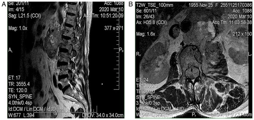
EXPERIMENTAL AND THERAPEUTIC MEDICINE 22: 1456, 2021                                                              3

Table I. Clinical and therapeutical parameters of the ESS and EESS patients.

                  Age		                    Surgical				                                                                 Treatment intent         Survival
Patient no.      (years) Year            intervention HP IHC Risk factors                                                and total dose          (months)

1 55 2004 TH+BSO ESS G3    ‑      Vagina   Adjuvant EBRT
							                                    TD=50 Gy
2 43 2004 TH+BSO  ESS      ‑      Vagina   Adjuvant EBRT                                                                                            168
							                                    TD=50 Gy
3 68 2006 TH+BSO  ESS      ‑     Vagina +  Adjuvant EBRT
						                          parameters TD=50 Gy
4 55 2009 TH+BSO ESS G3    ‑    Parameter, Adjuvant EBRT
						                           miometer  TD=50 Gy
5 66 2016 TH+BSO  ESS   HG‑ESS;		          Adjuvant EBRT                                                                                              23
					ER‑, PR‑ CD10+, 		                    TD=50 Gy
					VIM+, Ki‑67 30%
6 64 2020		 EESS			Palliative EBRT                                                                                                                    11
							                                    TD=30 Gy

HP, histopathology; IHC, immunohistochemistry; TH+BSO, total hysterectomy + bilateral salpingo‑oophorectomy; EBRT, external beam
radiotherapy; ESS, endometrial stromal sarcoma; EESS, extrauterine endometrial stromal sarcoma; HG‑ESS, high‑grade endometrial stromal
sarcoma.

Figure 1. Histopathological aspects of low‑grade EESS. (A) Proliferation of monotonous ovoid to spindle tumor cells, involving the adipose tissue. H&E stain;
magnification, x40. (B) Tumor cells are uniformly small, with oval‑round nuclei and they focally whorl around small vessels. H&E stain, magnification, x200.
EESS, extrauterine endometrial stromal sarcoma; H&E, hematoxylin and eosin.

Discussion                                                                      did not receive adjuvant radiotherapy (no EBRT group). For
                                                                                all patients in the EBRT group, adjuvant radiotherapy signifi‑
Most of the information concerning uterine sarcomas avail‑                      cantly improved the disease‑free survival (DFS) (P=0.008)
able in the medical literature is based on small series or case                 and pelvic failure‑free survival (PFFS) (P=0.004). This study
reports, which have analyzed the effects of adjuvant radio‑                     indicated that adjuvant radiotherapy significantly improved
therapy on the main histological subtypes. When grouping                        DFS and PFFS with tolerable adverse effects, especially
all of the histological subtypes, most studies have shown that                  in patients with stage IB to IIB disease. Despite several
adjuvant radiotherapy reduces the local and regional recur‑                     limitations (unequal number of patients in the two groups;
rence rates without having an overall survival advantage (4,6).                 retrospective single‑center study), this is the largest popula‑
Endometrial stromal sarcoma (ESS) patients receiving adju‑                      tion‑based study exploring adjuvant radiotherapy in resected,
vant radiotherapy have registered improved local control, as                    early‑stage LG‑ESS patients and could be a valuable reference
compared to patients undergoing surgical treatment alone (4).                   that provides guidance for radiotherapy treatment selection in
    Wang et al (7) retrospectively reviewed 152 patients with                   specific subgroups (7).
stage I to II resected low‑grade (LG)‑ESS; the patients were                        A retrospective analysis was conducted in 2009 and it
included and analyzed for 20 years (1998‑2018). Forty patients                  included 3,650 patients with uterine sarcoma; it was conducted
received adjuvant radiotherapy (EBRT group) and 112 patients                    using the National Oncology Database and had a median
4 REBEGEA et al: MANAGEMENT OF UTERINE AND EXTRAUTERINE STROMAL SARCOMAS
Figure 2. Immunohistochemistry staining. (A) Intense, diffuse positivity for CD10; magnification, x100. (B) Tumor cells show nuclear positivity for ER, with
variable intensity; magnification, x400. (C) Focal positivity for actin (clone HHF35); magnification, x200. (D) CD34 is negative in the tumor cells and positive
in the blood vessels network; magnification, x100. ER, estrogen receptor. HHF35, anti‑muscle actin antibody.
Figure 3. (A and B) Patient no. 6. January 2020 MRI examination; retroperitoneal heterogeneous tissue mass. MRI, magnetic resonance imaging.
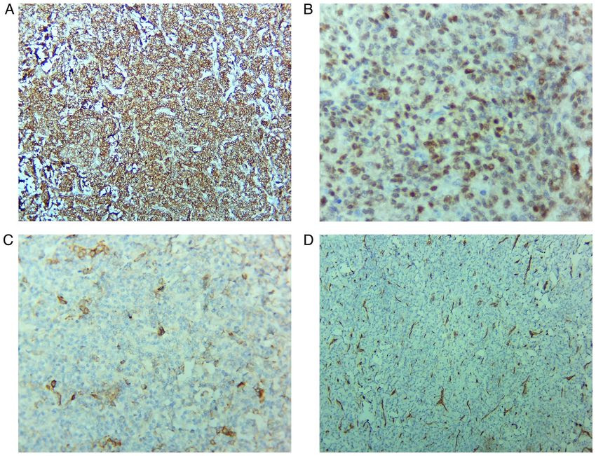
Figure 4. (A and B) Patient no. 6. March 2020 MRI; solid expansive gadolinophilic retroperitoneal process. MRI, magnetic resonance imaging.EXPERIMENTAL AND THERAPEUTIC MEDICINE 22: 1456, 2021 5
follow‑up period of 59 months, with a 5‑year overall survival Markers such as caldesmon, desmin, SMA, EMA, Melan‑A or
of 37%. Use of adjuvant radiotherapy was not predictive for pankeratin are negative (10‑12,15,16).
overall survival. For non‑metastatic cancer patients receiving Differential diagnosis for uterine LG‑ESS includes cellular
definitive surgery, the 5‑year local‑regional failure‑free leiomyoma, intravascular leiomyomatosis, leiomyosarcoma,
survival (LRFFS) was 87% (8). HG‑ESS, gastrointestinal stromal tumor (GIST), perivascular
ESS is a rare diagnosis, usually presenting as a leio‑ epithelioid cell tumor (PEComa), gland‑poor adenomyosis.
myoma of the uterus. The symptoms are nonspecific, patients Proper diagnosis requires attention to histological aspects
complaining most frequently of abnormal uterine bleeding. An and the use of an adequate panel of IHC markers (10‑12,16).
early diagnosis is essential because patient survival is directly Vroobel et al framed an immunopanel for the differential
related to the tumor stage. Uterine sarcomas most often affect diagnosis of extrauterine neoplasms, which can be helpful in
postmenopausal women (9). distinguishing LG‑ESS from the entities mentioned above,
The gross appearance of LG‑ESS is characterized by but also from granulosa cell tumor, desmoplastic small round
several confluent tumor masses having a tan to yellow cut cell tumor or even lymphoma (12). HG‑ESS needs to be
surface, sometimes presenting tumor plugs with worm‑like differentiated from epithelioid leiomyosarcoma, PEComa of
appearance which permeate the myometrium or which can be gynecological origin, epithelioid GIST with pelvic location,
found in blood vessels. The extrauterine spread of LG‑ESS or undifferentiated uterine sarcoma (UUS), mainly based on
is indicated by some palpable tumor cords in the parauterine histomorphology and immunophenotype (17).
tissues. In contrast, high grade (HG)‑ESS presents as large In the specialty literature, the ESS relapse pattern is
tumor masses (up to 9 cm in diameter) with extensive limited. Zhou et al (18) demonstrated that relapse of LG‑ESS
permeation of the myometrium; in some cases, the extra‑ was 12.3% (14 of 114 included patients) mainly due to distant
uterine spread can be more extensive than the uterine tumor metastasis (64.3%, 9/14) and only 5/14 were pelvic recur‑
itself (10‑13). rences. The median time of recurrences was 50 months (range,
The microscopic features of LG‑ESS are character‑ 6‑169 months). In this study, all patients performed hysterec‑
ized by an endometrial stromal nodule‑like cytology tomy, ovarian preservation was performed in 20/114 of cases,
(small cells, uniform round‑oval nuclei, scant cytoplasm) adjuvant radiotherapy was performed in 31.6% of cases,
with tongue‑like/island‑like invasion of the myometrium, 49.1% (56/114) patients received postoperative chemotherapy
sometimes also with lymphovascular invasion. It can have (median 3 courses) and 9.6% of patients received endocrine6 REBEGEA et al: MANAGEMENT OF UTERINE AND EXTRAUTERINE STROMAL SARCOMAS
to IV ESS. Adjuvant EBRT may be added for stage II‑IVA. Competing interests
Hormone therapy includes aromatase inhibitors, megestrol
acetate, or medroxyprogesterone acetate (25). The authors declare that they have no competing interests.
Case series of patients with ESS suggest long disease‑free
intervals in the absence of specific therapy and raise questions References
about the use of adjuvant EBRT that has been demonstrated
to reduce local recurrence rates but with limited effect on 1. Sampath S and Gaffney DK: Role of radiotherapy treatment
of uterine sarcoma. Best Pract Res Clin Obstet Gynaecol 25:
survival (6,26,27). 761‑772, 2011.
Regarding HG‑ESS, the role of adjuvant EBRT in 2. Jin Y, Pan L, Wang X, Dai Z, Huang H, Guo L, Shen K and
non‑metastatic disease is controversial (3). Lian L: Clinical characteristics of endometrial stromal sarcoma
from an academic medical hospital in China. Int J Gynecol
The treatment is multimodal, with a management requiring Cancer 20: 1535‑1539, 2010.
a multidisciplinary team, and a difference regarding the 3. Reed NS, Mangioni C, Malmström H, Scarfone G, Poveda A,
primary tumor location and staging of the tumor. Rarely ESS Pecorelli S, Tateo S, Franchi M, Jobsen JJ, Coens C, et al: Phase III
randomised study to evaluate the role of adjuvant pelvic radio‑
is initially present in an extrauterine site. IHC will help in therapy in the treatment of uterine sarcomas stages I and II: An
the detection of the specific tumor markers. In our cases, the European organisation for research and treatment of cancer gynae‑
IHC of LG‑ESS and HG‑ESS revealed a completely different cological cancer group study (protocol 55874). Eur J Cancer 44:
808‑818, 2008.
biological aggressiveness and clinical behavior. Management 4. Valduvieco I, Rovirosa A, Colomo L, De San Juan A,
was different regarding the primary tumor location and Pahisa J and Biete A: Endometrial stromal sarcoma. Is
staging of the tumor. Initial surgical approach is considered there a place for radiotherapy? Clin Transl Oncol 12: 226‑230, 2010.
5. Bacinschi XE, Zgura A, Safta I and Anghel R: Biomolecular
as the optimal treatment, but the aim of post‑operative radia‑ factors represented by Bcl‑2, p53, and tumor‑infiltrating lympho‑
tion treatment remains uncertain. EBRT may have broader cytes predict response for adjuvant anthracycline chemotherapy
indications than what is currently accepted in clinical practice. in patients with early triple‑negative breast cancer. Cancer Manag
Res 12: 11965‑11971, 2020.
The purpose of adjuvant EBRT in non‑metastatic disease is 6. Weitmann HD, Knocke TH, Kucera H and Pötter R: Radiation
controversial. therapy in the treatment of endometrial stromal sarcoma. Int J
Radiat Oncol Biol Phys 49: 739‑748, 2001.
7. Wang W, Sun S, Miao Z, Hou X, Zhang F and Hu K: Adjuvant
Acknowledgements radiotherapy improved survival in stage I to II low‑grade endo‑
metrial stromal sarcoma: A retrospective study of 152 cases.
Not applicable. Front Oncol 10: 608152, 2021.
8. Sampath S, Schultheiss TE, Ryu JK and Wong JY: The role of
adjuvant radiation in uterine sarcomas. Int J Radiat Oncol Biol
Funding Phys 76: 728‑734, 2010.
9. Jain R, Batra S, Ahmad A, Elahi AA, Gupta M and Saith P: Low
grade endometrial stromal sarcoma: A case report. Iran J Med
No funding was received. Sci 40: 81‑84, 2015.
10. Hoang L, Chiang S and Lee CH: Endometrial stromal sarcomas
Availability of data and materials and related neoplasms: New developments and diagnostic
considerations. Pathology 50: 162‑177, 2018.
11. Lipsich F, Causa Andrieu PI, Wernicke A, Patrono MG,
The information generated and analyzed in regards to the Napoli MN, Chacon CRB and Nicola R: Extra‑uterine
case reports is available from the corresponding author on endometrial stromal sarcoma arising from deep infiltrating
endometriosis. Clin Imaging 67: 250‑254, 2020.
reasonable request. 12. Vroobel KM, Karawita TS and Nafisa Wilkinson: New develop‑
ments in endometrial stromal sarcoma. Diagn Histopathol 23:
Authors' contributions 311‑322, 2017.
13. Ashraf‑Ganjoei T, Behtash N, Shariat M and Mosavi A:
Low grade endometrial stromal sarcoma of uterine corpus, a
LFR, DF, RMA, LG, AMI, MED, MC, EN, ALT, MSC, ML clinico‑pathological and survey study in 14 cases. World J Surg
and AIN contributed to the acquisition, analysis and inter‑ Oncol 4: 50, 2006.
14. Seagle BL, Shilpi A, Buchanan S, Goodman C and Shahabi S:
pretation of the data; they made substantial contributions to Low‑grade and high‑grade endometrial stromal sarcoma: A
the conception and design of the work, and supervised and national cancer database study. Gynecol Oncol 146: 254‑262,
substantially revised this work. All authors had equal contri‑ 2017.
15. Farhood AI and Abrams J: Immunohistochemistry of endome‑
butions, equal participation and the same rights to this article. trial stromal sarcoma. Hum Pathol 22: 224‑230, 1991.
All authors have read and agreed to the published version of 16. Zhu XQ, Shi YF, Cheng XD, Zhao CL and Wu YZ:
the manuscript. Immunohistochemical markers in differential diagnosis of
endometrial stromal sarcoma and cellular leiomyoma. Gynecol
Oncol 92: 71‑79, 2004.
Ethics approval and consent to participate 17. Ali RH and Rouzbahman M: Endometrial stromal tumours
revisited: An update based on the 2014 WHO classification.
J Clin Pathol 68: 325‑332, 2015.
The Ethics Committee of ‘Sfantul Apostol Andrei’ Emergency 18. Zhou J, Zheng H, Wu SG, He ZY, Li FY, Su GQ and Sun JY:
Clinical Hospital of Galati has granted approval for this study Influence of different treatment modalities on survival of patients
with the decision number 8540 from 09.04.2021. with low‑grade endometrial stromal sarcoma: A retrospective
cohort study. Int J Surg 23: 147‑151, 2015.
19. Puliyath G, Nair VR and Singh S: Endometrial stromal sarcoma.
Patient consent for publication Indian J Med Paediatr Oncol 31: 21‑23, 2010.
20. Hasiakos D, Papakonstantinou K, Kondi‑Paphiti A and Fotiou S:
Low‑grade endometrial stromal sarcoma of the endocervix.
Consent for participation was granted by all of the patients and Report of a case and review of the literature. Eur J Gynaecol
is part of the personal observation sheet. Oncol 28: 483‑486, 2007.EXPERIMENTAL AND THERAPEUTIC MEDICINE 22: 1456, 2021 7
21. Gadducci A, Cosio S, Romanini A and Genazzani AR: The 25. Amant F, Coosemans A, Debiec‑Rychter M, Timmerman D and
management of patients with uterine sarcoma: A debated clinical Vergote I: Clinical management of uterine sarcomas. Lancet
challenge. Crit Rev Oncol Hematol 65: 129‑142, 2008. Oncol 10: 1188‑1198, 2009.
22. Palombaa S, Falboa A, Mocciaroa R, Russob T and Zulloa F: 26. Mansi JL, Ramachandra S, Wiltshaw E and Fisher C: Endometrial
Laparoscopic treatment for endometrial cancer: A meta‑analysis stromal sarcomas. Gynecol Oncol 36: 113‑118, 1990.
of randomized controlled trials (RCTs). Gynecol Oncol 112: 27. Berchuck A, Rubin SC, Hoskins WJ, Saigo PE, Pierce VK and
415‑421, 2009. Lewis JL Jr: Treatment of endometrial stromal tumors. Gynecol
23. Lindner T, Pink D, Kretzschmar A, Mrozek A, Thuss‑Patience PC Oncol 36: 60‑65, 1990.
and Reichardt P: Hormonal treatment of endometrial stromal
sarcoma: A possible indication for aromatase inhibitors. J Clin
This work is licensed under a Creative Commons
Oncol 23 (Suppl 16): S9057, 2005.
24. Chu MC, Mor G, Lim C, Zheng W, Parkash V and Schwartz PE: Attribution-NonCommercial-NoDerivatives 4.0
Low grade endometrial stromal sarcoma: Hormonal aspects. International (CC BY-NC-ND 4.0) License.
Gynaecol Oncol 90: 170‑176, 2003.You can also read