3: Providing safe blood - Transfusion Handbook - JPAC
←
→
Page content transcription
If your browser does not render page correctly, please read the page content below
JPAC              Joint United Kingdom (UK) Blood Transfusion and Tissue
                  Transplantation Services Professional Advisory Committee
                                                                                                 PDF Generated
                                                                                                25/08/2021 05:47

 Transfusion Handbook
 3: Providing safe blood
 http://www.transfusionguidelines.org/transfusion-handbook/3-providing-safe-blood

3: Providing safe blood

 Essentials

        Unpaid volunteers, donating regularly, are key to the provision of safe and sufficient blood for
        transfusion.
        17 to 65 year olds can enrol as first-time blood donors and there is no upper age limit for regular
        donors (subject to an annual health check).
        To ensure the safety of the donor and recipient, a medical questionnaire covering health, lifestyle,
        travel history, medical history and medication is completed before each donation.
        The minimum mandatory infection screen on all donations is for hepatitis B and C, HIV, HTLV and
        syphilis, and extra tests are performed as required.
        The risk of transmission of prion diseases such as variant Creutzfeldt–Jakob disease (vCJD) is
        reduced by excluding at-risk donors (including recipients of a blood transfusion or tissue/organ
        transplant since 1980), removing white cells from donations (leucodepletion), importing plasma
        derivatives from countries with a low risk of vCJD and providing imported, virus-inactivated fresh
        frozen plasma (FFP) for patients born on or after 1 January 1996.
        Donations are routinely ABO and RhD typed and screened for clinically important blood group
        antibodies.
        Modern transfusion practice is based on the use of blood components rather than whole blood
        donations.
        Plasma derivatives are licensed medicines and include albumin solutions, coagulation factor
        concentrates and immunoglobulins.

Blood transfusion in the UK is now very safe indeed and most serious adverse events originate in the
hospital rather than the blood transfusion centre (see Chapter 5). However, ensuring a safe and effective
blood supply remains essential. This requires a combination of high-quality donor recruitment and selection,
infection screening, serological testing and blood component production (followed by rational clinical use).
The four UK Blood Services – NHS Blood and Transplant, Northern Ireland Blood Transfusion Service,
Scottish National Blood Transfusion Service and Welsh Blood Service – maintain common standards for
blood donation, testing and blood products. The Joint UKBTS Professional Advisory Committee (JPAC) is
responsible for producing the Guidelines for the Blood Transfusion Services in the UK, often known as the
Red Book (http://www.transfusionguidelines.org.uk/). In 2011 the UK Blood Services issued 2.1 million units
of red cells, 300 000 platelet doses, 288 000 units of fresh frozen plasma and 126 000 units of
cryoprecipitate.

                                                       page 1 of 14
Transfusion Handbook / 3: Providing safe blood
3.1: Blood donation
Unpaid volunteers who donate on a regular basis are a crucial element in the provision of a safe and
reliable supply of blood. Many studies show that altruistic donors have a lower prevalence of transfusion-
transmissible infections.
The minimum age for donation is 17 years. There is no upper age limit for regular donors, although they are
subject to annual health review after their 66th birthday. The upper age limit for first-time donors is 65 years.
The minimum body weight for blood donation is 50 kg (7 st 12 lb). Only 5% of eligible people are regular
blood donors and the Blood Services put much effort into improving recruitment, especially of donors from
minority ethnic groups.
3.1.1: Donor eligibility
Donors answer a series of questions before each donation relating to their health, lifestyle, travel history,
medical history and medication. This is to ensure the safety of both the donor and recipients. Donor
exclusion and deferral criteria are regularly reviewed in the light of scientific knowledge. For example, there
have been recent significant changes to the eligibility of ‘men who have sex with men’ (MSM) to donate
blood in the UK (see Chapter 5). Up-to-date eligibility criteria are given in the Red Book (http://www.
transfusionguidelines.org.uk/).
3.1.2: Frequency of donation
The normal interval between whole blood donations is 16 weeks (minimum 12 weeks) but no more than
three donations a year are collected from female donors because of their more precarious iron status.
Donors undergo a screening test for anaemia, usually the copper sulphate flotation test on a finger prick
sample. The minimum pre-donation Hb concentration is 125 g/L for female donors and 135 g/L for males.
Donors giving double red cell donations by apheresis must have a pre-donation Hb concentration of 140 g/L
and the minimum interval between donations is 26 weeks.
Donors can give platelets or plasma by apheresis on a cell separator with a maximum of 24 procedures in
12 months. The minimum interval between donations is 2 weeks and plasma donors are limited to 15 litres
a year.
3.1.3: Genetic haemochromatosis
Donors with this common genetic condition, which causes increased iron absorption from the diet, are
eligible to become blood donors if they meet all the other medical selection and age criteria. Regular blood
donation can be part of their maintenance treatment schedule to prevent iron overload.
3.2: Tests on blood donations
3.2.1: Screening for infectious agents
At each donation, the following mandatory tests are performed:
Hepatitis B – HBsAg
Human immunodeficiency virus – anti-HIV 1 and 2 and HIV NAT (nucleic acid testing)
Hepatitis C – anti-HCV and HCV NAT
Human T-cell lymphotropic virus – anti-HTLV I and II
page 2 of 14Transfusion Handbook / 3: Providing safe blood
Syphilis – syphilis antibodies.
Some donations are tested for cytomegalovirus (CMV) antibodies to provide CMV negative blood for
patients with certain types of impaired immunity (see Chapter 5).
Additional tests, performed in special circumstances, include:
Malarial antibodies
West Nile Virus antibodies
Trypanosoma cruzi antibodies.
3.2.2: Precautions to reduce the transfusion transmission of prion-associated diseases
These include variant Creutzfeldt–Jakob disease (vCJD – caused by the same agent as bovine
spongioform encephalopathy (BSE) in cattle – ‘mad cow disease’) and sporadic or inherited CJD. The
following are permanently deferred from blood donation:
Persons who have received a blood transfusion or tissue/organ transplant from a donor since 1980
Anyone who has received human pituitary-derived hormones, grafts of human dura mater or cornea,
sclera or other ocular tissue
Members of a family at risk of inherited prion diseases
Persons notified that they may be at increased risk of vCJD due to possible exposure to an infected
individual by surgical instruments, blood product transfusion or transplant of tissues or organs
Persons notified that they may be at increased risk because a recipient of their blood or tissues has
developed a prion-related disorder.
More information, including the latest data on transfusion-transmitted vCJD, can be obtained from the
National CJD Research and Surveillance Unit
(http://www.cjd.ed.ac.uk/index.html).
3.2.3: Blood groups and blood group antibodies
Every donation is tested to determine the ABO and RhD group of the red cells and the plasma is screened
to detect the most common blood group antibodies that might cause problems in a recipient. Some
donations are tested for a wider range of clinically significant blood groups (extended phenotyping) to allow
closer matching and reduce the development of alloantibodies in patients who need long-term red cell
transfusion support (see Chapter 8). Blood for neonatal or intrauterine use has a more extensive antibody
screen (see Chapter 10).
Some group O donations are screened for high levels of anti-A and anti-B antibodies to reduce the risk of
haemolytic reactions when group O plasma, platelets or other components containing a large amount of
plasma (e.g. red cells for intrauterine or neonatal exchange transfusion) are transfused to group A, B or AB
patients, especially neonates and infants.
3.2.4: Molecular blood group testing
The genes for most human blood groups have now been identified. Currently only a limited number of
patients undergo genotyping. These include recently transfused patients whose blood group is uncertain
and fetuses that require typing to define the risk from maternal antibodies. Routine DNA testing/genotyping
using rapid automated technology is likely to enter blood service and hospital laboratory practice in the next
decade.
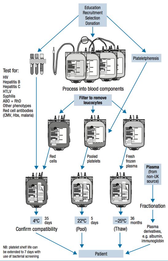
page 3 of 14Transfusion Handbook / 3: Providing safe blood 3.3: Blood products These are classified as blood components prepared in the blood transfusion centre (red cells, platelets, fresh frozen plasma and cryoprecipitate) or plasma derivatives manufactured from pooled plasma donations in plasma fractionation centres (such as albumin, coagulation factors and immunoglobulins). Plasma derivatives are covered by the Medicines Act and, like any other drug, must be prescribed by a licensed practitioner. Since 1999, as a vCJD risk-reduction measure, all plasma derivatives used in the UK are manufactured using donations from countries with a low risk of vCJD. 3.3.1: Blood components Whole blood is now rarely used for transfusion. Blood component therapy makes clinical sense as most patients require a specific element of blood, such as red cells or platelets, and the dose can then be optimised. Each component is stored under ideal conditions (e.g. red cells must be refrigerated, platelets must not) and the use of precious blood donations becomes more efficient. The use of blood components in clinical practice is covered in Chapters 7 to 10. The process of producing blood components and plasma derivatives is summarised in Figure 3.1. 3.3.2: Labelling of blood components 3.3.2.1: Blood component labels The content of blood pack labels attached at the transfusion centre is prescribed by the Blood Safety and Quality Regulations 2005 (BSQR). Key information is present in both eye-readable and barcoded form and allows the donor origin (via a unique donation number) and processing steps of the product to be traced as well as indicating the blood group, any special requirements (such as CMV negative or irradiated), expiry date and storage conditions. Work is in progress to review the content of blood component labels and improve their clarity. Up-to-date information is available in the Guidelines for the Blood Transfusion Services in the UK (http://www.transfusionguidelines.org.uk). 3.3.2.2: Blood compatibility labels These are attached to the pack in the hospital transfusion laboratory and uniquely identify the patient for whom the component has been selected. At the final bedside check, the donation number and other details on the compatibility label must match those on the blood pack label and the patient details must exactly match those on the recipient’s ID band (see Chapter 4 for detailed discussion of safe blood administration). 3.3.2.3: Specifications of blood components Whole blood donations of 405–495 mL (mean 470 mL) are collected into 63 mL of citrate phosphate dextrose (CPD) anticoagulant. All blood donations are filtered to remove white blood cells (pre-storage leucodepletion) to leave
Transfusion Handbook / 3: Providing safe blood

                page 5 of 14
Transfusion Handbook / 3: Providing safe blood
Red cells
These are used to restore oxygen carrying capacity in patients with anaemia or blood loss where alternative
treatments are ineffective or inappropriate. They must be ABO compatible with the recipient (see Table 2.2).
Clinical indications for red cell transfusion are discussed in Chapters 7 to 10.
Red cells in additive solution
In red cells in additive solution (Table 3.1) the majority of plasma is removed and replaced by 100 mL
saline, adenine, glucose and mannitol additive solution (SAG-M).
Table 3.1 Red cells in additive solution
Volume (mL) 220–340
Haematocrit (L/L) 0.5–0.7
Haemoglobin content (g) >40 (in more than 75% of units tested)
Residual plasma (mL) 5–30
Storage temperature 2–6°C
Shelf life Up to 35 days from donation
Irradiated red cells
Irradiated red cells are indicated for patients at risk of transfusion-associated graft-versus-host disease (TA-
GvHD – see Chapter 8). The component must be irradiated by gamma or X-rays within 14 days of donation
and it then has a shelf life of 14 days from irradiation.
Washed red cells
Indicated for patients with recurrent or severe allergic or febrile reactions to red cells, and severely IgA-
deficient patients with anti-IgA antibodies for whom red cells from an IgA-deficient donor are not available (
see Chapter 5). They are produced either manually (24-hour shelf life) or by a closed, automated system in
which the red cells are sequentially washed to remove most of the plasma (240×109
per transfusion.
page 6 of 14Transfusion Handbook / 3: Providing safe blood
Platelets have ABO antigens on their surface and may have reduced survival if transfused to an ABO-
incompatible recipient, although this is not usually clinically significant. They are usually only available in
groups O, A or B, with only a small number of group AB platelets produced.
Anti-A or anti-B antibodies in the plasma of platelet components may rarely cause haemolysis of the
recipient’s red cells, especially in babies and small children. Group O platelets should ideally only be given
to group O recipients. Selection of platelets for patients of other ABO groups is summarised in Table 3.2.
RhD negative platelet concentrates should be given to RhD negative patients where possible, especially to
RhD negative women of child-bearing potential. When RhD-incompatible platelets have to be given,
administration of anti-D immunoglobulin may prevent immunisation.
Platelets are produced in two ways (see Tables 3.2 and 3.3):
Whole blood donations are centrifuged and the buffy coats (between the red cell and plasma layers)
from four donations are pooled in the plasma of one of the donors (male, to reduce the risk of
transfusion-related acute lung injury (TRALI) – see Chapter 5).
An ATD of platelets is obtained from a single donor by apheresis (donors may give two or three
ATDs at a single session).
The UK Blood Services aim to provide more than 80% of platelet doses by apheresis to reduce the
exposure of patients to multiple donors (a vCJD risk-reduction measure).
Platelets are stored in temperature-controlled incubators (20–24°C) with constant agitation (refrigerated
platelets are rapidly removed from the circulation). The recent introduction of automated bacterial screening
has allowed some Blood Services to extend the shelf life from 5 to 7 days after donation.
Table 3.2 Platelets from pooled buffy coats
Number of donors per pack 4
Mean volume (mL) 300
Mean platelets (×109 per unit) 308 (range 165–500)
Anticoagulant CPD
Storage 20–24°C with agitation
Shelf life 5 days (7 days if bacterial screening)
Table 3.3 Platelets from apheresis donation
page 7 of 14Transfusion Handbook / 3: Providing safe blood
Number of donors per pack 1
Mean volume (mL) 199
Mean platelets (×109 per unit) 280 (range 165–510)
Anticoagulant Acid citrate dextrose
Storage 20–24°C with agitation
Shelf life 5 days (7 days if bacterial screening)
Irradiated platelets
Platelets may be irradiated to prevent TA-GvHD in susceptible patients. They retain their normal shelf life.
Platelets in additive solution
After ‘washing’ to remove most of the plasma the platelets are resuspended in 200 mL of platelet additive
solution (PAS). This component is indicated for patients with recurrent severe allergic or febrile reactions to
standard platelet transfusions. The shelf life is reduced to 24 hours after preparation and they must be
ordered specially from the Blood Service. Some platelets are lost in the washing process and the
component still contains around 10 mL residual plasma.
Human leucocyte antigen (HLA)-selected platelets
Indicated for patients refractory to random platelet components because of the development of HLA
antibodies after previous transfusions (see Chapter 9). The Blood Services maintain a panel of HLA-typed
platelet donors who donate by apheresis. The platelets are irradiated before issue to prevent TA-GvHD.
Human platelet antigen (HPA)-selected platelets
HPA-1a/5b negative platelets are kept in limited numbers at strategically placed stock-holding units in the
UK and are used for babies with neonatal alloimmune thrombocytopenia (NAIT) (see Chapter 10).
Plasma
Plasma is obtained from whole blood donations or component donation by apheresis. Only male donors are
used to reduce the risk of TRALI. The UK Departments of Health recommend that patients born on or after
1 January 1996 should only receive plasma sourced from countries with a low risk of vCJD. Imported
plasma is treated with a pathogen reduction process, such as methylene blue or solvent detergent
treatment, to reduce the risk of viral transmission.
page 8 of 14Transfusion Handbook / 3: Providing safe blood Plasma components of the same ABO group should be transfused to patients wherever possible. If ABO- identical plasma is not available, the selection criteria given in Table 2.2 are recommended. Plasma components do not need to be matched for RhD group as they contain no red cells or red cell stroma. They do not cause TA-GvHD and irradiation is not required. Fresh frozen plasma (FFP) Plasma is frozen soon after collection to maintain the activity of blood-clotting factors. It can be stored for up to 36 months at –25°C or below. Standard UK FFP is issued as single-donor packs which must be thawed before use, usually in a purpose-designed waterbath. Thawed units of FFP can be stored for up to 24 hours at 4°C before transfusion. Clotting factor levels vary widely between normal healthy donors and this variability is reflected in the concentrations found in individual packs of FFP. FFP (see Table 3.4) is indicated for the treatment of patients with bleeding due to multiple clotting factor deficiencies such as disseminated intravascular coagulation (DIC). It may also be used in patients with inherited clotting factor deficiencies (e.g. Factor V deficiency) where a clotting factor concentrate is not yet available. The recommended dose is 12–15 mL/kg (minimum of four units in a 70 kg adult). However, much larger doses may be needed to produce ‘therapeutic’ levels of coagulation factors and volume overload is a significant clinical problem. FFP is no longer indicated for the reversal of warfarin, as a specific and effective antidote is available (prothrombin complex). FFP carries a significant risk of severe allergic reactions (see Chapter 5) and should not be used as a plasma volume expander. Table 3.4 Fresh frozen plasma Number of donor exposures per pack 1 Mean volume (mL) 274 Mean Factor VIIIc (IU/mL) 0.83 (specification >0.7) Anticoagulant CPD Storage
Transfusion Handbook / 3: Providing safe blood

treatment reduces the concentration of fibrinogen and Factor VIIIc by 15–20%, but levels remain within the
defined specification. Levels of Protein S, an anticoagulant factor, are around 30% lower and this may be
important in patients with an increased risk of thromboembolism. UK guidelines recommend imported SD-
FFP for plasma exchange in patients with thrombotic thrombocytopenic purpura (TTP – see Chapter 11).

Table 3.5 Solvent detergent plasma (Octaplas®)

 Number of donor exposures per pack                 Maximum 1520 donors per batch

 Volume (mL)                                        200 (standardised)

 Mean Factor VIIIc (IU/mL)                          0.8 (specification >0.5)

 Mean fibrinogen (mg/mL)                            2.6 (range 1.5–4.0)

 Anticoagulant                                      Sodium citrate

 Storage
Transfusion Handbook / 3: Providing safe blood

                   Cryoprecipitate packs                          Cryoprecipitate pools

 Number of         1                                              5
 donors

 Mean volume       43                                             189
 (mL)

 Fibrinogen        396 (specification >140)                       1552 (specification >700)
 (mg/pack)

 Factor VIIIc      105 (specification >70)                        454 (specification >350)
 (IU/pack)

 Storage
Transfusion Handbook / 3: Providing safe blood
Mean granulocytes (×109/pack) 1.0 (10 packs = 1×10¹⁰)
Haematocrit (L/L) 0.45
Platelets (×109/pack) 70
Storage 20–24°C
Shelf life To midnight on day of collection
Pooled buffy coats (granulocytes pooled buffy coat derived in additive solution and plasma)
This component (see Table 3.8) was introduced in the UK in 2012. Although the manufacturing process is
more complicated, it has the advantages of lower volume, less red cell and plasma contamination and
resuspension in male donor plasma and additive solution to reduce the risk of TRALI. The dose is two
packs (20 donations) for an adult and 10–20 mL/kg for children.
Table 3.8 Granulocytes pooled buffy coat derived in additive solution and plasma
Mean volume per pack (mL) 207 (175–250) mL
Mean granulocytes (×1010/pack) 1.0 (1×10¹⁰)
Haematocrit (L/L) 0.15
Platelets (×109/pack) 499 (equivalent to 2.5 adult transfusion doses)
Storage 20–24°C (not agitated)
Shelf life To midnight on day following collection
Apheresis granulocytes
page 12 of 14Transfusion Handbook / 3: Providing safe blood
The collection of a therapeutic dose of apheresis granulocytes (Table 3.9) requires the donor to be pre-
treated with steroids and/or injections of granulocyte colony stimulating factor (G-CSF). Hence, their
collection is restricted to directed donors (usually a relative) for an individual patient, rather than UK Blood
Service volunteer donors, and the component is only available in certain clinical centres.
Table 3.9 Apheresis granulocytes
Mean volume per unit (mL) 312
Granulocytes per unit >1×10¹⁰
Haematocrit (L/L) 0.23
Platelets (×109 per unit) 111
Storage 20–24°C
Shelf life 24 hours from collection
Plasma derivatives
These are licensed medicinal products manufactured from human plasma donations. Some of the main
products used in hospital practice are listed below but the reader is referred to the British National
Formulary (BNF – http://bnf.org/bnf) and the individual Summary of Product Characteristics for more
detailed information about formulation and clinical indications. Although these products are manufactured
from large donor pools, sometimes thousands of donations, all now undergo multiple pathogen inactivation
steps to eradicate transfusion-transmitted viruses. Since 1999, all plasma derivatives used in the UK are
derived from imported plasma (a vCJD risk-reduction measure).
Human albumin solution
Human albumin solution (HAS) contains no clotting factors or blood group antibodies and crossmatching is
not required. The clinical indications for HAS are controversial. Crystalloid solutions or synthetic colloidal
plasma substitutes are alternatives for use as plasma expanders in acute blood or plasma loss. HAS should
not be used to ‘correct’ the low serum albumin level often associated with acute or chronic illness. Side
effects include occasional severe hypersensitivity reactions. HAS is available in two forms:
Isotonic solutions (4.5 and 5.0% in volumes of 50 to 500 mL): Often used to replace subacute
plasma volume loss caused by burns, pancreatitis or trauma, and as a replacement fluid in plasma
exchange.
Concentrated solutions (20% in volumes of 50 and 100 mL): Indications may include initiating
diuresis in hypoalbuminaemic patients with liver cirrhosis or nephrotic syndrome, removal of large
volumes of ascites in patients with portal hypertension and to assist the reduction of high bilirubin
levels by exchange transfusion in the newborn (unconjugated bilirubin binds to albumin).
page 13 of 14Transfusion Handbook / 3: Providing safe blood
Clotting factor concentrates
Single-factor concentrates are available for the treatment of most inherited coagulation deficiencies except
Factor V and Factor II (prothrombin). Most patients in the UK with severe haemophilia A are now treated
with recombinant Factor VIIIc, which carries no risk of viral or prion transmission.
Fibrinogen concentrate (Factor I) is, at present, only licensed in the UK for the treatment of congenital
hypofibrinogenaemia but there is encouraging international experience of its effectiveness in the much more
common setting of acquired hypofibrinogenaemia (e.g. DIC, traumatic haemorrhage, massive transfusion).
Many coagulation experts believe that it will replace the use of cryoprecipitate for this purpose in view of its
ease of administration, convenience of storage and standardised fibrinogen content.
Prothrombin complex concentrate (PCC) contains Factors II, VII, IX and X. It has replaced FFP as the
recommended treatment for rapid reversal of warfarin overdose, with elevated international normalised ratio
(INR) and severe bleeding, in view of its superior efficacy, ease of administration and lower risk of severe
allergic reactions or fluid overload. Modern formulations of PCC do not contain activated clotting factors and
have a low risk of causing thrombotic complications. PCC may also be used to treat bleeding due to the
coagulopathy associated with liver disease. The dose for reversal of warfarin is 25–50 IU/kg.
Immunoglobulin solutions
These are manufactured from large pools of donor plasma:
Normal immunoglobulin: contains antibodies to viruses that are common in the population.
Intramuscular normal immunoglobulin may be used to protect susceptible contacts against hepatitis
A, measles or rubella. High-dose intravenous immunoglobulin is used as replacement therapy in
patients with severe immunoglobulin deficiency and in the treatment of autoimmune diseases such
as idiopathic thrombocytopenic purpura (ITP).
Specific immunoglobulins: made from selected donors with high antibody levels to the target of
treatment. Examples include tetanus, hepatitis B and rabies immunoglobulins as well as anti-D
immunoglobulin for the prevention of maternal sensitisation to RhD in pregnancy (see Chapter 9).
page 14 of 14You can also read