Kikuchi-Fujimoto disease preceded by lupus erythematosus panniculitis: do these findings together herald the onset of systemic lupus erythematosus?
←
→
Page content transcription
If your browser does not render page correctly, please read the page content below
Volume 26 Number 8| Aug 2020|
Dermatology Online Journal || Case Report                                                                              26(8):6

Kikuchi-Fujimoto disease preceded by lupus erythematosus
panniculitis: do these findings together herald the onset of
systemic lupus erythematosus?
Anh Khoa Pham1, Stephanie A Castillo2, Dorothea T Barton1,2, William FC Rigby2,3, Marshall A Guill III1,2,
Roberta Lucas1,2, Robert E LeBlanc2,4
Affiliations: 1Department of Dermatology, Dartmouth-Hitchcock Medical Center, New Hampshire, USA, 2Geisel School of Medicine
at Dartmouth College, New Hampshire, USA, 3Section of Rheumatology, Department of Medicine, Dartmouth-Hitchcock Medical
Center, Lebanon, New Hampshire, USA, 4Department of Pathology and Laboratory Medicine, Dartmouth-Hitchcock Medical
Center, New Hampshire, USA
Corresponding Author: Robert E LeBlanc, MD, Department of Pathology and Laboratory Medicine, Dartmouth-Hitchcock Medical Center,
and Geisel School of Medicine, One Medical Center Drive, Lebanon, NH 03756, Email: robert.e.leblanc@hitchcock.org

                                                                    Introduction
  Abstract                                                          Kikuchi-Fujimoto Disease (KFD), also known as
  Kikuchi-Fujimoto disease (KFD), also known as                     histiocytic necrotizing lymphadenitis, is a rare
  histiocytic necrotizing lymphadenitis, is a rare                  disorder that must be distinguished from systemic
  disorder that must be distinguished from systemic                 lupus erythematosus (SLE). It is characterized by
  lupus erythematosus (SLE). Although a minority of                 painful cervical lymphadenopathy, leukopenia, and
  patients with KFD develop SLE, most patients have a               systemic symptoms including fever and malaise
  self-limited disease. Importantly, KFD can have skin              [1,2]. The etiology of KFD is unknown, but infectious
  manifestations resembling cutaneous lupus.                        and autoimmune etiologies have been postulated
  Therefore, the diagnosis of SLE should be predicated              [2]. A correlation between KFD and SLE is well-
  on a complete rheumatologic workup and not on the                 documented, with reports of SLE diagnoses
  constellation of skin disease and lymphadenitis.                  established before, during, and after KFD diagnoses
  Nonetheless, as our exceedingly rare case illustrates,            [3]. Furthermore, the histologic features of KFD are
  patients who do not initially meet diagnostic criteria            often indistinguishable from lupus lymphadenitis,
  for SLE require dermatologic follow-up. We present a              which supports the prevailing opinion that findings
  young adult woman who had a remote history of KFD                 of KFD in the lymph node of a patient with SLE
  and later presented with combined features of                     should be interpreted as a manifestation of lupus
  discoid lupus and lupus erythematosus panniculitis                and not KFD. The distinction between KFD and lupus
  (LEP). On subsequent rheumatologic workup, she                    lymphadenitis would be most important when
  fulfilled criteria for SLE. We discuss the differential           patients develop KFD without additional criteria to
  diagnosis of both KFD and LEP and emphasize how                   establish a diagnosis of SLE. This is important
  strong communication among dermatologists and                     because KFD is considered a benign and self-limited
  other healthcare providers is essential in the                    disorder. In contrast, SLE can be life-threatening,
  management of patients with KFD.                                  requiring coordination of care among a
                                                                    multidisciplinary network of healthcare providers [4].
                                                                    Therefore, the diagnosis of KFD should be made with
Keywords: Kikuchi-Fujimoto disease, systemic lupus                  the caveat that histologic findings can be
erythematosus, lupus erythematosus panniculitis, lupus              indistinguishable from those of lupus and patients
profundus                                                           should be monitored for the dermatologic and

                                                              -1-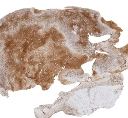
Volume 26 Number 8| Aug 2020|
Dermatology Online Journal || Case Report 26(8):6
nuclear antigens (ENA) and anti-double-stranded
DNA (anti-dsDNA) were negative, no antibodies
against cardiolipin or beta2-glycoprotein-I were
identified, and C3 level was within normal limits.
Altogether, the clinical, histologic, and serologic
findings supported the diagnosis of SLE.
The patient’s past medical history was also notable
for lymphadenitis at age 16. An excisional biopsy of
a tender left cervical lymph node had revealed non-
A B infectious necrotizing lymphadenitis with a paucity
of neutrophils, which suggested a differential
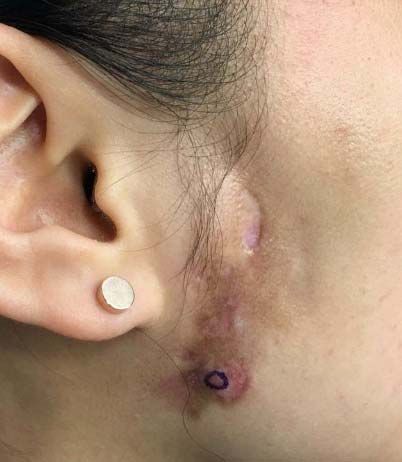
Figure 1. Discoid lupus overlying lupus erythematosus
panniculitis. A) An atrophic hyperpigmented plaque with sclerotic diagnosis of KFD versus lupus lymphadenitis (Figure
foci, erythema, and an underlying lipodystrophy. B) Follow-up 3). Neither hematoxylin bodies nor Azzopardi
photograph at six months with hydroxychloroquine, phenomenon were present to favor lupus and a
methotrexate, and topical tacrolimus therapy. battery of special stains, immunohistochemical
extracutaneous manifestations of both diseases stains, and in situ hybridization study, including
[5,6]. We present an exceedingly rare and illustrative Epstein-Barr encoding region, human herpes virus 8,
case of KFD-lupus overlap, review the pertinent acid-fast bacillus, Gömöri methenamine silver, and
literature on how KFD and SLE can be distinguished, periodic acid–Schiff helped to exclude an infectious
and discuss the implications for practicing etiology. Given her young age, ethnicity, isolated
dermatologists and dermatopathologists. lymphadenitis, and absence of additional clinical
findings at the time, a presumptive diagnosis of KFD
Case Synopsis
A 25-year-old woman of east Asian ancestry
presented to the dermatology clinic for cosmetic
laser treatment of a brown, atrophic, sclerotic, and
indurated plaque on her right preauricular region
(Figure 1A). This plaque was believed to be the
sequela of a two-year history of parotitis that was
refractory to systemic antibiotics and topical A B
corticosteroids. On questioning, the patient
endorsed a history of photosensitivity, complex oral
aphthae, fatigue, and arthralgia. Physical
examination revealed no additional discoid lesions,
evidence of scarring alopecia, or dermatitis. A punch
biopsy of the plaque showed combined features of
discoid lupus and lupus erythematosus panniculitis
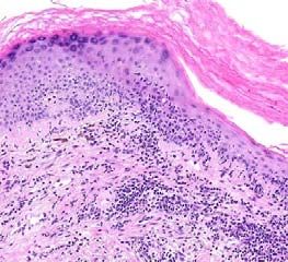
(LEP) including brisk lichenoid interface, C D
perivascular, periadnexal, and subcutaneous Figure 2. Lupus erythematosus panniculitis and discoid lupus. A)
lymphocytic inflammation with hyaline Interface, perivascular, and subcutaneous inflammation is
lipomembranous fat necrosis (Figure 2). The present. H&E, 2x. B) Florid interface dermatitis with basement
membrane alterations, pigment incontinence, epidermal atrophy,
patient’s complete blood count (CBC), complete
and scale. H&E, 100x. C) Hyaline lipomembranous fat necrosis in
metabolic panel (CMP), and lipid panel were within an area of resolving lobular panniculitis. H&E, 100x. D)
normal limits. An antinuclear antibody (ANA) titer Inflammation with histiocytes, lymphocytes, and plasma cells.
was 1:320 with a speckled pattern. Extractable Some lymphocytes encircle adipocytes. H&E, 400x.
-2-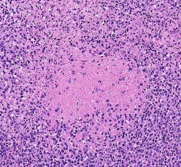
Volume 26 Number 8| Aug 2020|
Dermatology Online Journal || Case Report 26(8):6
had been established. Curiously, at the time KFD was Case Discussion
diagnosed, her ANA titer was 1:2560 with a speckled Kikuchi-Fujimoto disease was described almost
pattern. However, an elevated titer in isolation does simultaneously by Kikuchi and Fujimoto in 1972
not warrant a diagnosis of SLE by the American [9,10]. Although it has been documented in patients
College of Rheumatology or the Systemic Lupus of various racial and ethnicities, there is a noted
International Collaborating Clinics criteria [7,8]. The prevalence among women of east Asian ancestry
patient was lost to dermatologic follow-up for with a typical disease onset in the early twenties to
almost a decade before presenting with the mid-thirties [4]. Kikuchi-Fujimoto disease most
preauricular plaque. frequently involves the cervical lymph nodes,
On 6-month follow-up, her symptoms have although generalized lymphadenopathy and
responded well to hydroxychloroquine, involvement of retroperitoneal, peritoneal,
methotrexate, and intramuscular triamcinolone. mediastinal, inguinal, axillary, supraclavicular, and
Scarring, mild lipoatrophy, and pigmentary intraparotid nodes has been described [4,11-14].
alterations are evident at the right preauricular Hepatosplenomegaly has also been reported [13].
region despite treatment with systemic, topical, and Patients usually describe low-grade fever, malaise,
intralesional corticosteroids. However, the plaque and night sweats [4]. Laboratory abnormalities are
has not increased in diameter (Figure 1B). nonspecific and can include anemia, leukopenia,
A B C
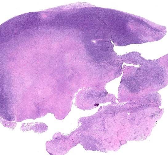
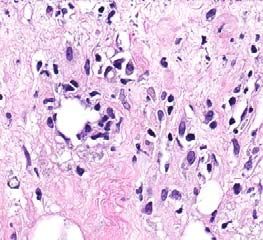
D E F
Figure 3. Necrotizing lymphadenitis. A) A large portion of lymph node architecture is effaced by necrosis. Some smaller, clearly
demarcated zones of necrosis are visible at the node periphery. H&E, 2x. B) CD3 immunostain highlights a preponderance of T-cells in
areas with necrosis. CD3, 2x. C) CD20 highlights relatively sparse B-cells in these areas. CD20, 2x. D) A roughly circumscribed, demarcated
focus of necrosis expanding the paracortex in the most preserved portion of the lymph node is surrounded by histiocytes. H&E, 100x. E)
Crescentic histiocytes (circled) adjacent to the necrosis are accompanied by immunoblasts, foamy histiocytes, and plasmacytoid cells.
H&E, 40x. F) Macrophages ingesting nuclear debris are present at the center of the necrosis. Neutrophils and eosinophils were not
identified. H&E, 40x.
-3-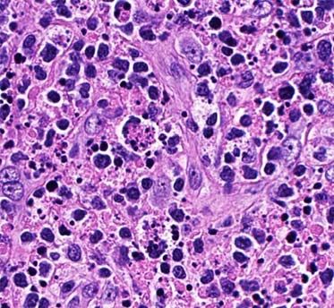
Volume 26 Number 8| Aug 2020|
Dermatology Online Journal || Case Report                                                                         26(8):6

presence of atypical lymphocytes on peripheral                  The combination of discoid lupus and LEP is an
blood smear, elevated liver function tests, elevated            uncommon         finding      in   cutaneous      lupus
lactate dehydrogenase, and elevated erythrocyte                 erythematosus, identified in only 2% of patients [23].
sedimentation rate [15]. Anti-nuclear antibodies                It has a female-to-male ratio of 4:1 [24].
have been described in patients with KFD. In one                Approximately one third of patients with LEP will
investigation, however, 23 of 33 patients with KFD              have discoid lupus and only 10%-24% of patients
and a positive ANA titer had or developed SLE.                  with LEP meet criteria for SLE [24-26]. The differential
Overall, 13-25% of patients with KFD-like disease will          diagnosis includes other autoimmune disease-
have a prior, concomitant, or subsequent diagnosis              associated panniculitides [27] and subcutaneous
of SLE [16-18]. Nonetheless, most patients with KFD             panniculitis-like T-cell lymphoma (SPTCL), which can
never meet criteria for SLE. Therefore, it is unclear           be distinguished by lymphoid cytologic atypia, a
whether KFD represents a forme fruste of SLE or an              preponderance of cytotoxic CD8-positive cells
etiologically distinct lymphadenopathy with both                surrounding adipocytes, Ki-67 hotspots, and T-cell
clinical and histologic overlap [2]. The concomitant            clonality [28,29]. In rare circumstances, KFD can also
occurrence of KFD in autoimmune diseases distinct               have skin manifestations that mimic lupus replete
from SLE [13,19,20] offers equivocal evidence to                with panniculitis. Of great importance to
support the latter postulate; however, SLE is the               dermatologists, some investigations have reported
most commonly reported autoimmune disease                       skin findings in 33% to 40% of patients with KFD,
association [12,13]. Nonetheless, a clinical work-up is         with a majority of biopsies revealing interface
required to exclude SLE in any patient receiving a              dermatitis [5]. Notably, Kim et al. presented a series
diagnosis of KFD regardless of their antecedent                 of 16 patients with cutaneous KFD after excluding
medical history.                                                from the study patients with concomitant or
                                                                previous diagnoses of SLE [6]. In their case series,
In KFD, lymph nodes show paracortical expansion by              vacuolar interface changes, necrotic keratinocytes,
demarcated histiocytic infiltrates with central                 superficial and deep lymphohistiocytic infiltrate,
necrosis.     Karyorrhectic     debris,    histiocytes          dermal mucin, and less frequent panniculitis were
(sometimes with crescentic nuclei), and enlarged                identified. In these exceedingly rare cases, a
lymphoid cells resembling plasmacytoid dendritic                complete rheumatologic workup and follow-up is
cells and immunoblasts are often present [15,16]. In            required to distinguish KFD with cutaneous
cases with little necrosis, these infiltrates are               manifestations from SLE.
sometimes confused with lymphoma [12].
                                                                Interestingly, Notaro et al. reported a case of SPTCL
Conspicuously absent from KFD are neutrophils,
                                                                in a patient with KFD [30]. SPTCL is conventionally
which are sometimes present in lupus                            understood to be an indolent cytotoxic T-cell
lymphadenitis. Lupus lymphadenitis and KFD share                lymphoma comprised of CD8-positive lymphocytes
the remainder of these histologic findings. However,            that rim adipocyte lobules. Although we now
the presence of neutrophils, hematoxylin bodies,                understand that a proportion of cases are hereditary,
and the Azzopardi phenomenon would favor lupus                  owing to an underpinning mutation in HAVCR2
[4]. Yet, many cases of lupus lymphadenitis,                    [31,32], the etiology of many cases remains elusive.
including the present example, are devoid of these              SPTCL occasionally shows striking histologic overlap
distinguishing     features    [3,21,22].   Extensive           with lupus panniculitis [33] and some patients,
effacement of nodal architecture, which was                     including those with wild type HAVCR2, have
identified in the present case and contrasts with the           underlying autoimmune diatheses including SLE
patchy, delineated foci of paracortical necrosis often          [34]. Although there is excellent overall survival, the
seen in KFD, is also more common in lupus [4].                  low SPTCL mortality rate is almost invariably
However, the amount and localization of necrosis                attributable to concomitant hemophagocytic
can vary widely in KFD [12].                                    lymphohistiocytosis (HLH), which can occur albeit

                                                          -4-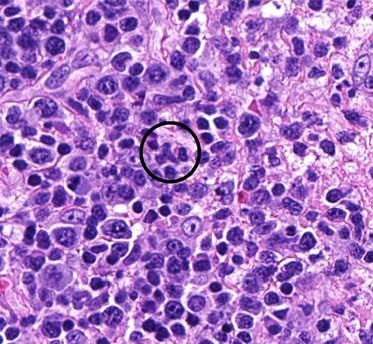
Volume 26 Number 8| Aug 2020|
Dermatology Online Journal || Case Report                                                                                                  26(8):6

with a much lower frequency in the settings of SLE,                             onset of a lymphadenitis that was clinically and
KFD, and SLE/KFD overlap [13,35,36]. Therefore,                                 histologically indistinguishable from KFD. The
patients presenting with KFD or LEP should also be                              development of cutaneous lupus erythematosus in a
monitored for the development of HLH and efforts,                               patient with concomitant or pre-existing KFD should
including T-cell clonality testing, can be considered                           prompt a workup for SLE. However, care should be
in select cases to help exclude SPTCL. T-cell clonality                         taken to avoid an over-diagnosis of SLE since skin
testing was not ordered in our case as the clinical and                         manifestations are common in KFD and the disease
histomorphologic findings argued against SPTCL. In                              is often self-limited. Exceedingly rare cases, such as
particular, the presence of interface changes,                                  ours, underscore how it is important for patients with
localization on the face, and lipoatrophy were more                             a KFD diagnosis to be followed closely since there is
in keeping with LEP. Although the patient described                             presently no method of determining which patients
by Notaro et al. had developed HLH as a sequela of                              will develop SLE. Nonetheless, ANA testing is
his concomitant SPTCL and KFD, he did not meet                                  indicated. Furthermore, when there is extensive
criteria for SLE.                                                               involvement of the subcutaneous fibroadipose
                                                                                tissue, SPTCL should be excluded since SPTCL is
                                                                                associated with a much greater risk of HLH than what
Conclusion                                                                      is generally reported in SLE, KFD, and LEP. It is
Although a correlation between KFD and SLE has                                  incumbent          on       dermatologists        and
been established in the hematology literature, it is                            dermatopathologists to be familiar with KFD and
important for dermatologists, particularly those who                            ensure that KFD patients presenting with skin
specialize in the care of patients with rheumatologic                           findings receive the appropriate management.
illnesses, to be familiar with KFD and consider it in
the differential diagnosis of SLE when patients
present with rash and lymphadenitis. Our patient                                Potential conflicts of interest
fulfilled clinical criteria for SLE years following the                         The authors declare no conflicts of interests.

References
1.   Ruaro B, Sulli A, Alessandri E, et al. Kikuchi-Fujimoto's disease                the Systemic Lupus International Collaborating Clinics
     associated with systemic lupus erythematous: difficult case report               classification criteria for systemic lupus erythematosus. Arthritis
     and literature review. Lupus. 2014;23:939-944. [PMID: 24739458].                 Rheum. 2012;64:2677-2686. [PMID: 22553077].
2.   Goldblatt F, Andrews J, Russell A, et al. Association of Kikuchi-          9.    Kikuchi M. Lymphadenitis showing focal reticulum cell
     Fujimoto's disease with SLE. Rheumatology (Oxford, England).                     hyperplasia with nuclear debris and phagocytes: a
     2008;47:553-554. [PMID: 18304938].                                               clinicopathological study. Nippon Ketsueki Gakkai Zasshi.
3.   Baenas DF, Diehl FA, Haye Salinas MJ, et al. Kikuchi-Fujimoto                    1972;35:379-380. https://ci.nii.ac.jp/naid/10008336899/en/.
     disease and systemic lupus erythematosus. Int Med Case Rep J.              10.   Fujimoto Y, Kozima Y, Yamaguchi M. Cervical subacute
     2016;9:163-167. [PMID: 27418858].                                                necrotizing lymphadenitis. A new clinicopathological entity.
4.   Behdadnia A, Allameh SF, Gharabaghi MA, et al. Systemic Kikuchi-                 Nihon        Naika        Gakkai      Zasshi.    1972;30:920-927.
     Fujimoto disease bordering lupus lymphadenitis: A fresh look?                    https://ci.nii.ac.jp/naid/10016261693/en/.
     Intractable Rare Dis Res. 2016;5:301-305. [PMID: 27904829].                11.   Bosch X, Guilabert A, Miquel R, et al. Enigmatic Kikuchi-Fujimoto
5.   Atwater AR, Longley BJ, Aughenbaugh WD. Kikuchi's disease: case                  disease: a comprehensive review. Am J Clin Pathol. 2004;122:141-
     report and systematic review of cutaneous and histopathologic                    152. [PMID: 15272543].
     presentations. J Am Acad Dermatol. 2008;59:130-136. [PMID:                 12.   Dorfman RF, Berry GJ. Kikuchi's histiocytic necrotizing
     18462833].                                                                       lymphadenitis: an analysis of 108 cases with emphasis on
6.   Kim JH, Kim YB, In SI, et al. The cutaneous lesions of Kikuchi's                 differential diagnosis. Semin Diagn Pathol. 1988;5:329-345. [PMID:
     disease: a comprehensive analysis of 16 cases based on the                       3217625].
     clinicopathologic,            immunohistochemical,            and          13.   Dumas G, Prendki V, Haroche J, et al. Kikuchi-Fujimoto disease:
     immunofluorescence studies with an emphasis on the differential                  retrospective study of 91 cases and review of the literature.
     diagnosis. Hum Pathol. 2010;41:1245-1254. [PMID: 20434191].                      Medicine (Baltimore). 2014;93:372-382. [PMID: 25500707].
7.   Hochberg MC. Updating the American College of Rheumatology                 14.   Turner RR, Martin J, Dorfman RF. Necrotizing lymphadenitis. A
     revised criteria for the classification of systemic lupus                        study of 30 cases. Am J Surg Pathol. 1983;7:115-123. [PMID:
     erythematosus. Arthritis Rheum. 1997;40:1725. [PMID: 9324032].                   6859386].
8.   Petri M, Orbai AM, Alarcon GS, et al. Derivation and validation of         15.   Hutchinson CB, Wang E. Kikuchi-Fujimoto disease. Arch Pathol Lab

                                                                          -5-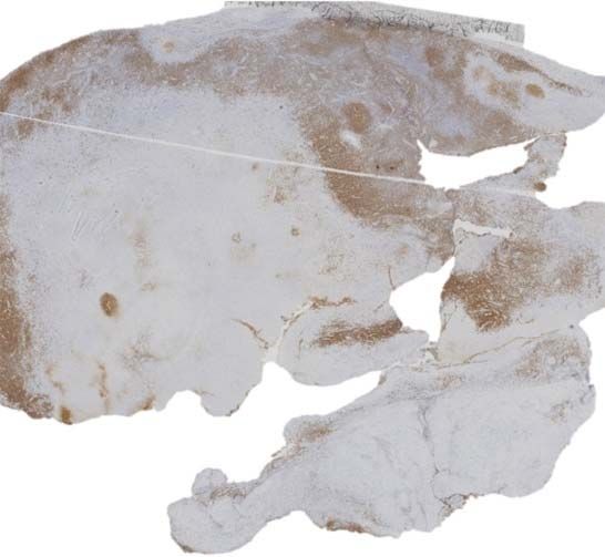
Volume 26 Number 8| Aug 2020|
Dermatology Online Journal || Case Report                                                                                                     26(8):6

    Med. 2010;134:289-293. [PMID: 20121621].                                          29524269].
16. Sopena B, Rivera A, Chamorro A, et al. Clinical association                   28. LeBlanc RE, Tavallaee M, Kim YH, et al. Useful Parameters for
    between Kikuchi’s disease and systemic lupus erythematosus: A                     Distinguishing Subcutaneous Panniculitis-like T-Cell Lymphoma
    systematic literature review. Semin Arthritis Rheum. 2017;47:46-52.               From Lupus Erythematosus Panniculitis. Am J Surg Pathol.
    [PMID: 28233572].                                                                 2016;40:745-754. [PMID: 26796503].
17. el-Ramahi KM, Karrar A, Ali MA. Kikuchi disease and its association           29. Sitthinamsuwan P, Pattanaprichakul P, Treetipsatit J, et al.
    with systemic lupus erythematosus. Lupus. 1994;3:409-411.                         Subcutaneous Panniculitis-Like T-Cell Lymphoma Versus Lupus
    [PMID: 7841995].                                                                  Erythematosus Panniculitis: Distinction by Means of the
18. Kucukardali Y, Solmazgul E, Kunter E, et al. Kikuchi-Fujimoto                     Periadipocytic Cell Proliferation Index. Am J Dermatopathol.
    Disease: analysis of 244 cases. Clin Rheumatol. 2007;26:50-54.                    2018;40:567-574. [PMID: 29742552].
    [PMID: 16538388].                                                             30. Notaro E, Shustov A, Chen X, et al. Kikuchi-Fujimoto Disease
19. Ohta A, Matsumoto Y, Ohta T, et al. Still's disease associated with               Associated With Subcutaneous Panniculitis-Like T-Cell
    necrotizing lymphadenitis (Kikuchi's disease): report of three                    Lymphoma. Am J Dermatopathol. 2016;38:e77-80. [PMID:
    cases. J Rheumatol. 1988;15:981-983. [PMID: 3418650].                             26844615].
20. Wilkinson CE, Nichol F. Kikuchi-Fujimoto disease associated with              31. Gayden T, Sepulveda FE, Khuong-Quang DA, et al. Germline
    polymyositis. Rheumatology (Oxford). 2000;39(11):1302-1304.                       HAVCR2 mutations altering TIM-3 characterize subcutaneous
    [PMID: 11085822].                                                                 panniculitis-like T cell lymphomas with hemophagocytic
21. Gordon JK, Magro C, Lu T, et al. Overlap between systemic lupus                   lymphohistiocytic syndrome. Nat Genet. 2018;50(12):1650-1657.
    erythematosus and Kikuchi Fujimoto disease: a clinical pathology                  [PMID: 30374066].
    conference held by the Department of Rheumatology at Hospital                 32. Polprasert C, Takeuchi Y, Kakiuchi N, et al. Frequent germline
    for Special Surgery. HSS J. 2009;5:169-177. [PMID: 19609622].                     mutations of HAVCR2 in sporadic subcutaneous panniculitis-like
22. Zuo Y, Foshat M, Qian YW, et al. A Rare Case of Kikuchi Fujimoto's                T-cell lymphoma. Blood Adv. 2019;3:588-595. [PMID: 30792187].
    Disease with Subsequent Development of Systemic Lupus                         33. Bosisio F, Boi S, Caputo V, et al. Lobular panniculitic infiltrates with
    Erythematosus. Case Rep Rheumatol. 2012;2012:325062. [PMID:                       overlapping histopathologic features of lupus panniculitis (lupus
    23346446].                                                                        profundus) and subcutaneous T-cell lymphoma: a conceptual and
23. Peters MS, Su WP. Lupus erythematosus panniculitis. Med Clin                      practical dilemma. Am J Surg Pathol. 2015;39:206-211. [PMID:
    North Am. 1989;73:1113-1126. [PMID: 2671535].                                     25118815].
24. Martens PB, Moder KG, Ahmed I. Lupus panniculitis: clinical                   34. Willemze R, Jansen PM, Cerroni L, et al. Subcutaneous
    perspectives from a case series. J Rheumatol. 1999;26:68-72.                      panniculitis-like T-cell lymphoma: definition, classification, and
    [PMID: 9918242].                                                                  prognostic factors: an EORTC Cutaneous Lymphoma Group Study
25. Watanabe T, Tsuchida T. Lupus erythematosus profundus: a                          of 83 cases. Blood. 2008;111:838-845. [PMID: 17934071].
    cutaneous marker for a distinct clinical subset? Br J Dermatol.               35. Ahn SS, Lee B, Kim D, et al. Evaluation of macrophage activation
    1996;134:123-125. [PMID: 8745897].                                                syndrome in hospitalised patients with Kikuchi-Fujimoto disease
26. Ng PP, Tan SH, Tan T. Lupus erythematosus panniculitis: a                         based on the 2016 EULAR/ACR/PRINTO classification criteria. PLoS
    clinicopathologic study. Int J Dermatol. 2002;41:488-490. [PMID:                  One. 2019;14:e0219970. [PMID: 31318961].
    12207763].                                                                    36. Liu AC, Yang Y, Li MT, et al. Macrophage activation syndrome in
27. Santos-Briz A, Calle A, Linos K, et al. Dermatomyositis panniculitis:             systemic lupus erythematosus: a multicenter, case-control study
    a clinicopathological and immunohistochemical study of 18                         in China. Clin Rheumatol. 2018;37:93-100. [PMID: 28409239].
    cases. J Eur Acad Dermatol Venereol. 2018;32:1352-1359. [PMID:

                                                                            -6-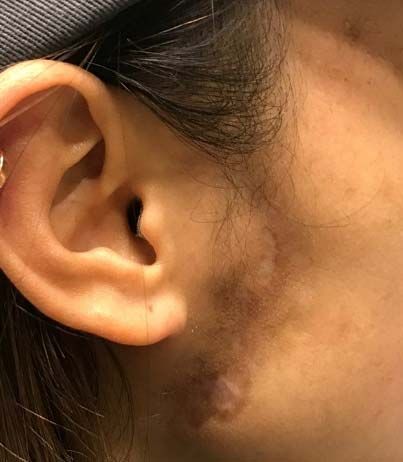
You can also read