Retinopathy From a Green Laser Pointer - A Clinicopathologic Study
←
→
Page content transcription
If your browser does not render page correctly, please read the page content below
CLINICAL SCIENCES

Retinopathy From a Green Laser Pointer
A Clinicopathologic Study
Dennis M. Robertson, MD; Jay W. McLaren, PhD; Diva R. Salomao, MD; Thomas P. Link, CRA

Objective: To report retinopathy following exposure to             5 mW. Retinopathy was observed 24 hours after laser ex-
light from a commercially available class 3A green laser           posure. This was characterized by a yellowish discolora-
pointer.                                                           tion at the level of the retinal pigment epithelium (RPE)
                                                                   in the subfoveal region and at the site superior to the
Methods: A 55-year-old woman with a ring melanoma                  macula where the retina received 15 minutes of laser ex-
was scheduled for enucleation. The eye (visual acuity 20/          posure. Each site developed granular changes at the level
20) had a healthy-appearing macular retina. The retina             of the RPE within 5 days of exposure. Histologic study
was exposed to light from a commercially available class           showed RPE damage in the exposed subfoveal and para-
3A green laser: 60 seconds to the fovea, 5 minutes to a            foveal regions.
site 5° below the fovea, and 15 minutes to a site 5° su-
perior to the fovea. Color photographs were obtained be-           Conclusion: A class 3A green laser pointer caused vis-
fore and after exposure. The eye was enucleated 20 days            ible retinopathy in the human eye with exposures as short
after exposure.                                                    as 60 seconds.

Results: Laser power measurements averaged less than               Arch Ophthalmol. 2005;123:629-633

                                W
                                                        E REPORT THE DE -         patient was unable to discern any visual
                                                        velopment of clini-       abnormality and could not discern a sco-
                                                        cally recognizable        toma on tangent screen or Amsler grid test-
                                                        retinopathy follow-       ing 6 and 20 days after exposure. Twenty-
                                                        ing exposure of the       four hours after exposure, fundus
                                human eye to light from a commercially            examination revealed a distinct yellow-
                                available class 3A green laser pointer. A         ish discoloration in the subfoveal region
                                55-year-old woman with a ring mela-               at the level of the retinal pigment epithe-
                                noma that involved the ciliary body was           lium (RPE). An area of yellowish discol-
                                scheduled for enucleation. The eye had a          oration was also recognized at the retinal
                                healthy-appearing macular retina and good         site exposed to the laser for 15 minutes.
                                visual function (visual acuity 20/20). The        A delicate granular irregularity devel-
                                patient agreed to have her retina exposed         oped in each site at the level of the pig-
                                to laser light from the green laser pointer       ment epithelium and was recognizable at
                                before enucleation. Continuous expo-              the 6- and 20-day follow-ups. Histologic
                                sure was directed to the fovea for 1 minute,      study of the enucleated eye showed a cho-
                                to the retina 5° below fixation for 5 min-        roidal melanoma in the ciliary body, and
                                utes, and to the retina 5° above fixation for     thick sections revealed apical displace-
                                15 minutes. The retina was evaluated oph-         ment of the nuclei of some RPE cells in
                                thalmoscopically and the fundus was docu-         the subfoveal region. The pigment gran-
                                mented by color photography before, 24            ules were also displaced into the outer re-
                                hours after, and 6 and 20 days after laser        ceptor layer, and many intracytoplasmic
                                exposure. Transient pink afterimages were         granules displayed irregular shapes and
Author Affiliations:
Department of Ophthalmology,
                                observed by the patient for approxi-              density characteristics of melanofuscin.
Mayo Clinic, Mayo Foundation,   mately 4 minutes after the laser expo-            Displacement of some of the pigment
and Mayo Medical School,        sure. The patient’s visual acuity was re-         epithelial cells into the subretinal space
Rochester, Minn.                corded as 20/20 the day after exposure and        was also observed at the exposed site.
Financial Disclosure: None.     20/20 at 6 and 20 days after exposure. The        Although in this experiment a class 3A

                 (REPRINTED) ARCH OPHTHALMOL / VOL 123, MAY 2005     WWW.ARCHOPHTHALMOL.COM
                                                             629

                                   ©2005 American Medical Association. All rights reserved.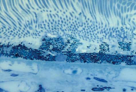
green laser pointer caused retinopathy with exposures                 low the center of the Amsler grid. The pupil of the eye that con-
as short as 60 seconds, the recognized ophthalmoscopic                tained the tumor was dilated to 8 mm with 2% cyclopentolate
and histologic abnormalities were unaccompanied by                    hydrochloride and 10% phenylephrine hydrochloride. The other
visually perceptible abnormalities.                                   eye was doubly patched. The eye that contained the mela-
                                                                      noma was subjected to 3 durations of exposure from the laser
    In a previous study,1 we reported the absence of reti-
                                                                      pointer.
nal injury following retinal exposures of laser light from                The retina was exposed to light from a handheld green la-
commercially available class 3A red laser pointers with               ser marketed as a laser pointer (LightVision Technologies Corp,
powers of 1, 2, and 5 mW. Three human eyes were ex-                   Kaoyuan, Taiwan). Light output was continuous (not pulsed)
posed to light from these laser pointers for 1, 5, and 15             and specified by the manufacturer as less than 5 mW at 532
minutes. We documented no functional, ophthalmo-                      nm. The beam power was measured with a radiometer (IL 700,
scopic, histologic, or ultrastructural abnormalities that             SEE-100 probe; International Light Inc, Newburyport, Mass).
could be attributed to the laser exposures. We con-                   From information relating to the retinal hazards of intrabeam
cluded that the risk to the adult human eye from tran-                viewing of lasers specified by the American National Standard
sient exposure to light from these red laser sources was              for the Safe Use of Lasers,4 we calculated maximum permis-
                                                                      sible exposure at various exposure times.
negligible, although 2 credible reports2,3 have been pub-
                                                                          Exposures were administered as follows. The patient fix-
lished of visible retinal abnormalities after exposure to             ated her gaze for 60 seconds directly at the laser beam as it passed
red laser pointers in 2 young patients, one 11 years old              through the center of the aperture in the Amsler grid. Then the
and the other 19 years old. Green laser pointers have been            patient fixated her gaze for 5 minutes on the fixation target 2½
used interchangeably with red laser pointers by some lec-             squares below the aperture and the laser beam. The last expo-
turers, and green laser pointers are increasingly being used          sure was a 15-minute fixated gaze on the fixation target 2½
by amateur astronomers as pointers to deep sky objects.               squares above the aperture and the laser beam. Normal blink-
Unlike the beam of the red laser, which cannot be seen                ing was allowed during the exposures. During each exposure,
well in ordinary night atmosphere, the beam from the                  the patient’s fixation was confirmed by one of us (D.M.R.), and
green laser can be easily seen in the deep night sky, where           the laser beam was maintained in the central 2 mm of the pa-
                                                                      tient’s widely dilated pupil. After each exposure the patient was
it can point to a single star. Additionally, since the green
                                                                      asked to report any recognized afterimages or photopsias. Im-
laser is visible in the daytime, when directed to outdoor             mediately after responding to this request, the patient was in-
objects of interest, the green laser pointer has proved use-          structed to gaze at the center of a standard Amsler grid and re-
ful to some instructors of outdoor painting, landscape                port any defects in the grid. The patient wore corrective eyewear
design, architecture, and construction. Since the retina              for this last assessment.
is increasingly more sensitive to shorter wavelengths, we                 The patient returned the following day, 6 days after expo-
were interested in learning if the green laser pointer could          sure, and again 20 days after exposure for measurement of the
cause retinopathy in the human eye.                                   Snellen visual acuity, ophthalmoscopic examination with slit-
                                                                      lamp biomicroscopy aided by a Hruby lens and the 90-diopter
                                                                      (D) and 60-D fundus viewing lenses, and color photographic
                     REPORT OF A CASE                                 documentation of the fundus. The retina was examined by ocu-
                                                                      lar coherence tomography (OCT) 24 hours after laser expo-
A 55-year-old woman with a ring melanoma of the cili-                 sure. The central visual field was studied 24 hours and 20 days
ary body was scheduled for enucleation. The eye was nor-              after laser exposure with the Amsler grid and tangent screen
                                                                      evaluations with a 1-mm white target. Sites in the fundus that
motensive and had an uncorrected visual acuity of 20/
                                                                      were exposed to the laser light were carefully inspected for ab-
20. The patient consented to participate in an experiment             normalities. These sites included the fovea and the RPE com-
during which a green laser pointer would expose her retina            plex 5° superior and 5° inferior to the fovea.
to light for intervals of up to 15 minutes. The study was
approved by our institutional review board before the ex-
periment, and our patient was fully informed of the na-                                           RESULTS
ture of the experiment and gave verbal and written in-
formed consent to participate.                                        The beam power of our green laser was variable and be-
                                                                      tween 3 and 7 mW, although the manufacturer stated that
                          METHODS                                     the light output was less than 5 mW. The maximum per-
                                                                      missible exposure is 0.39 mW for exposures between 1
An apparatus was designed to direct the laser beam from a class       and 15 minutes, assuming a 7-mm limiting aperture (pu-
3A green laser pointer through a hole (5 mm in diameter cre-          pil diameter).4 Retinal exposure from our laser ex-
ated with a simple paper punch) in the center of a black Ams-         ceeded this limit by 8 to 18 times.
ler grid and then into the patient’s pupil to target the retina.         Pretreatment evaluation of the fundus of our patient
This device was similar to that used by Robertson et al1 (as shown    revealed the presence of a small choroidal nevus be-
in their Figure 1). The apparatus was arranged on a slitlamp          neath the inferior retinal vascular arcade. The central
so the patient’s head could be positioned comfortably during          macula appeared normal (Figure 1A). The visual acu-
the exposure. A paper clip and a rubber band held the switch
of the laser on continuously. The front aperture of the laser
                                                                      ity was 20/20 uncorrected. After exposures to the laser
pointer was fixed 15 cm from the estimated location of the pos-       pointer, the patient observed pink discoloration within
terior pole of the eye (macular retina). Two 1-mm white fixa-         her central visual field, which faded within 4 minutes of
tion targets were placed on the Amsler grid, one 2½ squares           each laser exposure. The visual acuity was 20/25 within
above and one 2½ squares below the center of the Amsler grid,         3 minutes of exposure. No functional disturbance in vi-
thereby subtending angles approximately 5° above and 5° be-           sual acuity or the central visual field could be discerned

                   (REPRINTED) ARCH OPHTHALMOL / VOL 123, MAY 2005       WWW.ARCHOPHTHALMOL.COM
                                                               630

                                       ©2005 American Medical Association. All rights reserved.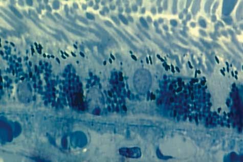
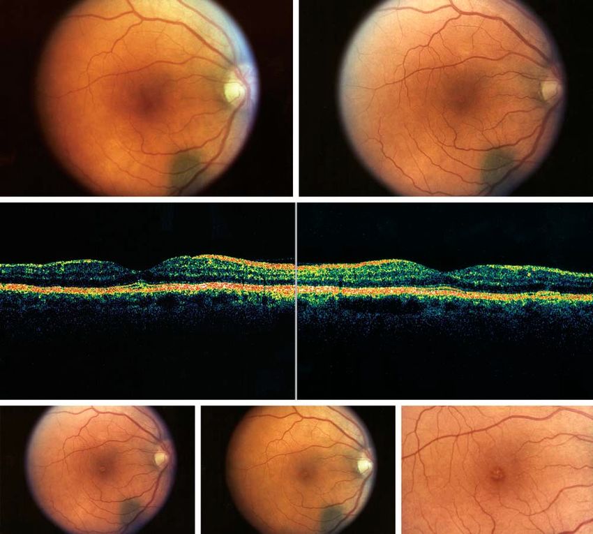
A                                                                                   B

   C

   D                                                       E                                                       F

Figure 1. Composite showing the fundus photographs and optical coherence tomographic images before and after exposure to the green laser pointer.
A, Normal-appearing macula. An incidental choroidal nevus is visible inferior to the disc. B, Twenty-four hours after exposure to the green laser pointer, subtle
yellowish discoloration at the level of the retinal pigment epithelium (site of 60-second exposure to the laser pointer) is apparent; abnormal yellowish discoloration
superior to the fovea where the site was continuously exposed for 15 minutes to the laser beam is also apparent. C, Ocular coherence tomogram that shows tissue
thickening at the level of the retinal pigment epithelium in the subfoveal region (top) and the area of the fundus superior to the fovea where the retina was exposed
to the laser for 15 minutes (bottom, arrow). D, Six days after exposure to the green laser pointer, the 2 sites identified in panel B now exhibit a delicate granular
irregularity at the level of the retinal pigment epithelium. E, Twenty days following exposure. The 2 sites identified in panels B and D are less evident. The
abnormality in the foveal region shows a light creamy discoloration. F, Foveal region shows irregular discoloration (original magnification ⫻2).

at subsequent visits 24 hours, 6 days, and 20 days after                               rior to the fovea where the retina was exposed to the la-
laser exposure. The uncorrected visual acuity was 20/20                                ser for 15 minutes (Figure 1C). Each of these 2 sites de-
at each of these follow-up visits.                                                     veloped a delicate granular irregularity at the level of the
    Twenty-four hours after laser exposure, an ophthal-                                pigment epithelium that was visible at the 6-day fol-
moscopically distinct yellowish discoloration appeared                                 low-up (Figure 1D). Some of this granularity persisted
in the subfoveal region at the level of the RPE (site of 60-                           at this follow-up, but by 20 days the abnormality at the
second exposure) (Figure 1B). No abnormality was vis-                                  fovea was characterized primarily by a more delicate
ible in the fundus at the site of the 5-minute exposure,                               creamy discoloration (Figure 1E and F). The eye was
but a distinct abnormal yellowish discoloration was ap-                                enucleated 20 days after laser exposure.
parent superior to the fovea where the retina had been                                     The eye was received fresh from the operating room.
exposed to the laser beam for 15 minutes (Figure 1B).                                  It was sectioned at the equator. The anterior segment was
An OCT examination 24 hours after exposure sug-                                        placed in 10% buffered formalin and fixed for 48 hours
gested tissue thickening at the level of the RPE in both                               before gross examination. The posterior segment was ex-
the subfoveal location and the area of the fundus supe-                                amined and dissected immediately. A small choroidal ne-

                      (REPRINTED) ARCH OPHTHALMOL / VOL 123, MAY 2005                      WWW.ARCHOPHTHALMOL.COM
                                                                  631

                                               ©2005 American Medical Association. All rights reserved.
Figure 2. Composite showing both light microscopic and electron
A microscopic findings in the retina 20 days after laser exposure. A, Epoxy
resin (Epon)–embedded thick section shows focal displacement of the retinal
pigment epithelial cells into the subretinal space. No abnormalities are seen
in the photoreceptor cells (epoxy resin–embedded toluidine blue stain,
original magnification ⫻100). B, Transmission electron microscopic study
shows clumping of pigment granules within the retinal pigment epithelial
cells and dispersed in the subretinal space. The cross-sectional area of the
outer segments appears normal (lead citrate, original magnification ⫻2500).
C, Transmission electron microscopic study shows apical displacement of
the nuclei of some pigment epithelial cells. Some pigment granules are
irregular in shape and show densities characteristic of melanofuscin (epoxy
resin–embedded toluidine blue stain, original magnification ⫻100).
vea were placed in glutaraldehyde and examined by trans-
mission electron microscopy.
Gross examination of the anterior segment showed
B clear cornea that measured 12 ⫻ 12 mm. The iris con-
tained a mass from 9- to 12-o’clock (5 ⫻ 3 ⫻ 2 mm) that
adhered to the posterior corneal surface and extended
posteriorly to the ciliary body. The anterior segment was
sectioned clockwise, and all sections were embedded for
histologic examination. Microscopically, a malignant
melanoma, mixed cell type (spindle and epithelioid), was
forming a predominant mass in the iris root that in-
vaded anteriorly the trabecular meshwork and ex-
tended posteriorly to invade the ciliary body muscle. How-
ever, isolated tumor cells were observed in the trabecular
meshwork and angle structures at approximately 360°.
This morphologic impression was confirmed by melan-A
immunostain, a melanoma marker.
Examination by transmission electron microscopy of
well-fixed tissue from the region of the fovea showed api-
cal displacement of the nuclei in some of the RPE cells
in addition to focal clumping of pigment granules within
the RPE cell cytoplasm. Many of the pigment granules
had irregular shapes and demonstrated densities char-
acteristic of melanofuscin granules. Distinct displace-
ment of RPE cells also occurred into the subretinal space
in some sections (Figure 2A-C). We were unable to iden-
tify any abnormalities in the choriocapillaries. The outer
segments of the photoreceptors appeared to be normal
except for some minimal disruption of the lamellae at-
tributed to prefixation autolysis (present both in the pos-
terior pole and a control site nasal to the disc). We could
C not identify abnormalities in the other sites exposed to
the laser.
COMMENT
In this experiment, we documented the development of
retinopathy in a human eye after purposeful exposure to
light from a green laser pointer. In previous studies with
red laser pointers, we failed to produce any evidence of
retinopathy despite exposures of the retina to continu-
ous light for up to 15 minutes.1 The fact that we were
able to demonstrate green laser pointer–induced reti-
nopathy with exposure times as short as 60 seconds may
not be surprising, since the human retina is much more
sensitive to shorter than longer wavelengths. Also, mela-
vus (2 mm) was noted inferior to the fovea. No other gross nin in the pigment epithelium absorbs more energy at
abnormalities were noted. Small portions of the fovea, shorter wavelengths than longer wavelengths.5
the macular region approximately 5 mm superior and in- The appearance of the lesion after 60 seconds of green
ferior to the fovea, and the nasal retina opposite the fo- laser exposure was similar to the clinical appearance of
(REPRINTED) ARCH OPHTHALMOL / VOL 123, MAY 2005 WWW.ARCHOPHTHALMOL.COM
632
©2005 American Medical Association. All rights reserved.solar retinopathy in patients who have stared at a solar mal injury that affects primarily the RPE. Although it re-
eclipse. The yellowish discoloration that was visible oph- mains comforting that the patient did not experience any
thalmoscopically probably represented a change at the visual abnormalities up to 20 days following laser expo-
level of the RPE where the pigment epithelium had re- sure, nevertheless the inducement of ophthalmoscopi-
ceived a mild thermal injury. Clinically, the discolora- cally visible photic damage along with the induced his-
tion did not appear at the inner retina in the region of tologic abnormalities suggests the need for caution with
greatest concentration of xanthophyll; an OCT study dem- the use of laser pointers and, more particularly, the green
onstrated thickening at the level of the pigment epithe- laser pointer. Fortunately, the risks to the human eye from
lium, which suggests that the ophthalmoscopically vis- transient exposure to light from laser pointers are mini-
ible abnormality was at the level of the pigment mized by the normal blink and aversion reflexes that oc-
epithelium. Why retinopathy was not visible 5° below fixa- cur within fractions of a second of exposure. Neverthe-
tion where the retina was exposed for 5 minutes cannot less, exposure of the retina to light from a commercially
be readily explained, but the presence of a relatively broad available green laser pointer carries a risk that is real; this
area of retinopathy superior to the fovea where the retina risk appears to exceed the risk from commercially avail-
had been exposed for 15 minutes suggests that the pa- able red laser pointers.
tient may have had difficulty maintaining precise fixa-
tion on the larger white target in the mounted apparatus Submitted for Publication: February 2, 2004; final re-
as opposed to the more precise foveal fixation when the vision received August 18, 2004; accepted September 28,
eye was gazing directly at the center of the laser beam. 2004.
Microsaccades, micronystagmus, and slow drifts in eye Correspondence: Dennis M. Robertson, MD, Mayo Clinic,
position during fixation of a small target for more than a 200 First St SW, Rochester, MN 55905.
few seconds can spread the area of retina exposed to a Funding/Support: This study was supported in part by
laser.6 Perhaps the greater excursions of the eye during a grant from Research to Prevent Blindness, Inc, New York,
fixation on the white target distributed the laser expo- NY.
sure across a greater area on the retina and allowed heat Acknowledgment: We acknowledge Cheryl Hann, MS,
dissipation so that the retina was not injured at 5 min- for her assistance with the transmission electron micros-
utes but was injured over a relatively broad region of 500 copy and Bonnie Ronken for her secretarial assistance.
to 700 µm after an exposure of 15 minutes.
Our patient was unable to recognize any defect in cen- REFERENCES
tral vision despite attempted efforts to identify a sco-
toma with the tangent screen using 1-mm targets and the 1. Robertson DM, Lim TH, Salomao DR, Link TP, Rowe RL, McLaren JW. Laser point-
Amsler grid study. The inability of the patient to recog- ers and the human eye: a clinicopathologic study. Arch Ophthalmol. 2000;118:
nize functional changes in vision may reflect either a true 1686-1691.
absence of functional damage or simply our inability to 2. Sell CH, Bryan JS. Maculopathy from handheld diode laser pointer. Arch Ophthalmol.
1999;117:1557-1558.
detect subtle changes in the central visual field function 3. Zamir E, Kaiserman I, Chowers I. Laser pointer maculopathy. Am J Ophthalmol.
with the testing methods used. The histologic study in- 1999;127:728-729.
dicated some damage to the RPE represented by displace- 4. American National Standard for Safe Use of Lasers. ANSI A136.1-2000. Orlando,
ment of the nuclei away from the basement membrane, Fla: Laser Institute of America; 2000.
dispersion of pigment granules, the development of mela- 5. Mainster MA. Wavelength selection in macular photocoagulation: tissue optics,
thermal effects, and laser systems. Ophthalmology. 1986;93:952-958.
nofuscin changes near the site of maximum exposure at 6. Ness JW, Zwick H, Stuck BE, et al. Retinal image motion during deliberate fixa-
the fovea, and displacement of RPE cells into the sub- tion: implications to laser safety for long duration viewing. Health Phys. 2000;
retinal space. These findings are consistent with ther- 78:131-142.
(REPRINTED) ARCH OPHTHALMOL / VOL 123, MAY 2005 WWW.ARCHOPHTHALMOL.COM
633
©2005 American Medical Association. All rights reserved.You can also read