A Chinese multicenter retrospective study of isolated increased nuchal translucency associated chromosome anomaly and prenatal diagnostic ...
←
→
Page content transcription
If your browser does not render page correctly, please read the page content below
www.nature.com/scientificreports
OPEN A Chinese multicenter retrospective
study of isolated increased
nuchal translucency associated
chromosome anomaly and prenatal
diagnostic suggestions
Hua Jin1,8, Juan Wang2,8, Guoying Zhang3, Hongyan Jiao4, Jiansheng Zhu5, Zhimin Li2,6,
Chen Chen7, XuanPing Zhang2, Huan Huang3* & JiaYin Wang2*
Extensive researches involving fetuses with multiple ultrasound anomalies have been conducted
over the years, but only few were focused on the isolated increased nuchal translucency (NT). On
top of that, these limited number of researches were all designed as single-arm studies and the
control group was missing. In this study, we conducted a multicenter, retrospective study using
amniotic fluid samples collected from 1197 pregnant women having fetuses with isolated increased
NT (INT group) or normal NT values (NNT group). Copy number variation sequencing (CNV-seq) was
performed to determine their chromosome status and pathogenic variations were validated using
SNP array. Overall, 59 chromosome aneuploidies, 34 pathogenic CNVs and 23 copy number variants
of unknown significance (VOUS CNVs) were discovered. the INT group had a significantly higher
proportion of aneuploidy (19.44%) and pathogenic CNV (8.33%) than the control group (3.49% and
2.30% respectively), and 88.89% of the pathogenic CNVs were related to heart defects. Additionally,
more male fetuses were presented in the INT group (68.51%), but they did not have a higher risk
(Relative Risk = 1.03) of carrying pathogenic chromosome variations than female fetuses. Our
results demonstrated that fetuses with isolated increased NT had a distinct pattern of chromosome
abnormality and majority of detected pathogenic CNVs could be linked to the congenital heart
disease. Furthermore, because a considerable proportion of pathogenic CNVs were detected, we
strongly recommend to perform a joint test of karyotyping and CNV analysis in prenatal diagnosis for
fetuses with isolated increased NT in order to decrease the incident of missed diagnosis.
For several decades, ultrasonography was widely used in prenatal diagnosis as a first-tier screening method
and the thickness of nuchal translucency (NT) measured between 11+0 and 13+6 weeks in the first trimester was
considered as an important indicator for Down s yndrome1,2. In the clinical management guideline published by
The American College of Obstetricians and Gynecologists (ACMG) and Society for Maternal Fetal Medicine, an
increased NT is determined when the thickness of nuchal translucency is more than 3.0 mm or above the 99th
percentile of the crown-rump length. The risk of trisomy 21 can be elevated to 1 in 46 when NT > 99th, whereas
the risk was 1 in 8806 when NT
www.nature.com/scientificreports/
NNT Group (N = 1089) INT Group (N = 108)
Anomalies N % N % RR† P
Normal 1004 92.19 77 71.30 0.77 < 0.001***
Aneuploidy 38 3.49 21 19.44 5.57 < 0.001***
Pathogenic CNV 25 2.30 9 8.33 3.62 < 0.001***
VOUS CNV 22 2.02 1 1.23 0.61 0.429
Table 1. Incidence of aneuploidy and CNV in NNT and INT groups. Abbreviations: NNT: normal nuchal
translucency; INT: increased nuchal translucency; CNV, copy-number variation; VOUS, variants of unknown
significance; RR, relative risk. † Relative risk is calculated as % cases of the anomaly in the INT Group/% cases
of the corresponding anomaly in the NNT Group, e.g. RR of aneuploidy: 19.44%/3.49% = 5.57. The NNT group
is used as the reference. ***P < 0.001.
shown that copy number variation (CNV) could also be detected in fetuses with increased NT4–9 and the cause
of increase by pathogenic CNVs (pCNVs) was through blood and lymph circulation d isruption10.
Fetus with increased NT is a frequently observed symptom. While majority studies primarily concentrated
on fetuses with multiple ultrasound indications, very few were focused on the ones with isolated increased NT.
Previous publications demonstrated that on top of the increased incidents of chromosome aneuploidies. vari-
ous percentages of pathogenic CNVs ranging from 0% to 12.8%11–13 and high prevalence of congenital heart
defects (CHD) and neurodevelopmental disorders were could also be found in fetuses with isolated increased
NT14. Unfortunately, these researches were all designed as a single-arm study, and without a proper control
group, results can only be compared with external historical data of other related work, which made it difficult
to diminish the effects caused by different enrollment criteria among studies. In this work, we conducted a
multicenter retrospective study with a control group to investigate chromosomal characteristics of fetuses with
isolated increased NT and next-generation sequencing (NGS) based CNV-seq method was chosen to identify
chromosome abnormalities because of its high sensitivity and s pecificity15,16. Our results showed that 8.33% of
the fetuses in the isolated increased NT group carried pCNVs, which was significantly different (P < 0.001, relative
risk = 3.62) than those in the normal NT group. Additionally, discrepancy in the distribution of pCNVs across
chromosomes between the two groups was also observed and the risk of CHD was higher in fetuses with isolated
increased NT. Furthermore, majority of cases in the isolated NT group were male (68.5%, 74/108), but their rela-
tive risk of having chromosome anomalies was the same (relative risk = 1.03) as female fetuses of the same group.
Results
Sample information. In this study, we analyzed 1197 amniotic fluid samples of high-risk Chinese pregnant
women with singleton pregnancy. Samples were successfully tested using next-generation sequencing platform
and they were all free of maternal contamination. Fetuses with normal NT and without any other ultrasound soft
marker abnormalities was assigned as the normal NT group (NNT), whereas the ones with isolated increased
NT was stratified as the isolated increased NT group (INT). The NNT group contained 1089 samples, the INT
group had 108 cases and the mean maternal age was 32.43 ± 5.71 years and 30.19 ± 4.89 years respectively.
Detection of chromosomal abnormalities. Overall, a total of 116 anomalies was detected among
1197 samples, 59 (4.93%) of which had chromosome aneuploidies, 34 (2.84%) carried pathogenic CNVs and
23 (1.95%) had copy number variants of unknown significance (VOUS CNVs). The INT group contained 21
(19.44%, 21/108) aneuploidy cases, 9 (8.33%, 9/108) pCNVs, 1 (0.93%, 1/108) VOUS CNVs and 77 (71.29%,
77/108) normal cases, whereas the ratio was 3.49% (38/1089), 2.30% (25/1089), 2.02% (22/1089) and 92.19%
(1004/1089) in the NNT group respectively (Table 1). In comparison with the NNT group, the ratio of fetuses
with normal karyotype, chromosomal aneuploidy and pCNV was significantly different in the INT group (all
P < 0.001) and the relative risk of normal, aneuploidy and pCNV was 0.77, 5.57 and 3.62 respectively. The ratio of
cases with VOUS CNVs, however, was not significantly different between the two groups (P = 0.429).
Comparison of chromosomal variations between NNT and INT group. The 108 samples in the
INT group, can be further divided into two sub-groups on the basis of NT thickness (Table 2). In the 3–4 mm
NT sub-group, the percentage of chromosomal aneuploidy, pathogenic CNV and VOUS CNV cases was 14.81%
(12/81), 7.41% (6/81) and 1.23% (1/81) respectively and it was 33.33% (9/27), 11.11% (3/27) and 0.00% (0/27)
in the NT > 4 mm sub-group. Statistical analysis did not show any significant difference in the rate of pathogenic
CNV and VOUS CNV between the two groups (P = 0.546, 0.562 respectively). The P value of chromosome ane-
uploidy was 0.035, demonstrating a sign of increase with increased NT thickness. Moreover, the distribution of
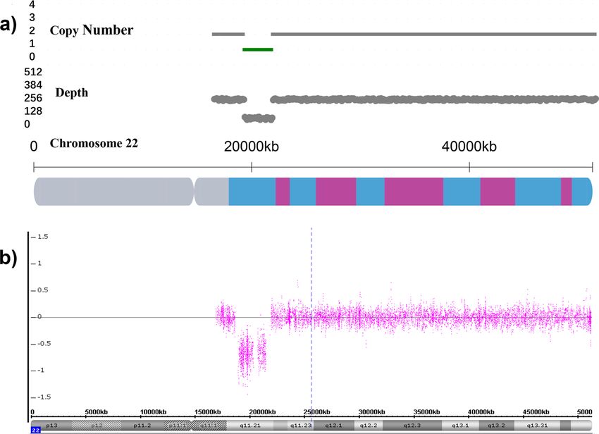
aneuploidy in the INT group was also different to that of the NNT group (Fig. 1a). In the INT group, trisomy
21 occupied 80.95% (17/21) of the aneuploidies and the rest of 4 cases were X0 (2/21, 9.52%), trisomy 18 (1/21,
4.76%) and trisomy 22 (1/21, 4.76%). The anomalies in the NNT group, on the other hand, were spread out
between trisomy 21 (14/38, 36.84%), trisomy X (8/38, 21.05%), trisomy 18 (5/38, 13.16%), X0 (5/38, 13.16%),
XYY (5/38, 13.16%) and trisomy 12 (1/38, 2.63%).
In terms of pathogenic CNVs, a total of 25 pCNVs in the NNT group and 9 pCNVs in the INT group were
discovered (Table 3). One example of the pCNV detected by CNV-seq and SNP array were illustrated in Fig. 2.
In the NNT group (Fig. 1b), 64% of the pathogenic CNVs were located on chromosome 16 (5/25, 20.00%),
Scientific Reports | (2021) 11:5596 | https://doi.org/10.1038/s41598-021-85108-6 2
Vol:.(1234567890)www.nature.com/scientificreports/
NT 3–4 mm (N = 81) NT > 4 mm (N = 27)
Anomalies N % N % RR† P
Normal 62 76.54 15 55.56 0.73 0.037*
Aneuploidy 12 14.81 9 33.33 2.25 0.035*
Pathogenic CNV 6 7.41 3 11.11 1.50 0.546
VOUS CNV 1 1.24 0 0 0 0.562
Table 2. Incidence of aneuploidy and CNV discovered in NT 3–4 mm and > 4 mm sub-groups. Abbreviations:
NT: nuchal translucency; CNV, copy-number variation; VOUS, variants of unknown significance; RR,
relative risk. † Relative risk is calculated as % cases of the anomaly in the NT > 4 mm group/% cases of the
corresponding anomaly in the NT 3–4 mm group, e.g. RR of aneuploidy: 33.33%/14.81% = 2.25. NT 3–4 mm
group is used as the reference. * P < 0.05.
Figure 1. Distribution of chromosome aneuplodies and pathogenic CNVs in NNT and INT groups. (a)
Number of chromosome aneuploidies showed distinct distribution patterns in NNT (blue) and INT groups
(orange); (b) Number of pathogenic CNVs revealed that distribution of CNVs was very different between NNT
(blue) and INT (orange) groups.
chromosome 17 (5/25, 20.00%) and chromosome 22 (7/25, 28.00%). The top 3 frequently observed pCNVs
were 22q11 duplication syndrome (5/25, 20.00%), 16p11.2 duplication syndrome (3/25, 12.00%) and Charcot-
Marie-Tooth syndrome type 1A (2/25, 8.00%). In comparison, 77.78% of pCNVs in the INT group were located
(Fig. 1b) on chromosome 2 (2/9, 22.22%), 16 (2/9, 22.22%) and 22 (3/9, 33.33%), and 22q11 deletion syndrome
(also known as the DiGeorge syndrome) had the highest occurrence rate (3/9, 33.33%).
Discussion
Increased NT detected by ultrasonography at 11+0 to 13+6 weeks is considered to be an effective indicator for
fetal anomalies. Currently, it is used to predict up to 50% of major fetal a bnormalities20 and it was associate
with high levels of several adverse outcomes including miscarriage, fetal anomalies and genetic syndromes21–24.
The mechanism of nuchal elevation is diversified. Cystic hygroma is a type of manifestation characterized by
fluid-filled cavities (cysts) and it was found to be associated with trisomy 13, trisomy 18 and Turner’s syndrome
(45, X0). The nuchal elevation exsited in non-45,X fetuses with cystic hygroma was caused by the proliferation
of lymphatic v essels25,26. In Turner’s syndrome, however, the primary cause of cystic hygroma is local aplasia of
lymphatic vessels, which interrupts the drainage into the jugular lymph s acs27. By contrast, nuchal oedemas, a
second type of NT elevation appearing as a non-echogenic area of over 3.5 mm below the skin and outside the
cervical vertebral column in ultrasonic scans28, is closely associated with trisomy 21. High level of precipitate
around bundles of collagen fibrils and over-expression of collagen type VI in nuchal skin of fetuses could be
observed28,29. Significant increase of hyaluronan was also found in fetuses with trisomy 21, suggesting that it may
be involved in the pathogenesis of increased N T30.
Scientific Reports | (2021) 11:5596 | https://doi.org/10.1038/s41598-021-85108-6 3
Vol.:(0123456789)www.nature.com/scientificreports/
Reported cases with CHD
and neurodevelopmental
Group Sample ID Fetus sex CNV position†
Size (Mb) Chromosomal disorder ‡
Source of evidence disorders§
NNT 00433 F chr2:g.10001_24135000dup 24.13 Partial trisomy 2p Lurie et al.43 CHD, ID
NNT 00376 F chr3:g.196965295_197415294dup 0.50 3q29 duplication syndrome #611936 ID
NNT 00745 F chr4:g.10001_4015500del 4.00 Wolf-Hirschhorn syndrome #194190 CHD, ID
NNT 00806 M chr5:g.10001_11160000del 11.15 Cri du Chat Syndrome #123450 ID, rare CHD
NNT 00117 F chr5:g.60001_6560000del 6.50 Cri du Chat Syndrome #123450 ID, rare CHD
NNT 00925 M chr8:g.12510001_23560000dup 11.05 8p23.1 duplication syndrome ORPHA:251076 CHD, ID
NNT 00324 M chr12:g.60001_34810001dup 34.75 Trisomy 12p ORPHA:1699 CHD, ID
NNT 00823 M chr15:g.31976624_32526623del 0.55 15q13.3 deletion syndrome #612001 ID, rare CHD
16p13.11 microduplication
NNT 01229 M chr16:g.15460001_16310000dup 0.85 ORPHA:261243 ID, rare CHD
syndrome
16p11.2p12.2 microduplication
NNT 00271 M chr16:g.25160001_26560000dup 1.40 ORPHA:261204 rare ID, rare Schizophrenia
syndrome
NNT 00702 F chr16:g.29460001_30210000dup 0.75 16p11.2 duplication syndrome #614671 ID; Schizophrenia
NNT 00692 F chr16:g.29610001_30210000dup 0.60 16p11.2 duplication syndrome #614671 ID, Schizophrenia
NNT 00319 M chr16:g.29610001_30160000del 0.60 16p11.2 deletion syndrome #611913 ID, ASD
NNT 00234 M chr17:g.1_25400000dup 25.40 Trisomy 17p ORPHA:261290 ID, rare CHD
Charcot-Marie-Tooth syndrome
NNT 00300 M chr17:g.14050001_15500000dup 1.45 #118220 HMSNs
type 1A
Charcot-Marie-Tooth syndrome
NNT 00457 M chr17:g.14100001_15500000dup 1.40 #118220 HMSNs
type 1A
NNT 01424 M chr17:g.16250001_22750000dup 6.50 Potocki-Lupski syndrome #610883 Autism, CHD, ID
Renal cysts and diabetes syn-
NNT 01532 M chr17:g.34800001_36250000del 1.45 #137920 rare ID
drome
NNT 00837 F chr22:g.18600001_21900004dup 3.30 22q11 duplication syndrome #608363 ID, rare CHD
NNT 01069 F chr22:g.18900001_21500004dup 2.60 22q11 duplication syndrome #608363 ID, rare CHD
NNT 00593 M chr22:g.18900001_25150004dup 6.25 22q11 duplication syndrome #608363 ID, rare CHD
NNT 00317 F chr22:g.18950001_2145000dup 2.50 22q11 duplication syndrome #608363 ID, rare CHD
NNT 00952 F chr22:g.20700005_21500004dup 0.80 22q11 duplication syndrome #608363 ID, rare CHD
NNT 00042 F chr22:g.18850001_21600004del 2.75 DiGeorge syndrome #188400 ADHD, CHD, rare Autism
NNT 00395 M chr22:g.45700005_51150004del 5.45 Phelan-McDermid syndrome #606232 rare ID
INT 10003 M chr2:g.61087852_61537851del 0.45 2p16.1-p15 deletion syndrome #612513 ADHD, Autism, ID
INT 00608 M chr2:g.178879852_184979851del 6.09 2q31.2 deletion syndrome #612345 ID, rare CHD
Interstitial 6q microdeletion 44
INT 00178 M chr6:g.88610001_100310000del 11.7 Vignoli et al. Autism, CHD, ID
syndrome
45
INT 00305 F chr6:g.128060001_171010000dup 42.95 Partial duplication of 6q Conrad et al. CHD, ID
16p13.11 microduplication
INT 00523 F chr16:g.14860001_16660000dup 1.80 ORPHA:261243 ADHD, CHD, ID
syndrome
46
INT 00218 F chr18:g.41460001_77210000dup 35.75 Partial trisomy 18q Cereda et al. CHD. ID
INT 00136 F chr22:g.18900001_21700004del 2.80 DiGeorge syndrome #188400 ADHD, CHD, rare Autism
INT 00936 M chr22:g.18900001_21500004del 2.60 DiGeorge syndrome #188400 ADHD, CHD, rare Autism
INT 00451 M chr22:g.18800001_21700004del 2.90 DiGeorge syndrome #188400 ADHD, CHD, rare Autism
Table 3. The list of pathogenic CNVs identified in the normal NT and enlarged NT groups. Abbreviations:
NNT, normal NT; INT, increased NT; M, male; F, female; CNV, copy-number variation; ORPHA, Orphanet
database; ADHD, attention deficit hyperactivity disorder; ASD, autism spectrum disorder; CHD, congenital
heart disease; HMSNs, hereditary motor and sensory neuropathies; ID, intellectual disability. † The positions
of CNV are written in accordance with the International System for Human Cytogenomic Nomenclature
(ISCN, 2016). ‡ The name of the disorder is written as the entry name of OMIM (Online Mendelian Inheritance
in Man) database, Orphanet database or descriptions in the cited articles. § Symptom description of the
corresponding pathogenic CNV was obtained from OMIM database, Orphanet database or cited articles.
Different from previous researches, we focused on the fetuses with isolated increased NT in this work. By
doing so, we could then investigate their molecular characteristics and provide a strong basis for guiding the
choice of subsequent diagnostic strategies in clinical settings. Overall, we analyzed and compared chromosomeal
variations between NNT control group (1089 samples) and INT group (108 samples). For chromosome ane-
uploidy, previous researches demonstrated that the rate of aneuploidy was positively correlated with increased
NT thickness. The percentage of chromosomal defects was 3.7% when NT value was between 2.5–3.5 mm and it
was increased to 21.1% or above 60% when the NT thickness reached 3.5–4.5 mm or over 6.5 mm r espectively12.
In our work, we also demonstrated a significant increase in the percentage of aneuploidies and the incident rate
was increased from 14.81% to 33.33% in NT 3.0–4.0 mm and NT > 4.0 mm subgroups. Moreover, the differences
Scientific Reports | (2021) 11:5596 | https://doi.org/10.1038/s41598-021-85108-6 4
Vol:.(1234567890)www.nature.com/scientificreports/
Figure 2. CNV-seq analysis and SNP array confirmation of the fetus with Di George syndrome (sample ID:
00936). (a) CNV‐seq analysis showed the presence of 22q11 deletion (green) in the amniotic fluid sample; (b)
SNP array validated the 22q11 deletion in the fetus.
in the distribution of aneuploidy between the NNT and INT groups suggested that the increased NT value was
closely associated with trisomy 21, whereas aneuploidies of 47, XXX and 47, XYY were less likely to cause the
elevation.
Unlike chromosome aneuploidy, CNVs were frequently found in human genomes31 and based on its influ-
ence to human health, it is classified as benign, VOUS and pathogenic. Since the clinical significance of VOUS
CNVs is not clear, we only focused on assessing the relationship between pathogenic CNVs in this work. The
potential biological effects of pathogenic CNVs on the relevant diseases were believed to be related to the dele-
tion or duplication of morbid genes. For example, Kerzendorfer et al.32 Discovered that deletion of SLBP or
NELFA genes may contribute to the clinical features of Wolf-Hirschhorn syndrome, such as growth retardation
and microcephaly. Both of these genes are involved in histone biogenesis and all patient cell lines with variable
deletion showed delayed progression from S-phase to M-phase of the cell cycle and defective DNA replication
and reduced levels of chromatin-associated histones after DNA replication were also observed. Medina et al.33
Demonstrated that the CTNND2 gene was closely related to the mental retardation phenotype of cri-du-chat
syndrome by showing the strong correlation between hemizygous loss of CTNND2 and severe mental retardation.
CTNND2 is a neuronal specific protein and expressed during early development and involved in cell motility.
These properties may support its role in the mental retardation of the syndrome when presented in only one copy.
Moreover, micro RNA (MIR) also seem to be associated with CNV related disease. Weber et al. Closely examined
a case of 8p23.1 duplication syndrome through SNP array analysis, he found that dosage sensitive genes such as
SOX7, TNKS1, MIR124-1 and MIR598 were located in the core duplicated interval. Both MIR124-1 and MIR598
genes have been implicated in neuropsychiatric disorders and so he suggested that these two MIRs might be a
contributing factor to autism spectrum disorder in the 8p23.1 duplication syndrome p atient34. Finding the mor-
bids genes, however, is a difficult and ongoing task because the deleted or duplicated region may contain many
genes. For example, 3q29 duplication syndrome is a CNV related disease, most patients have eye abnormalities,
intellectual disability and small head. It has an additional copy of 1.6 Mb at position 29 on chromosome 3 and
this duplicated segment contains about 20 genes. Although some of these genes are thought to be involved in
brain and eye development, it is still unknown which specific genes, when abnormally copied, are related to the
varied signs and symptoms of the syndrome.
Scientific Reports | (2021) 11:5596 | https://doi.org/10.1038/s41598-021-85108-6 5
Vol.:(0123456789)www.nature.com/scientificreports/
In this work, our data showed that the percentage of pCNVs detected in fetuses in the INT group was 8.33%,
which was consistent to the previously reported range of 6.86% to 9.09% in NT > 99th s amples12,35, and it was
significantly higher than that of the NNT group. However, no significant difference in pCNV incident rate was
found between 3.0–4.0 mm and > 4 mm sub-groups and this suggested that, unlike chromosome aneuploidy,
the occurrence of pathogenic CNVs would not increase further when NT value was above 3 mm. In addition,
previous literatures reported that fetus with increased NT may have an increased risk of C HD4 and different
36
hypotheses on the aetiology of the increased NT have been suggested . Since our enrollment criteria excluded
fetuses with other ultrasound anomalies including heart defects, we therefore looked into potential symptoms
which could be caused by the identified pathogenic CNVs. The description of symptoms were collected from
OMIM (Online Mendelian Inheritance in Man) and ORPHANET database, whereas the rest of information was
obtained from appropriate literatures.
In our data, the most common pathogenic CNVs detected in the INT group was Di George syndrome, which
was well-known to cause malformation of the heart. Other pCNVs, such as chromosome 2q31.2 deletion syn-
drome, 6q microdeletion syndrome, partial 6q duplication, 16p13.11 microduplication syndrome and partial
trisomy 18q, of the same group were also reported to cause heart defects with the exception of chromosome
2p16.1-p15 deletion syndrome (1/9, 11.11%). In contrast, only 9 out of 25 pathogenic CNVs in the NNT group,
namely partial trisomy 2p, Wolf-Hirschhorn syndrome, 8p23.1 duplication syndrome, trisomy 12p, chromosome
15q13.3 deletion syndrome, 16p13.11 microduplication syndrome, Trisomy 17p, Potocki-Lupski syndrome and
Di George syndrome, could be linked to venticular septal defect, atrial septal defect or hypoplastic left heart,
whereas the rest of 16 (64%) pCNVs have not yet established a clear association to CHD. What’s more, difference
in types of pCNVs was also discovered between the two groups, 68.00% (17/25) of pCNVs in the NNT group had
microduplications, whereas 66.67% (6/9) of pCNVs in the INT group were microdeletions. However, due to the
limited number of samples, this finding needs to be further validated using larger cohort and its potential impli-
cations also need to be investigated. Furthermore, it was reported that the gender of fetus may also play a role in
the increased NT t hickness37 and we have investigated this claim in our work. The results showed that 68.51%
(74/108) of the INT group were male fetuses and 31.48% (34/108) were female fetuses which was in concordance
with the previous finding. However, the ratio of pathogenic chromosome anomalies in males and females was
almost identical (P = 0.94), 24.32% (18/74) and 23.53% (8/34) respectively. Male fetuses did not have higher risk
(RR = 1.03, female is used as the reference) of carrying pathogenic chromosome variations than female fetuses.
Despite our best efforts to distinguish nuchal hygroma from nuchal oedemas using ultrasonography, we
could not completely rule out the possible inclusion of nuchal hygroma samples, which is a limitation of this
study. Although the cases of trisomy 18 and Tuner sydrome in the INT group showed no signs of cysts under
ultrasound examination, miscarriage tissues were not collected and examined and so the exact nature of the
NT elevation was undetermined. Also, this work only focused on the effects of chromosomal aneuploidy and
copy number variation on the increased NT, which was mainly caused by incorrect seperation of chromosome
or chromosome lost.
during the maturation process of germ cells or early cleavage of fertilized eggs and non-allelic homologous
recombination respectively. Environmental factors such as malnutrition may also associate with heart or neural
abnormal development, but it is not examined in this work. According to previous literatures, prenatal malnutri-
tion might have an impact on genetic selection, which would increase the chance of obtaining de novo genetic
mutations in germ cells and transmit to the offspring if the starvation occurs before fertilization or elevate the
probability of passing on the alleles of neuropsychiatric disorders to the next generation when pregnant women
under nutritional s tress38,39. In addition, transcriptome analysis using mouse model under prenatal nutritional
deficient environment revealed altered gene expression profile and discovered 15 key genes related to autism
and schizophrenia40–42.
In summary, NT thickness was originally proposed to evaluate the risk of Down syndrome and fetuses with
isolated increased NT anomaly are usually diagnosed with the karyotyping alone, but we demonstrated in this
work that fetuses with isolated increased NT also had additional 8.33% of pathogenic CNVs on top of chromo-
some aneuploidies, and 88.89% of which appeared to be associated with heart defects. Although no signs of CHD
were discovered by the ultrasound throughout the entire pregnancy, these fetuses with pCNVs would be highly
likely to develop severe symptoms during infancy or childhood. Therefore, it is most beneficial to integrate CNV
analysis and karyotyping in prenatal diagnosis to avoid missed diagnosis.
Material and methods
Sample collection. This was a joint retrospective study with prenatal diagnosis centers across four Chinese
provinces Anhui, Jiangsu, Shandong and Shannxi. The enrollment criteria was: 1. women underwent amniocen-
tesis procedure between 16 + 0 to 18 + 6 weeks; 2. singleton pregnancy; 3. pregnant women without inheritable
risk, tumor, pre-eclampsia, and prior risk of abnormal pregnancy outcome; 4. crown-rump length was between
45 and 84 mm; 5. NT was measured between 11 + 0 and 13 + 6 weeks; 6. fetuses without any ultrasound anoma-
lies (NNT group); 7. fetuses with increased NT (INT group) and no other ultrasound soft marker abnormalities
(e.g. nuchal hygroma, mild ventricular expansion, intracardiac echogenic focus, hyperechogenic bowel, absence
or dysplasia of nasal bone, short long bones, single umbilical artery, choroid plexus cyst, increased cisterna
magna and hydronephrosis etc.); 8. CNV-seq analysis was carried out using amniotic fluids; 9. pathogenic CNVs
detected by CNV-seq were confirmed by CMA.
In total, 1197 pregnant women between January 2017 and March 2019 were enrolled, their archived clinical
records and sequencing results were retrieved from the Jinan Maternal and Child Health Care Hospital, the First
Affiliated Hospital of Nanjing Medical University, the Shijiazhuang Maternal and Child Care Service Hospital and
the Maternity and Child Health Hospital of Anhui Province, Affiliated Maternity and Child Health Hospital of
Scientific Reports | (2021) 11:5596 | https://doi.org/10.1038/s41598-021-85108-6 6
Vol:.(1234567890)www.nature.com/scientificreports/
Anhui Medical University. Pregnant women were stratified into 2 categories on the basis of the NT thickness, the
normal NT group (NNT group) and the isolated increased NT group (INT group). The NNT group consists of
fetuses with the NT value less than 3 mm. The INT group, on the other hand, contained fetuses with NT ≥ 3 mm.
For 59 pregnant women with chromosome aneuploidies, 9 of which experienced miscarriage, 43 had induced
abortion, and 7 with fetuses carrying 47, XYY or 47, XXX continued their pregnancy and gave birth. Six pregnant
women with cases of DiGeorge syndrome and Cri du Chat Syndrome chose to take induced abortion, whereas
the rest of CNV cases continued their pregnancy and gave birth to a living infant.
CNV‑seq. CNV-seq was performed by following manufacture’s protocol. In short. total genomic DNA was
isolated from amniotic fluid samples using the Amp Genomic DNA Kit (TIANGEN Biotech, Beijing, China)
according to the user’s manual. Next generation sequencing was performed as previously described17. In short,
genomic DNA was fragmented to the average size of 200 bp and 2.5 ng of fragmented DNA was used for the
sequencing library construction. Barcoded sequencing adaptors were ligated to the DNA fragments and ampli-
fied by the polymerase chain reaction (PCR). The PCR product was then purified using magnetic beads and the
constructed libraries were pooled and sequenced with NextSeq 550AR platform (Annoroad Technology, China).
Finally, 8–10 million of 35 bp single-end raw reads were generated for each sample.
After sequencing quality control and trimming, short reads were aligned to the human reference genome
(hg19) with the BWA aligner. Unique reads were counted for each of the 100 kb window. GC bias of per window
read counts was corrected using the LOWESS model. The normal-karyotype database (NKD), derived from
a group of 1000 samples with normal karyotype confirmed by G-banded karyotype analysis, was used as the
background. Algorithms used for the bioinformatics analysis was detailed in the previous literature18. According
to the recommendations of the ACMG standards and guidelines19, CNVs detected by the analysis were classified
as pathogenic, variants of unknown significance (VOUS) or benign. Benign CNVs was usually considered as
population polymorphism, thus samples with benign CNVs were classified as normal in this study.
CMA validation. SNP array was used in all four institutes to verify the pathogenic CNVs identified by
the CNV-seq analysis. Briefly, Affymetrix Genechip CytoScan 750 K SNP Array (ThermoFisher, Shanghai,
China) was used according to the user’s manual for the CNV identification with the average inter probe dis-
tance of 100 kb. The accompanied data analysis software Chromosome Analysis Suite (ChAS, version 1.2.1,
ThermoFisher, https://www.thermofisher.com/cn/zh/home/life-science/microarray-analysis/microarray-analy
sis-instruments-software-services/microarray-analysis-software/chromosome-analysis-suite.html) was used to
calculate the log2 intensity ratio.
Statistical analysis. The differences between groups were examined using the chi-square test by Statistical
Product and Service Solutions software (SPSS, version 22, IBM, https://www.ibm.com/analytics/spss-statistics
-software). A significant P value was defined as 0.05.
Ethical approval. This study was approved by the institutional ethics committee of each hospital, including
the Jinan Maternal and Child Health Care Hospital, the First Affiliated Hospital of Nanjing Medical University,
the Shijiazhuang Maternal and Child Care Service Hospital and the Maternity and Child Health Hospital of
Anhui Province, Affiliated Maternity and Child Health Hospital of Anhui Medical University, with the exemp-
tion of patient’s informed consent because only retrospective data was used. All experiments were performed in
accordance with relevant regulations and details.
Data availability and material
Sequence data of this study are available upon reasonable request.
Received: 23 April 2020; Accepted: 15 February 2021
References
1. Malone, F. D. et al. First-trimester or second-trimester screening, or both, for Down’s syndrome. N. Engl. J. Med. 353, 2001–2011
(2005).
2. Nicolaides, K. H., Azar, G., Snijders, R. J. & Gosden, C. M. Fetal nuchal oedema: associated malformations and chromosomal
defects. Fetal Diagn. Ther. 7(2), 123–131 (1992).
3. Hellmuth, S. G. et al. Increased nuchal translucency thickness and risk of neurodevelopmental disorders. Ultrasound Obstet.
Gynecol. 49, 592–598 (2017).
4. Brady, P. D. et al. A prospective study of the clinical utility of prenatal chromosomal microarray analysis in fetuses with ultrasound
abnormalities and an exploration of a framework for reporting unclassified variants and risk factors. Genet. Med. 16, 469–476
(2014).
5. Evangelidou, P. et al. Implementation of high resolution whole genome array CGH in the prenatal clinical setting: advantages,
challenges, and review of the literature. Biomed. Res. Int. 2013, 346762 (2013).
6. Hillman, S. C. et al. Use of prenatal chromosomal microarray: prospective cohort study and systematic review and meta-analysis.
Ultrasound Obstet. Gynecol. 41, 610–620 (2013).
7. Faas, B. H. et al. Non-targeted whole genome 250 K SNP array analysis as replacement for karyotyping in fetuses with structural
ultrasound anomalies: evaluation of a one-year experience. Prenat. Diagn. 32, 362–370 (2012).
8. Srebniak, M. I. et al. Genomic SNP array as a gold standard for prenatal diagnosis of foetal ultrasound abnormalities. Mol.
Cytogenet. 5, 14 (2012).
9. Rooryck, C. et al. Prenatal diagnosis using array-CGH: a French experience. Eur. J. Med. Genet. 56, 341–345 (2013).
Scientific Reports | (2021) 11:5596 | https://doi.org/10.1038/s41598-021-85108-6 7
Vol.:(0123456789)www.nature.com/scientificreports/
10. Clur, S. A., Ottenkamp, J. & Bilardo, C. M. The nuchal translucency and the fetal heart: a literature review. Prenat. Diagn. 29,
739–748 (2009).
11. Huang, J. et al. Is high fetal nuchal translucency associated with submicroscopic chromosomal abnormalities on array CGH?.
Ultrasound Obstet. Gynecol. 43, 620–624 (2014).
12. Lund, I. C., Christensen, R., Petersen, O. B., Vogel, I. & Vestergaard, E. M. Chromosomal microarray in fetuses with increased
nuchal translucency. Ultrasound Obstet. Gynecol. 45, 95–100 (2015).
13. Leung, T. Y. et al. Identification of submicroscopic chromosomal aberrations in fetuses with increased nuchal translucency and
apparently normal karyotype. Ultrasound Obstet. Gynecol. 38, 314–319 (2011).
14. Egloff, M. et al. Diagnostic yield of chromosomal microarray analysis in fetuses with isolatedincreased nuchal translucency: a
French multicenter study. Ultrasound Obstet. Gynecol. 52, 715–721 (2018).
15. Dong, Z. et al. Low-pass whole-genome sequencing in clinical cytogenetics: a validated approach. Genet. Med. 18, 940–948 (2016).
16. Liang, D. et al. Copy number variation sequencing for comprehensive diagnosis of chromosome disease syndromes. J. Mol. Diagn.
16, 519–526 (2014).
17. Liang, D. et al. Non-invasive prenatal testing of fetal whole chromosome aneuploidy by massively parallel sequencing. Prenat.
Diagn. 33, 409–415 (2013).
18. Qi, H. et al. High resolution global chromosomal aberrations from spontaneous miscarriages revealed by low coverage whole
genome sequencing. Eur. J. Obstet. Gynecol. Reprod. Biol. 224, 21–28 (2018).
19. Kearney, H. M. et al. American College of Medical Genetics standards and guidelines for interpretation and reporting of postnatal
constitutional copy number variants. Genet. Med. 13, 680–685 (2011).
20. Rossi, A. C. & Prefumo, F. Accuracy of ultrasonography at 11–14 weeks of gestation for detection of fetal structural anomalies: a
systematic review. Obstet. Gynecol. 122, 1160–1167 (2013).
21. Senat, M. V. et al. Pregnancy outcome in fetuses with increased nuchal translucency and normal karyotype. Prenat. Diagn. 22,
345–349 (2002).
22. Souka, A. P., Von Kaisenberg, C. S., Hyett, J. A., Sonek, J. D. & Nicolaides, K. H. Increased nuchal translucency with normal
karyotype. Am. J. Obstet. Gynecol. 192, 1005–1021 (2005).
23. Baer, R. J. et al. Risk of selected structural abnormalities in infants after increased nuchal translucency measurement. Am. J. Obstet.
Gynecol. 211(675), e1-19 (2014).
24. Tiyatha, S., Sirilert, S., Sekararithi, R. & Tongsong, T. Association between unexplained thickened nuchal translucency and adverse
pregnancy outcomes. Arch. Gynecol. Obstet. 298, 97–101 (2018).
25. Chitayat, D., Kalousek, D. K. & Bamforth, J. S. Lymphatic abnormalities in fetuses with posterior cervical cystic hygroma. Am. J.
Med. Gen. 33, 352–356 (1989).
26. von Kaisenberg, C. S. et al. Morphological classification of nuchal skin in fetuses with trisomy, 21, 18 and 13 at 12–18 weeks and
in a trisomy 16 mouse. Anat. Embryol. 197, 105–124 (1998).
27. Brand-Saberi, B. et al. Alterations of the fetal extracellular matrix in the nuchal oedema of Down’s syndrome. Ann. Anat. 176,
539–547 (1994).
28. Brand-Saberi, B., Epperlein, H. H., Romanos, G. E. & Christ, B. Distribution of extracellular matrix components in nuchal skin
from fetuses carrying trisomy 18 and trisomy 21. Cell Tissue Res. 277, 465–475 (1994).
29. von Kaisenberg, C. S. et al. Collagen type VI gene expression in the skin of trisomy 21 fetuses. Obstet. Gynaecol. 91, 319–323 (1998).
30. Böhlandt, S. et al. Hyaluronan in the nuchal skin of chromosomally abnormal fetuses. Hum. Reprod. 15(5), 1155–1158 (2000).
31. Zarrei, M., MacDonald, J. R., Merico, D. & Scherer, S. W. A copy number variation map of the human genome. Nat. Rev. Genet.
16, 172–183 (2015).
32. Kerzendorfer, C. et al. Characterizing the functional consequences of haploinsufficiency of NELF-A (WHSC2) and SLBP identifies
novel cellular phenotypes in Wolf-Hirschhorn syndrome. Hum. Mol. Genet. 21, 2181–2193 (2012).
33. Medina, M., Marinescu, R. C., Overhauser, J. & Kosik, K. S. Hemizygosity of delta-catenin (CTNND2) is associated with severe
mental retardation in cri-du-chat syndrome. Genomics 63, 157–164 (2000).
34. Weber, A., Köhler, A., Hahn, A. & Müller, U. 8p23.1 duplication syndrome: narrowing of critical interval to 1.80 Mbp. Mol.
Cytogenet. 7, 94 (2014).
35. Pérez, S. P. Contribution of chromosomal microarray analysis in fetuses with increased nuchal translucency: a prospective obser-
vational study. Gynecol. Obstet. (Sunnyvale) 8, 479 (2018).
36. Haak, M. C. & van Vugt, J. M. Pathophysiology of increased nuchal translucency: a review of the literature. Hum. Reprod. Update
9, 175–184 (2003).
37. Timmerman, E., Pajkrt, E. & Bilardo, C. M. Male gender as a favorable prognostic factor in pregnancies with increased nuchal
translucency. Ultrasound Obstet. Gynecol. 34, 373–378 (2009).
38. St Clair, D. et al. Rates of adult schizophrenia following prenatal exposure to the Chinese famine of 1959–1961. JAMA 294, 557–562
(2005).
39. Xu, M. et al. Prenatal malnutrition and adult schizophrenia: further evidence from the 1959–1961 Chinese famine. Schizophr. Bull.
35, 568–576 (2009).
40. Chen, J., Zhao, X., Cui, L., He, G. & Xu, M. Genetic regulatory subnetworks and key regulating genes in rat hippocampus perturbed
by prenatal malnutrition: implications for major brain disorders. Aging (Albany NY). 12, 8434–8458 (2020).
41. Li, H., Wang, X., Lu, X., Zhu, H. & Xu, M. Co-expression network analysis identified hub genes critical to triglyceride and free
fatty acid metabolism as key regulators of age-related vascular dysfunction in mice. Aging (Albany NY). 11, 7620–7638 (2019).
42. Yan, X., Zhao, X., Li, J., He, L. & Xu, M. Effects of early-life malnutrition on neurodevelopment and neuropsychiatric disorders
and the potential mechanisms. Prog. Neuropsychopharmacol. Biol. Psychiatry 83, 64–75 (2018).
43. Lurie, I. W. et al. Trisomy 2p: analysis of unusual phenotypic findings. Am. J. Med. Genet. 55, 229–236 (1995).
44. Vignoli, A., Scornavacca, G. F., Peron, A., La Briola, F. & Canevini, M. P. Interstitial 6q microdeletion syndrome and epilepsy: a
newpatient and review of the literature. Am. J. Med. Genet. A. 161A, 2009–2015 (2013).
45. Conrad, B. A., Higgins, R. R. & Pierpont, M. E. Duplication 6q22–>qter: definition of the phenotype. Am. J. Med. Genet. 78,
123–126 (1998).
46. Cereda, A. & Carey, J. C. The trisomy 18 syndrome. Orphanet. J. Rare Dis. 7, 81 (2012).
Author contributions
H.H. and J.W. laid out the overall study design. Data collection and analysis were performed by H.J., G.Z., H.J.,
and J.Z. Data analysis was done by J.W., Z.L., C.C. and X.Z. The first draft of the manuscript was written by H.J.
and J.W., and all authors commented on previous versions of the manuscript. All authors read and approved
the final manuscript.
Funding
The funding of this study is provided by the National Key R&D Program of China (Grant number:
2016YFC1000307-12).
Scientific Reports | (2021) 11:5596 | https://doi.org/10.1038/s41598-021-85108-6 8
Vol:.(1234567890)www.nature.com/scientificreports/
Competing interests
The authors declare no competing interests.
Additional information
Correspondence and requests for materials should be addressed to H.H. or J.W.
Reprints and permissions information is available at www.nature.com/reprints.
Publisher’s note Springer Nature remains neutral with regard to jurisdictional claims in published maps and
institutional affiliations.
Open Access This article is licensed under a Creative Commons Attribution 4.0 International
License, which permits use, sharing, adaptation, distribution and reproduction in any medium or
format, as long as you give appropriate credit to the original author(s) and the source, provide a link to the
Creative Commons licence, and indicate if changes were made. The images or other third party material in this
article are included in the article’s Creative Commons licence, unless indicated otherwise in a credit line to the
material. If material is not included in the article’s Creative Commons licence and your intended use is not
permitted by statutory regulation or exceeds the permitted use, you will need to obtain permission directly from
the copyright holder. To view a copy of this licence, visit http://creativecommons.org/licenses/by/4.0/.
© The Author(s) 2021
Scientific Reports | (2021) 11:5596 | https://doi.org/10.1038/s41598-021-85108-6 9
Vol.:(0123456789)You can also read