A set of serum markers detecting systemic inflammation in psoriatic skin, entheseal, and joint disease in the absence of C- reactive protein and ...
←
→
Page content transcription
If your browser does not render page correctly, please read the page content below
Sokolova et al. Arthritis Research & Therapy         (2020) 22:26
https://doi.org/10.1186/s13075-020-2111-8

 RESEARCH ARTICLE                                                                                                                               Open Access

A set of serum markers detecting systemic
inflammation in psoriatic skin, entheseal,
and joint disease in the absence of C-
reactive protein and its link to clinical
disease manifestations
Maria V. Sokolova1,2, David Simon2,3, Kemal Nas4, Mario M. Zaiss1,2, Yubin Luo3, Yi Zhao4, Jürgen Rech1,2 and
Georg Schett1,2*

  Abstract
  Background: C-reactive protein (CRP) is often normal in patients with psoriatic disease. Herein, we aimed to define
  markers of systemic inflammation in patients with monomorphic and polymorphic psoriatic skin, entheseal, and
  joint disease.
  Methods: Three-step approach: (i) selection of serum markers elevated in psoriatic arthritis compared healthy
  controls from a panel of 10 different markers reflecting the pathophysiology of psoriatic disease; (ii) testing of these
  selected markers as well as C-reactive protein (CRP) in a larger cohort of 210 individuals- 105 healthy controls and
  105 patients with psoriatic disease with either monomorphic skin (S), entheseal (E) or joint (A) involvement or
  polymorphic disease with various combinations of skin, entheseal and joint disease (SE, SA, EA, SEA); (iii) testing
  whether tumor necrosis factor (TNF) and interleukin (IL)-17 inhibitor therapy normalizes these markers.
  Results: CRP was not elevated or was rarely elevated in the subgroups (S 0%, E 0%, A 20%, SE 7%, SA 33%, EA 27%,
  SEA 33%) despite active psoriatic disease. In sharp contrast, beta-defensin 2 and lipocalin-2 levels were elevated in
  the majority of patients with monomorphic skin (93% and 73%) and entheseal (both 53%), but not joint disease
  (27% and 20%). Conversely, elevations of calprotectin and IL-8 were found in the majority of patients with
  monomorphic joint disease (both 73%). IL-22 was elevated in all three monomorphic disease manifestations (S 60%,
  E 46%; A 60%). Furthermore, the vast majority of patients with polymorphic psoriatic disease (SE, SA, EA, SEA)
  showed widespread marker elevation. IL-17- and TNF inhibitor treatment significantly lowered all 5 markers of
  inflammation in PsA patients.
  Conclusions: Systemic inflammation is detectable in the majority of patients with psoriatic disease, even if CRP is
  normal. The respective marker pattern depends on the manifestation of psoriatic disease with respect to skin,
  entheseal, and joint involvement.
  Keywords: Psoriasis, Psoriatic arthritis, Enthesitis, Serum markers, Inflammation

* Correspondence: georg.schett@uk-erlangen.de
1
 Department of Internal Medicine 3-Rheumatology and Immunology,
Friedrich-Alexander University Erlangen-Nuremberg and Universitätsklinikum
Erlangen, Ulmenweg 18, 91054 Erlangen, Germany
2
 Deutsches Zentrum fur Immuntherapie (DZI), Erlangen, Germany
Full list of author information is available at the end of the article

                                       © The Author(s). 2020 Open Access This article is distributed under the terms of the Creative Commons Attribution 4.0
                                       International License (http://creativecommons.org/licenses/by/4.0/), which permits unrestricted use, distribution, and
                                       reproduction in any medium, provided you give appropriate credit to the original author(s) and the source, provide a link to
                                       the Creative Commons license, and indicate if changes were made. The Creative Commons Public Domain Dedication waiver
                                       (http://creativecommons.org/publicdomain/zero/1.0/) applies to the data made available in this article, unless otherwise stated.
Sokolova et al. Arthritis Research & Therapy   (2020) 22:26                                                       Page 2 of 8

Background                                                     in substantial concentrations in the serum suggesting
Psoriatic disease is a systemic inflammatory condition         that they may qualify for measuring systemic inflamma-
that affects the skin, the entheses, and the joints [1].       tion in psoriatic disease. Finally, apart from IL-23/IL-17
Psoriatic skin, entheseal, and joint disease share a com-      pathway and innate immune system activation, angio-
mon pathophysiology that is based on innate immune             genesis is a key feature of the psoriatic disease [24] and
system activation as well as the generation of pathogenic      elevated levels of vascular endothelial growth factor have
T cells that trigger inflammation. The immune path-            been reported in PsA [14].
ology in psoriasis and psoriatic arthritis (PsA) is con-         Based on these considerations, we cross-sectionally
trolled by the combination of genetic factors and              and longitudinally assessed a selected pathophysiology-
extrinsic triggers, such as mechanical load, that precipi-     based marker panel in a cohort of healthy controls and
tate inflammation. Inflammation in the context of psori-       well-characterized psoriatic patients, who exhibited
atic disease is considered being of systemic nature, not       isolated or combined organ manifestations of psoriatic
lastly because both conditions are associated with             disease including the skin, the entheses, and the joints.
increased cardiovascular risk, osteoporosis, and metabolic
abnormalities [1].                                             Methods
   Despite its systemic nature, the quantification of the      Healthy controls
burden of systemic inflammation in psoriatic disease is        Healthy subjects were recruited through a field cam-
challenging. Hence, the levels of C-reactive protein           paign. The recruitment was prospective aiming for each
(CRP), which is commonly used to quantify systemic in-         10 females and 10 males in 6 different decades (21–30,
flammation, are often low or absent in psoriatic disease       31–40, 41–50, 51–60, 61–70, and 71–80 years) [25]. A
[2]. The lack of a robust CRP signal in psoriasis and PsA      detailed history taking and clinical examination was
is owed to the fact that interleukin (IL)-6, the essential     done in all subjects by skilled rheumatologists (AH/JR)
stimulator of CRP, plays no critical role in these diseases,   to rule out tenderness, stiffness, swelling, and bony
which is illustrated by the very limited therapeutic effect    swelling. Subjects must have been free of present or past
of IL-6 neutralization in psoriatic disease [3]. Lack of       signs of rheumatic disease and of cancer, diabetes melli-
CRP, however, does not mean that systemic inflamma-            tus, cardiovascular disease (angina, myocardial infarc-
tion is absent but rather indicate that different markers      tion, stroke) as well as chronic renal, gastrointestinal,
are needed that allow better quantification of systemic        and hepatic disease. Subjects had to be tested negative
inflammation in psoriasis and PsA. Notably, the respect-       for rheumatoid factor or anti-cyclic citrullinated protein
ive contribution of psoriatic skin, entheseal, and joint       antibodies (ACPA). The presence of psoriasis or a family
disease to specific mediators of inflammation may be           history of psoriasis was also not allowed.
different. Thus, the production of individual mediators
may be context- and tissue-dependent and therefore dif-        Patients
ferent whether skin, entheses, or joints are preferentially    We recruited each 15 patients in the following forms of
affected [4–6].                                                mono- and poly-symptomatic psoriatic disease: (1) iso-
   Certain mechanistic principles may help to choose the       lated psoriatic skin disease (skin, S) (2) isolated enthesitis
right marker panel for measuring systemic inflammatory         in patients with personal or family history of psoriasis
activity in psoriasis and PsA: IL-23/IL-17 activation as       (entheses, E), (3) isolated arthritis in patients with per-
well as alarmin-triggered innate immune system activa-         sonal or family history of psoriasis (arthritis, A), (4)
tion are hallmarks of the disease. Direct measurement of       psoriatic skin disease with enthesitis (SE), (5) psoriatic
IL-23 and IL-17A, however, is challenging as these medi-       skin disease with arthritis (SA), (6) arthritis and enthesi-
ators act locally and often reach only very low concen-        tis in patients with personal or family history of psoriasis
trations in the serum [7–11]. Hence, conventional assays       (EA), and (7) the full spectrum with psoriatic skin dis-
can detect these cytokines only in a fraction of patients      ease, arthritis, and enthesitis (SEA). Psoriatic skin disease
and even if so, only at picomolar concentrations. In con-      (S) was defined as active plaque psoriasis confirmed by
trast, IL-17 induced proteins such as beta-defensin 2,         the dermatologist. Enthesitis (E) was defined as tender-
lipocalin 2, and IL-22 reach much higher concentrations        ness at least one entheseal site of the SPARCC score
in the serum and may therefore allow more reliable             over at least 6 weeks in patients with active psoriasis (SE,
assessment [12–16]. Also, molecules associated with            SEA) or patients with personal or family history of psor-
innate immune system activation, such as the alarmins          iasis (E, EA). The presence of entheseal inflammation
calprotectin, cathelicidin/LL-37, and pentraxin 3 trigger-     has to be confirmed by a positive Power Doppler signal
ing macrophage activation [17–19] as well as IL-8 [20–         at least one entheseal site. Arthritis (A) was defined as
23], relevant for neutrophil activation have been re-          joint swelling and pain for at least 6 weeks in patients
ported to be involved in psoriatic disease and are found       with active psoriasis (SA, SEA) or patients with personal
Sokolova et al. Arthritis Research & Therapy   (2020) 22:26                                                     Page 3 of 8

or family history of psoriasis (A, EA). To reduce the level    not include routine CRP, 10 healthy controls and 10
of complexity psoriatic patients with an axial disease         patients with active polymorphic psoriatic arthritis (in-
were not included in this study. For practical reasons,        cluding skin, entheseal, and joint disease, SEA) were
we accepted very minor skin abnormalities (PASI up to          tested for all 10 parameters (see above) to find a differ-
1) in the subgroups without skin disease. All healthy          ence between healthy controls and disease. Cut-offs for
subjects and patients gave written informed consent.           positive values were defined as the normal value plus 3
The ethical committee of the University Clinic of              standard deviations. In the validation step, 105 controls
Erlangen approved the study.                                   and 105 disease samples (each 15 from the 7 aforemen-
                                                               tioned disease patterns) were tested for the 5 parameters
Clinical assessments                                           (beta-defensin 2, lipocalin 2, IL-22, IL-8, calprotectin),
Demographic (age, sex, body mass index, smoking                which showed significant differences between controls
status) and disease activity parameters for skin (psoriasis    and disease, as well as for CRP. And finally, in the longi-
area severity index, PASI), entheseal disease (Spondy-         tudinal step, each 10 PsA patients (A, SA or SEA) receiv-
loarthritis Research Consortium of Canada, SPARCC),            ing treatment with either TNF- or IL-17 inhibition were
and joints disease (swollen joint count 66) were collected     tested for the parameters before treatment and 3 months
in all patients.                                               after initiation of treatment.

Serum analyses                                                 Statistical analysis
High-sensitivity CRP was measured by turbimetric assay         Data were collected, organized, and analyzed through
with an Optilyte Analyzer from The Binding Site                SPSS software for statistics (IBM SPSS 21.0, IBM cor-
(Birmingham, UK). In addition, the following markers           poration®, Armonk, NY, USA). With respect to demo-
were analyzed by enzyme-linked immune sorbent assay:           graphic and disease-specific characteristics, categorical
(1) calprotectin (S100A8/S100A9 heterodimers; R&D              variables are presented as numbers and percentages,
Diagnostics, cat. no. DS8900; normal range 481–6540            continuous variables are provided as mean ± standard
ng/mL; intra-assay precision 5.6 ng/mL; inter-assay pre-       deviation (SD), if not stated otherwise. Assumptions of
cision 6.1 ng/mL); (2) IL-22 (R&D Diagnostics, no.             normally distributed continuous variables were tested
D2200; normal range 0–53.3 pg/mL; intra-assay preci-           using quantile-quantile plots, Kolmogorov-Smirnov, and
sion 56.5 pg/mL; inter-assay precision 63.0 pg/mL); (3)        Shapiro-Wilk test. For comparison of the above-
IL-8 (R&D Diagnostics, no. HS800; normal range 2.8–            mentioned serum parameters between healthy controls
16.5 pg/mL; intra-assay precision 5.5 pg/mL; inter-assay       and the respective disease groups, unpaired Student’s t
precision 5.0 pg/mL; (4) lipocalin 2 (R&D Diagnostics,         test was applied. For comparison of baseline pre-
no. DLCN20; normal range 2.8–16.5 pg/mL; intra-assay           treatment values and post-treatment values, paired Stu-
precision 1.14 ng/mL; inter-assay precision 1.05 ng/mL);       dent’s t test was used. P values ≤ 0.05 were considered
(5) beta-defensin 2 (Alpha Diagnostic, catalog no. 100–        statistically significant.
250-BD-2; no information on precision and normal
range provided by manufacturer), [6] IL-17 (R&D Diag-          Results
nostics, no. HS170; normal range not detectable − 0.4          Characteristics of patients and controls
pg/mL; intra-assay precision 2.03 pg/mL; inter-assay pre-      Totally, 105 healthy subjects and 105 patients with the
cision 2.09 pg/mL), [7] IL-23 (R&D Diagnostics, no.            psoriatic disease were analyzed in this study. Their
D2300B; normal range not detectable − 40.5 pg/mL;              demographic and disease-specific characteristics are
intra-assay precision 180 pg/mL; inter-assay precision         summarized in Tables 1 and 2. Patients with the psori-
203 pg/mL); (8) VEGF (R&D Diagnostics, no. DVE00;              atic disease were recruited according to their pattern of
normal range 62–707 pg/mL; intra-assay precision 29.2          disease manifestation with monomorphic skin (S), enthe-
pg/mL; inter-assay precision 32.8 pg/mL), [9] LL37             seal (E), and joint (A, arthritis) or polymorphic (SE, SA,
(Cusabio, no. E14948; no information on precision and          EA, SEA) disease. Each disease pattern was represented
normal range provided by manufacturer), (10) pentraxin         by 15 patients. Demographic characteristics were com-
3 (R&D Diagnostics, no. DPTX30B; normal range not              parable among all groups and within each disease mani-
detectable − 1.36 ng/mL pg/mL; intra-assay precision           festations, the activity of the skin, entheseal, and joint
2.12 ng/mL; inter-assay precision 2.05 ng/mL).                 disease was also comparable.
  Measurements were done in a blinded way in dupli-
cates by scientists not involved in the clinical patient as-   Selection of serum markers of inflammation elevated in
sessment (MS, KN, YL) in a three-step approach: (i) first      psoriatic arthritis
signal finding step, (ii) second validation step, and (iii)    In the first step, 10 healthy controls and 10 patients with
longitudinal step. In the signal finding step (which did       PsA (skin and joint involvement) were randomly selected
Sokolova et al. Arthritis Research & Therapy (2020) 22:26 Page 4 of 8
Table 1 Clinical parameters
HC S E A SE SA EA SEA
N = 105 N = 105
N = 15 N = 15 N = 15 N = 15 N = 15 N = 15 N = 15
Age 53 ± 11 53 ± 8 56 ± 9 52 ± 10 54 ± 8 57 ± 8 56 ± 9 56 ± 8
Sex (F) 49.5% 53.3% 40.0 53.3% 46.6% 46.6% 49.5% 40.0%
BMI 28 ± 2.4 30 ± 3.2 28 ± 3.1 29 ± 2.6 28 ± 3.2 30 ± 2.7 29 ± 3.5 29 ± 2.5
PASI – 8.8 ± 3.0 0.1 ± 0.3 0.2 ± 0.3 7.5 ± 2.9 9.2 ± 3.2 0.2 ± 0.3 8.0 ± 2.9
SPARCC – 0±0 4.2 ± 1.8 0±0 2.8 ± 1.3 0±0 3.5 ± 2.2 3.2 ± 1.3
SJC – 0±0 0±0 6.1 ± 2.8 0±0 5.6 ± 2.6 6.2 ± 2.6 6.4 ± 2.8
HC healthy controls; monomorphic psoriatic disease manifestations: S skin disease, E enthesitis, A arthritis; polymorphic psoriatic disease manifestations: SE skin
disease + enthesitis, SA skin disease + arthritis, EA enthesitis + arthritis, SEA skin + enthesitis + arthritis, BMI body mass index, PASI psoriasis area severity index,
SPARCC Spondyloarthritis Research Consortium of Canada Enthesitis Index, SJC swollen joint count. All values except sex (% females) indicate means ± SEM
and tested for 10 different serum parameters associated elevated CRP (> 5 mg/L) were as follows: S 0%, E 0%, A
with (i) IL-17/IL-23 activation (lipocalin 2, beta- 20%, SE 7%, SA 33%, EA 27%, and SEA 33%, indicating
defensin2, IL-17A, IL-22, and IL-23), (ii) innate immune that only a subset of patients with arthritis, but not
cell activation (calprotectin, pentraxin 3, LL-37/cathelici- patients with skin or entheseal disease show elevated
din, IL-8), and (iii) angiogenesis (VEGF). Among them, 5 CRP (Fig. 2). Also, CRP level was only correlated to the
markers (lipocalin 2, beta-defensin2, IL-22, calprotectin, extent of arthritis but not skin disease or enthesitis
and IL-8) were significantly elevated in PsA patients (Additional file 3: Table S1). In sharp contrast, beta-
compared to controls, while the others were either not defensin 2 levels (> 1.88 ng/mL) and lipocalin 2 (> 24.7
significantly different (pentraxin 3, LL-37/cathelicidin, ng/mL) were elevated in the majority of patients with
VEGF) or not detectable in a substantial proportion of monomorphic skin (93% and 73%, respectively) and
controls or patients (IL-17A, IL-23) (Fig. 1). Based on entheseal (both 53%), but not joint disease (27% and
these data, we pursued 5 markers (lipocalin 2, beta- 20%, respectively). Serum levels of both proteins were
defensin 2, IL-22, calprotectin, and IL-8) in addition to significantly correlated to the extent of skin disease and
CRP assessment in a larger patient cohort with different to a lesser extent also entheseal disease (Additional file 3:
patterns of psoriatic disease. Table S1). Conversely, elevations of calprotectin (>
3.58 μg/mL) and IL-8 (> 10.3 pg/mL) were found in the
Serum markers indicating different patterns of psoriatic majority of patients with joint disease (73%) and were
disease correlated to the extent of arthritis (Additional file 3:
We next analyzed 105 controls and 105 patients with Table S1). IL-22 was elevated (> 17.1 pg/mL) in all three
the psoriatic disease (each 15 in the 7 aforementioned manifestations of psoriatic disease. Reflecting a combin-
disease patterns) for systemic levels of lipocalin 2, beta- ation of the findings the vast majority of patients with
defensin 2, IL-22, calprotectin, and IL-8 as well as CRP. polymorphic disease manifestation (SE, SA, EA, SEA)
As expected, CRP levels were normal in the majority of showed widespread marker elevation. The concentration
individuals. The respective percentages of patients with of biomarkers in all the subsets of patients is shown in
Table 2 Laboratory parameters
HC S E A SE SA EA SEA
N = 105 N = 105
N = 15 N = 15 N = 15 N = 15 N = 15 N = 15 N = 15
CRP 3.1 ± 0.1 3.4 ± 0.3 3.1 ± 0.3 6.4 ± 1.6*** 3.0 ± 0.3 5.2 ± 0.9*** 5.5 ± 1.1*** 6.7 ± 1.4***
LC2 8.7 ± 0.5 86 ± 14*** 44 ± 7*** 33 ± 6*** 93 ± 18*** 71 ± 10*** 50 ± 6*** 76 ± 11***
*** *** *** *** *** ***
BD2 0.4 ± 0.1 10 ± 2.8 2.4 ± 0.4 1.5 ± 1.4 10 ± 2.7 9.9 ± 2.2 1.7 ± 0.4 9.3 ± 3.1***
IL-22 8.6 ± 0.2 19 ± 2.6*** 20 ± 2.6*** 13 ± 2.3*** 17.4 ± 1.7*** 29 ± 5.5*** 27 ± 3.8*** 41 ± 5.6***
*** *** *** *** *** ***
IL-8 4.4 ± 0.1 11 ± 2.2 10 ± 1.3 25 ± 6.9 14 ± 2.1 18 ± 4.1 20 ± 5.0 25 ± 6.6***
CP 1.3 ± 0.1 3.9 ± 0.5*** 4.4 ± 0.6*** 8.2 ± 1.0*** 4.9 ± 0.6*** 10 ± 1.6*** 7.2 ± 1.3*** 8.5 ± 1.6***
HC healthy controls; monomorphic psoriatic disease manifestations: S skin disease, E enthesitis, A: arthritis; polymorphic psoriatic disease manifestations: SE skin
disease + enthesitis, SA skin disease + arthritis, EA enthesitis + arthritis, SEA skin + enthesitis + arthritis, CRP C-reactive protein, LC2 lipocalin 2, BD2 beta-defensin 2,
IL interleukin, CP calprotectin and IL-8. All values indicate means ± SEM. Asterisks indicate significances (p < 0.01) compared to healthy controlsSokolova et al. Arthritis Research & Therapy (2020) 22:26 Page 5 of 8
Fig. 1 Identification of inflammation markers elevated in psoriatic arthritis (step 1): serum levels of the respective markers were measured in 10
healthy controls (HC) and 10 patients with psoriatic arthritis (PsA). The respective markers were lipocalin 2, beta-defensin 2, calprotectin, pentraxin
3, LL-37 (cathelicidin), vascular endothelial growth factors (VEGF) as well as interleukins (IL)-8, -17A, -22, and -23. Significances between HC and
PsA are indicated and calculated by unpaired Student’s test
Tables 1 and 2. Results for individual patients in all 5 Patients in both groups were balanced for skin (PASI
markers plus CRP are shown in Additional file 1: Figure 9.6 ± 1.2 vs. 8.9 ± 0.6; p = 0.64), joint (SJC 5.5 ± 0.8 vs.
S1. Specificities, sensitivities, and predictive values for 6.8 ± 1.0; p = 0.34), and entheseal (SPARCC 1.9 ± 0.7 vs.
the individual parameters are shown in Additional file 4: 1.5 ± 0.5; p = 0.67) manifestations. Both treatment regi-
Table S2. mens significantly lowered the elevated levels of lipocalin
2, beta-defensin2, IL-22, calprotectin, and IL-8 (Fig. 3
Effects of cytokine blockers on systemic markers of and Additional file 5: Table S3). While the effects on IL-
inflammation 22, calprotectin, and IL-8 were similar, IL-17 inhibition
Finally, we assessed how the treatment of PsA with IL- showed a more pronounced lowering of lipocalin 2 and
17 or TNF inhibition affects elevated markers of inflam- beta-defensin 2 levels.
mation. To do this, we compared baseline and 6-month
follow-up serum samples in PsA patients (with skin and Discussion
joint involvement) starting on either IL-17- (N = 10, These data show the feasibility of measurement of sys-
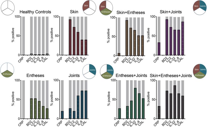
secukinumab) or TNF- (N = 10, adalimumab) inhibition. temic inflammation in patients with psoriatic disease, ifSokolova et al. Arthritis Research & Therapy (2020) 22:26 Page 6 of 8 Fig. 2 Inflammation markers in various subsets of patients with the psoriatic disease (step 2). Significantly different markers from step 1 (beta- defensin 2, BD2; lipocalin 2, LC2; interleukin-22, IL-22; interleukin-8, IL-8; calprotectin, CAL) as well as C-reactive protein (CRP) were analyzed in 105 healthy controls and each 15 patients with either monomorphic psoriatic disease of the skin (red), the entheses (green), or the joint (blue) or polymorphic disease of with skin/entheses, skin/joints, entheses/joints, or all 3 manifestations. Y-axis indicates % of positive (> 3 SD over mean; > 0.5 mg/dl in case of CRP) patients for the respective markers Fig. 3 Effect of anti-cytokine treatment on inflammation markers (step 3). Patients with psoriatic arthritis received treatment with either tumor necrosis factor inhibitor adalimumab (TNFi; N = 10) or interleukin-17 inhibitor secukinumab (IL-17i; N = 10). Serum was analyzed for beta-defensin 2 (BD2), lipocalin 2 (LC2), interleukin (IL)-22, interleukin (IL)-8, and calprotectin (CAL) at baseline and 3 months follow-up
Sokolova et al. Arthritis Research & Therapy   (2020) 22:26                                                                            Page 7 of 8

not focusing the analysis on the acute phase response.          markers to structural damage, as well as their potential
The results also suggest that various subsets of psoriatic      role in defining patients that progress from psoriatic skin
disease show distinct marker profiles with differences for      to musculoskeletal disease.
skin, entheseal, and joint disease.
   This study particularly underlines that CRP is not a per-    Conclusion
fectly useful marker for assessing systemic inflammation        These data indicate that a distinct set of markers allows
in psoriatic disease. Skin and entheseal disease are not as-    measuring systemic inflammation in patients with psori-
sociated with CRP elevations despite active inflammation.       atic disease in the absence of elevated C-reactive protein.
If present, CRP elevations (> 5 mg/L) are confined to syn-      Furthermore, different clinical subsets of psoriatic disease
ovial disease (joint swelling). However, only a fraction of     based on skin, entheseal and joint involvement are charac-
about one-third of patients with arthritis shows elevated       terized by specific inflammation marker profiles. Treat-
CRP. As mentioned, these findings reflect the generally         ment of psoriatic disease with cytokine inhibitors reduces
low or absent IL-6 signature in psoriatic disease, which is     these elevated levels of systemic inflammation markers.
required for mounting the production of acute-phase
proteins in hepatocytes.                                        Supplementary information
   In contrast, beta-defensin 2 and lipocalin 2 are both        Supplementary information accompanies this paper at https://doi.org/10.
IL-17 regulated mediators, which are produced by innate         1186/s13075-020-2111-8.
immune cells. Both are found at much higher levels than
                                                                 Additional file 1: Figure S1. Individual levels of inflammation markers
IL-17 in the circulation and appear to be strongly associ-       in patients with different subsets of psoriatic disease.
ated with skin and to some extent also with entheseal            Additional file 2: Figure S2. Marker Spidergram.
disease, while the joint disease is more inconsistently as-      Additional file 3: Table S1. Correlation between serum markers and
sociated with elevated levels (< 30%) (Additional file 2:        disease activity parameters.
Figure S2). These results confirm earlier data showing           Additional file 4: Table S2. Sensivity and specificity of the serum
elevated beta-defensin levels in psoriasis patients and its      markers.
association with the extent of skin involvement [12].            Additional file 5: Table S3. Effects of TNF- and IL-17A inhibition on the
                                                                 serum levels of the markers.
Hence, beta-defensin and lipocalin 2 provide an oppor-
tunity to quantify systemic inflammation elicited by
psoriatic skin and entheseal disease.                           Abbreviations
                                                                A: Arthritis; BD: Beta defensin; CP: Calprotectin; CRP: C-reactive protein;
   In contrast, the elevation of the alarmin calprotectin and   DAPSA: Disease activity in psoriatic arthritis; E: Enthesitis; IL: Interleukin;
the neutrophil chemo-attractant mediator IL-8 in the            LC: Lipocalin; PASI: Psoriasis area severity index; PSA: Psoriatic arthritis; S: Skin
serum were preferentially driven by psoriatic joint disease     disease; SJC: Swollen joint count; SPARCC: Spondyloarthritis Research
                                                                Consortium of Canada; VEGF: Vascular endothelial growth factor
and to some extent also entheseal disease rather than skin
disease. These findings support the role of neutrophil in-      Acknowledgements
flux and activation in the musculoskeletal manifestations       Nothing to acknowledge.
of psoriatic disease and provide instruments to measure
                                                                Authors’ contributions
this process, which occurs independently from acute             DS, YL, X, KN, and JR recruited the patients. MS and MZ performed the
phase reactants. While IL-17A and IL-23 were barely de-         measurements and analyzed the data. MS and GS wrote the manuscript. All
tectable in the circulation, we found IL-22 in relevant con-    authors read and approved the final manuscript.

centrations across all manifestations of psoriatic disease.     Funding
   Overall, these results offer a new possibility to measure    This work was supported by CRC1181 (project INST 90/925-1), FOR 2886
systemic inflammation in psoriatic disease complementing        (project SCHE 1583/15-1), and project SCHE 1583/14-1, all supported by the
                                                                German Research Foundation, the project Metarthros and Mascara supported
previous findings of altered bone biomarkers in psoriatic       by the Bundesministerium für Bildung und Forschung (BMBF), the IMI
arthritis [26, 27]. As such, these data address the unmet       funded project RTCure (115142), the ERC grant 4D NanoScope from the
need of immunological markers to monitor disease activ-         European Union (to GS) and PARTNER fellowship program (to MS).
ity in psoriatic disease. Such an approach can complement
                                                                Availability of data and materials
the use of well-established clinical instruments to quantify    The datasets used and/or analyzed during the current study are available
the extent of skin, entheseal, and joint disease. In conse-     from the corresponding author on reasonable request.
quence, these markers may not only support the classifica-
                                                                Ethics approval and consent to participate
tion of different endotypes in psoriatic disease but may        All patients provided written informed consent and the institutional review
also improve the characterization of the activity of the dis-   board (IRB)/ethics committee (Ethik-Kommission der Friedrich-Alexander-
ease as well as its response to treatment. Finally, these       Universität Erlangen-Nürnberg) approved the analysis of proteins in stored
                                                                serum samples for the patients.
findings will also allow addressing new research questions,
such as the characterization of axial disease (which has        Consent for publication
not been included into this study), the relation of these       Not applicable
Sokolova et al. Arthritis Research & Therapy (2020) 22:26 Page 8 of 8
Competing interests 19. Bevelacqua V, Libra M, Mazzarino MC, et al. Long pentraxin 3: a marker
The authors declare that they have no competing interests. of inflammation in untreated psoriatic patients. Int J Mol Med. 2006;
18(3):415–23.
Author details 20. Chandran V, Abji F, Perruccio AV, Gandhi R, Li S, Cook RJ, Gladman DD.
1
Department of Internal Medicine 3-Rheumatology and Immunology, Serum-based soluble markers differentiate psoriatic arthritis from
Friedrich-Alexander University Erlangen-Nuremberg and Universitätsklinikum osteoarthritis. Ann Rheum Dis. 2019;78:796–801.
Erlangen, Ulmenweg 18, 91054 Erlangen, Germany. 2Deutsches Zentrum fur 21. Fiocco U, Sfriso P, Oliviero F, et al. Synovial effusion and synovial fluid
Immuntherapie (DZI), Erlangen, Germany. 3Division of Rheumatology and biomarkers in psoriatic arthritis to assess intraarticular tumor necrosis factor-
Immunology, Sakarya University School of Medicine, Sakarya, Turkey. α blockade in the knee joint. Arthritis Res Ther. 2010;12(4):R148.
4
Department of Rheumatology and Immunology, West China Hospital, 22. Abdel-Hamid MF, Aly DG, Saad NE, Emam HM, Ayoub DF. Serum levels of
Sichuan University, Chengdu, China. interleukin-8, tumor necrosis factor-α and γ-interferon in Egyptian psoriatic
patients and correlation with disease severity. J Dermatol. 2011;38(5):442–6.
Received: 4 November 2019 Accepted: 23 January 2020 23. Arican O, Aral M, Sasmaz S, Ciragil P. Serum levels of TNF-alpha, IFN-gamma,
IL-6, IL-8, IL-12, IL-17, and IL-18 in patients with active psoriasis and
correlation with disease severity. Mediat Inflamm. 2005;2005(5):273–9.
24. Biniecka M, Connolly M, Gao W, et al. Redox-mediated angiogenesis in the
References hypoxic joint of inflammatory arthritis. Arthritis Rheumatol. 2014;66(12):
1. Ritchlin CT, Colbert RA, Gladman DD. Psoriatic arthritis. N Engl J Med. 2017; 3300–10.
376(10):2095–6. 25. Berlin A, Simon D, Tascilar K, et al. The ageing joint-standard age- and sex-
2. Vadakayil AR, Dandekeri S, Kambil SM, Ali NM. Role of C-reactive protein as related values of bone erosions and osteophytes in the hand joints of
a marker of disease severity and cardiovascular risk in patients with healthy individuals. Osteoarthr Cartil. 2019;27(7):1043–7.
psoriasis. Indian Dermatol Online J. 2015;6(5):322–5. 26. Chandran V, Cook RJ, Edwin J, et al. Soluble biomarkers differentiate
3. Mease PJ, Gottlieb AB, Berman A, Drescher E, Xing J, Wong R, Banerjee S. patients with psoriatic arthritis from those with psoriasis without arthritis.
The efficacy and safety of clazakizumab, an anti-interleukin-6 monoclonal Rheumatology (Oxford). 2010;49(7):1399–405.
antibody, in a phase IIb study of adults with active psoriatic aryhritis. 27. Chandran V, Shen H, Pollock RA, Pellett FJ, Carty A, Cook RJ, Gladman DD.
Arthritis Rheumatol. 2016;68(9):2163–73. Soluble biomarkers associated with response to treatment with tumor necrosis
4. Villanova F, Di Meglio P, Nestle FO. Biomarkers in psoriasis and psoriatic factor inhibitors in psoriatic arthritis. J Rheumatol. 2013;40(6):866–71.
arthritis. Ann Rheum Dis. 2013;72(Suppl 2):ii104–10.
5. Ritchlin CT. Strategies for biomarker development in psoriatic disease: a
report from the GRAPPA 2010 annual meeting. J Rheumatol. 2012;39(2): Publisher’s Note
423–6. Springer Nature remains neutral with regard to jurisdictional claims in
6. FitzGerald O, Mease PJ. Biomarkers: project update from the GRAPPA 2012 published maps and institutional affiliations.
annual meeting. J Rheumatol. 2013;40(8):1453–4.
7. Soderstrom C, Berstein G, Zhang W, Valdez H, Fitz L, Kuhn M, Fraser S. Ultra-
sensitive measurement of IL-17A and IL-17F in psoriasis patient serumand
skin. AAPS J. 2017;19(4):1218–22.
8. Siebert S, Sweet K, Dasgupta B, Campbell K, McInnes IB, Loza MJ.
Responsiveness of serum C- reactive protein, interleukin- 17A, and
interleukin- 17F levels to Ustekinumab in psoriatic arthritis: lessons from two
phase III, multicenter, double- blind, Placebo- Controlled Trials. Arthritis
Rheumatol. 2019;71(10):1660–9.
9. Uluçkan Ö, Jimenez M, Karbach S, et al. Chronic skin inflammation leads to
bone loss by IL-17-mediated inhibition of Wnt signaling in osteoblasts. Sci
Transl Med. 2016;8(330):330ra37.
10. Kyriakou A, Patsatsi A, Vyzantiadis TA, Sotiriadis D. Serum levels of TNF-α, IL-
12/23p40, and IL-17 in plaque psoriasis and their correlation with disease
severity. J Immunol Res. 2014;2014:467541.
11. Arican O, Aral M, Sasmaz S, Ciragil P. Serum levels of TNF-a, IFN-c, IL-6, IL-8,
IL-12, IL-17, and IL-18 in patients with active psoriasis and correlation with
disease severity. Mediat Inflamm. 2005;2005:273–9.
12. Kolbinger F, Loesche C, Valentin MA, et al. β-Defensin 2 is a responsive
biomarker of IL-17A-driven skin pathology in patients with psoriasis. J
Allergy Clin Immunol. 2017;139(3):923–32.
13. Romaní J, Caixàs A, Ceperuelo-Mallafré V, et al. Circulating levels of lipocalin-
2 and retinol-binding protein-4 are increased in psoriatic patients and
correlated with baseline PASI. Arch Dermatol Res. 2013;305(2):105–12.
14. Shimauchi T, Hirakawa S, Suzuki T, et al. Serum interleukin-22 and vascular
endothelial growth factor serve as sensitive biomarkers but not as
predictors of therapeutic response to biologics in patients with psoriasis. J
Dermatol. 2013;40(10):805–12.
15. Lo YH, Torii K, Saito C, Furuhashi T, Maeda A, Morita A. Serum IL-22
correlates with psoriatic severity and serum IL-6 correlates with
susceptibility to phototherapy. J Dermatol Sci. 2010;58(3):225–7.
16. Michalak-Stoma A, Bartosińska J, Kowal M, Juszkiewicz-Borowiec M,
Gerkowicz A, Chodorowska G. Serum levels of selected Th17 and Th22
cytokines in psoriatic patients. Dis Markers. 2013;35(6):625–31.
17. Hannson C, Eriksson C, Alenius GM. S-calprotectin (S100A8/S100A9): a
potential marker of inflammation in patients with psoriatic arthritis. J
Immunol Res. 2014;2014:696415.
18. Hwang YJ, Jung HJ, Kim MJ, et al. Serum levels of LL-37 and inflammatory
cytokines in plaque and guttate psoriasis. Mediat Inflamm. 2014;2014:
268257.You can also read