THE ROLE OF BRONCHOSCOPY IN PATIENTS WITH SARS-COV-2 PNEUMONIA
←
→
Page content transcription
If your browser does not render page correctly, please read the page content below
ERJ OPEN RESEARCH
                                                                                                                   ORIGINAL RESEARCH ARTICLE
                                                                                                                   M. ARENAS-DE LARRIVA ET AL.

The role of bronchoscopy in patients with SARS-CoV-2
pneumonia
Marisol Arenas-De Larriva1, Roberto Martín-DeLeon 2, Blanca Urrutia Royo3, Iker Fernández-Navamuel4,
Andrés Gimenez Velando4, Laura Nuñez García4, Carmen Centeno Clemente5, Felipe Andreo García 5,
Albert Rafecas Codern6, Carmen Fernández-Arias6, Virginia Pajares Ruiz6, Alfons Torrego Fernández6,
Olga Rajas7, Gorane Iturricastillo8, Ricardo Garcia Lujan9, Lorena Comeche Casanova10,
Albert Sánchez-Font11, Ricardo Aguilar-Colindres12, Roberto Larrosa-Barrero13, Ruth García García14,
Rosa Cordovilla14, Ana Núñez-Ares15, Andrés Briones-Gómez16, Enrique Cases Viedma16, José Franco17,
Javier Cosano Povedano18, Manuel Luis Rodríguez-Perálvarez 19, Jose Joaquin Cebrian Gallardo20,
Manuel Nuñez Delgado21, María Pavón-Masa22, Maria del Mar Valdivia Salas23 and Javier Flandes 24

1
 Dept of Bronchoscopy and Interventional Pulmonology, Hospital Universitario Reina Sofía, IMIBIC, Córdoba, Spain. 2Dept of
Pulmonology, Hospital Universitario Reina Sofía, IMIBIC, Córdoba, Spain. 3Pulmonary Dept, Thorax Clinic Institute, Hospital
Universitari Germans Trias i Pujol, Badalona, Spain. 4Bronchoscopy and Interventional Pulmonology Unit, Pulmonology Dept, Hospital
Fundacion Jimenez Diaz, ISS-FJD, CIBERES, Madrid, Spain. 5Interventional Pulmonology Unit, Pulmonary Dept, Thorax Clinic Institute,
Hospital Universitari Germans Trias i Pujol, UAB, IGTP, Badalona, Spain. 6Respiratory Dept, Hospital Santa Creu i Sant Pau, Barcelona,
Spain. 7Interventional Pulmonology Unit, Pulmonology Dept, Hospital Universitario de la Princesa, Instituto de Investigación Princesa,
Madrid, Spain. 8Pulmonology Dept, Hospital Universitario de la Princesa, Instituto de Investigación Princesa, Madrid, Spain. 9Dept of
Interventional Pulmonology, Hospital Universitario 12 Octubre and Hospital Univesitario Quirónsalud Madrid, Madrid, Spain. 10Dept of
Pulmonology, Hospital Universitario Quirónsalud Madrid, Madrid, Spain. 11Pulmonology Dept, Hospital del Mar, CIBERES, UAB, IMIM
(Hospital del Mar Medical Research Institute), Barcelona, Spain. 12Pulmonology Dept, Hospital del Mar, CIBERES, Barcelona, Spain.
13
   Pulmonary Dept, Hospital Universitario Clínico San Carlos, Madrid, Spain. 14Interventional Pulmonology Unit, Pulmonary Dept,
Salamanca University Hospital, Salamanca, Spain. 15Interventional Pulmonology Unit, Pulmonary Dept, Albacete, Spain.
16
   Interventional Pulmonology Unit, Pulmonary Dept, Hospital Universitario y Politécnico La Fe, Valencia, Spain. 17Pneumology Service,
Clinic University Hospital, INCLIVA Health Research Institute, Valencia, Spain. 18Bronchoscopy and Interventional Pulmonology Unit,
Pulmonology Dept, Hospital Universitario Reina Sofía, IMIBIC, Córdoba, Spain. 19Maimónides Institute of Biomedical Research of
Córdoba (IMIBIC), CIBERehd, Córdoba, Spain. 20Dept of Pulmonology, Agencia sanitaria Costa del Sol, Marbella, Spain. 21Dept of
Bronchoscopy and Interventional Pulmonology, Hospital Álvaro Cunqueiro, CHUVI, Vigo, Spain. 22Dept of Interventional Pulmonology,
Hospital Universitario Virgen Macarena, Seville, Spain. 23Dept of Interventional Pulmonology, Hospital Universitario Santa Lucía,
Cartagena, Spain. 24Bronchoscopy and Interventional Pulmonology Unit, Pulmonology Dept, Hospital Fundación Jimenez Diaz,
ISS-FJD, CIBERES, Madrid, Spain.

                                                                      Corresponding author: Marisol Arenas-De Larriva (arlam23@hotmail.com)

                                        Shareable abstract (@ERSpublications)
                                        Bronchoscopy is part of the armamentarium against #COVID19. It allows diagnosis, facilitates
                                        mechanical ventilation and provides prognostic information. This information could be used to
                                        refine healthcare pathways in order to improve outcomes. https://bit.ly/2QuAQOt

                                        Cite this article as: Arenas-De Larriva M, Martín-DeLeon R, Urrutia Royo B, et al. The role of
                                        bronchoscopy in patients with SARS-CoV-2 pneumonia. ERJ Open Res 2021; 7: 00165-2021
                                        [DOI: 10.1183/23120541.00165-2021].

                                        Abstract
    Copyright ©The authors 2021         Background The role of bronchoscopy in coronavirus disease 2019 (COVID-19) is a matter of debate.
                                        Patients and methods This observational multicentre study aimed to analyse the prognostic impact of
    This version is distributed under
    the terms of the Creative
                                        bronchoscopic findings in a consecutive cohort of patients with suspected or confirmed COVID-19.
    Commons Attribution Non-            Patients were enrolled at 17 hospitals from February to June 2020. Predictors of in-hospital mortality were
    Commercial Licence 4.0. For         assessed by multivariate logistic regression.
    commercial reproduction rights      Results A total of 1027 bronchoscopies were performed in 515 patients (age 61.5±11.2 years; 73% men),
    and permissions contact
                                        stratified into a clinical suspicion cohort (n=30) and a COVID-19 confirmed cohort (n=485). In the clinical
    permissions@ersnet.org
                                        suspicion cohort, the diagnostic yield was 36.7%. In the COVID-19 confirmed cohort, bronchoscopies were
                                        predominantly performed in the intensive care unit (n=961; 96.4%) and major indications were: difficult
                                        mechanical ventilation (43.7%), mucus plugs (39%) and persistence of radiological infiltrates (23.4%).

https://doi.org/10.1183/23120541.00165-2021                                                                        ERJ Open Res 2021; 7: 00165-2021
ERJ OPEN RESEARCH ORIGINAL RESEARCH ARTICLE | M. ARENAS-DE LARRIVA ET AL.
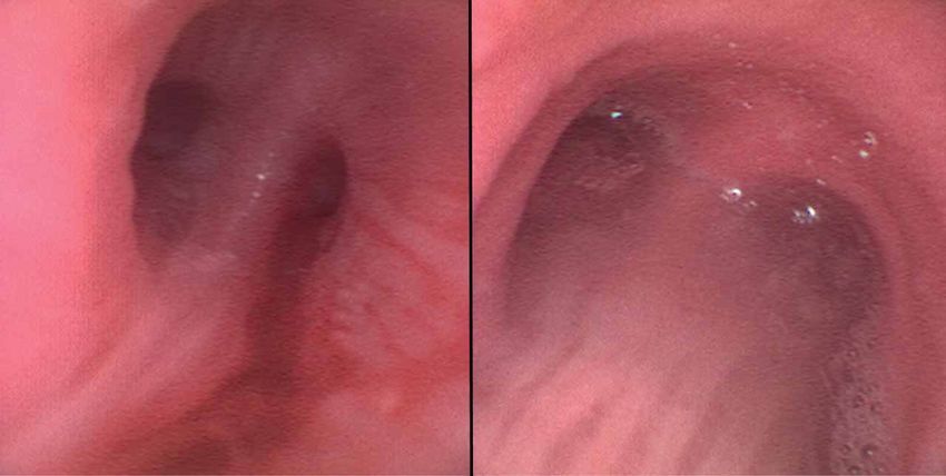
This article has supplementary 147 bronchoscopies were performed to rule out superinfection, and diagnostic yield was 42.9%. There
material available from were abnormalities in 91.6% of bronchoscopies, the most frequent being mucus secretions (82.4%),
openres.ersjournals.com haematic secretions (17.7%), mucus plugs (17.6%), and diffuse mucosal hyperaemia (11.4%). The
independent predictors of in-hospital mortality were: older age (OR 1.06; pERJ OPEN RESEARCH ORIGINAL RESEARCH ARTICLE | M. ARENAS-DE LARRIVA ET AL.
positive RT-PCR of SARS-CoV-2 in nasopharyngeal swab specimens who underwent bronchoscopy to
rule out superinfection or for therapeutic purposes formed the RT-PCR-confirmed cohort.
Data were recorded in an anonymised electronic datasheet using the REDCap (Research Electronic Data
Capture) platform [9]. Study investigators received online training at baseline to homogenise the data
collection, and they were granted access with a unique username/password. All clinical information was
extracted from reliable electronic medical data sources. Demographic characteristics and comorbidities
(graded with the Charlson comorbidity index as absent if 0–1, mild if 2 or severe if ⩾3 [10]), clinical
symptoms and diagnostic tests of COVID-19 were recorded. Blood tests and radiological features were
considered within the 48 h prior to bronchoscopy. Imaging findings obtained in chest computed
tomography (CT) were reported according to the COVID-RADS classification as typical, fairly typical,
atypical or normal [11]. Bronchoscopic findings and procedures were also registered. Patients were
followed until hospital discharge or death. The main outcome evaluated was in-hospital mortality at
90 days after bronchoscopy.
Sample size calculation
The sample size was calculated using EPIDAT version 4.2 (Xunta de Galicia, Spain). The following
assumptions were made to study a theoretical relationship between endoscopic findings and outcomes:
• The prevalence of an endoscopic feature indicating poor prognosis: 20%.
• In-hospital mortality in patients showing an endoscopic feature indicating poor prognosis: 40%
(obtained from the upper range of mortality reported in previous series of critically ill patients [12, 13]).
• In-hospital mortality in patients without an endoscopic feature indicating poor prognosis: 25%
(obtained from the lower range of mortality reported in previous series of critically ill patients [12, 13]).
• Statistical power: 80%
• α error: 5%
• Incomplete or unavailable data: 5%
Under these premises, the minimum sample size required was 483 patients with RT-PCR-confirmed
COVID-19. The study finally comprised 515 patients, including 488 RT-PCR-confirmed cases.
Statistical analysis
Categorical variables were described as frequency tables and percentages. Continuous variables were
described using mean and standard deviation, except for those with an asymmetric distribution, in which
median and interquartile range (IQR) were used. To identify clinical, radiological and endoscopic features
associated with in-hospital mortality at 90 days, the first bronchoscopy performed in each patient with
RT-PCR-confirmed COVID-19 was considered. Univariate and multivariate logistic regression was used.
Variables with pERJ OPEN RESEARCH ORIGINAL RESEARCH ARTICLE | M. ARENAS-DE LARRIVA ET AL.
TABLE 1 Clinical characteristics of 515 patients admitted to the hospital with suspected or confirmed
COVID-19 who required a bronchoscopy
Variable Clinical suspicion RT-PCR-confirmed p-value
cohort (n=30) cohort (n=485)
Age, mean±SD 59.2±15.5 61.7±10.9 0.390
Women 36.7% (11) 26.4% (128) 0.219
Previous medical history
Diabetes 20% (6) 22.5% (109) 0.752
Hypertension 36.7% (11) 47.6% (231) 0.243
Cardiovascular 13.3% (4) 10.9% (53) 0.684
Bronchopulmonary 23.3% (7) 14% (68) 0.161
Neoplasms 30% (9) 9.3% (45) 0.002
Charlson comorbidity indexERJ OPEN RESEARCH ORIGINAL RESEARCH ARTICLE | M. ARENAS-DE LARRIVA ET AL.
TABLE 1 Continued
Variable Clinical suspicion RT-PCR-confirmed p-value
cohort (n=30) cohort (n=485)
Corticosteroids 3.3% (1) 70.4% (338)ERJ OPEN RESEARCH ORIGINAL RESEARCH ARTICLE | M. ARENAS-DE LARRIVA ET AL.
TABLE 2 Therapeutic indications and findings in 997 bronchoscopies performed in 485 hospitalised patients
with RT-PCR-confirmed SARS-CoV-2 pneumonia
Indications
Atelectasis 7% (70)
Mucus plugs 39% (389)
Haemoptysis 6% (60)
Radiological progression 10% (100)
Persistence of radiological infiltrates 23.4% (233)
Difficult mechanical ventilation 43.7% (436)
Impossible weaning from mechanical ventilation 6.3% (63)
Findings
Normal 8.4% (84)
Diffuse mucosal hyperaemia 11.4% (114)
Thick mucus secretion 59.9% (597)
Fluid mucus secretion 22.5% (224)
Mucus plugs 17.6% (175)
Haematic secretions 17.7% (176)
Intrabronchial clots 6% (60)
Location of mucus plugs (n=175)
Trachea 24% (42)
Main right bronchus 31.4% (55)
Main left bronchus 33.5% (59)
Right superior bronchus 18.3% (32)
Right middle bronchus 24% (42)
Right inferior bronchus 45.1% (79)
Left superior bronchus 16% (28)
Left inferior bronchus 36.6% (64)
Location of intrabronchial clots (n=60)
Trachea 31.7% (19)
Main right bronchus 55% (33)
Main left bronchus 41.7% (25)
Right superior bronchus 15% (9)
Right middle bronchus 21.7% (13)
Right inferior bronchus 40% (24)
Left superior bronchus 10% (6)
Left inferior bronchus 20% (12)
Therapy
Aspiration 82.3% (821)
Removal with grasp forceps 1.4% (14)
Cannula placement 0.3% (3)
Bronchial occlusion 0.2% (2)
Cryotherapy 0.1% (1)
Endobronchial selective intubation 0.1% (1)
Intrabronchial drugs
Saline solution 60.2% (600)
Mesna 5.1% (51)
Hypertonic solution 14.5% (145)
N-acetylcysteine 6% (60)
Hyaluronic acid (+hypertonic solution) 6.5% (65)
Others 0.9% (9)
Samples
Bronchial aspiration 43% (429)
Combined bronchial aspiration and bronchoalveolar lavage 24.3% (242)
Bronchoalveolar lavage 5.8% (58)
Bronchial washing 11% (110)
Microbiological agents
Bacteria 27.2% (271)
Fungi 12.8% (128)
Virus 3.6% (36)
Data are presented as % (n).
https://doi.org/10.1183/23120541.00165-2021 6ERJ OPEN RESEARCH ORIGINAL RESEARCH ARTICLE | M. ARENAS-DE LARRIVA ET AL.
most frequent being mucus secretions (82.4%), mucus plugs (17.6%), haematic secretions/clots (23.7%)
and diffuse mucosal hyperaemia (11.4%) (figure 1). The most frequent therapy consisted in atelectasis
resolution or mucus aspiration (82.3%). Among 147 bronchoscopies performed to rule out superinfection,
the microbiological samples were obtained from: BAS (11.6%), BAL (10.9%), bronchial washing (52.5%),
and BAS in combination with BAL (21.7%). The diagnostic yield was 42.9%, including 71
microbiological isolations which are detailed as supplementary material.
Impact of endoscopic findings on outcomes
All patients with RT-PCR-confirmed COVID-19, either in nasopharyngeal swab or in lower respiratory
tract specimens, were included to evaluate clinical, radiological and endoscopic features associated with
mortality (n=496). Univariate and multivariate logistic regression analyses to predict in-hospital mortality
at 90 days are shown in table 3. The independent predictors of in-hospital mortality were: older age (OR
1.06, 95% CI 1.03–1.08; pERJ OPEN RESEARCH ORIGINAL RESEARCH ARTICLE | M. ARENAS-DE LARRIVA ET AL.
TABLE 3 Clinical, radiological and endoscopic predictors of in-hospital mortality at 90 days among patients with RT-PCR-confirmed COVID-19
admitted to the hospital who required a first bronchoscopy (n=496)
Variables Univariate analysis Multivariate analysis Multivariate analysis
(initial model) (final model)
OR (95% CI) p-value OR (95% CI) p-value OR (95% CI) p-value
Age 1.05 (1.03–1.08)ERJ OPEN RESEARCH ORIGINAL RESEARCH ARTICLE | M. ARENAS-DE LARRIVA ET AL.
1.0
p=0.038
0.8
Cumulative survival %
0.6
0.4
0.2
0.0
0 15 30 45 60 75 90
Interval from bronchoscopy to hospital discharge/death (days)
Group; number at risk 30 days 60 days 90 days
With bloody secretions 50 10 5
Without bloody secretions 168 57 24
FIGURE 2 Kaplan–Meier curve showing the influence of haematic secretions in the distal bronchial tract on
mortality in 496 patients with RT-PCR-confirmed COVID-19 admitted to the hospital.
clinical practice, as illustrated in the present study. According to our results, BAS alone should be avoided
but other options including BAL, bronchial washing or BAL in combination with BAS, would be equally
valid. In contrast, guidelines are broadly homogeneous regarding protocols to protect healthcare personnel
[17, 20, 21]. In brief, bronchoscopies in patients with suspected or confirmed COVID-19 should be
performed in negative-pressurised or in adequately ventilated rooms. The involved healthcare personnel
may be experienced and reduced to the minimum (two or three people depending on the procedure).
Disposable bronchoscopes are advised. Individual enhanced third-degree protection elements are required
( protective glasses or face shield, FFP3 face masks, protective clothing, gloves, etc.). Unfortunately, some
of these recommendations are difficult to implement in real clinical practice, particularly in secondary
hospitals, which were overwhelmed during the peak of the pandemic. Negative-pressurised rooms are
anecdotal in ICUs where most therapeutic endoscopies need to be performed. These structural deficiencies
should be urgently amended by healthcare authorities to protect medical staff from COVID-19
transmission. In any case, the decision to perform (or not perform) a bronchoscopy in a patient with
COVID-19 should be taken after a careful weighing of potential benefits against the potential risk of
disease transmission to healthcare personnel.
Critically ill patients with COVID-19 usually require prolonged mechanical ventilation. Bronchoscopy may
help to prevent, diagnose or resolve ventilator-related complications. This is the first multicentre study
describing the indications and procedures in this setting. The presence of mucus plugs was the only
indication independently associated with worse outcomes (60% increased mortality rates as compared with
other indications), although it is tightly related to other indications such as atelectasis, superinfection and
difficult mechanical ventilation. It is paramount to optimise ventilation to prevent excess secretions and to
perform frequent aspirations through the endotracheal tube [20].
There are well established clinical, analytical and radiological predictors of poor outcomes in patients with
COVID-19 including (but not limited to) older age, men, increased comorbidities, lymphopenia, increased
D dimer and serum ferritin, and extent of pneumonia in the chest CT [22, 23]. This is the first study
sufficiently powered to analyse the impact of bronchoscopic findings on outcomes among hospitalised
patients with COVID-19. The presence of diffuse mucosal hyperaemia was associated with reduced
https://doi.org/10.1183/23120541.00165-2021 9ERJ OPEN RESEARCH ORIGINAL RESEARCH ARTICLE | M. ARENAS-DE LARRIVA ET AL.
in-hospital mortality rates, as it is likely a typical feature of an earlier phase of COVID-19, indicating acute
inflammation [24]. This situation may still be reversible with or without anti-inflammatory drugs such as
corticosteroids [25]. However, the disappearance of this endoscopic sign under persistent respiratory
insufficiency may indicate a poor prognosis. The presence of haematic secretions in the distal bronchial
tract was an independent predictor of increased in-hospital mortality. In contrast to diffuse mucosal
hyperaemia, haematic secretions could translate into irreversible damage of the capillaries and the
interstitial/alveolar space, which characterises the most advanced and severe forms of COVID-19 [26–28].
Indeed, the presence of haematic secretions identified a subgroup of very sick patients (16%) with
in-hospital mortality above 60%. Further studies focused on this subpopulation are needed to delineate
more aggressive and life-saving therapies.
The present study is limited by its ambispective design which precluded a protocolised clinical
management of the study population. Although laboratory and radiological assessment of patients with
COVID-19 varied among different institutions, making it difficult to extract solid conclusions regarding
these parameters, the study adequately captured the heterogeneity in real clinical practice. On the other
hand, the number of patients in the clinical suspicion cohort was limited as this indication is uncommon
and not accepted by some experts [14]. Finally, a potential relationship between ventilator-derived trauma
and some bronchoscopic findings in critically ill patients could not be ruled out.
In conclusion, bronchoscopy is pivotal as part of the armamentarium against COVID-19. In carefully
selected patients with clinical and radiological suspicion of SARS-CoV-2 pneumonia who test negative in
nasopharyngeal swabs, a lower respiratory tract specimen may provide an acceptable diagnostic yield, also
including the identification of alternative microbiological agents or superinfection. In critically ill patients
with COVID-19, bronchoscopy allows removal of mucus plugs and intrabronchial clots, and the resolution
of atelectasis, thereby improving mechanical ventilation. Finally, haematic secretions in the respiratory tract
and absence of diffuse mucosal hyperaemia are poor prognostic features.
Acknowledgements: We acknowledge Donna Pringle for professional English language polishing.
Data availability: Upon publication, data collected for the study will be made available for others in a Mendeley
repository.
Conflict of interest: M. Arenas-De Larriva reports a travel grant from Novartis and lecture fees from Ferrer outside
the submitted work. R. Martín-DeLeon has nothing to disclose. B. Urrutia Royo has nothing to disclose.
I. Fernández-Navamuel has nothing to disclose. A. Gimenez Velando has nothing to disclose. L. Nuñez García has
nothing to disclose. C. Centeno Clemente has nothing to disclose. F. Andreo García has nothing to disclose.
A. Rafecas Codern has nothing to disclose. C. Fernández-Arias has nothing to disclose. V. Pajares Ruiz has nothing
to disclose. A. Torrego Fernández has nothing to disclose. O. Rajas has nothing to disclose. G. Iturricastillo has
nothing to disclose. R. Garcia Lujan has nothing to disclose. L. Comeche Casanova has nothing to disclose.
A. Sánchez-Font has nothing to disclose. R. Aguilar-Colindres has nothing to disclose. R. Larrosa-Barrero has
nothing to disclose. R. García García has nothing to disclose. R. Cordovilla has nothing to disclose. A. Núñez-Ares
has nothing to disclose. A. Briones-Gómez has nothing to disclose. E. Cases Viedma reports lecture fees from
Ambu outside the submitted work. J. Franco has nothing to disclose. J. Cosano Povedano reports a travel grant
from Izasa scientific outside the submitted work. M.L. Rodríguez-Perálvarez reports lecture fees from Novartis,
Astellas and Intercept outside the submitted work. J.J. Cebrian Gallardo has nothing to disclose. M. Nuñez Delgado
has nothing to disclose. M. Pavón-Masa has nothing to disclose. M. Valdivia Salas has nothing to disclose.
J. Flandes has nothing to disclose.
Support statement: The present study was funded by the Spanish Society of Pulmonology and Thoracic Surgery:
extraordinary grant PII 2020 for research in COVID-19. Funding information for this article has been deposited with
theCrossref Funder Registry.
References
1 Huang C, Wang Y, Li X, et al. Clinical features of patients infected with 2019 novel coronavirus in Wuhan,
China. Lancet 2020; 395: 497–506.
2 Fennelly KP. Particle sizes of infectious aerosols: implications for infection control. Lancet Respir Med 2020; 8:
914–924.
3 Wang W, Xu Y, Gao R, et al. Detection of SARS-CoV-2 in different types of clinical specimens. JAMA 2020; 323:
1843–1844.
4 Lentz RJ, Colt H. Summarizing societal guidelines regarding bronchoscopy during the COVID-19 pandemic.
Respirology 2020; 25: 574–577.
https://doi.org/10.1183/23120541.00165-2021 10ERJ OPEN RESEARCH ORIGINAL RESEARCH ARTICLE | M. ARENAS-DE LARRIVA ET AL.
5 Mondoni M, Sferrazza Papa GF, Rinaldo R, et al. Utility and safety of bronchoscopy during the SARS-CoV-2
outbreak in Italy: a retrospective, multicentre study. Eur Respir J 2020; 56: 2002767.
6 Torrego A, Pajares V, Fernandez-Arias C, et al. Bronchoscopy in patients with COVID-19 with invasive
mechanical ventilation: a single-center experience. Am J Respir Crit Care Med 2020; 202: 284–287.
7 Chang SH, Jiang J, Kon ZN, et al. Safety and efficacy of bronchoscopy in critically ill patients with
coronavirus disease 2019. Chest 2021; 159: 870–872.
8 Kanne JP, Bai H, Bernheim A, et al. COVID-19 Imaging: what we know now and what remains unknown.
Radiology 2021; 299: 204522.
9 Harris PA, Taylor R, Minor BL, et al. The REDCap consortium: building an international community of
software platform partners. J Biomed Inform 2019; 95: 103208.
10 Charlson ME, Pompei P, Ales KL, et al. A new method of classifying prognostic comorbidity in longitudinal
studies: development and validation. J Chronic Dis 1987; 40: 373–383.
11 Salehi S, Abedi A, Balakrishnan S, et al. Coronavirus disease 2019 (COVID-19) imaging reporting and data
system (COVID-RADS) and common lexicon: a proposal based on the imaging data of 37 studies. Eur Radiol
2020; 30: 4930–4942.
12 Grasselli G, Zangrillo A, Zanella A, et al. Baseline characteristics and outcomes of 1591 patients infected with
SARS-CoV-2 admitted to ICUs of the Lombardy region, Italy. JAMA 2020; 323: 1574–1581.
13 Bhatraju PK, Ghassemieh BJ, Nichols M, et al. COVID-19 in critically ill patients in the Seattle region - case
series. N Engl J Med 2020; 382: 2012–2022.
14 Ora J, Puxeddu E, Cavalli F, et al. Does bronchoscopy help the diagnosis in COVID-19 infection? Eur Respir J
2020; 56: 2001619.
15 Jackson T, Deibert D, Wyatt G, et al. Classification of aerosol-generating procedures: a rapid systematic
review. BMJ Open Respir Res 2020; 7: e000730.
16 Ai T, Yang Z, Hou H, et al. Correlation of chest CT and RT-PCR testing for coronavirus disease 2019 (COVID-19)
in China: a report of 1014 cases. Radiology 2020; 296: E32–E40.
17 Wahidi MM, Shojaee S, Lamb CR, et al. The use of bronchoscopy during the coronavirus disease 2019
pandemic: CHEST/AABIP guideline and expert panel report. Chest 2020; 158: 1268–1281.
18 Luo F, Darwiche K, Singh S, et al. Performing bronchoscopy in times of the COVID-19 pandemic: practice
statement from an international expert panel. Respiration 2020; 99: 417–422.
19 Steinfort DP, Herth FJF, Irving LB, et al. Safe performance of diagnostic bronchoscopy/EBUS during the
SARS-CoV-2 pandemic. Respirology 2020; 25: 703–708.
20 Yang H, Chen H, Gao B, et al. Expert panel consensus statement on the applications and precaution
strategies of bronchoscopy in patients with COVID-19. Endosc Ultrasound 2020; 9: 211–219.
21 Cordovilla R, Alvarez S, Llanos L, et al. SEPAR and AEER consensus recommendations on the use of
bronchoscopy and airway sampling in patients with suspected or confirmed COVID-19 infection. Arch
Bronconeumol 2020; 56: Suppl. 2, 19–26.
22 Zhou F, Yu T, Du R, et al. Clinical course and risk factors for mortality of adult inpatients with COVID-19 in
Wuhan, China: a retrospective cohort study. Lancet 2020; 395: 1054–1062.
23 Colombi D, Villani GD, Maffi G, et al. Qualitative and quantitative chest CT parameters as predictors of
specific mortality in COVID-19 patients. Emerg Radiol 2020; 27: 701–710.
24 Batah SS, Fabro AT. Pulmonary pathology of ARDS in COVID-19: a pathological review for clinicians. Respir
Med 2021; 176: 106239.
25 Cano EJ, Fuentes XF, Campioli CC, et al. Impact of corticosteroids in coronavirus disease 2019 outcomes:
systematic review and meta-analysis. Chest 2021; 159: 1019–1040.
26 Fox SE, Akmatbekov A, Harbert JL, et al. Pulmonary and cardiac pathology in African American patients with
COVID-19: an autopsy series from New Orleans. Lancet Respir Med 2020; 8: 681–686.
27 Bradley BT, Maioli H, Johnston R, et al. Histopathology and ultrastructural findings of fatal COVID-19
infections in Washington State: a case series. Lancet 2020; 396: 320–332.
28 Menter T, Haslbauer JD, Nienhold R, et al. Postmortem examination of COVID-19 patients reveals diffuse
alveolar damage with severe capillary congestion and variegated findings in lungs and other organs
suggesting vascular dysfunction. Histopathology 2020; 77: 198–209.
https://doi.org/10.1183/23120541.00165-2021 11You can also read