MRI Evaluation of the Brain in Children with Attention Deficit and Hyperactivity Disorder; How to Hear the Whispers Early? - Mums.ac.ir
←
→
Page content transcription
If your browser does not render page correctly, please read the page content below
http:// ijp.mums.ac.ir
                                                                          Original Article (Pages: 9379-9390)

  MRI Evaluation of the Brain in Children with Attention Deficit
  and Hyperactivity Disorder; How to Hear the Whispers Early?
                *Nadia F. El Ameen1, Mohamed A. Ibrahim1, Samir M. Mouner21

¹Assistant Professor of Radiology, Radiology Department, Faculty of Medicine, El Minia University, El Minia,
Egypt.
²Lecturer of Pediatric Neurology, Pediatric Neurology Unit, Faculty of Medicine, El Minia University, El Minia,
Egypt.

Abstract
Background
Attention deficit and hyperactivity disorder (ADHD) is a disorder of the brain characterized by
periods of inattention, hyperactivity and impulsive behavior. We aimed to evaluate the role of MRI of
the brain in children with ADHD.
Materials and Methods
This prospective study included 100 children with clinical diagnosis of attention deficit and
hyperactivity disorder according to the criteria of IDC-10. There were 79 males and 21females. Their
ages ranged between 3 and 14 years old. A cohort of patients were referred from pediatric neurology
unit to radiology department in El Minia University hospital, El Minia, Egypt, in the period between
January 2017 and December 2017. All of them underwent MRI evaluation of the brain after approval
of the ethical committee of our institution and completion of informed consent.
Results
MRI examination was positive in 66/100 patients and negative in 34 /100 patients. Corpus callosum
dysgenesis was the most common finding in 19/66 patients, followed by temporal lobe pathology in
14/66 patients. Fronto-parietal or cerebellar atrophy was found in 11/66 patients. Tuber cinereum
lesions, hippocampus sclerosis, heterotopia, pachygyria, hemimegalencephaly, Joubert syndrome and
pineal cyst were a spectrum of findings among the remaining positive patients.
Conclusion
MRI of the brain in children with attention deficit and hyperactivity disorder will be the pivot for
diagnosis. Not all patients had cerebellar or fronto-parietal atrophy as presumed before. Temporal
lobe, corpus callosum and tuber cinereum must be looked for carefully.
Key Words: Attention deficit and hyperactivity disorder, Brain, Children, MRI.

*Please cite this article as: El Ameen NF, Ibrahim MA, Mouner SM. MRI Evaluation of the Brain in Children
with Attention Deficit and Hyperactivity Disorder; How to Hear the Whispers Early? Int J Pediatr 2019; 7(5):
9379-90. DOI: 10.22038/ijp.2019.37060.3228

*Corresponding Author:
Dr. Nadia F. El Ameen (M.D), Asst. Prof. of Radiology. Radiology Department, Faculty of Medicine, El Minia
University, El Minia, Egypt.
Email: nadia.elameen@yahoo.com AND nadia.elameen@hotmail.com
Received date: Nov.14, 2018; Accepted date: Jan.12, 2019

Int J Pediatr, Vol.7, N.5, Serial No.65, May. 2019                                                       9379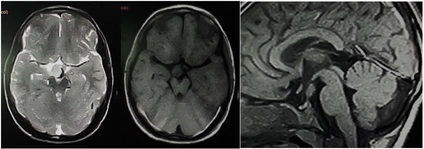
MRI of the Brain in Children with ADHD

1- INTRODUCTION                                      79 males and 21 females. Their ages
                                                     ranged between 3 and 14 years old. They
    Attention-deficit/hyperactivity disorder
                                                     all referred from pediatric neurology unit
(ADHD) is a clinically diagnosed
condition. It is characterized by chronic            to MRI unit at the radiology department in
                                                     El Minia University hospital, Egypt, from
excessive hyperactivity, impulsivity and
                                                     January 2017 to December 2017.
inattention. It affect 5-7% of children and
adolescents population. For a long time, it          2-2. Ethical consideration
was classified as hyperkinetic disorder              A cohort patients were included after
(HKD). There are three subtypes of
                                                     approval of ethical committee of our
ADHD        which      are   predominantly
                                                     institution. Parents of the recruited
inattentive, predominantly hyperactive/              children have signed a written informed
impulsive and combined type (1). Clinical
                                                     consent before MRI examination and
diagnosis of ADHD requires the presence              before anesthesia.
of at least 6 symptoms of hyperactivity
/impulsivity and at least 6 symptoms of              2-3. Inclusion and exclusion criteria
inattention which are necessary for making           All included children have six or more
the clinical decision of whether or not to           symptoms          suggesting      attention-
treat. The high degree of heterogeneity in           deficit/hyperactivity disorder according to
ADHD brings attention to the presence of             Wolraich et al., persisted for 6 months to a
many underlying causes for the condition.            degree that is inconsistent with the
Neuro-radiological investigations began as           developmental level and negatively
a starting point for diagnosis of ADHD               impacts the social and academic activities
using structural and functional MRI (1- 3).          of the child (3).
Neuroimaging techniques are increasingly
being applied to the study of attention-             2-4. Methods
deficit/hyperactivity disorder. Imaging of           2-4-1. Patient preparation
the brain anatomy of ADHD has become
                                                     Before MRI examination, all patients’
the main stay for diagnosis. For a decade,
                                                     parents were routinely questioned about
most studies have focused only on frontal-
                                                     any conditions that contraindicate MRI
striatal regions and detection of smaller
                                                     examination such as metallic prosthesis,
volume of the brain in the affected
                                                     clips or implants. They changed into
patients. As most published studies
                                                     cotton gown for examination. Patients’
showed, there is a 3% to 4% global
                                                     parents were asked about any condition
reduction in brain volume with abnormally
                                                     that would interfere with anesthesia in
small caudate nuclei (1). Our study aimed
                                                     patients that need anesthesia. An
to answer two important questions: 1- Are
                                                     experienced anesthesia consultant (A.H.)
there any brain anatomic abnormalities
                                                     supervised all of the anesthetic procedure
associated with ADHD? 2- Are there any
                                                     using IV anesthetic material (Ketamine 1-2
developmental disorders that could be
                                                     mg/Kg or Propofol 0.5% 1-2 mg /kg), after
associated with the disease?
                                                     complete fasting of the children for at least
                                                     6 hours before the procedure.
2- MATERIALS AND METHODS
2-1. Study design and population                     2-4-2. MRI technique

    Our study is a prospective study                 MRI examination was performed for all
including 100 consecutive patients with              patients using a 1.5 T Gyroscan Achieva
clinical symptoms and signs supporting the           (Philips Medical Systems, Netherlands), in
clinical diagnosis of ADHD. There were               supine position. Images were acquired in
                                                     the axial, coronal, and sagittal planes using

Int J Pediatr, Vol.7, N.5, Serial No.65, May. 2019                                           9380
El Ameen et al.

head coil. A multi planner fast field echo             Two experienced neuro-radiologists (N.
(FFE) localizer upon which the                         F.), and (M.I.) with more than 10 years’
remaining pulse sequences were planned                 experience in analysis and interpretation of
(localizing scan) was used. MRI protocol               MRI brain images interpreted all MRI
for imaging the brain included: axial and              data. All data were statistically described
coronal T2WI (TR 3200, TE 90, FOV 25,                  in terms of frequencies and percentage
slice thickness 3 mm, gap 1–2 mm, NSA 3                when appropriate. Correlation between
and matrix 304 × 512). Axial and sagittal              MRI findings and clinical data was
T1WI (TR 2700, TE 108, FOV 19, slice                   calculated using Chi-square test for
thickness 3 mm, gap 0.5 mm NSA 3 and                   qualitative data with the significant
matrix 304 × 512). Axial FLAIR (TR                     correlation set at p-value ≤ .05. All
6750, TE 79, FOV 23, slice thickness                   statistical calculations were done using
3 mm, gap 1–2 mm, NSA 3 and matrix                     computer programs IPM SPSS software
304 × 512). Sagittal and coronal T1WI                  version 20.0.
with thin sections (2-3mm) with small
field of view was done for patients with               3-RESULTS
hypothalamic lesions, repeated after                       Our study included 100 consecutive
contrast administration of a standard dose             patients. There were 79 % males and 21%
(0.2      mmol/kg)      of    gadopentetate            females. Their ages ranged between 3 and
dimeglumine. Coronal 3D VIBE was used                  14 years old. Their baseline characteristics
for confirmation of hippocampal sclerosis              and relevant history were tabulated in
in some patients (TR 63/TE 7000; flip                  (Table.1). They were presented clinically
angle, 15°; field of view, 400 mm; slice               by stigmata suggesting ADHD. Poor social
thickness, 3 mm; section gap, 0.6 mm;                  relationships, hyperactivity and poor
number of slices, 32-40; image matrix 346              behavioral inhibition were the most
x 512 ; bandwidth, 490 Hz/pixel; 1 signal              common clinical presentation among our
acquisition; scanning time, 24-28 seconds).            patient cohort (Table.2). MRI examination
                                                       showed positive diagnostic data in (66%)
2-5. Data Analyses
                                                       of the patients and negative results in 34%
                                                       of the patients.

 Table-1: Baseline characteristics of patient cohort, (n=100)
Data                                                                  Percent, total=100
Gender:
                                                                             79
    Male
                                                                             21
    Female
Locality area
    Urban                                                                    63
    Rural                                                                    37
Relevant history
                                                                             36
     Obstructed labor
                                                                             24
     Febrile convulsion
                                                                             16
     Congenital infection

Family history
                                                                             12
     Positive
                                                                             83
     Negative
                                                                              5
     Not sure

Int J Pediatr, Vol.7, N.5, Serial No.65, May. 2019                                            9381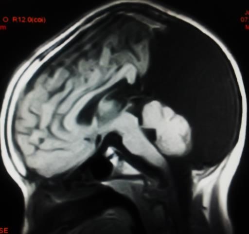
MRI of the Brain in Children with ADHD

Table-2: Clinical presentation of ADHD patients (n=100)
Clinical presentation*                                                       Number
Poor social relationships.                                                    (82)
Hyperactivity                                                                 (74)
Poor behavioral inhibition.                                                   (54)
Aggressive behavior.                                                          (48)
Conductive disorders.                                                         (46)
Lack of self-regulation.                                                      (46)
Cognitive disorder.                                                           (45)
Antisocial personality disorder.                                              (44)
Impaired response control                                                     (37)
Learning disorders                                                            (24)
Compulsive behaviors                                                          (24)
Poor academic performance.                                                    (23)
Anxiety and depression symptoms.                                              (23)
Separation fears.                                                             (22)
Perfectionism.                                                                (12)
 *More than one symptom in one patient.

Corpus callosum dysgenesis was the most              (Figure.1). Seven patients showed
common finding in 19/66 patients                     reduction of the girth of the rostral
presenting 28.7 %. Ten patients had                  segment of the body with normal genu and
reduction of girth of all segments of corpus         splenium (dysgenesis of corpus callosum).
callosum including body, genu and                    Two patients showed hypoplasia of
splenium (hypoplasia- corpus callosum)               posterior segment and splenium.

                                 (a)                      (b)
Fig.1: A 4- year- old boy with hyperactivity and inappropriate response. a) Axial FLAIR shows
reduction of the volume of white matter that showed abnormal bright signal, more pronounced at the
frontal and occipital regions. b) Sagittal T1WI showed hypoplastic corpus callosum with marked
reduction of the thickness of the fronto-rostral segments (Final diagnosis hypoplastic corpus
callosum).

Temporal lobe pathology was the second               (Figure.2) was seen in 5 patients, two of
common pathology encountered among                   them had associated temporal lobe
our patients (14/66 patients). They include          atrophy. Two patients had temporal lobe
unilateral atrophic temporal lobe in four            space occupying lesions. One of them
patients and bilateral atrophic temporal             proved to have astrocytoma and the other
lobe in three patients. Arachnoid cyst               one could not be traced and final diagnosis

Int J Pediatr, Vol.7, N.5, Serial No.65, May. 2019                                           9382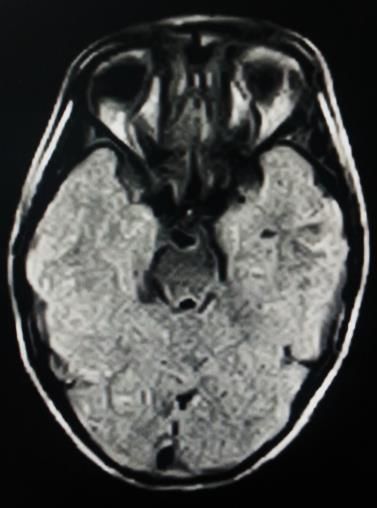
El Ameen et al.
not known. Atrophic brain was atrophy (Figure.3), and the other two had
encountered in 11/66 patients. Isolated associated findings. One had associated
frontoparietal atrophic change was the with rhombencephalic (Figure.4), and the
most common as it was seen in seven other one had associated Dandy Walker
patients. Cerebellar atrophy was seen in 4 variant.
patients. Two had isolated cerebellar
(a) (b) (c)
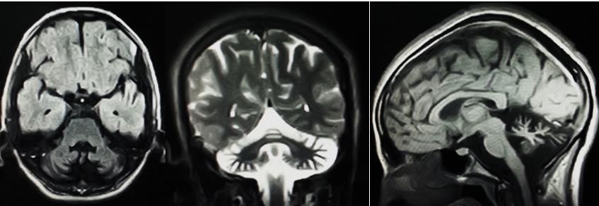
Fig.2: A 6- year- old male presented with hyperactivity and abnormal movement. a) Axial FLAIR
showed widened left temporal arachnoid space with relative reduction in the size of left temporal
lobe. b) Axial T2 showed high SCF of the cyst. c) Sagittal T1 showed the relative reduction of the left
temporal lobe size (Final diagnosis left temporal lobe arachnoid cyst).
(a) (b) (c)
Fig.3: A 4- year- old male presented with hyperactivity, impaired response control, abnormal
movement. a) Axial FLAIR showed markedly hypoplastic right cerebellar hemisphere with
hypoplastic vermis. b) Sagittal T1WI showed markedly hypoplastic cerebellum, note the normal size
and segmentation of corpus callosum, normal pituitary and normal tuber cinereum. c) Coronal T2WI
showed the hypoplastic right cerebellar hemisphere and hypoplastic vermis (Final diagnosis right
cerebellar hypoplasia).
Int J Pediatr, Vol.7, N.5, Serial No.65, May. 2019 9383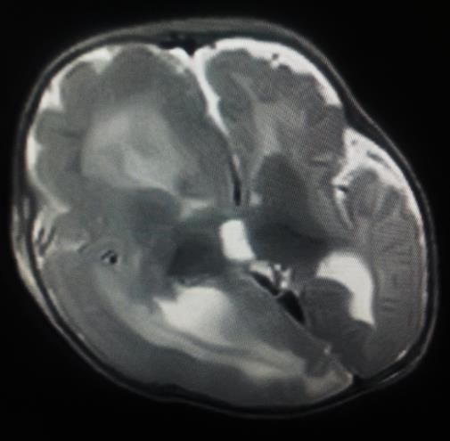
MRI of the Brain in Children with ADHD

              (a)                                    (b)                                (c)
Fig.4: A 7- year- old male presented with impaired response control, ataxia and gait disturbance.
a) Axial FLAIR showed markedly hypoplastic cerebellar hemispheres that are seen fused at the mid
line. b) Sagittal T1WI showed the hypoplastic cerebellum associated with large cystic simulating
lesion. c) Coronal T2WI showed the hypoplastic cerebellum fused at mid line (Final diagnosis
cerebellar hypoplasia with rhombencephalic).

Tuber cinereum lesions were depicted in                    use of three-dimensional (3D) Volumetric
seven patients, five of them were finally                  Interpolated Breath-hold Examination
diagnosed as hamartoma of tuber cinereum                   (VIBE) sequence for more confident
(Figure.5), diagnosis was confirmed by                     diagnosis, and measurement of the volume
MRS (not done in our facility), and                        of hippocampus to confirm the diagnosis.
assigned for follow-up. Cavernoma of                       An electroencephalogram (EEG) was done
tuber cinerium (Figure.6) was seen in two                  for these patients for further evaluation and
patients. They were diagnosed after                        they were positive. 3D VIBE showed high
detection of signal void of calcifications                 accuracy in diagnosis and short time
and marginal low signal of hemosiderin                     utilization. Two patients showed bilateral
within the lesions. Hippocampus sclerosis                  hippocampal sclerosis and three showed
was diagnosed in 5 patients. It needs the                  unilateral hippocampal sclerosis.

            (a)                              (b)                                  (c)
Fig.5: A 5- year- old boy with aggressive antisocial behavior and hyperactivity. a) Axial FLAIR and
b) Axial T1WI show an isointense signal lesion at the right side of mid line filling the right side of the
supra-seller cistern. c) Sagittal T1WI showed markedly thickened tuber cinereum. Note the normal
girth and segmentation of corpus callosum (Final diagnosis hamartoma of tuber cinerium).

Int J Pediatr, Vol.7, N.5, Serial No.65, May. 2019                                                  9384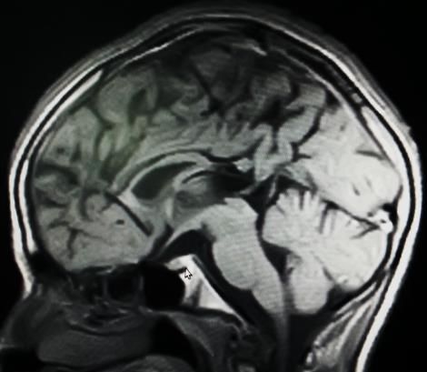
El Ameen et al.

                  (a)                         (b)                         (c)
Fig.6: A 6- year- old boy with hyperactivity, impulsive action and inappropriate response. a) Axial
FLAIR and b) axial T2WI show a well-defined low signal lesion at the right side of interpeduncular
cistern. c) Sagittal T1WI showed markedly thickened tuber cinerium with marginal signal void of
calcification (Final diagnosis cavernous hemangioma of tuber cinerium).

Heterotopia, Pachygyria, Leigh syndrome,               hemimegalencephaly, and the other one
and hemimegalencephaly (HME) were                      had      associated    corpus     callosum
seen in two patients for each. Regarding               dysgenesis. Patients with Leigh syndrome
heterotopia, one patient had focal cortical            (Figure.8) showed abnormal high signal in
dysplasia and one patient had sub-                     basal ganglia and confirmed using
ependymal band heterotopia. As regards                 laboratory investigations that showed high
HME patients (Figure.7), one had isolated              lactate levels.

                   (a)                               (b)                         (c)
Fig.7: A 3- year- old male presented with hyperactivity and impaired response. a) Axial and b)
coronal T2 relatively large sized cerebral hemisphere with smooth gyral appearance that is seen more
notable at the parieto-occipital regions. c) Sagittal T1WI support the same findings, note the
hypoplastic corpus callosum (Final diagnosis Hemimegalencephaly with Lissencephaly).

Int J Pediatr, Vol.7, N.5, Serial No.65, May. 2019                                             9385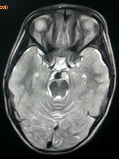
MRI of the Brain in Children with ADHD

               (a)                                   (b)                                (c)
Fig.8: A 7- year- old female child presented with abnormal gait, poor academic performance and
anxiety and depression symptoms. a) Axial T1 showed bilateral nearly symmetrical abnormal low
signal involving both putamen. b and c) Axial and coronal T2WI showed bilateral high signal in the
same area of lentiform nucleus at the putamen (Final diagnosis Leigh disease).

Concerning Joubert syndrome (Figure.9),                          follow-up. All MRI findings were
it was an unusual case with near normal                          tabulated      in   (Table.3).     Clinico-
mentality, and complaining of ataxia, and                        radiological correlation between the
antisocial behavior with marked academic                         clinical presentation of patients and the
delay. Pineal cyst was seen in one patient,                      radiological findings were detected in MRI
it was 1 cm in diameter and assigned for                         of the brain.

               (a)                                         (b)                                      (c)

Fig.9: A 14- year old female presented with impaired response control, learning disorders and ataxia.
a) Axial FLAIR showed atrophic cerebellum and markedly hypoplastic middle cerebellar peduncles,
vermis and molar tooth appearance of mid brain. b) Coronal T2WI showed the atrophic cerebellum
and atrophic middle cerebellar peduncle. c) Sagittal T1WI showed markedly hypoplastic cerebellum,
note the normal size and segmentation of corpus callosum and normal pituitary (Final diagnosis
Joubert syndrome).

Int J Pediatr, Vol.7, N.5, Serial No.65, May. 2019                                                        9386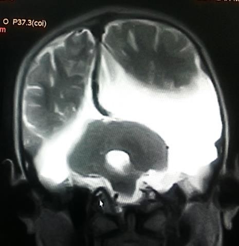
El Ameen et al.

Table-3: Final MRI diagnosis of 100 patients with ADHD child
Findings                                                                            Number
Corpus callosum dysgenesis                                                            19
Temporal lobe pathology:                                                              14
         Atrophy                                                                       7
         Arachnoid cyst                                                                5
         Space occupying lesion                                                        2
Brain atrophy:                                                                        11
                                                                                       7
        Fronto-parietal atrophy
                                                                                       4
        Cerebellar atrophy
Tuber cinerium lesion:                                                                7
        Hamartoma                                                                     5
        Cavernous hemangioma                                                          2
Hippocampus sclerosis                                                                 5
Pachygyria                                                                            2
Heterotopia                                                                           2
Hemimegalencephaly                                                                    2
Leigh syndrome                                                                        2
Joubert syndrome                                                                      1
Pineal body cyst                                                                      1
Unremarkable MRI                                                                     34
Total                                                                                100

4- DISCUSSION                                        79 males, and 21 females. In spite of the
                                                     limited number of patients we had higher
    In the last decade, Attention-
                                                     incidence of disease among male patients
deficit/hyperactivity disorder (ADHD)
became         the       most         common         than females which agrees with Edmond et
                                                     al., who stated that boys are likely to be
neuropsychiatric disorder among children.
                                                     involved with ADHD around three times
It occurs in approximately 3-9% of the
childhood population. ADHD has been                  more than girls, which is almost
                                                     approximately the same incidence in our
conceptualized as a neurological disorder
                                                     study (5). We found that family history
of the prefrontal cortex and its
connections.      In    fact,    the     dorsal      was positive in 17% of our patient cohort
                                                     which is a considerable incidence
frontostriatal circuits have been linked to
                                                     percentage making heredofamilial element
cognitive     control,    whereas       fronto-
cerebellar circuits have been linked to              must be searched for, which is concordant
                                                     with Mulder et al., and others who found
timing. Neurobiological dysfunction of
                                                     that familiar vulnerability presents in
these circuits could lead to symptoms of
                                                     families that had history of ADHD patients
ADHD (4, 5). However, like all
                                                     (6- 9). In our study, clinical presentations
psychiatric disorders are based on
                                                     of ADHD had a wide spectrum including:
symptoms. They have a wide range of
causes      and       susceptibilities.     So       poor social relationships, hyperactivity and
                                                     poor behavioral inhibition. Impaired
neurobiological investigations are a
                                                     response      control,    poor     academic
necessary point for diagnosis. They have
been the basis for an increasing number of           performance and learning disorders are
                                                     also recorded. We tried to depict the
structural    as     well     as    functional
                                                     structural and anatomic brain changes and
neuroimaging studies (1). The aim of our
study was to answer a simple question:               correlate them with the presenting clinical
                                                     manifestations. Our results showed that
what are the anatomic substrates
                                                     patients with corpus callosum dysgenesis
associated with combined type of ADHD?
Our study was conducted on 100 children,             have poor self-regulation and impulsive

Int J Pediatr, Vol.7, N.5, Serial No.65, May. 2019                                           9387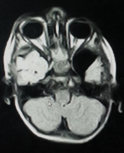
MRI of the Brain in Children with ADHD

behavior. This could be explained by the             performance and learning disorders are
neurobiological function of corpus                   usually associated with temporal lobe
callosum. Corpus callosum fiber tracts are           pathology or reductions in volume of
connecting between the two cerebral                  frontal or temporal cortex. This is in
hemispheres. It allows for transfer and              agreement with Castellanos, Cortese et al.,
integration of sensory, motor and cognitive          and Angriman et al.’s reports which
information.      Disruption      of     these       confirmed that most ADHD patients have
connections usually leads to impulsivity             reduction in the pre-frontal and occipital
and cognitive disorder which is consistent           cortex volume (1, 4, 17). In our study, we
with Neal et al., and others who stated that         have seen heterotopia, pachygyria and
corpus callosum dysgenesis             usually       hemimegalencephaly in two patients for
correlated with impaired response control            each. MRI can easily diagnose migration,
(10 -12). Corpus callosum dysgenesis                 proliferation and sulcation defects and
group of patients have near normal                   differentiate between them and the
mentality. They present the most common              simulate picture of vasculitis. In patients
pathological finding (28.7%) of our patient          with focal cortical dysplasia sub-
cohort. This could be explained by the fact          ependymal heterotopia appeared as
that agenesis and dysgenesis of corpus               abnormal signal of gray matter within the
callosum is one of the most frequent brain           periventricular white matter and within the
malformations. It is a heterogeneous                 parietal subcortical white matter.
condition that may be seen as an isolated
                                                     The        proliferation       defect      in
entity or as one manifestation of congenital         hemimegalencephaly and pachygyria can
syndrome which is in agreement with
                                                     be diagnosed by the discrepancy between
Doherty et al., and Mohapatra et al., who
                                                     the size of both cerebral hemispheres in
stated that some patients with agenesis or           hemimegalencephaly          patients     and
dysgenesis of corpus callosum may show
                                                     alteration of the shape and thickness of the
no developmental delay, and normal
                                                     gyri in pachygyria patients. This is in
intelligence with mild behavioral or social          agreement with Abdel Razek et al., who
problems as well as the attention-deficit-
                                                     stated that interruption of normal
hyperactivity disorder (ADHD) (10,
                                                     developmental sequences either due to
13,14). As regards hypothalamic region               defective proliferation, migration, or
affection, our results showed that thought
                                                     organization of the cortex can be easily
and attention problems are correlated with
                                                     diagnosed by neuroimaging and MRI
the presence of hypothalamic lesion                  which was proved to be a valuable tool in
regardless of the underlying pathological
                                                     their diagnosis, and differentiation
type. This agrees with Castellanos who
                                                     between them and vasculitis which may
stated      that     cortico-striato-thalamo-        have simulating picture for focal cortical
cortical (CSTC) circuits are responsible for
                                                     dysplasia picture. This is also in agreement
selection, initiation, and execution of
                                                     with Duerden et al. who found that there is
complex motor and cognitive responses.               significant increase in cortical thickness in
Also, this is consistent with the results of
                                                     patients with ADHD (18-20). As regarding
Fortier et al., and Van der Meer et al. who
                                                     Joubert syndrome, there was an interesting
stated that the biological changes through           case where the patient presented with anti-
hypothalamic-pituitary-adrenal (HPA) axis
                                                     social behavior suggesting ADHD
in case of increased circulating cortisol
                                                     associated with ataxia. Using MRI imaging
level may result in several psychiatric              of the brain we detected the atrophic
disorders and significant behavioral
                                                     cerebellum, and markedly hypoplastic
changes (1, 15, 16). Our results showed
                                                     middle      cerebellar    peduncles.     The
that poor memory, poor academic

Int J Pediatr, Vol.7, N.5, Serial No.65, May. 2019                                           9388
El Ameen et al.
characteristic molar tooth appearance of proved to be very useful. Indeed, not all
mid brain also noted that is concordant patients of ADHD had cerebellar or
with what was stated by Abdel Razek and frontoparietal atrophy as presumed. Other
Castillo in their article about hind brain regions of the brain must be searched
malformations where they classified carefully according to the specific clinical
Joubert syndrome as a combined cerebellar presentation. Functional MRI including
and brain stem malformation (21). Finally, DWI and MRS as well as tractography
we can say the small number of the sample must be supervised in these patients to
that was one of the limitations of this study evaluate the white matter tracts in future
did not allow us to make a full judgement studies.
on all brain changes associated with
ADHD. Since the study was limited to 6- CONFLICT OF INTEREST: None.
children’s population which needs 7- REFERENCES
anesthesia and full consent from parents to
do the MRI examination. We also 1. Castellanos FX. Anatomic magnetic
resonance imaging studies of attention
considered that not using functional MRI
deficit/hyperactivity disorder. Dialogues Clin
including Diffusion-weighted imaging
Neurosci 2002; 4 (4): 444–48.
(DWI), Magnetic resonance spectroscopy
2. Gong Q. MRI Shows Brain
(MRS), and tractography is one of the
Differences Among ADHD Patients. RSNA
most important limitations in this work. In press release. 2017, Nov. 22.
fact, we will consider them soon in another
3. Wolraich M, Brown L , Brown TR,
coming work, as recent advances in MRI
DuPaul G, Earls M, Feldman HM, et al.
could be used in evaluation of different ADHD: Clinical Practice Guideline for the
metabolic diseases, autism and ADHD Diagnosis, Evaluation, and Treatment of
simulating condition such as Gusher Attention-Deficit/Hyperactivity Disorder in
disease. Many authors such as Abdel Children and Adolescents. Pediatrics. 2011
Razek et al., and Lea et al. stated that there Nov; 128(5): 1007–22.
is significant difference in the apparent 4. Cortese S and Castellanos FX.
diffusion coef- ficient (ADC) value of Neuroimaging of Attention-
normal brain, and brain of children with Deficit/Hyperactivity Disorder: Current
metabolic diseases like Gaucher disease. Neuroscience-Informed Perspectives for
This may help in differentiation between Clinicians. Curr Psychiatry Rep. 2012; 14 (5):
them and ADHD patients who require a 10.
completely different treatment pathway 5. Emond V, Joyal C, Poissant H.
(22-25). Also, the application of other Structural and functional neuroanatomy of
functional MRI studies using BOLD attention-deficit hyperactivity disorder
technique in evaluation of brain function (ADHD). Encephale. 2009; 35 (2):107-14.
of children and adolescents with ADHD 6. Mulder MJ, Baeyens D, Davidson
which may have altered regional brain MC, Casey BJ, van den Ban E, van Engeland
H, Durston S. Familial vulnerability to ADHD
function and be associated with executive
affects activity in the cerebellum in addition to
dysfunction as stated by Li et al., may help
the prefrontal systems. J Am Acad Child
our understanding of the relationships Adolesc Psychiatry. 2008; 47(1):68-75.
between neural substrate and executive
7. Durston S, van Belle J, de Zeeuw P.
function in ADHD (26).
Differentiating frontostriatal and fronto-
cerebellar circuits in attention-
5- CONCLUSION deficit/hyperactivity disorder. Biol
According to the results, MRI Psychiatry. 2011; 69 (12):1178-84.
evaluation of the brain in patients with 8. Amos SP, Homan GJ, Sollo
attention-deficit/hyperactivity disorder N, Ahlers-Schmidt CR, Engel M, Rawlins P.
Int J Pediatr, Vol.7, N.5, Serial No.65, May. 2019 9389MRI of the Brain in Children with ADHD The Relationship of Personality Style Childhood ADHD: 2013. Current and Attention Deficit Hyperactivity Disorder i Developmental Disorders Reports. 2014; n Children. Kans J Med. 2017; 10 (2):26-9. 1(1):29–40. 9. Sobanski E, Banaschewski 18. Abdel Razek AA, Kandell AY, T, Asherson P, Buitelaar J, Chen W, Franke Elsorogy LG, Elmongy A, Basett AA. B, et al. Emotional lability in children and Disorders of cortical formation: MR imaging adolescents with ADHD: clinical correlates features. AJNR Am J Neuroradiol 2009; 30: 4- and familial prevalence. J Child Psychol 11. Psychiatry. 2010; 51(8):915-23. 19. Abdel Razek AA, Alvarez H, Bagg S, 10. Neal JB, Filippi CC and Mayeux R. Refaat S, Castillo M. Imaging spectrum of Morphometric variability of neuroimaging CNS vasculitis. Radiographics 2014; 34:873- features in Children with Agenesis of the 94. Corpus Callosum. BMC Neurology.2015; 20. Duerden EG, Tannock R, Dockstader 15:116. C. Altered cortical morphology in 11. Yaroglu KS. sensorimotor processing regions in adolescents Attention Deficit Hyperactivity Disorder in a and adults with attention-deficit/hyperactivity Patient with Congenital Mirror disorder. Brain Res. 2012; 1445:82–91. Movement Disorder and Colpocephaly. Iran J 21. Abdel Razek AA1, Castillo M. Pediatr. 2015; 25(5):e1787. Magnetic Resonance Imaging of 12. McNally MA, Crocetti D, Mahone Malformations of Midbrain-Hindbrain. J EM, Denckla MB, Suskauer SJ, Mostofsky Comput Assist Tomogr. 2016; 40 (1):14-25. SH. Corpus callosum segment circumference 22. Abdel Razek AA, Abd El-Gaber N, is associated with response control in children Abdalla A, Fathy A, Azab A, Rahman AA. with attention-deficit hyperactivity disorder. Apparent diffusion coefficient vale of the brain Journal of Child Neurol. 2010; 25(4):453-62. in patients with Gaucher's disease type II and 13. Doherty D, Tu S, Schilmoeller K, type III. Neuroradiology 2009; 51: 773-9. Schilmoeller G. Health-related issues in 23. Abdel Razek A, Mazroa J2, Baz H. individuals with agenesis of the corpus Assessment of white matter integrity of callosum. Child: Care Health Dev. 2006; 32: autistic preschool children with diffusion 333–42. weighted MR imaging. Brain Dev. 2014; 36 14. Mohapatra S, Panda UK, Sahoo AJ , (1):28-34. Dey S and Rath N. Neuropsychiatric 24. Lea SE, Matt AR, Patros CHG, Tarle manifestations in a child with agenesis of the SJ, Arrington EF, Grant DM. Working corpus callosum J Neurosci Rural Pract. 2015 Memory and Motor Activity: A Comparison Jul-Sep; 6(3): 456–457. Across Attention-Deficit/Hyperactivity 15. Fortier MÈ, Sengupta SM, Grizenko Disorder, Generalized Anxiety Disorder, and N, Choudhry Z, Thakur G, Joober R. Genetic Healthy Control Groups. Behav Ther. 2018; evidence for the association of the 49(3):419-34. hypothalamic-pituitary-adrenal (HPA) axis 25. Razek AA, Abdalla A, Ezzat A, with ADHD and methylphenidate treatment Megahed A, Barakat T. Minimal hepatic response. Neuromolecular Med. 2013; encephalopathy in children with liver 15(1):122-32. cirrhosis: diffusion-weighted MR imaging and 16. Van der Meer D, Hoekstra PJ, van proton MR spectroscopy of the brain. Donkelaar M, Bralten J, Oosterlaan Neuroradiology 2014; 56: 885-91. J, Heslenfeld D, et al. Predicting attention- 26. Li F, He N, Li Y, Chen L, Huang X, deficit/hyperactivity disorder severity from Lui S, Guo L, Kemp G J and Gong Q. Intrinsic psychosocial stress and stress-response genes: Brain Abnormalities in Attention Deficit a random forest regression approach. Hyperactivity Disorder: A Resting-State Psychiatry. 2017; 7 (6):1145. Functional MR Imaging Study. 17. Angriman M, Beggiato A, Cortese S. Radiology. 2014; 272(2):514-23. Anatomical and Functional Brain Imaging in Int J Pediatr, Vol.7, N.5, Serial No.65, May. 2019 9390
You can also read