Clinical aspects for differential diagnosis of Kawasaki disease shock syndrome: a case control study - BMC Pediatrics
←
→
Page content transcription
If your browser does not render page correctly, please read the page content below
Park et al. BMC Pediatrics    (2021) 21:25
https://doi.org/10.1186/s12887-020-02488-w

 RESEARCH ARTICLE                                                                                                                                    Open Access

Clinical aspects for differential diagnosis of
Kawasaki disease shock syndrome: a case
control study
Woo Young Park, Sang Yun Lee*, Gi Beom Kim, Mi Kyoung Song, Hye Won Kwon, Eun Jung Bae,
Eun hwa Choi and June Dong Park

  Abstract
  Background: Because of the absence of a specific diagnostic test and pathognomonic clinical features, physicians
  must rely on the presence of specific clinical criteria and laboratory data that support the diagnosis of KD. To help
  clinicians distinguish KD, KDSS, septic shock, and TSS earlier, we suggest differential diagnosis and treatment
  guideline.
  Methods: Medical records of immunocompetent patients who were admitted to the pediatric department with a
  diagnosis of KDSS, septic shock or TSS (SS group) were retrospectively reviewed. In addition, KD patients were
  selected by seasonal matching to each case of KDSS patient by date of admission (± 2 weeks).
  Results: There were 13 patients with KDSS, 35 patients with SS group, and 91 patients with KD. In comparison
  between KDSS and septic shock group, KDSS group had significantly higher rate of coronary aneurysm incidence,
  and higher left ventricle dysfunction rate. In comparison between KDSS and TSS, patients with KDSS had a
  significantly higher erythrocyte sedimentation rate (ESR) and significantly lower creatinine. Receiver operation
  characteristic curve revealed that the optimal ESR cut off value for determining the KDSS was 56.0 (sensitivity 75.0%,
  specificity of 100.0%) and the optimal creatinine cut off value for determining the TSS was 0.695 (sensitivity 76.9%,
  specificity 84.6%).
  Conclusions: Clinical symptoms, laboratory finding, echocardiography, and culture studies can be used to
  differentiate KD, KDSS, septic shock and TSS.
  Keywords: Kawasaki disease shock syndrome, Kawasaki disease, Septic shock, Toxic shock syndrome

Background                                                                              artery complications. Therefore, accurate and timely
Kawasaki disease (KD) is an acute systemic inflammatory                                 diagnosis of KD is critical. However, because of the
vasculitis of early childhood, predominantly involving                                  absence of a specific diagnostic test and pathognomonic
medium-sized arteries [1]. About 15–25% of untreated                                    clinical features, physicians must rely on the presence of
children will develop coronary artery abnormalities [2].                                specific clinical criteria and laboratory data that support
Early detection and prompt initiation of therapy with                                   the diagnosis of KD, while excluding other illnesses that
high dose intravenous immunoglobulin (IVIG) plus                                        could mimic the disease [3].
aspirin can reduce the incidence of serious coronary                                      As the COVID-19 pandemic continues, there have
                                                                                        been reports of multi-system inflammatory syndrome in
* Correspondence: saeng123@hanmail.net                                                  children (MIS-C) that shares clinical features with KD
Department of Pediatrics, Seoul National University Children’s Hospital, 101,           [4]. Recognition of sudden increase of severe Kawasaki-
Daehak-ro, Jongno-gu, Seoul, Republic of Korea

                                         © The Author(s). 2021 Open Access This article is licensed under a Creative Commons Attribution 4.0 International License,
                                         which permits use, sharing, adaptation, distribution and reproduction in any medium or format, as long as you give
                                         appropriate credit to the original author(s) and the source, provide a link to the Creative Commons licence, and indicate if
                                         changes were made. The images or other third party material in this article are included in the article's Creative Commons
                                         licence, unless indicated otherwise in a credit line to the material. If material is not included in the article's Creative Commons
                                         licence and your intended use is not permitted by statutory regulation or exceeds the permitted use, you will need to obtain
                                         permission directly from the copyright holder. To view a copy of this licence, visit http://creativecommons.org/licenses/by/4.0/.
                                         The Creative Commons Public Domain Dedication waiver (http://creativecommons.org/publicdomain/zero/1.0/) applies to the
                                         data made available in this article, unless otherwise stated in a credit line to the data.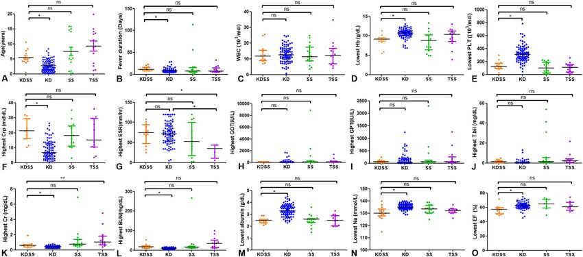
Park et al. BMC Pediatrics   (2021) 21:25                                                                          Page 2 of 9

like disease in the North America and European coun-            lymphadenopathy (≥ 1.5 cm in diameter), usually unilat-
tries during the COVID-19 pandemic is alarming given            eral. A diagnosis of incomplete or atypical KD was used
the unknown etiology of KD. Also, it is timely to address       for patients with a history of fever lasting for more than
the differences in clinical manifestations between various      five days who had less than four of the five typical clin-
Kawasaki mimic diseases as some patients with KD may            ical features of KD but showing evidence of coronary
also present with hypotension or shock, known as                artery lesion on echocardiography or compatible labora-
“Kawasaki disease shock syndrome” (KDSS). Differenti-           tory findings suggested by AHA [7]. KDSS was defined
ating between KDSS and septic shock or toxic shock              as a patient with KD complicated by hypotension with-
syndrome (TSS) in early stages of clinical diagnosis is         out evidence of infection [5]. Hypotension was defined
challenging [1]. Although KDSS and septic shock or TSS          as a systolic blood pressure < 5th percentile for a
show similar clinical presentation, early diagnosis is crit-    patient’s age. All patients with TSS were diagnosed by
ical in patients with unstable conditions because differ-       infection specialists using the diagnostic criteria pro-
ent clinical management is required for treatment.              vided by the Centers for Disease Control and Prevention
   Therefore, we retrospectively investigated patients          (Supplement Table 1) [8]. Septic shock was defined as
admitted with diagnosis of KDSS, septic shock or TSS            sepsis and cardiovascular organ dysfunction. Cardiovas-
(SS group), and KD among immunocompetent patients.              cular organ dysfunction means hypotension (< 5th per-
Additionally, we tried to suggest KDSS differential diag-       centile for age) despite > 40 ml/kg fluid bolus in 1 h or
nosis and treatment guideline.                                  vasoactive requirement to maintain blood pressure des-
                                                                pite > 40 ml/kg fluid bolus in 1 h or two or more signs of
Methods                                                         abnormal perfusion (increased lactate, metabolic acid-
Patient selection                                               osis, decreased urine output (< 0.5 mL/kg/hr), capillary
A computerized search program was used to search for            refill > 5 s) [9]. Organ damage was defined as damage in
patients diagnosed with KD or KDSS or SS that occurred          major organs including brain, heart, lung, liver, and kid-
in immunocompetent patients from January 2004 to                ney due to hypoperfusion and sequential organ failure
August 2019. These patients were classified into three          assessment score was used as an index [10]. The coron-
groups: KDSS group, KD group, and SS group. 13                  ary artery aneurysm was defined as Z score ≥ 2.5. In the
patients satisfied the KDSS criteria [5]. In KD group,          present study, patients with septic shock and TSS were
control subjects were chosen for each case patient and          assigned into the septic shock (SS) group.
matched to its control by date of admission (± 2 weeks)
as the matching factor to control for the possibility of        Data collection
seasonal variation. Patients with incomplete KD and             For all patients with KD, we accessed the following retro-
refractory KD were also included in the KD group.               spectively collected data: demographic data (age, gender,
Ninety-one patients met the inclusion criteria [6]. To          associated symptoms and number of days of fever at pres-
search for patients with septic shock group, we searched        entation), laboratory values (white blood cell and differen-
“Septic shock” and “Toxic shock syndrome” from dis-             tial counts, platelet count, erythrocyte sedimentation rate
charge records and immunocompromised patients were              (ESR), and concentrations of hemoglobin, C-reactive pro-
excluded. A total of 29 patients met the inclusion cri-         tein (CRP), aspartate aminotransferase (AST), alanine ami-
teria for the septic shock or toxic shock syndrome.             notransaminase (ALT), and γ-glutamyl transpeptidase
  This study used clinical data retrieved from Seoul            (GGT), creatinine, blood urea nitrogen (BUN), albumin,
National University Hospital Patients Research Environ-         electrolytes), laboratory evaluations of coagulation function,
ment system. This study was approved by the Institu-            response to IVIG, oral prednisolone, methylprednisolone
tional Review Board of the Seoul National University            pulse therapy, and infliximab, and echocardiographic data.
Hospital (IRB number: 1910–051-1068, approved date:             The laboratory data were compared to the worst values
Oct 14th, 2019).                                                between 3 groups.

Definitions                                                     Statistical analysis
Diagnosis of KD was based on the diagnostic criteria            All data analyses were performed using SPSS statistics
provided by the American Heart Association (AHA) [7].           25.0 (IBM, Armonk, NY, USA). Data is presented as
The diagnostic criteria were the presence of a fever last-      median and ranges if not normally distributed. Not nor-
ing for at least five days and at least four of the following   mally distributed data was compared between groups
five typical clinical features of KD: bilateral bulbar con-     using the Mann-Whitney test, Fisher’s exact test, or
junctival injection without exudate, erythema and crack-        Chi-square test. One-way ANOVA was performed for
ing of lips, strawberry tongue, and/or erythema of oral         continuous variables if normally distributed. Kruskal-
and pharyngeal mucosa, polymorphous rash and cervical           Wallis test was used to compare the laboratory data
Park et al. BMC Pediatrics     (2021) 21:25                                                                                                      Page 3 of 9

among more than two groups. Area under the ROC                                Table 1 Demographics and clinical characteristics of patients
(receiver operating characteristic) curve was used as an                      with Kawasaki disease shock syndrome and Kawasaki disease
accuracy index for the diagnosis of KDSS. As appropri-                        (continuous variables were described as median and range)
ate, p < 0.05 was considered significant.                                                                      KDSS (n=13)       KD (n=91)          P value
                                                                              Age, (years)                     5.1 (0.5–10.6)    2.3 (0.2–8.4)      0.000
Results                                                                       Male (%)                         46.2% (6/13)      58.2% (53/91)      0.411
From January 2004 to August 2019, 13 patients were                            Fever duration (days)            11 (8–23)         8 (3–29)           0.002
diagnosed as a KDSS. Over the same time, 13 patients
                                                                              Total hospital day (days)        18 (6–31)         6 (3–18)           0.000
were admitted with a diagnosis of TSS and 16 patients
were admitted with a diagnosis of septic shock. In                            Follow up duration (months)      10 (1–105)        13 (0–175)         0.723
addition, 91 patients were treated as KD. Demographics                        ICU care (%)                     53.8% (7/13)      0.0% (0/91)        0.000
and clinical characteristics for KDSS group and control                       ICU care duration (days)         4 (0–11)          0                  0.000
groups are shown in supplement Table 2.                                       Inotropic drugs (%)              92.3% (12/13)§    1.1% (1/91)        0.000
  All patients in the KDSS and KD group, and 17 of 29                         Respiratory support
patients in the SS group received an echocardiographic
                                                                                 - No support                  30.8% (4/13)      100% (91/91)       0.000
assessment. Laboratory values and ejection fraction for
KDSS group and control groups are shown in Fig. 1 and                            - Oxygen delivery             53.8% (7/13)      0.0% (0/91)        0.000
supplement Table 3. Kawasaki features, associated symp-                          - Mechanical ventilation      15.4% (2/13)      0.0% (0/91)        0.015
toms and treatment for KDSS group and control groups                          Mortality (%)                    0.0% (0/13)       0.0% (0/91)
are shown in supplement Table 4.                                              KDSS Kawasaki disease shock syndrome, KD Kawasaki disease, SS Septic shock
                                                                              and toxic shock syndrome, ICU Intensive care unit

KDSS vs. KD
Demographics and clinical characteristics of patients                           Kawasaki features and associated symptoms of Kawasaki
with Kawasaki disease shock syndrome and Kawasaki                             disease shock syndrome and Kawasaki disease are shown
disease are shown in Table 1. Patients with KDSS had a                        in Table 2. There was no statistically significant difference
significantly older age (p = 0.000), longer fever duration                    in Kawasaki features between KDSS and KD groups.
(p = 0.002), longer hospital day (p = 0.000), longer ICU                      Patients in the KDSS group had significantly higher
admission days (p = 0.000), and higher rate of using ino-                     gastrointestinal symptoms (p = 0.000), respiratory
tropic drugs (p = 0.000) than patients with KD. How-                          symptoms (p = 0.000), systemic pain (p = 0.000),
ever, there were no significant differences in sex-                           pleural effusion (p = 0.00), and organ damage (p =
distribution between the two groups.                                          0.000) than those in the KD group.

 Fig. 1 Comparison of Age, Fever duration, Laboratory Results and Ejection fraction in 3 Different Patient Groups. Statistics were calculated with
 GraphPad Prism software. Horizontal lines represent median values for each group and vertical lines represent interquartile range. *, **: statistically
 significant, (p < 0.05), as determined by the Kruskal-Wallis test. ns: not significant
Park et al. BMC Pediatrics (2021) 21:25 Page 4 of 9
Table 2 Kawasaki features and associated symptoms of Compared with patients with hemodynamically normal
Kawasaki disease shock syndrome and Kawasaki disease KD, left ventricle dysfunction represented by reduced
KDSS(n=13) KD(n=91) P value ejection fraction (< 55%) was more common in the
Kawasaki features KDSS group (p = 0.000). In addition, there was signifi-
Conjunctival injection 92.3% (12/13) 91.2% (83/91) 1.000 cantly (p = 0.040) more coronary artery dilatation in the
KDSS group than in the KD group. In the KDSS group,
Oropharyngeal changes 84.6% (11/13) 81.3% (74/91) 1.000
during the acute phase, five patients showed transient
Polymorphous rash 76.9% (10/13) 84.6% (77/91) 0.442
coronary artery dilatation and three patients had persist-
Cervical lymphadenopathy 69.2% (9/13) 44.0% (40/91) 0.088 ent coronary artery dilatation. In the KD group, during
Extremity changes 92.3% (12/13) 67.0% (61/91) 0.102 the acute phase of Kawasaki disease, 19 patients showed
Associated symptoms transient coronary artery dilatation and four patients
Gastrointestinal symptoms 84.6% (11/13) 26.4% (24/91) 0.000 had persistent coronary artery dilatation at the last
echocardiography.
Respiratory symptoms 76.9% (10/13) 9.9% (9/91) 0.000
Neurologic symptoms 15.4% (2/13) 2.2% (2/91) 0.066
KDSS vs SS group
Systemic pain 53.8% (7/13) 3.3% (3/91) 0.000 Demographics and clinical characteristics of patients
Pleural effusion 76.9% (10/13) 2.2% (2/91) 0.000 with Kawasaki disease shock syndrome and septic shock
Organ damage 92.3% (12/13) 20.9%(19/91) 0.000 group are shown in Table 4. There were no significant
KD Kawasaki disease, KDSS Kawasaki disease shock syndrome, SS Septic shock differences in sex-distribution, age, fever duration, total
and toxic shock syndrome hospital day, ICU admission days, or rate of using ino-
tropic drugs between the two groups.
Diagnosis and treatment of Kawasaki disease shock Kawasaki features and associated symptoms of
syndrome and Kawasaki disease are shown in Table 3. patients with Kawasaki disease shock syndrome and
KD group had significantly higher rate of initial diagno- septic shock group are shown in Table 5. Patients
sis of KD than those in the KDSS group and KDSS with KDSS had significantly higher rate of conjunc-
group initially showed only 1–2 of typical Kawasaki tival injection (p = 0.000), oropharyngeal changes (p =
symptoms. However, 3 or more symptoms were seen in 0.000), cervical lymphadenopathy (p = 0.000), and
all patients eventually. In addition, patients in the KDSS extremity changes (p = 0.001) than patients with SS
group had significantly higher IVIG resistance rate than group. In addition, patients with KDSS had a signifi-
those in the KD group. cantly higher rate of respiratory symptoms (p = 0.033)
Compared to those in the KD group, patients in the KDSS and pleural effusion (p = 0.033) than patients with SS
group had lower hemoglobin level (p = 0.000), lower platelet group.
counts (p = 0.00), higher C-reactive protein level (p = 0.000), Diagnosis and treatment of Kawasaki disease shock
higher total bilirubin level (p = 0.012), higher creatinine level syndrome and septic shock group are shown in Table 6.
(p = 0.001), higher BUN level (p = 0.000), lower albumin Patients with KDSS had significantly higher rates of
level (p = 0.00), and lower sodium level (p = 0.000) (Fig. 1, usage 2nd IVIG (p = 0.000), and methylprednisolone
supplementary Table 3). pulse therapy (p = 0.002) than patients with SS group.
Table 3 Diagnosis, and treatment of Kawasaki disease shock syndrome and Kawasaki disease
KDSS(n=13) KD(n=91) P value
Diagnosis
Initial diagnosis of KD 23.1% (3/13) 80.2% (73/91) 0.000
Complete KD 46.2% (6/13) 67.0% (61/91) 0.214
Incomplete KD 38.5% (5/13) 29.7% (27/91) 0.532
Treatment
1st IVIG 84.6% (11/13) 98.9% (90/91) 0.041
2nd IVIG 76.9% (10/13) 19.8% (18/91) 0.000
Oral prednisolone 7.7% (1/13) 11.0% (10/91) 1.000
Methylprednisolone pulse therapy 38.5% (5/13) 11.0% (10/91) 0.020
Infliximab 15.4% (2/13) 0.0% (0/91) 0.015
Antibiotics 92.3% (12/13) 15.4% (14/91) 0.000
IVIG Intravenous immunoglobulin, KD Kawasaki disease, KDSS Kawasaki disease shock syndrome, SS Septic shock and toxic shock syndromePark et al. BMC Pediatrics (2021) 21:25 Page 5 of 9
Table 4 Demographics and clinical characteristics of patients addition, there was no coronary artery aneurysm case
with Kawasaki disease shock syndrome and septic shock group in SS group. (Fig. 1, Supplement Table 3).
(continuous variables were described as median and range)
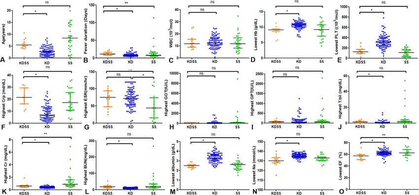
KDSS (n=13) SS (n=29) P value KDSS vs TSS
Age, (years) 5.1 (0.5–10.6) 7.6 (0.3–19.9) 0.185 There were no significant differences in age, sex-
Male (%) 46.2% (6/13) 65.5% (19/29) 0.237 distribution, and laboratory findings between KDSS and
Fever duration (days) 11 (8–23) 7 (1–114) 0.068
TSS groups except for ESR and creatinine. Patients with
KDSS had a significantly higher ESR (p = 0.019) and sig-
Total hospital day (days) 18 (6–31) 13 (7–225) 0.419
nificantly lower creatinine (p=0.007) than patients with
Follow up duration (months) 10 (1–105) 2.5 (0–119) 0.124 TSS. (Supplement Table 3) Receiver operation character-
ICU care (%) 53.8% (7/13) 62.1% (18/29) 0.616 istic (ROC) curve revealed that the optimal ESR cut off
ICU care duration (days) 4 (0–11) 3 (0–20) 0.591 value for determining the KDSS was 56.0 which had a
Inotropic drugs (%) 92.3% (12/13)§ 86.2% (25/29) 0.153 sensitivity of 75.0% and a specificity of 100.0% (Figs. 2
Respiratory support
and 3a, AUC, 0.894; 95% CI, 0.757–1.000, P=0.003) and
the optimal creatinine cut off value for determining the
- No support 30.8% (4/13) 48.3% (14/29) 0.289
TSS was 0.695 which had as sensitivity of 76.9% and a
- Oxygen delivery 53.8% (7/13) 10.3% (3/29) 0.005 specificity of 84.6% (Figs. 2 and 3b, AUC, 0.802; 95% CI,
- Mechanical ventilation 15.4% (2/13) 41.4%(12/29) 0.159 0.620–0.983, P=0.009).
Mortality (%) 0.0% (0/13) 10.3%(3/29) 0.540
KDSS Kawasaki disease shock syndrome, KD Kawasaki disease, SS Septic shock Discussion
and toxic shock syndrome, ICU Intensive care unit Because there is no pathognomonic clinical feature or
diagnostic test for KD, patients with KDSS are frequently
There was no difference in antibiotic treatment rates misdiagnosed. Its clinical presentation may be mistaken
between the two groups. 12 of the 13 patients in the for septic shock or TSS, leading to delay in treatment
KDSS were initially treated with antibiotics as they were [11]. In COVID-19 pandemic period, cases for MIS-C
indistinguishable from septic shock. have been reported and shared clinical features with KD,
Compared with KDSS patients, patients in septic KDSS, and TSS [4]. Case definitions for emerging
shock group had higher level of creatinine (p = 0.006) inflammatory condition during COVID-19 pandemic
and lower ESR level (p=0.008). However, there were from the World Health Organization, Royal College of
no significant differences in hemoglobin, platelet Pediatrics and Child Health, and Centers for Disease
count, CRP level, liver function, BUN, albumin or Control and Prevention are similar in many ways to KD
sodium level between KDSS and SS groups. In [12–14]. Therefore, it has become more important to
accurately distinguish among various Kawasaki mimick-
ing diseases.
Table 5 Kawasaki features and associated symptoms of Considering the symptoms of KDSS that look different
Kawasaki disease shock syndrome, and Septic shock group from KD and similar symptoms to other diseases such as
KDSS(n=13) SS (n=29) P value TSS or MIS-C, the pathophysiological cause becomes
Kawasaki features curious. Despite 4 decades of investigation, the cause of
Conjunctival injection 92.3% (12/13) 10.3% (3/29) 0.000
KD remains unknown [7]. Recently updated three major
pathophysiologic components of KD are a genetic pre-
Oropharyngeal changes 84.6% (11/13) 17.2% (5/29) 0.000
disposition, immunomodulation through both habitual
Polymorphous rash 76.9% (10/13) 69.0% 1 (20/29) 0.722 exposures and environmental factors, and contact with
Cervical lymphadenopathy 69.2% (9/13) 3.4%(1/29) 0.000 the disease trigger or triggers [15]. In this background,
Extremity changes 92.3% (12/13) 34.5% (10/29) 0.001 exposure to the still unidentified trigger such as SARS-
Associated symptoms CoV-2 might result in the development of KD in a gen-
Gastrointestinal symptoms 84.6% (11/13) 82.8% (24/29) 1.000
etically susceptible child, with at least a partial contribu-
tion from immune-modulating factors. Multiple factors
Respiratory symptoms 76.9% (10/13) 41.4% (12/29) 0.033
may act sequentially or simultaneously as predisposing,
Neurologic symptoms 23.1% (3/13) 51.7% (15/29) 0.083 immune-modulating, or triggering agents, altering both
Systemic pain 53.8% (7/13) 37.9% (11/29) 0.335 individual risk as well as the incidence of KD in the
Pleural effusion 76.9% (10/13) 41.4% (12/29) 0.033 population across countries or regions [15]. Recent re-
Organ damage 92.3% (12/13) 82.8% (24/29) 0.647 ports of MIS-C suggest that MIS-C may have a different
KD Kawasaki disease, KDSS Kawasaki disease shock syndrome, SS Septic shock
racial predilection, affecting primarily people of African
and toxic shock syndrome American, Caribbean, and Hispanic ancestry [4].Park et al. BMC Pediatrics (2021) 21:25 Page 6 of 9
Table 6 Diagnosis, and treatment of Kawasaki disease shock syndrome, and Septic shock group
KDSS(n=13) SS (n=29) P value
Diagnosis
Initial diagnosis of KD 23.1% (3/13) 0%(0/29) 0.025
Complete KD 46.2% (6/13) 0%(0/29) 0.000
Incomplete KD 38.5% (5/13) 3.4%(1/29) 0.007
Treatment
1st IVIG 84.6% (11/13) 51.7% (15/29) 0.084
2nd IVIG 76.9% (10/13) 17.2% (5/29) 0.000
Oral prednisolone 7.7% (1/13) 0.0%(0/29) 0.310
Methylprednisolone pulse therapy 38.5% (5/13) 0.0%(0/29) 0.002
Infliximab 15.4% (2/13) 0.0%(0/29) 0.091
Antibiotics 92.3% (12/13) 100.0%(29/29) 0.310
IVIG Intravenous immunoglobulin, KD Kawasaki disease, KDSS Kawasaki disease shock syndrome, SS Septic shock and toxic shock syndrome
The pathophysiology of KDSS is unknown, but it is hy- Because there is no diagnostic test for KD, the diagno-
pothesized that the “overexpression” of proinflammatory sis is made based on patient’s symptoms. However,
cytokines, in combination with an intense and systemic symptoms of KD do not appear at once and may appear
inflammation, might lead to multiple organ damage and in other diseases such as TSS and septic shock, making
failure in KDSS. These clinical and laboratory findings it difficult to diagnose. Thus, for many years, studies to
suggest greater underlying inflammation with a more in- distinguish KD from other diseases have been con-
tense systemic vasculitis, capillary leak, and more pro- ducted. Zandstra, Judith, et al. [18] reported that com-
found myocardial involvement [1, 5, 11, 16]. bination of plasma markers, myeloid-related protein 8/
Gámez-González et al. [17] reported that patients with 14, CRP, and human neutrophil-derived elastase may as-
KDSS seem to have more gastrointestinal symptom, incom- sist in distinguishing KD from other infection. However,
plete presentation, IVIG resistance and worse cardiac out- in our study, there was no significant difference in CRP
comes. These findings were also confirmed in our study. In between KDSS and SS group. These inflammatory
addition, in our study, patients with KDSS seem to have markers are still insufficient to confirm KD and further
more respiratory symptoms and systemic pain as well. studies are needed.
Fig. 2 Comparison of Age, Fever duration, Laboratory Results and Ejection fraction in 4 Different Patient Groups. Statistics were calculated with
GraphPad Prism software. Horizontal lines represent median values for each group and vertical lines represent interquartile range. *, **: statistically
significant, (p < 0.05), as determined by the Kruskal-Wallis test. ns: not significantPark et al. BMC Pediatrics (2021) 21:25 Page 7 of 9 Fig. 3 Receiver operation chaaracterisic (ROC) curve and cutoff value for determining disease. a The optimal ESR cutoff for determining the KDSS ≥ 56.0, sensitivity: 75.0% and specificity: 100.0%, AUC, 0.894; 95% CI, 0.757–1.000, P=0.003, b The optimal creatinine cutoff for determining the TSS ≥ 0.695, sensitivity: 76.9% and specificity: 84.6%, AUC, 0.802; 95% CI, 0.620–0.983, P=0.009 In actual clinical trials, it was difficult to distinguish differential diagnosis. In laboratory tests, highest ESR between KDSS and TSS. Therefore, we studied the dif- and creatinine level showed significant difference and ferential diagnosis between KDSS and TSS and per- possibility as useful marker for differential diagnosis in formed analysis among subgroups. In comparison of two groups. (Figs. 2 and 3) These results are novel find- clinical manifestations between KDSS and TSS, no pa- ings, and different from previous studies [1]. Since the tients with TSS showed conjunctival injection or cervical diagnostic criteria for TSS include elevated creatinine, it lymphadenopathy, which could be the points of is not surprising that the TSS group has a significantly Fig. 4 Diagnosis and treatment guideline suggested for Kawasaki disease shock syndrome. *Extra-cardiac symptoms were included gastrointestinal, respiratory, neurologic symptoms and systemic pain. **Coronary artery abnormality was defined from AHA scientific statement (7). IVIG = Intravenous immunoglobulin, NS = Normal saline, TNF = Tumor necrosis factor
Park et al. BMC Pediatrics        (2021) 21:25                                                                                                    Page 8 of 9

higher creatinine level. Since ESR level is proportional to                    Abbreviations
the intensity of inflammation, it is estimated that more                       AHA: American Heart Association; AST: Aspartate aminotransferase;
                                                                               ALT: Alanine aminotransaminase; BUN: Blood urea nitrogen; CRP: C-reactive
severe inflammatory immune response occurs in KDSS.                            protein; ESR: Erythrocyte sedimentation rate; GGT: γ-glutamyl transpeptidase;
  According to our comparison among each groups,                               IVIG: Intravenous immunoglobulin; ICU: Intensive care unit; KD: Kawasaki
specific symptoms and laboratory findings of each                              disease; KDSS: Kawasaki disease shock syndrome; MIS-C: Multi-system
                                                                               inflammatory syndrome in children; SS group: Septic shock or toxic shock
group are summarized in Supplement Table and Figure.                           syndrome; TSS: Toxic shock syndrome
Based on our clinical experiences, KDSS and SS group
had many common findings. However, the KDSS group                              Acknowledgements
                                                                               Not applicable.
had Kawasaki symptoms, coronary dilation, and left
ventricle dysfunction. These findings can help us differ-                      Authors’ contributions
entiate KDSS from SS group. Based on our results, we                           WYP: He is first author of this manuscript. He designed this study, analyzed
suggested a differential diagnosis and treatment guide-                        and interpreted data, and drafted the manuscript and finally approved for
                                                                               submission. GBK, MKS, HWK, EJB, EHC and JDP: They all analyzed and
line for KDSS (Fig. 4).                                                        interpreted data and revised the manuscript and finally approved for
                                                                               submission. SYL: He is corresponding author of this manuscript. He designed
Study limitations                                                              this study and analyzed and interpreted data, and revised the manuscript
                                                                               and finally approved for submission.
This study has several limitations. Our study was retro-
spective in nature and our case number was too small to                        Funding
analyze independent risk factors. This was a case-control                      This work received no financial support.
study. KD patients were selected by seasonal matching
                                                                               Availability of data and materials
to each case patient based on the date of admission                            The datasets used and/or analyzed during the current study are available
within two weeks before and after. In addition, some pa-                       from the corresponding author on reasonable request.
tients were diagnosed by clinical manifestations. Thus,
there could be a possibility of patient-selection bias.                        Ethics approval and consent to participate
                                                                               The ethics committee of the Institutional Review Board of the Seoul National
                                                                               University Hospital approved this retrospective study (IRB number: 1910–051-
Conclusions                                                                    1068, approved date: Oct 14th, 2019) and we could access the medical
KDSS should be considered for patients with fever,                             records data with this approval. Given the retrospective and grave nature of
                                                                               our study, the need for retrospective parental consent was waived by the
Kawasaki symptoms, and hypotension. Diagnosis and                              Institutional Review Board of the Seoul National University Hospital.
treatment for patients with KDSS may be more com-
plicated because atypical type, gastrointestinal, respira-                     Consent for publication
tory, and neurological symptoms, and treatment                                 Not applicable.

resistant type are more common. Clinical symptoms,                             Competing interests
laboratory finding, echocardiography, and serology or                          The authors declare that they have no competing interests.
culture studies can be used to differentiate KDSS, SS
                                                                               Received: 9 September 2020 Accepted: 22 December 2020
and TSS. This study also suggested a guideline for
diagnosis and treatment of KDSS.
                                                                               References
                                                                               1. Lin Y-J, Cheng M-C, Lo M-H, Chien S. Early differentiation of Kawasaki
                                                                                   disease shock syndrome and toxic shock syndrome in a pediatric intensive
Supplementary Information                                                          care unit. J Pediatr Infect Dis. 2015;34(11):1163–7.
The online version contains supplementary material available at https://doi.
                                                                               2. Singh S, Sharma D, Bhattad S, Phillip S. Recent advances in Kawasaki
org/10.1186/s12887-020-02488-w.
                                                                                   disease–proceedings of the 3rd Kawasaki disease summit, Chandigarh, 2014.
                                                                                   Indian J Pediatr. 2016;83(1):47–52.
 Additional file 1: Supplement Table 1. Centers for Disease control            3. Yun SH, Yang NR, Park SJ. Associated symptoms of Kawasaki disease. Kcj.
 and prevention TSS Diagnostic Criteria. Supplement Table 2.                       2011;41(7):394–8.
 Demographics and clinical characteristics of patients with Kawasaki           4. Whittaker E, Bamford A, Kenny J, Kaforou M, Jones CE, Shah P, et al. Clinical
 disease shock syndrome, Kawasaki disease, Septic shock and Toxic shock            characteristics of 58 children with a pediatric inflammatory multisystem
 syndrome. (continuous variables were described as median and range).              syndrome temporally associated with SARS-CoV-2. Jama. 2020. https://doi.
 Supplement table 3. Laboratory and Echocardiographic characteristics              org/10.1001/jama.2020.10369.
 of patients with, Kawasaki disease shock syndrome, Kawasaki disease,          5. Kanegaye JT, Wilder MS, Molkara D, Frazer JR, Pancheri J, Tremoulet AH,
 Septic shock and Toxic shock syndrome. Supplement Table 4.                        et al. Recognition of a Kawasaki disease shock syndrome. Pediatrics. 2009;
 Symptoms, diagnosis, and treatment of, Kawasaki disease shock                     123(5):e783–9.
 syndrome, Kawasaki disease, Septic shock and Toxic shock syndrome.            6. Dominguez SR, Friedman K, Seewald R, Anderson MS, Willis L, Glode MP.
 Additional file 2: Supplement Figure. Specific symptoms and                       Kawasaki disease in a pediatric intensive care unit: a case-control study.
 laboratory findings of each group. AKI = Acute kidney injury, CAL =               Pediatrics. 2008;122:e786–90.
 Coronary artery lesion, ESR = Erythrocyte sediment rate, LV = Left            7. McCrindle BW, Rowley AH, Newburger JW, Burns JC, Bolger AF, Gewitz M,
 ventricle, GI = Gastrointestinal, GOT = Glutamic oxalacetic transaminase,         et al. Diagnosis, treatment, and long-term Management of Kawasaki
 Cr = Creatinine, BUN = Blood Urea Nitrogen, HD=Hospital day, ICU=                 Disease: a scientific statement for health professionals from the American
 Intensive care unit, KD = Kawasaki disease, KDSS = Kawasaki disease               Heart Association. Circulation. 2017;135(17):e927–99.
 shock syndrome, SS = Septic shock and toxic shock syndrome.                   8. Centers for Disease Control and Prevention. Toxic shock syndrome (other
                                                                                   than streptococcal) (TSS) 2011 case definition. 2011. https://wwwn.cdc.gov/
Park et al. BMC Pediatrics (2021) 21:25 Page 9 of 9
nndss/conditions/toxic-shock-syndrome-other-than-streptococcal/case-
definition/2011/. Accessed 5 Sept 2020.
9. Martin K, Weiss SL. Initial resuscitation and management of pediatric septic
shock. Minerva Pediatr. 2015;67(2):141.
10. Fujishima S. Organ dysfunction as a new standard for defining sepsis.
Inflamm Regen. 2016;36(1):24.
11. Taddio A, Rossi ED, Monasta L, Pastore S, Tommasini A, Lepore L, et al.
Describing Kawasaki shock syndrome: results from a retrospective study and
literature review. Clin Rheumatol. 2017;36:223–8.
12. World Health Organization. Multisystem inflammatory syndrome in children
and adolescents with COVID-19. 2020. https://www.who.int/news-room/
commentaries/detail/multisystem-inflammatory-syndrome-in-children-and-
adolescents-with-covid-19/. Accessed 5 Sept 2020.
13. Royal College of Paediatrics and Child Health. Guidance: paediatric
multisystem inflammatory syndrome temporally associated with COVID-19.
2020. https://www.rcpch.ac.uk/resources/guidance-paediatric-multisystem-
inflammatory-syndrome-temporally-associated-covid-19-pims. Accessed 5
Sept 2020.
14. Centers for Disease Control and Prevention. Emergency preparedness and
response: health alert network. 2020 https://emergency.cdc.gov/han/2020/
han00432.asp. Accessed 5 Sept 2020.
15. McCrindle BW, Manlhiot C. SARS-CoV-2–related inflammatory multisystem
syndrome in children: different or shared etiology and pathophysiology as
Kawasaki disease? Jama. 2020. https://doi.org/10.1001/jama.2020.10370.
16. Gatterre P, Oualha M, Dupic L, Iserin F, Bodemer C, Lesage F, et al. Kawasaki
disease: an unexpected etiology of shock and multiple organ dysfunction
syndrome. Intensive Care Med. 2012;38(5):872–8.
17. Gámez-González LB, Murata C, Muñoz-Ramírez M, Yamazaki-Nakashimada M.
Clinical manifestations associated with Kawasaki disease shock syndrome in
Mexican children. Eur J Pediatr. 2013;172:337–42.
18. Zandstra J, van de Geer A, Tanck MW, van Stijn-Bringas DD, Aarts CE, Dietz
SM, et al. Biomarkers for the discrimination of acute Kawasaki disease from
infections in childhood. Front Pediatr. 2020;8:355.
Publisher’s Note
Springer Nature remains neutral with regard to jurisdictional claims in
published maps and institutional affiliations.You can also read