18F FDG uptake of axillary lymph nodes after COVID-19 vaccination in oncological PET/CT: frequency, intensity, and potential clinical impact
←
→
Page content transcription
If your browser does not render page correctly, please read the page content below
European Radiology
https://doi.org/10.1007/s00330-021-08122-2

    MOLECULAR IMAGING

[18F]FDG uptake of axillary lymph nodes after COVID-19 vaccination
in oncological PET/CT: frequency, intensity, and potential clinical
impact
Stephan Skawran 1,2 & Antonio G. Gennari 1,2 & Manuel Dittli 1,2 & Valerie Treyer 1,2 & Urs J. Muehlematter 1,2 &
Alexander Maurer 1,2 & Irene A. Burger 1,2,3 & Cäcilia Mader 1,2 & Olivia Messerli 2,4 & Hannes Grünig 1,2 &
Catherine Gebhard 1,2 & Martin W. Huellner 1,2 & Alessandra Curioni-Fontecedro 2,5 & Christoph Berger 2,6 &
Michael Messerli 1,2

Received: 6 April 2021 / Revised: 15 May 2021 / Accepted: 2 June 2021
# The Author(s) 2021

Abstract
Objectives To assess the frequency, intensity, and clinical impact of [18F]FDG-avidity of axillary lymph nodes after vaccination
with COVID-19 vaccines BNT162b2 (Pfizer-BioNTech) and mRNA-1273 (Moderna) in patients referred for oncological FDG
PET/CT.
Methods One hundred forty patients referred for FDG PET/CT during February and March 2021 after first or second vaccination
with Pfizer-BioNTech or Moderna were retrospectively included. FDG-avidity of ipsilateral axillary lymph nodes was measured
and compared. Assuming no knowledge of prior vaccination, metastatic risk was analyzed by two readers and the clinical impact
was evaluated.
Results FDG PET/CT showed FDG-avid lymph nodes ipsilateral to the vaccine injection in 75/140 (54%) patients with a mean
SUVmax of 5.1 (range 2.0 – 17.3). FDG-avid lymph nodes were more frequent in patients vaccinated with Moderna than Pfizer-
BioNTech (36/50 [72%] vs. 39/90 [43%] cases, p < 0.001). Metastatic risk of unilateral FDG-avid axillary lymph nodes was rated
unlikely in 52/140 (37%), potential in 15/140 (11%), and likely in 8/140 (6%) cases. Clinical management was affected in 17/140
(12%) cases.
Conclusions FDG-avid axillary lymph nodes are common after COVID-19 vaccination. The avidity of lymph nodes is more
frequent in Moderna compared to that in Pfizer-BioNTech vaccines. To avoid relatively frequent clinical dilemmas, we recom-
mend carefully taking the history for prior vaccination in patients undergoing FDG PET/CT and administering the vaccine
contralateral to primary cancer.
Key Points
• PET/CT showed FDG-avid axillary lymph nodes ipsilateral to the vaccine injection site in 54% of 140 oncological patients
  after COVID-19 vaccination.
• FDG-avid lymphadenopathy was observed significantly more frequently in Moderna compared to patients receiving
  Pfizer-BioNTech-vaccines.
• Patients should be screened for prior COVID-19 vaccination before undergoing PET/CT to enable individually tailored
  recommendations for clinical management.

Stephan Skawran and Antonio Gennari share first authorship.

* Michael Messerli                                                      4
                                                                            Department of Dermatology, University Hospital Zurich,
  michael.messerli@usz.ch                                                   Zürich, Switzerland
                                                                        5
1
                                                                            Department of Medical Oncology and Hematology, University
     Department of Nuclear Medicine, University Hospital Zurich,            Hospital Zurich, Zurich, Switzerland
     Rämistrasse 100, CH-8091 Zurich, Switzerland                       6
2
                                                                            Division of Infectious Diseases and Children’s Research Centre,
     University of Zurich, Zurich, Switzerland                              University Children’s Hospital Zurich, Zurich, Switzerland
3
     Department of Nuclear Medicine, Kantonsspital Baden,
     Baden, Switzerland
Eur Radiol

Keywords COVID-19 vaccines . Lymph nodes . Fluorodeoxyglucose F18 . Positron-emission tomography

Abbreviations                                                      Materials and methods
CT          Computed tomography
[18F]FDG F18-Fluorodeoxyglucose                                    Study design and population
PET         Positron emission tomography
SUVmax      Maximum standardized uptake value                      In this retrospective cross-sectional single-center study, we
VOI         Volume of interest                                     included all patients who underwent a clinically indicated
                                                                   [18F]FDG-PET/CT after vaccination with either BNT162b2
                                                                   ( C o m i r n at y ®, Pf i ze r / B i o N Te ch ) o r m R N A - 1 27 3
                                                                   (Moderna®, Moderna Biotech) during the study period from
                                                                   2nd of February 2021 to 19th of March 2021. Clinical infor-
                                                                   mation including age, sex, body mass index (BMI), and on-
                                                                   cological diagnosis was recorded (Table 1). Dates of the first
                                                                   and, if applicable, second vaccination as well as the vaccine
Introduction                                                       administered were recorded. Written informed consent for the
                                                                   scientific use of medical data was obtained from all patients.
The COVID-19 virus pandemic has deeply affected world-             The study was approved by the local ethics committee and
wide healthcare systems. To this day, the detrimental effect       was conducted in compliance with ICH-GCP rules and the
the virus exerts on the respiratory system is a major concern      Declaration of Helsinki.
for global health, particularly in smokers, elderly, or patients
with malignant tumors [1]. Among the latter, the fatality rate     PET acquisition and image reconstruction
was higher in infected patients, even after adjusting for con-
founders [2]. Therefore, cancer patients were prioritized to       Examinations were performed on a latest-generation PET/CT
receive BNT162b2 (Pfizer-BioNTech) and mRNA-1273                   scanner (GE Discovery MI, GE Healthcare) using a standard-
(Moderna) vaccines.                                                ized clinical protocol with a BMI-adapted [18F]FDG dosage
   Pain at the site of injection site and ipsilateral axillary     protocol as described in detail [10]. Participants fasted for at
lymphadenopathy were acknowledged as COVID-19 vac-                 least 4 h prior to the [18F]FDG tracer injection. The [18F]FDG
cines’ side effects [3, 4]. Similar findings have been reported    uptake time was set to 60 min. A CT scan was obtained from
with other vaccines (e.g., H1N1, papillomavirus) on imaging        the vertex of the skull to the mid-thighs or feet (e.g., in case of
studies [5]. Cases of cancer patients with metabolically active    lower extremity melanoma) and used for anatomical localiza-
axillary lymph nodes are burgeoning in the literature after        tion of [18F]FDG uptake as well as attenuation correction. The
F18-fluorodeoxyglucose ([ 18F]FDG) positron emission               CT scan was acquired using automated tube dose modulation
tomography/computed tomography (PET/CT) [6], sometimes             (range 15–100 mA) with 120 kV. Following the CT acquisi-
leading to unnecessary lymph node core needle biopsy [6, 7].       tion, the PET images were acquired covering the identical
Different expert consensus opinions for the management of          anatomical region. The PET acquisition time was 2 min per
axillary adenopathy in patients with recent COVID-19 vacci-        bed position, with 6–11 bed positions per patient (depending
nation undergoing imaging (e.g., from the Society of Breast        on patient size), using an overlap of 23% (17 slices). The PET
Imaging) were recently published [8]. A very recent publica-       was acquired in 3D mode and slice thickness was 2.79 mm.
tion from Israel with 728 vaccinated patients reported an in-      PET reconstructions were generated using penalized likeli-
creased rate of FDG-avid axillary lymph nodes in 46% of            hood reconstruction (Q.Clear, GE Healthcare) with a β-value
patients undergoing PET/CT after vaccination with Pfizer-          of 450. All PET datasets were reconstructed with a 256 × 256
BioNTech [9]. Further evidence is sparse with only case re-        pixel matrix.
ports or small cohort studies published and in-depth knowl-
edge about the impact of prior vaccination on PET imaging,         [18F]FDG PET/CT data analysis
especially using other vaccines than Pfizer-BioNTech is cur-
rently lacking.                                                    One reader (A.G.G., 6 years of experience in radiology)
   Accordingly, the aims of this study were to analyze the         reviewed all PET data sets. Commercial image analysis soft-
overall frequency and intensity of [18F]FDG PET/CT avid            ware (Advantage Workstation Version 4.7, GE Healthcare)
axillary lymph nodes ipsilateral to COVID-19 vaccination in-       was used for the review. The reader measured FDG avidity
jection site, to compare Pfizer-BioNTech and Moderna vac-          by drawing a semi-automated cubicle volume of interest
cines’ reactogenicity, and to assess potential clinical impact.    (VOI) around the most avid axillary lymph nodes bilaterally.
Eur Radiol
Table 1 Demographic data of study subjects (n = 140)
Female/male, n (%) 39 (28%)/101 (72%)
Age, years 67 ± 13 (25–94)
Body weight, kg 75 ± 17 (40–137)
Body height, m 1.73 ± 0.09 (1.48–1.93)
BMI, kg/m2 24.9 ± 4.8 (15.8–45.8)
Blood glucose level at time of injection, mg/dL 105 ± 19 (67–203)
Injected FDG activity, MBq 201 ± 63 (86–335)
PET/CT scan post injection time, min 60 ± 7 (46–89)
Time interval between vaccination and PET/CT, days 17 ± 11 (0–48)
Type of primary disease
Melanoma and other skin cancers 45 (32%)
Lung and mediastinal tumors 35 (25%)
Lymphoma 16 (11%)
Head and neck cancer 14 (10%)
Paraneoplastic syndrome 6 (4%)
Colorectal cancer 5 (4%)
Breast cancer 4 (3%)
Pancreatic cancer 4 (3%)
Cholangiocarcinoma 4 (3%)
Mesothelioma 2 (1%)
Urogenital cancer 2 (1%)
Cancer of unknown origin 2 (1%)
Esophageal cancer 1 (1%)
Values are given as absolute numbers and percentages in parenthesis or mean ± standard deviation (range)
BMI, body mass index; MBq, Mega-Becquerel; PET/CT, positron emission tomography/computed tomography
FDG avidity was measured as the maximum standardized lymphadenopathy to assess metastatic risk. For that purpose, the
uptake value (SUVmax) within the VOI (i.e., decay corrected readers had access to clinical information on primary tumor/
radioactivity per volume [kBq/mL], divided by the initially primary oncological malignancy and other clinical information
injected activity [MBq] and multiplied by body weight [kg]). (e.g., primary stage, previous pathology findings). Metastatic risk
The absolute difference of SUVmax between the data pairs of was assessed as follows: In a patient without a history of previous
axillary lymph nodes was calculated. A positive reaction was vaccination based on localization and intensity of FDG-avid axil-
defined as unilateral FDG-avidity of axillary lymph nodes lary lymphadenopathy, this finding would be (1) unlikely to rep-
ipsilateral to the prior vaccination being (a) at least one visu- resent metastasis, e.g., abdominal primary without
ally depicted lymph node on maximum intensity projection infradiaphragmal metastasis; (2) potentially represent metastasis,
(MIP), and (b) having a difference in SUVmax > 0.5 (avidity e.g., lymphoma with active axillary lymph nodes; (3) likely to
ipsilateral lymph nodes − avidity contralateral lymph node). represent metastasis, e.g., breast cancer with ipsilateral [18F]FDG
Only cases with positive reactions were included in quantita- avid lymph nodes. Furthermore, on a per-patient basis, the impact
tive analyses of avidity. Two patients were excluded because on clinical management was as follows: In consensus with a
of extensive bilateral axillary FDG-uptake in the lymph nodes board-certified medical oncologist (A.C.F., 11 years of experience
due to active lymphoma and the fact that bilaterally FDG-avid in oncology), cases with [18F]FDG avid lymph nodes were
lymph nodes are very unlikely after (unilateral) vaccination. reviewed, simulating a scenario without knowledge of previous
vaccination. Cases, where the finding would have resulted in
Assessment of metastatic risk and clinical impact of change of clinical management, were recorded and recommenda-
[18F]FDG avid lymph nodes tions formulated accordingly.
In a second reading session, two readers (S.S., 5 years of experi- Statistical analysis
ence in radiology and M.M., 9 years of experience in radiology
and 3 years in PET reading and board-certified in radiology and All statistical analyses were performed in the open-source
nuclear medicine) reviewed all cases with FDG-avid statistics software R (version 3.6.1, R Foundation forEur Radiol
Statistical Computing) [11]. Categorical variables are correlation between FDG-uptake and days after vaccination
expressed as frequency distribution. Continuous variables (rho = −0.268; p value = 0.001).
are presented as mean ± standard deviation if normally dis-
tributed or median (range) otherwise. Assessment of group Differences of FDG-avidity according to vaccines used
differences was determined using an unpaired t-test after en- and 1. vs. 2. vaccination
suring a normal distribution of the data using the Shapiro-
Wilk test. For non-normally distributed data, a Wilcoxon- The mean FDG-uptake in the axillary lymph nodes was
test or chi-square-test was used. F18-FDG avidity of axillary SUVmax 5.1 ± 3.4 (range 2.0 to 17.3) in 39/90 patients vacci-
lymph nodes was plotted over time interval after vaccination nated with Pfizer-BioNTech and SUVmax 5.1 ± 2.7 (2.0 to
and a locally weighted scatterplot smoothing curve was fitted 11.6) in 36/50 patients vaccinated with Moderna, p value =
using R. Spearman’s coefficient of rank correlation was cal- 0.542 (Table 3). Patients that received Moderna vaccines pre-
culated. For all comparisons, a p value of < 0.05 was consid- sented more frequently with FDG-avid lymphadenopathy (36/
ered to be statistically significant. 50; 72%) as compared to patients that received Pfizer-
BioNTech (39/90; 43%), p value < 0.001, see Table 3.
Patients receiving the second vaccination were significantly
Results older, p value = 0.024, and received Pfizer-BioNTech more
frequently, p-value < 0.001, Table 4. The single highest avid-
Patient characteristics ity was measured on the 14th day after the first vaccination
(SUVmax of 10.1) and on the 5th day after the second vaccina-
Demographic data of the study patients (n = 140) is given in tion (SUVmax of 17.3) (Fig. 1).
Table 1. One hundred forty patients that were vaccinated with
COVID-19 vaccines prior to undergoing [18F]FDG PET/CT for Metastatic risk assessment and impact on clinical
re-/staging of oncological diseases were retrospectively included management
in the study. The average BMI was 24.9 ± 4.8 (range 15.8–45.8)
kg/m2 and the mean injected [18F]FDG activity was 200.9 ± 62.7 Assessing the metastatic risk of unilateral axillary lymphade-
(range 85.7–334.7) MBq. Ninety patients (64%) were vaccinated nopathy based on oncological diagnosis and intensity of FDG
with Pfizer-BioNTech and 50 patients (36%) were vaccinated uptake, we found FDG-avid lymph nodes (n = 75) unlikely to
with Moderna vaccines. Forty-eight patients (34%) were vacci- represent metastasis in 52/75 (69%; 37% of total study group)
nated for the first time and 92 patients (66%) received the second cases, potentially to represent metastasis in 15/75 (20%; 11%
vaccination. The mean time interval between vaccination and of total study group) cases, and likely to represent metastasis in
[18F]FDG PET/CT scanning was 17 ± 11 (range 0–48) days. 8/75 (11%; 6% of total study group) cases, respectively.
Underlying etiologies most frequently were melanoma (45/140 Reviewing the 75 patients with FDG uptake in the ipsilat-
patients), lung and mediastinal tumors (35/140 patients), lympho- eral axillary lymph nodes, in a total of 17/140 (12%), we
ma (16/140 patients), and head and neck cancer (14/140 patients) identified a potential impact on clinical management, mainly
with the remaining explicitly given in Table 1. due to additional focused sonography of the region and fine-
needle aspiration to exclude malignancy, as described in detail
FDG-avidity of the axillary lymph nodes ipsilateral to in Table 5. In these patients, the underlying etiologies were
COVID-19 vaccination melanoma (12/17), breast cancer (1/17), pharyngeal cancer
(1/17), paraneoplastic syndrome (1/17), chronic lymphocytic
Overall, the average SUVmax of axillary lymph nodes ipsilat- leukemia (1/17), and lymphoma (1/17).
eral to the injection was 3.3 ± 3.0 (range 0.3–17.3). There A representative case of PET/CT with [18F]FDG-avid ax-
were 75/140 (54%) patients presenting with [18F]FDG-avid illary lymphadenopathy is given in Fig. 2.
lymph nodes ipsilateral to the vaccine injection with an aver-
age SUVmax 5.1 ± 2.1 (range 2.0 to 17.3). The frequencies of
[18F]FDG-avid lymph nodes after vaccination discretized to Discussion
the time delay in weeks are given in Table 2. During the first 7
days after vaccination, 22/31 (71%) patients presented with In this study, we sought to assess the overall frequency and
FDG-avid lymph nodes (Pfizer-BioNTech, n = 18; Moderna, intensity of [18F]FDG-PET/CT avid axillary lymph nodes ip-
n = 4). After 28 days post vaccination, 9/24 (38%) patients silateral to COVID-19 vaccination injection site, to further
(Pfizer-BioNTech, n = 7; Moderna, n = 2) presented with compare Pfizer-BioNTech and Moderna vaccines’
unilateral FDG-uptake in the axillary lymph nodes. reactogenicity, as well as to assess potential clinical impact.
Spearman’s coefficient of rank correlation showed a weak One hundred and forty patients that were vaccinated withEur Radiol
Table 2 Patients with reaction to COVID-19 vaccination in regard to time delay between vaccination and PET/CT scan
Increased [18F]FDG uptake in axillary lymph node
Total patients SUVmax Yes No SUVmaxa
Overall 140 (100%) 3.3 ± 3.0 (0.3–17.3) 75 (54%) 65 (46%) 5.1 ± 2.1 (2.0–17.3)
Days 0–7 31 (22%) 5.2 ± 4.3 (0.3–17.3) 22 (71%) 9 (29%) 6.8 ± 4.1 (2.0–17.3)
Days 8–14 33 (24%) 3.3 ± 2.8 (0.4–10.1) 17 (52%) 16 (48%) 5.2 ± 2.8 (2.2–10.1)
Days 15–21 35 (25%) 2.8 ± 2.1 (0.4–9.5) 18 (51%) 17 (49%) 4.3 ± 1.9 (2.2–9.5)
Days 22–28 17 (12%) 2.6 ± 2.0 (0.7–8.2) 9 (53%) 8 (47%) 3.8 ± 2.0 (2.0–8.2)
Day > 28 24 (17%) 2.1 ± 1.6 (0.4–6.8) 9 (38%) 15 (62%) 3.9 ± 1.4 (2.3–6.8)
Values are given as absolute numbers and percentages in parenthesis or mean ± standard deviation (range)
PET/CT, positron emission tomography/computed tomography; SUVmax, maximum standardized uptake value
a
i.e., SUVmax of all patients (n = 75) with increased FDG uptake
COVID-19 vaccines prior to undergoing [18F]FDG PET/CT The diagnostic dilemma posed by lymphadenopathy after
for a variety of oncological indications were included. COVID-19 vaccination is highly dependent on the indication
The major findings of our study are as follows: First, 54% for FDG PET/CT. Oncological diseases that predominantly
of patients presented with avid axillary lymph nodes ipsilater- manifest in lymph nodes such as lymphoma; malignancies that
al to COVID-19 vaccination injection site. Second, FDG-avid tend to involve axillary, supraclavicular, or cervical lymph
axillary lymphadenopathy was most frequently seen on day nodes, such as upper extremity or trunk melanoma, breast cancer,
1–7 after vaccination (71% of patients) and showed a negative and head and neck cancer, as well as generally advanced-stage
correlation with time after vaccination, but after 28 days and cancers, will be particularly prone to interpretive challenges.
longer, still 38% of patients presented with FDG-avid lymph Indeed, in our cohort, we observed an impact on patient man-
nodes. Third, the peak of lymph node activity tended to be agement most frequently in melanoma patients followed by
earlier after the 2. vaccination as compared to the 1. vaccina- breast cancer patients, head and neck cancer, and lymphoma.
tion. Fourth, patients vaccinated with Moderna presented with Various case reports have recognized the clinical implications
FDG-avid lymphadenopathy significantly more frequently resulting from FDG-avid lymph nodes as well, e.g., in breast
compared to patients receiving Pfizer-BioNTech vaccines cancer [12], melanoma [13, 14], and lung cancer [15], and all
(i.e., 72% vs. 43%). Fifth, the FDG-avid lymph nodes had a suggest history taking and raising awareness to consider the dif-
potential impact on patient management (e.g., additional ferential diagnosis of reactive lymphadenopathy.
sonography/fine needle aspiration, earlier follow-up PET im- Lymphadenopathy after COVID-19 vaccination was re-
aging) in 12% (17/140) of patients. ported in 64 patients receiving the Pfizer-BioNTech vaccine
Table 3 Vaccine based analysis of patients’ characteristics and FDG PET/CT findings of study cohort (n = 140)
Characteristics Pfizer-BioNTech (n = 90) Moderna (n = 50) p valuea
Patient age, years 67 ± 13 (33–94) 67 ± 14 (25–91) 0.896
Sex, male 64 (71%) 37 (74%) 0.716
Number of vaccinations < 0.001
1. vaccination 18 (20%) 30 (60%)
2. vaccination 72 (80%) 20 (40%)
Injection site 0.940
Right arm 18 (20%) 9 (18%)
Left arm 72 (80%) 41 (82%)
Day post vaccination, days 18 ± 13 (0–48) 16 ± 8 (0–40) 0.478
Patients with avid lymph nodes 39 (43%) 36 (72%) 0.001
SUVmax of avid lymph node 5.1 ± 3.4 (2.0–17.3) 5.1 ± 2.7 (2.1–11.6) 0.542
Values are means ± standard deviations (range), or frequencies (percentages)
a
Wilcoxon test for paired non-parametric data and chi-squared-test for non-paired, non-parametric dataEur Radiol
Table 4 Vaccination date-based analysis of patient characteristics and FDG PET/CT findings of study cohort (n = 140)
Characteristics Post 1. vaccination (n = 48) Post 2. vaccination (n = 92) p valuea
Patient age, years 64 ± 13 (33–91) 68 ± 13 (25–94) 0.024
Sex, male 35 (73%) 66 (72%) 0.883
Vaccine < 0.001
Pfizer-BioNTech 18 (38%) 72 (78%)
Moderna 30 (62%) 20 (22%)
Injection site 0.859
Right arm 10 (21%) 18 (20%)
Left arm 38 (79%) 74 (80%)
Day post vaccination, days 15 ± 9 (0–40) 18 ± 12 (0–48) 0.268
Patients with avid lymph nodes 27 (56%) 48 (52%) 0.647
Pfizer-BioNTech 7 (39%) 32 (44%) 0.787
Moderna 20 (67%) 16 (80%) 0.682
SUVmax of avid lymph node 4.1 ± 1.7 (2.1–8.2) 5.4 ± 3.5 (2.0–17.3) 0.393
Values are means ± standard deviations (range), or frequencies (percentages)
a
Wilcoxon test for paired non-parametric data and chi-squared-test for non-paired, non-parametric data
Fig. 1 Scatter plot of maximum
standardized uptake values
(SUVmax) of lymph nodes
ipsilateral to COVID-19 vaccina-
tion and local polynomial regres-
sion fitting curves separated for
first (red) and second (green)
vaccination. Note: Based on
qualitative and quantitative as-
sessment (see Materials and
Methods section), the lowest
SUVmax in a patient deemed to
have FDG-avid lymphadenopa-
thy was 2.0 (orange line)Eur Radiol
Table 5 Cases (17/140) of patients where metabolically avid lymph adenopathy in the axilla ipsilateral to COVID-19 vaccination led to change of management
N Age Day p. Vacc., Oncological SUVmax History, imaging finding Impact on management recommendation
vacci. site diagnosis
1# 75 8 M, left Melanoma 8.6 Previously metastatic lymph nodes left cervical; new left axillary FDG-avid lymph node. Additional sonography / fna
2# 69 13 M, left Melanoma 4.4 Metastatic melanoma with unclear primary; metabolic active lymph node left axilla. Additional sonography / fna
3# 55 14 M, left Breast cancer 10.1 Operated right breast cancer with lymphadenectomy right axilla; new left axillary FDG-avid lymph Additional sonography / fna
node.
4# 49 15 M, left Melanoma 5.5 Melanoma resected left neck; new FDG-avid left axillary lymph node. Additional sonography / fna
5# 76 27 M, Melanoma 5.3 Melanoma resected left arm; new right axillary FDG-avid lymph node. Additional sonography / fna
right
6* 77 2 M, left Melanoma 10.0 Melanoma resected left neck; new left axillary FDG-avid lymph node. Additional sonography / fna
7* 53 3 M, left Melanoma 11.6 Melanoma resected left arm; new left axillary FDG-avid lymph node. Earlier PET imaging restaging
8* 41 4 PfB, Paraneoplastic 14.4 Highly FDG-avid left axillary lymph node. fna of lymph nodes
left syndrome
9* 33 5 PfB, Lymphoma 17.3 Unclear swelling of lymph nodes neck; highly FDG-avid left axillary lymph node. fna of lymph nodes
left
10* 79 6 PfB, Melanoma 5.3 Melanoma resected left shoulder; new right axillary FDG-avid lymph node. Additional sonography / fna
right
11* 77 6 PfB, Melanoma 10.9 Metastatic melanoma with unclear primary; FDG-avid left axillary lymph node. Additional sonography / fna
left
12* 67 15 M, left Pharyngeal cancer 9.5 Extensive pharyngeal cancer with lymph node metastasis level I - V cervical plus left axillary. Earlier PET imaging restaging
13* 73 17 M, CLL 7.4 Mildly FDG-avid lymphadenopathy supra- and infradiaphragmal; imaging shows enhanced Changing site of initially planned lymph
right FDG-avidity in right axillary lymph nodes node dissection
14* 64 23 M, Melanoma 2.6 Melanoma metastasis right chest wall; new right axillary FDG-avid lymph node. Additional sonography / fna
right
15* 44 26 PfB, Melanoma 8.2 Metastatic melanoma; new left axillary FDG-avid lymph node. Additional sonography / fna
left
16* 65 30 PfB, Melanoma 3.6 Melanoma resected right neck; new left axillary FDG-avid lymph node. Additional sonography / fna
left
17* 75 39 PfB, Melanoma 3.4 Melanoma resected parietal right; new right axillary FDG-avid lymph node Additional sonography / fna
right
FDG, fluorodeoxyglucose; fna, fine needle aspiration; PET, positron emission tomography; vacci., vaccination; Vacc., vaccine (PfB = Pfizer-BioNTech; M = Moderna)
#
Post 1. vaccination
*
Post 2. vaccinationEur Radiol
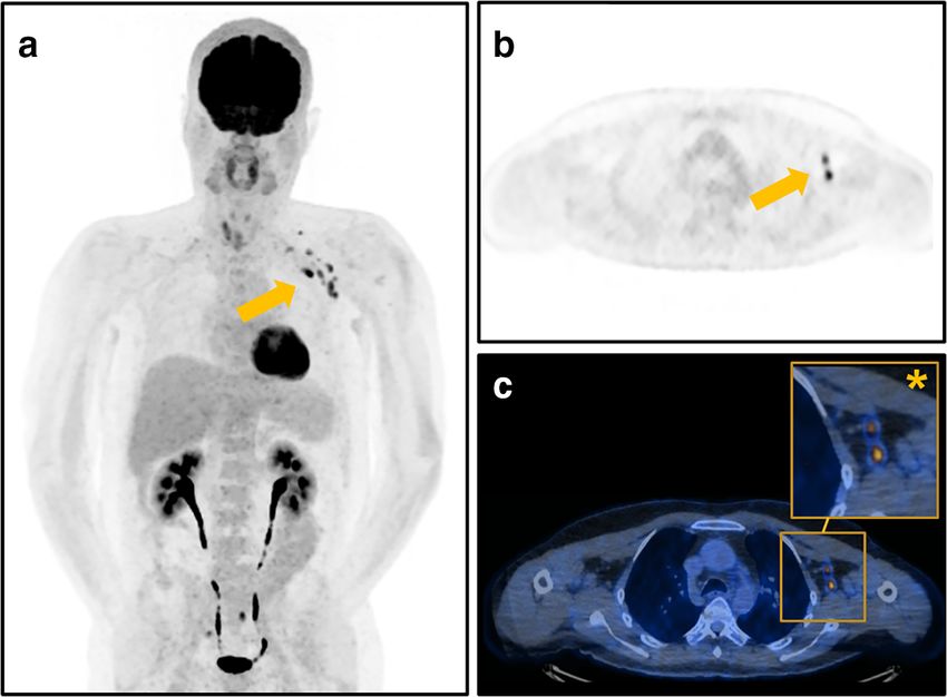
Fig. 2 Representative images of a
53-year-old patient who
underwent [18F]FDG PET/CT for
restaging after resection of a mel-
anoma of the left arm with FDG-
avid axillary lymphadenopathy
(arrows) after COVID-19 vacci-
nation. The patient was scanned 3
days after the second vaccination
with Moderna administered on
the left side with SUVmax 11.6 of
axillary lymph nodes. a
Maximum intensity projection
showing FDG-avid axillary
lymph nodes, (b) axial PET im-
age, and (c) fused PET/CT images
with magnified image of the left
axilla (asterisk). Due to the site of
the primary lesion on the left arm
and the FDG-avid lymph nodes,
the patient was scheduled earlier
for FDG PET/CT follow-up
compared to 6 patients in the placebo arm [16]. Further data vaccination in this group as compared to Pfizer-BioNTech which
from the US Center for Disease Control and Prevention was mostly administered twice already; however, as we observed
(CDC) on local reactions after the Moderna COVID-19 vac- consistently higher rates of FDG-avid lymph nodes after
cines reported that axillary swelling and tenderness was the Moderna vaccination after the 1. and 2. vaccinations, we feel that
second most common reported local reaction, after pain at site this does not affect this finding. Third, we were not able to obtain
of injection. Among patients 18–64 years old, 12% receiving pathological proof of non-malignant (i.e., reactive) changes at the
the vaccine reported axillary swelling and tenderness after the site of axillary lymphadenopathy, but by including only patients
1. vaccination, as compared to 5% in the placebo arm, and with unilateral active lymphadenopathy, we limited this potential
16% after the 2. dose, compared to 4% in the placebo arm [3, bias. Fourth, we have not obtained follow-up imaging to assess
16]. This clinically detectable finding may only represent “the how long the FDG-activity is persisting. Future studies may
tip of the iceberg” as we found strikingly more patients with a assess this topic as the vaccines are likely to remain in use for
reaction in the axillary lymph nodes on FDG PET/CT (up to the coming time. Fifth, the retrospective assessment of metastatic
80% after 2nd Moderna vaccination). risk in our study evaluated a scenario assuming no history of
Both in vitro as well as clinical data suggest that the two previous vaccination. Although this does not necessarily reflect
mRNA vaccines investigated are more immunostimulatory factual recommendations and decisions of tumor boards, it pro-
and therefore inherently more immunogenic as compared to vides a more accurate estimation of the frequency of diagnostic
other traditional vaccine agents [17], which may potentially predicaments after COVD-19 vaccination.
account for the more frequent and longer-lasting lymphade- In conclusion, metabolically active lymphadenopathy after
nopathy observed on imaging as compared to other agents COVID-19 vaccination is commonly seen in FDG PET/CT and
(e.g., H1N1 with 29% [5] vs. 54% in our study). the frequency may differ depending on the vaccine administered.
A very recent study from Israel [9] reported high rates of This may lead to diagnostic and even therapeutic dilemmas. To
FDG-avid axillary lymphadenopathy after vaccination with tackle this dilemma, we recommend to explicitly ask patients for
Pfizer-BioNTech (36% after the 1. dose, 54% after the 2. dose, prior vaccination before undergoing PET/CT. To avoid potential-
overall 46%). This number is in line with the results from our ly confounding FDG-uptake axillary lymph node, in oncological
study where we observed 39% of patients with avid lymph patients with lateralized upper body primary, the COVID-19 vac-
nodes after the 1. dose, 44% after the 2. dose and overall 43%, cines should be administered in the contralateral arm.
respectively. Interestingly, this number is well exceeded in pa-
tients that received Moderna vaccines with 67% after the 1.
vaccination and even 80% after the 2. vaccination (overall 72%). Acknowledgements The authors would like to thank Josephine Trinckauf,
Corina Weyermann, Marlena Hofbauer, Victoria Schober, Edlira Loga,
Our study has some limitations. First, the study group is rel-
Melanie Thüringer, Nina Bächle, Sabrina Epp, Ana-Mari Gaspar, Michèle
atively small, but this is reflected by the currently limited access Hug, Juliana Koller, Eirini Leivaditaki, and Danijel Tomic for their excellent
to vaccination agents in our country. Second, as Moderna was support. Furthermore, Dr. Michael Messerli would like to thank Prof. G. K.
introduced later in our country, we have more patients after the 1. von Schulthess for his invaluable support of this work.Eur Radiol
Funding Open Access funding provided by Universität Zürich. 4. Polack FP, Thomas SJ, Kitchin N et al (2020) Safety and efficacy of
the BNT162b2 mRNA Covid-19 Vaccine. N Engl J Med 383:
2603–2615
Declarations 5. Burger IA, Husmann L, Hany TF, Schmid DT, Schaefer NG (2011)
Incidence and intensity of F-18 FDG uptake after vaccination with
Guarantor The scientific guarantor of this publication is Dr. Michael H1N1 vaccine. Clinical Nuclear Medicine 36:848–853
Messerli. 6. Ozutemiz C, Krystosek LA, Church AL et al (2021)
Lymphadenopathy in COVID-19 vaccine recipients: diagnostic di-
Conflict of interest Dr. Stephan Skawran is supported by a grant from lemma in oncology patients. Radiology. https://doi.org/10.1148/
the Palatin-Foundation, Switzerland. Dr. Michael Messerli and Dr. Irene radiol.2021210275:210275
Burger received a research grant from the Iten-Kohaut Foundation, 7. Mortazavi S (2021) Coronavirus Disease (COVID-19) Vaccination
Switzerland. Dr. Michael Messerli and Dr. Huellner are supported by a associated axillary adenopathy: imaging findings and follow-up
grant from the CRPP AI Oncological Imaging Network of the University recommendations in 23 women. AJR Am J Roentgenol. https://
of Zurich. Dr. Martin W. Huellner received grants from GE Healthcare doi.org/10.2214/AJR.21.25651
and a fund by the Alfred and Annemarie von Sick legacy for translational 8. Seely JM, Barry MH (2021) The Canadian Society of Breast
and clinical cardiac and oncological research. Imaging/ Canadian Association of Radiologists’
Recommendations for the management of axillary adenopathy in
Statistics and biometry No complex statistical methods were necessary patients with recent COVID-19 vaccination. Can Assoc Radiol J.
for this paper. https://doi.org/10.1177/0846537121998949:846537121998949
9. Cohen D, Krauthammer SH, Wolf I, Even-Sapir E (2021)
Hypermetabolic lymphadenopathy following administration of
Informed consent Written informed consent was obtained from all pa-
BNT162b2 mRNA Covid-19 vaccine: incidence assessed by
tients in this study.
[(18)F]FDG PET-CT and relevance to study interpretation. Eur J
Nucl Med Mol Imaging. https://doi.org/10.1007/s00259-021-
Ethical approval Institutional Review Board approval was obtained. 05314-2
10. Messerli M, Stolzmann P, Egger-Sigg M et al (2018) Impact of a
Methodology Bayesian penalized likelihood reconstruction algorithm on image
• retrospective quality in novel digital PET/CT: clinical implications for the assess-
• observational study ment of lung tumors. EJNMMI Phys 5:27
• performed at one institution 11. Core Team R (2019) R: a language and environment for statistical
computing. Austria, Vienna
Open Access This article is licensed under a Creative Commons 12. Brown AH, Shah S, Groves AM, Wan S, Malhotra A (2021) The
Attribution 4.0 International License, which permits use, sharing, adap- challenge of staging breast cancer with PET/CT in the era of
tation, distribution and reproduction in any medium or format, as long as COVID vaccination. Clin Nucl Med. https://doi.org/10.1097/
you give appropriate credit to the original author(s) and the source, pro- RLU.0000000000003683
vide a link to the Creative Commons licence, and indicate if changes were 13. Ulaner GA, Giuliano P (2021) 18F-FDG-Avid Lymph Nodes After
made. The images or other third party material in this article are included COVID-19 Vaccination on 18F-FDG PET/CT. Clin Nucl Med 46:
in the article's Creative Commons licence, unless indicated otherwise in a 433–434
credit line to the material. If material is not included in the article's 14. Avner M, Orevi M, Caplan N, Popovtzer A, Lotem M, Cohen JE
Creative Commons licence and your intended use is not permitted by (2021) COVID-19 vaccine as a cause for unilateral lymphadenop-
statutory regulation or exceeds the permitted use, you will need to obtain athy detected by 18F-FDG PET/CT in a patient affected by mela-
permission directly from the copyright holder. To view a copy of this noma. Eur J Nucl Med Mol Imaging. https://doi.org/10.1007/
licence, visit http://creativecommons.org/licenses/by/4.0/. s00259-021-05278-3
15. Nawwar AA, Searle J, Hopkins R, Lyburn ID (2021) False-positive
axillary lymph nodes on FDG PET/CT resulting from COVID-19
immunization. Clin Nucl Med. https://doi.org/10.1097/RLU.
0000000000003657
References 16. Local reactions, systemic reactions, adverse events, and serious
adverse events: Pfizer-BioNTech COVID-19 vaccine. Centers for
1. Pontone G, Scafuri S, Mancini ME et al (2021) Role of computed Disease Control and Prevention Available from: https://www.cdc.
tomography in COVID-19. J Cardiovasc Comput Tomogr 15:27– gov/vaccines/covid-19/info-by-product/pfizer/reactogenicity.html;
36 Accessed March 2021.
2. Ribas A, Sengupta R, Locke T et al (2021) Priority COVID-19 17. Mingos M, Howard S, Giacalone N, Kozono D, Jacene H (2016)
vaccination for patients with cancer while vaccine supply is limited. Systemic immune response to vaccination on FDG-PET/CT. Nucl
Cancer Discov 11:233–236 Med Mol Imaging 50:358–361
3. Baden LR, El Sahly HM, Essink B et al (2021) Efficacy and safety
of the mRNA-1273 SARS-CoV-2 vaccine. N Engl J Med 384:403– Publisher’s note Springer Nature remains neutral with regard to jurisdic-
416 tional claims in published maps and institutional affiliations.You can also read