A patient with typical Wallenberg syndrome: a case report and literature review - TMR Publishing Group
←
→
Page content transcription
If your browser does not render page correctly, please read the page content below
CASE REPORT                                                                   doi: 10.12032/TMRND20200603010

TMR Non-Drug Therapy

A patient with typical Wallenberg syndrome: a case report and
literature review
Min Zhang1, Hong You1∗

1
 Sino-French Department of Neurological Rehabilitation, Gansu Provincial Hospital, Lanzhou 730000, China.

*Corresponding to: Hong You. Sino-French Department of Neurological Rehabilitation, Gansu Provincial Hospital, No.204
Donggang West Road, Lanzhou 730000, China. E-mail: lzyouhonginedin@163.com.

Highlights

        This case report extends knowledge of clinical characteristics of Wallenberg syndrome. Increases awareness
        of Wallenberg syndrome etiologies, diagnosis, and comprehensive management. It provides a new reference
        value for clinical application.

Submit a manuscript: https://www.tmrjournals.com/ndt                                  TMR | June 2020 | vol. 3 | no. 2 | 63
doi: 10.12032/TMRND20200603010                                                   CASE REPORT
Abstract
Wallenberg syndrome is a special type of medulla oblongata infarction with many and severe clinical dysfunction,
which is dorsolateral medullary syndrome. Wallenberg syndrome can have various initial symptoms due to different
damaged parts. Typical clinical manifestations of the syndrome include dizziness, vomiting, dysphagia, cross
sensory disturbance, ataxia, etc. Based on the complexity of functional anatomy, infarction in the medulla
oblongata can produce various types of clinical symptoms or signs depending on the location. We describe the
clinical comprehensive management of a 71-year-old man who presented with typical Wallenberg syndrome.
Through early diagnosis and comprehensive clinical management, the prognosis of patient can be effectively
improved.
Keywords: Wallenberg syndrome, Management, Prognosis, Case report, Literature review

Statement of ethics:
   The patient consented to the publication of this paper. This case report was approved by the Ethics Committee of
   Gansu Provincial Hospital.
Acknowledgments:
   This study was supported by a grant from the Gansu Provincial Hospital in China (No. 18GSSY4-31).
Abbreviations:
   MRI, magnetic resonance imaging; MRA, magnetic resonance angiography; PICA, posterior inferior cerebellar
   artery.
Competing interests:
    The authors declare that there is no conflict of interest.
Citation:
    Min Zhang, Hong You. A patient with typical Wallenberg syndrome: a case report and literature review. TMR
    Non-Drug Therapy 2020, 3 (2): 63–68.
Executive editor: Rui-Wang Zhao.
Submitted: 30 December 2019, Accepted: 17 April 2020, Online: 03 June 2020.
TMR | June 2020 | vol. 3 | no. 2 | 64                              Submit a manuscript: https://www.tmrjournals.com/ndt
CASE REPORT doi: 10.12032/TMRND20200603010
conscious, indwelling gastric tube for nasal feeding,
Introduction poor sleep, no abnormality in stool and urine, and no
obvious weight loss.
Wallenberg syndrome is a special type of medulla He had a history of hypertension for more than 10
oblongata infarction with many and severe clinical years, with the highest hypertension being 190/100
dysfunction, which is dorsolateral medullary syndrome mmHg. He did not take medicine regularly and his
[1, 2]. Typical clinical manifestations of the syndrome blood pressure was not monitored regularly. Denial of
include dizziness, vomiting, dysphagia, cross sensory the history of diabetes, coronary heart disease and
disturbance, ataxia, etc [3]. In terms of etiology of other chronic diseases, denial of the history of
Wallenberg’s syndrome, middle-aged and elderly infectious diseases, denial of major surgery and trauma,
patients mainly suffer from atherosclerosis, and young denial of blood transfusion history, denial of food and
patients are mainly cardiac embolism. Common risk drug allergy history. The vaccination history is
factors are the same as ischemic stroke, including unknown. More than 50 years of smoking history,
hypertension, diabetes, hyperlipidemia, coronary heart denied the family history of genetic diseases.
disease, atrial fibrillation, etc [4]. Analyses of lesions
via magnetic resonance imaging (MRI) have revealed Physical examination (including rehabilitation
that various types of clinical symptoms or signs may assessment)
be present depending on the location of a lesion in the (1) Physical examination: T: 36.3°C, P: 68 times/min,
medulla oblongata [5]. This article reports the clinical R: 17 times/min, BP: 150/90 mmHg. Clear mind,
experience of comprehensive management of a typical normal development, moderate nutrition, wheelchair
Wallenberg syndrome patient. pushed into the ward, physical examination
cooperation, answer the right question, slurred speech.
Case Presentation General physical examination showed no abnormality,
while cardiopulmonary examination and abdominal
Current medical history examination showed no abnormality.
A 71-year-old man was admitted to hospital for (2) Rehabilitation assessment: clear mind, nasal
“sudden dizziness with dysphagia and unstable feeding diet, slurred speech, bilateral nasolabial sulcus
standing for 1 month”. One month ago, the patient basically symmetrical, tongue extension basically
suffered from sudden dizziness at night, accompanied centered, uvula basically centered, pharyngeal reflex
by unstable standing, unconscious loss at that time, no weakened, soft palate raised poorly, left side obvious.
nausea, vomiting and other discomfort. Then the No obvious abnormality is found in muscle strength
patient was rushed to the local county hospital. The and muscle tension of limbs. The left facial numbness,
blood pressure was measured at 190/100 mmHg and right limb pain and temperature sensation decreased,
no obvious abnormality was found in cranial CT at that limb tendon reflex was normal and pathological sign
time, and the dizziness symptom of patient was was negative. Bilateral finger-nose test and
relieved after antihypertensive treatment. The patient heel-knee-shin test are satisfactory, but the left side is
returned home for recuperation. Next morning, the slightly worse. The mini-mental state examination
patient suffered from dizziness again, accompanied by score was 26 points. Balance check does not cooperate
dysphagia, nausea, vomiting, general weakness, (dizziness cannot cooperate), Watian drinking water
unstable standing, etc., and then had blurred test grade 5, assessment of daily living ability score
consciousness. Therefore, he called for emergency (Modified Barthel Index): 45 points, severe functional
medical help and was admitted to Gansu Provincial defect.
Hospital for treatment. No bleeding focus was found
after cranial CT. Considering diagnosis of “cerebral Laboratory and imaging examination
infarction”, he was treated with blood circulation (1) Laboratory examination: routine blood, urine and
promotion, microcirculation improvement, antiplatelet, feces, coagulation function, blood sugar, electrolyte of
lipid regulation, stomach protection, dehydration and liver and kidney function and blood lipid are basically
intracranial pressure reduction. The cranial MRI normal. (2) Imaging examination: cranial MRI, left
examination revealed left medulla oblongata infarction. medulla oblongata acute infarction (Figure 1); cranial
Pulmonary infection occurred during hospitalization, magnetic resonance angiography (MRA), left vertebral
aspiration pneumonia was considered, and the patient artery not shown (Figure 2). The whole brain digital
improved after indwelling gastric tube and substraction angiography examination was rejected by
anti-infection treatment. At present, there are still patient.
dizziness and discomfort, dysphagia, unstable standing
and other functional disorders. For further Diagnosis
rehabilitation treatment, we have admitted the patient (1) Clinical diagnosis: ① brain stem infarction
to our department with “brainstem infarction (medulla convalescence (left medulla oblongata), ②
oblongata)”. At the time of admission, the patient was hypertension grade 3, extremely high risk. According
Submit a manuscript: https://www.tmrjournals.com/ndt TMR | June 2020 | vol. 3 | no. 2 | 65doi: 10.12032/TMRND20200603010 CASE REPORT
to the patient’s medical history and imaging findings, syndrome. Furthermore, it is considered that the causes
the diagnosis is clear. of this syndrome may be hypertension, smoking
(2) Functional diagnosis: dysphagia, ataxia and history for many years, as well as the age is also a
dysfunction of daily living. Watian drinking water test factor.
grade 5, pharyngeal reflex weakened, etc., diagnosed
as dysphagia. The left side of bilateral finger-nose test Differential diagnosis
and heel-knee-tibia test is slightly worse, and the (1) Brain stem hemorrhage is usually more acute and
diagnosis is ataxia. Modified Barthel Index score: 45 severe, usually coma occurs earlier, most patients are
points, severe functional defect, diagnosed as critically ill, and the mortality rate is extremely high.
dysfunction of daily living. Obvious hemorrhage foci can be seen in brain stem on
(3) Clinical syndrome: Wallenberg syndrome. The cranial CT and can be clearly identified on imaging. (2)
cranial MRI examination of this patient showed that Cerebral hemispheric infarction, such as basal ganglia,
the left medulla oblongata infarction foci, and the usually starts with limb weakness, which can be
patient had 4 major symptoms [2, 6, 7], including gradually aggravated or accompanied by dysphagia,
dysphagia, ataxia, numbness of the affected lateral part, usually pseudobulbar paralysis and pharyngeal reflex.
and decreased pain and temperature sensation of the Cranial MRI examination can clearly identify the
healthy limb. The clinical diagnosis was Wallenberg infarct site.
Figure 1 Cranial MRI shows left medulla oblongata infarction (black arrow indication)
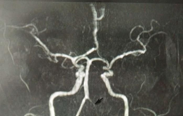
Figure 2 Cranial MRA shows left vertebral artery not shown (black arrow indication)
TMR | June 2020 | vol. 3 | no. 2 | 66 Submit a manuscript: https://www.tmrjournals.com/ndtCASE REPORT doi: 10.12032/TMRND20200603010
by vertebral artery dissection [10, 13, 14]. In patients
Comprehensive management with dorsolateral medulla oblongata syndrome, only a
few patients are caused by PICA occlusion. Most of
After admission, patient was thoroughly evaluated the causes of PICA ischemia are stenosis or occlusion
(liver function, renal function, electrolyte, prothrombin of intracranial segment or origin of vertebral artery, or
time activity, blood routine , );monitor patient’s blood thrombus shedding at origin of vertebral artery [10, 15,
pressure, and provide nasal feeding diet, and 16]. Cranial MRA of this patient showed a loss of left
giveantihypertensive (amlodipine), antiplatelet vertebral artery signal. Although no further digital
(aspirin), anti-vertigo (betahistine), and promotion of substraction angiography examination of the whole
nerve repair (bossgate) treatments. brain was performed, it could still reflect that there
For swallowing dysfunction, there are mainly basic were some problems in the vertebral artery.
training, indirect swallowing training and feeding Considering that the responsible vessel for this
training [8, 9]. Basic training includes sensory occurrence is the left vertebral artery, the etiological
stimulation (including touch, temperature and taste type is macroatherosclerosis, and qualitative diagnosis
stimulation) and muscle training of mouth and face. is ischemic cerebrovascular disease.
Indirect swallowing training is to train patients to Wallenberg syndrome can have various initial
swallow related decomposition actions under the symptoms due to different damaged parts, but
condition of no eating, and to achieve the purpose of vertigo/dizziness is common in clinic [3]. Dizziness is
safe and effective swallowing by means of some the first symptom of this patient, which indicates
specific manipulations and actions. During the feeding vestibular nucleus is involved. Nausea and vomiting,
training, choose the appropriate body position, and dysphagia, ataxia, and cross sensory disturbance
select the appropriate one-mouthful food intake gradually appear as the disease progresses [17, 18]. At
according to the actual swallowing function of the the initial stage of the disease, due to incomplete
patient, and increase it according to the patient’s symptoms or careless physical examination, it is easy
reaction during the training [8]. Electric standing bed to be misdiagnosed as posterior circulation ischemia,
training can improve the patient’s adaptability to body delay relating examination and treatment, and even
position changes, enhance the proprioceptive input and cause sudden death of patients due to respiratory or
balance of lower limbs, and prevent complications. cardiac arrest.
Exercise therapy and balance function training There are 8 types of Wallenberg syndrome sensory
improve limb movement and coordination ability, and disturbance [19]: (1) lateral contralateral limb sensory
improve balance and coordination functions; disturbance of lesion, which is crossed sensory
occupational therapy improves patients’ coordination disturbance; (2) the pain and temperature sensation of
ability and fine function, and improves their daily bilateral face and contralateral lesion decreased; (3)
activities. sensory disturbance of the lesion to the lateral part and
After nearly one month of rehabilitation treatment, hemi-body; (4) sensory disturbance only at the lateral
the patient’s swallowing function was significantly part of the lesion; (5) somatosensory disturbance on
improved. The Watian drinking water test was grade 2. opposite side of lesion only; (6) bilateral
The gastric tube was pulled out and the oral intake was somatosensory disorders; (7) bilateral facial sensory
good. Sitting balance level 3, standing balance level 2. disturbance; (8) lesion-to-lateral sensory disturbance.
Berg Balance Scale score: 31 points. Patient can walk Combined with the clinical symptoms and signs of this
a short distance with the help of family members. patient, the type of sensory disturbance of this patient
Modified Barthel Index score: 75 points, mild is analyzed as typical cross sensory disturbance. The
functional defect. previous reports of patients with dorsolateral medulla
oblongata syndrome presenting as pure sensory
Discussion disturbance [20]. In addition, this patient has no
consciousness disorder, but there are also patients with
Wallenberg syndrome, also known as posterior inferior consciousness disorder, which may be caused by
cerebellar artery (PICA) syndrome and dorsolateral gradual aggravation of edema after infarction,
medullary syndrome, is the most common posterior compression of brainstem and influence on
circulation ischemic stroke syndrome [2]. In 1895, cerebrospinal fluid circulation [21].
German neurologist Adolf Wallenberg described the If the patient lacks typical clinical manifestations,
clinical manifestations of the syndrome. Of all some scholars suggest that the possibility of
ischemic stroke patients, about 20% are posterior Wallenberg syndrome can be indicated when the
circulation stroke, and about half of them can be patient meets the following two conditions: (1) cranial
represented as syndrome, which is mostly male and is MRI indicates that the focus is in medulla oblongata,
more common in the middle-aged and elderly and dysarthria and dysphagia must be one of them; (2)
population [10–12]. About 75% of patients are caused cranial MRI indicates that the lesion is located at the
by atherosclerosis, 17% by cardiac embolism, and 8% dorsolateral medulla oblongata, pain and temperature
Submit a manuscript: https://www.tmrjournals.com/ndt TMR | June 2020 | vol. 3 | no. 2 | 67doi: 10.12032/TMRND20200603010 CASE REPORT
sensation disorder, ataxia and Horner syndrome must adult with cerebellar infarct. Acta Neurol Taiwan
be one of them [22]. This patient met the above 2008, 17: 243–247.
requirements and was diagnosed accordingly. 14. Sun H, Zhou Y, Bai C, et al. Clinical features and
To sum up, this case extends our knowledge of etiology of Wallenberg syndrome. Chin J
clinical characteristics of Wallenberg syndrome, and Gerontol 2005, 25: 382–383.
increases awareness of its etiologies, diagnosis, and 15. Collado A, Santamaria J, Ribalta T, et al.
comprehensive management, and effectively improves Giant-cell arteritis presenting with ipsilateral
the prognosis of patient. hemiplegia and lateral medullary syndrome. Eur
Neurol 1989, 29: 266–268.
References 16. Tehrani ASS, Desanto JR, Kattah JC.
Neuroimaging "HINTS" of the lateral medullary
1. Kim K, Lee HS, Jung YH, et al. Mechanism of syndrome. J Neuroophthalmol 2017, 37: 403–404.
medullary infarction based on arterial territory 17. Arshad Q, Roberts RE, Ahmad H, et al. Patients
involvement. J Clin Neurol 2012, 8: 116. with chronic dizziness following traumatic head
2. Day GS, Swartz RH, Chenkin J, et al. Lateral injury typically have multiple diagnoses involving
medullary syndrome: a diagnostic approach combined peripheral and central vestibular
illustrated through case presentation and literature dysfunction. Clin Neurol Neurosurg 2017, 155:
review. CJEM 2014, 16: 164–170. 17–19.
3. Jia J. Neurology. Beijing: People’s Medical 18. Xu Z, Wang C, Li J, et al. Research progression
Publishing House 2013. on dysphagia in patients with cerebralinfarction.
4. Hu J, Xu R, Lu X, et al. Clinical analysis of Chin J Rehabil 2017, 32: 148–150.
Wallenberg’s syndrome. Chin J Contemp Neurol 19. Kim JS, Lee JH, Lee MC. Patterns of sensory
Neurosurg 2019, 19: 41–46. dysfunction in lateral medullary infarction:
5. Ogawa K, Suzuki Y, Oishi M, et al. Clinical study clinical-MRI correlation. Neurology 1997, 49:
of 46 patients with lateral medullary infarction. J 1557–1563.
Stroke Cerebrovasc Dis 2015, 24: 1065–1074. 20. Yamaoka Y, Kishishita S, Takayama Y, et al. A
6. De Bruyn D, Van Aken E, Herman K. A rare case report of a case involving body lateropulsion with
of concomitant sicca keratopathy and ipsilateral numbness of the ipsilesional fingers caused by a
central facial palsy in Wallenberg's dorsolateral small infarction in the dorsal part of the middle
medullary syndrome. GMS Ophthalmol Cases medulla. Case Rep Neurol 2018, 10: 54–59.
2017, 7: Doc08. 21. Lee SH, Kim JS. Acute diagnosis and
7. Aydogdu I, Ertekin C, Tarlaci S, et al. Dysphagia management of stroke presenting dizziness or
in lateral medullary infarction (Wallenberg’s vertigo. Neurol Clin 2015, 33: 687–698.
syndrome): an acute disconnection syndrome in
premotor neurons related to swallowing activity?
Stroke 2001, 32: 2081–2087.
8. Rosenvinge SK, Starke ID. Improving care for
patients with dysphagia. Age Ageing 2005, 34:
587–593.
9. Battel I, Koch I, Biddau F, et al. Efficacy of
botulinum toxin type-A and swallowing treatment
for oropharyngeal dysphagia recovery in a patient
with lateral medullary syndrome. Eur J Phys
Rehabil Med 2017, 53: 798–801.
10. Kim JS. Pure lateral medullary infarction:
clinical-radiological correlation of 130 acute,
consecutive patients. Brain. 2003, 126:
1864–1872.
11. Kameda W, Kawanami T, Kurita K, et al. Lateral
and medial medullary infarction: a comparative
analysis of 214 patients. Stroke 2004, 35:
694–699.
12. Ma N. Analysis of clinical and imaging features
of cerebral infarction in patients with posterior
circulation. Guide China Med 2015, 10: 50–51.
13. Jao T, Liu HM, Tang SC, et al. Dissection of the
posterior inferior cerebellar artery in a young
TMR | June 2020 | vol. 3 | no. 2 | 68 Submit a manuscript: https://www.tmrjournals.com/ndtYou can also read


