EVIDENCE OF THE FIRST CLINICAL CASE OF EQUINE NEUROINVASIVE WEST NILE DISEASE IN SERBIA, 2018 - Acta Veterinaria
←
→
Page content transcription
If your browser does not render page correctly, please read the page content below
Acta Veterinaria-Beograd 2019, 69 (1), 123-130
                                                     UDK: 636.1:616.98:579.833.2(497.11)”2018”
Case report                                                         DOI: 10.2478/acve-2019-0009

EVIDENCE OF THE FIRST CLINICAL CASE OF EQUINE
NEUROINVASIVE WEST NILE DISEASE IN SERBIA, 2018

MEDIĆ Strahinja1, LAZIĆ Sava2, PETROVIĆ Tamaš2, PETRIĆ Dušan3,
SAMOJLOVIĆ Milena2, LAZIĆ Gospava2, LUPULOVIĆ Diana2*

1
 “Vetlab” doo, Veterinary laboratory for clinical pathology, Savska 31, Belgrade, Serbia;
2
 Scientific Veterinary Institute “Novi Sad”, Department of virology, Rumenački put 20, Novi Sad, Serbia;
3
 University of Novi Sad, Faculty of Agriculture, Laboratory for medical and veterinary entomology, Trg
Dositeja Obradovića 8, Novi Sad, Serbia

(Received 19 November 2018, Accepted 21 February 2019)

        During July 2018, the first clinical case of neurological West Nile virus (WNV) infection
        was reported in a Belgian sports mare in Belgrade, Serbia. Typical symptoms, such as
        hypersensitive skin reaction, disorientation, weakness, ataxia and the loss of equilibrium
        were reported. Detection of WNV IgM antibodies by commercial ELISA in the serum
        samples of the diseased mare strongly indicated acute infection. The ELISA positive
        results were confirmed by VNT. Hematological and biochemical parameters were in
        the reference range. The only finding was a minor lymphopenia. WNV RNA was not
        detected by RT-qPCR in the blood sample extracted seven days after the disease had
        broken out. The horse improved clinically in two weeks while other horses at the same
        premises remained asymptomatic. The clinical, serological, biochemical and molecular
        analyses applied confirmed the first clinical case of neuroinvasive WNV infection in
        horses in Serbia. The West Nile virus has been circulating in Serbia in the last decade
        in mosquitoes, birds, and horses, but no evidence of equine WNV clinical cases were
        registered so far.
        Key words: West Nile virus, neuroinvasive disease, horses, Serbia

                                        INTRODUCTION

West Nile virus (WNV), the causative agent of West Nile fever, is one of the most
spread mosquito-borne viruses, belonging to the family Flaviviridae, genus Flavivirus.
Its enzootic transmission cycle is maintained in nature between mosquitoes and birds,
while humans and horses can be only dead-end hosts. Horses and humans can be
infected by a mosquito bite, primarily from the genus Culex. In horses, WNV infection
is in most cases unapparent, and only 10 % of infected animals show clinical signs of
neurological disorder with up to 50% of lethality rate [1].

*Corresponding author: e-mail: diana@niv.ns.ac.rs

                                                                                                   123
Acta Veterinaria-Beograd 2019, 69 (1), 123-130
The occurrence of WNV disease was for a long time limited to Sub-Saharan Africa
with only sporadic outbreaks elsewhere. But, the situation dramatically changed in the
1990s, when the epidemiology of WNV infection appeared to be more severe, with
human fatalities and cases of equine encephalitis. Starting in 2010, an elevated number
of large epizootics in horses was recorded in Hungary, Italy, Spain, Greece, Romania,
Portugal, etc. [2].
In this paper are presented the clinical, serological, biochemical and molecular analyses
applied to confirm the first reported clinical case of neuroinvasive WNV infection in
horses in Serbia.
CASE PRESENTATION
During midsummer 2018, a 7-year-old mare, with a suspect case of neurological
disorder was reported to a local veterinary practitioner. The Belgian sport mare spent
three months on pasture in Horgoš (Vojvodina Province), in the northern part of
Serbia, accompanied by a gelding from the same stable. Both horses returned from
Horgoš to Belgrade on July 3rd, and the first symptoms of clinical manifestation of
encephalitis in the mare started on July 15th (Figure 1). The suspicion of WNV infection
was established immediately, in accordance to OIE (World Organisation for Animal
Health) guidance [3]. The complete physical examination, vaccination status, history
of traveling, localization of the grazing area, data of previous medical treatment and
clinical course of the disease were undertaken.
The diseased mare exhibited muscle twitching and hypersensitivity, first in the region
of the back, and later on the neck, head and limb regions. The mare had an inconstant
fever (38.5-39.0), normal inspiration (40/min) and normal heart rate (15-20/min).
On the fifth day after the first symptoms appeared, lameness of the left rear limb
has been noticed. Disorientation, weakness, ataxia and the loss of equilibrium started
to develop gradually from the fifth day on. The first signs of somnolence started on
the sixth day of the onset of symptoms and they developed during the next two days
with head pressing and paralysis of the lower lip. Dysphagia and dysuria have started
in the evening of the sixth day, and they have lasted for the next three days. The
mare received supportive therapy, compiled of antibiotic Penstrep 1600000 IJ, Dexason
(0,05 mg/kg i.v), Flunixin (1,0 mg/kg i.m.)and Ringer solution (10 L/day). The first signs
of health improvement were noticed on the morning of the eighth day when urinary
function recovered followed by appetite normalization in the evening. During the next
two days, the mare still showed hypersensitivity to light, and the symptoms of ataxia
disappeared gradually. On the tenth and eleventh day no neurological signs have been
detected during the clinical examination and the recovery process started. The horse
improved clinically in two weeks. No other horses at the same premises, including the
gelding that was on pasture together with the infected mare, did show any signs of
neurological disorders. The mare was not vaccinated against WNV.
124Medić et al.: Evidence of the first clinical case of equine neuroinvasive West Nile disease in Serbia, 2018
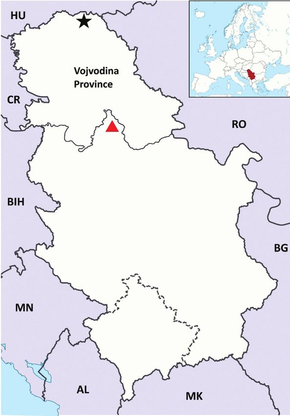
Figure 1. Localization of the study area– Map of Serbia
( – Horgoš, grazing area of the horses; – the City of Belgrade, the location where
the first WNV clinical case was diagnosed)
Laboratory examination
a) Sampling
The blood samples from the diseased mare were collected twice. First sampling was
conducted one week after the onset of clinical symptoms and the second sampling
was carried out three weeks later, to assess the seroconversion rate. In both occasions,
blood samples were collected in two tubes, in a plain serum tube for serological analyzes
and in a tube with EDTA for biochemical and molecular testing. Additionally, blood
samples from 7 apparently asymptomatic horses that shared the same stall with the
infected mare in Belgrade (including the gelding that also was in Horgoš on pasture)
were subjected to serological examination.
125Acta Veterinaria-Beograd 2019, 69 (1), 123-130 b) Hematological and biochemical testing The complete blood count (CBC) analysis has been performed on hematology analyzer ADVIA® 120, Siemens, USA. The clinical chemistry profile was carried out on the Olympus AU400 system. c) Serological tests Sera samples from the infected mare were tested for the presence of WNV IgM antibodies by a commercial capture ELISA (INGEZIM West Nile IgM, Ingenasa, Spain), according to manufacturer’s instruction. For the confirmation of the obtained ELISA results and to exclude cross-reactivity with other Flaviviruses, the virus neutralization test (VNT) was applied. A VN titer higher or equal to 1:10 was considered positive. For VNT, VERO cells (ATCC CCL-81) and WNVisolate SRB-Novi Sad/12 (NCBI GenBank No: KC407673) were used, following the prescribed procedure by OIE Manual [6]. Furthermore, blood samples from 7 other healthy horses were also tested on the presence of anti-WNV IgM antibodies with the aforementioned commercial ELISA. d) Molecular detection of WNV RNA Detection of WNV RNA was carried out in the first sampled blood specimen with EDTA and tested by one-step real-time reverse transcription-polymerase chain reaction (RT-qPCR),with primers and probe previously described by Linke et al.[4]. Briefly,RNA was extracted with TRI reagent Solution (Invitrogen, Thermo Fisher Scientific), and one step RT-qPCR was conducted using the commercial kit RNA UltraSense™ One-Step qRT-PCR System (Applied Biosystems™, Thermo Fisher Scientific), following manufacturer’s instruction. In both sera samples from the suspicious mare anti-WNV, IgM antibodies were detected by ELISA and confirmed positive by VNT. The established neutralizing antibody titer differs in one 2-fold dilution in the first and second sera specimens (1:160 vs. 1:80), respectively. Positive WNV IgM finding was confirmed at the National Referent Laboratory for WNV infection (Veterinary Specialized Institute “Kraljevo”) in Kraljevo, by the commercial antibody capture ELISA (ID Screen West Nile IgM capture, ID.Vet, France). Hematological and biochemical parameters were in the reference range. The only finding was a minor lymphopenia, most likely due to the dexamethasone therapy. WNV RNA was not detected in EDTA blood sample by RT-qPCR. Sera samples from 7 cohabitating horses reacted negatively in ELISA. This is the first report of the clinical case of neuroinvasive WN disease described in horses in Serbia. Differential diagnosis excluded EHV-1, rabies, and USUV. Typical neurological signs of the disease found in the infected mare were compatible with already documented reports of WNV outbreaks among horses in Canada, Portugal, Spain, Greece, and many other countries [5-8]. Detection of WNV IgM antibodies 126
Medić et al.: Evidence of the first clinical case of equine neuroinvasive West Nile disease in Serbia, 2018

by capture ELISA in both sera samples of the diseased mare strongly indicated an
acute course of infection, which is in accordance to literature data that IgM antibodies
appear between the 7th and 10th day post-infection [3]. The ELISA positive results
were confirmed by VNT, which is the “OIE gold standard” for serological WNV
examinations [3].The obtained negative RT-qPCR result in our analysis was expected
because viremia in horses lasts only 4-6 days and disappears with the onset of clinical
symptoms [9]. No significant differences were detected in the biochemical parameters
and CBC, which was the finding reported also by other authors [5].
West Nile virus has been circulating in our region for at least a decade, and after the
alarming reports of re-emergence of WNV disease in Europe, our research group
conducted the first serological investigation of WNV in horses in Serbia. The presence
of WNV antibodies was detected by ELISA and plaque reduction neutralization test
(PRNT) in 12% (46/349) and 28.6% (72/252) blood sera of Serbian horses sampled
during 2009/2010 and 2011, respectively [10,11]. In another study, WNV antibodies
were present in 3.97% horses, 0.93% dogs, 0.31% poultry and 1.36% man out of 3618
tested sera samples [12]. Furthermore, our studies show that WNV also circulates in
Serbia among mosquitoes and wild birds [13,14]. Human WNV clinical outbreaks
are recorded each year in Serbia, starting in 2012, when the first human epidemic
case was reported [15]. National WNV surveillance programme funded by Veterinary
Directorate started in 2014 and was successful in detection of the WNV presence in
sentinel animals, wild birds and mosquitoes before human outbreaks in each season
[16]. There is no available vaccine for the prevention of WNV infection in horses in
Serbia and horses are not vaccinated. West Nile virus infection is on the list of diseases
that are obliged to be notified to veterinary authorities in Serbia.
It is not surprising that this summer Serbia was faced for the first time with the clinical
case of equine West Nile fever. The transmission season started earlier this year due
to an extremely hot spring and lots of rainy days during the summer that were suitable
for mosquito’s activity [17]. European Center for Disease Prevention and Control
announced 276outbreaks among equids in EU Member states till 8thNovember 2018.
Reports on the number of human cases in Europe were also alarming, including
Serbia, where already 385 confirmed WNV human cases and 35 deaths were reported
during this year, with the highest number of cases in Belgrade area [18].
It was hard to presume if the mare got infected on pasture in Horgoš or at the stable in
Belgrade because it’s known that the incubation period lasts from 3 to 15 days [3]. The
mare returned from grazing on 3rd July while the first clinical signs appeared twelve
days later. Both locations, Horgoš and especially Belgrade area, are highly affected with
WNV[18]. Entomological results revealed that most of the WNV positive mosquito
pools were found in the Vojvodina Province and in the City of Belgrade, indicating
that the virus circulates in this area intensively [16,19].
This is the first clinical case of WNV infection in horses, reported during July 2018 in
Serbia. According to the previously conducted studies and national surveillance data,

                                                                                                           127
Acta Veterinaria-Beograd 2019, 69 (1), 123-130
WNV has already been circulating in Serbia in the last decade in mosquitoes, birds,
and horses but no evidence of equine encephalitis caused by WNV was registered
so far. It is necessary to improve the collaboration between veterinary practitioners
and veterinary institutes and to raise awareness among horse owners regarding the
reporting of the cases of this serious zoonotic disease.
Acknowledgments
This work was supported by grant TR31084, funded by the Ministry of Education,
Science and Technological Development of the Republic of Serbia.
Authors’ contributions
MS conducted hematological and biochemical examinations, participated in writing
and performed sampling; LS performed virus neutralization test and revised critically
the manuscript; PT performed molecular testing and contributed to the drafting of
manuscript; PD was involved in data collection and commented on the manuscript;
SM carried out ELISA and participated in writing; LG analyzed the results and data;
LD made contribution to design, was involved in writing and gave the final approval
of the version to be published.
Declaration of conflicting interests
The author(s) declared no potential conflicts of interest with respect to the research,
authorship, and/or publication of this article.
Statement of Informed Consent
The owner understood procedure and agrees that results related to investigation or
treatment of their companion animals could be published in this journal.
REFERENCES
1. Calistri P, Giovannini A, Hubalek Z, Ionescu A, Monaco F, Savini G, Lelli R: Epidemiology
of West Nile in Europe and in the Mediterranean basin. Open Virol J 2010, 22:29-37.
2. Angenvoort J, Brault AC, Bowen RA, Groschup MH: West Nile Viral Infection of Equids.
Vet Microbiol 2013, 167:168–180.
3. OIE (World Organisation for Animal Health): Manual of Diagnostic Tests and Vaccines
2018, Chapter 2.1.24, West Nile fever. Available from: http://www.oie.int/fileadmin/
Home/eng/Health_standards/tahm/2.01.24_WEST_NILE.pdf
4. Linke S, Heinz E, Niedrig M, Nitsche A, Pauli G: Detection of West Nile virus lineages 1
and 2 by real-time PCR. J Virol Meth 2007, 146:355-358.
5. Abutarbush SM, O’Connor BP, Clark C, Sampieri F, Naylor JM: Clinical West Nile virus
infection in 2 horses in western Canada. Can Vet J 2004, 45: 315–317.
128Medić et al.: Evidence of the first clinical case of equine neuroinvasive West Nile disease in Serbia, 2018
6. Barros S, Ramosa F, Fagulhaa T, Duartea M, Henriquesa AM , Waapa H , Luisa T, Costab
T, Amadorc R, Quintansc S, Fevereiroa M: West Nile virus in horses during the summer
and autumn seasons of 2015 and 2016, Portugal. Vet Microbiol 2017, 212:75-79.
7. García-Bocanegra IJ, Jaén Téllez A, Napp S, Arenas –Montes A, Fernández- Morente M,
Fernández –Molera V, Arenas A: Monitoring of the West Nile Virus epidemic in Spain
between 2010 and 2011. Transbound Emerg Dis 2012, 59:448–455.
8. Bouzalas IG, Diakakis N, Chaintoutis SC, Brellou GD, Papanastassopoulou M, Danis
K, Vlemmas I, Seuberlich T, Dovas CI: Emergence of Equine West Nile Encephalitis in
Central Macedonia, Greece, 2010. Transbound Emerg Dis 2016, 63: 219-227.
9. Bunning ML, Bowen RA, Cropp CB, Sullivan KG, Davis BS, Komar N, Baker D, Hettler
DL, Holmes DA, Biggerstaff BJ, Mitchell CJ 2002: Experimental infection of horses with
West Nile virus. Emerg Infect Dis 2002, 8:380–386.
10. Lupulovic D, Martín-Acebes MA, Lazic S, Alonso-Padilla J, Blázquez AB, Escribano-
Romero E, Petrovic T, Saiz JC: First serological evidence of West Nile virus activity in
horses in Serbia. Vector Borne Zoonotic Dis 2011, 11: 1303-1305.
11. Medić S, van den Hoven R, Petrović T, Lupulović D, Nowotny N: Serological evidence of
West Nile virus infection in the horse population of northern Serbia. J. Infect. Dev. Ctries.
2014, 8:914-918.
12. Đuričić B, Vasić A, Rogožarski D, Vojinović D, Elezović Radovanović M, Manić M, Marić
J, Prokić N, Ilić Ž, Novotny N, Gligić A: Seroepizootiological-epidemiological investigation
and mapping of West Nile infection in the Republic of Serbia. Acta Vetereinaria Belgrade
2013, 63:569-579
13. Petrović T, Blazquez A, Lupulović D, Lazić G, Escribano-Romero E, Fabijan D, Kapetanov
M, Lazić S, Saiz JC: Monitoring West Nile virus (WNV) infection in wild birds in Serbia
during 2012: first isolation and characterisation of WNV strains from Serbia. Euro Surveill
2013, 18:pii = 20622.
14. Petrić D, Petrovic T, Hrnjakovic Cvjetkovic I, Zgomba M, Milosevic V, Lazic G, Lupulović
D, Lazić S, Dondur D, Vaselek S, Živulj A, Kisin B, Molnar T, Janku D, Pudar D, Radovanov
J, Kavran M, Kovačević G, Plavšić B, Jovanović Galović A, Vidić M, Ilić S, Petrić M: West
Nile virus circulation in Vojvodina, Serbia: Mosquito, bird, horse and human surveillance.
Mol Cell Probes 2017, 31:28–36.
15. Popović N, Milošević B, Urošević A, Poluga J, Lavadinović L, Nedeljković J, Jevtović D,
Dulović O: Outbreak of West Nile virus infection among humans in Serbia, August to
October 2012. Euro Surveill 2013, 18:pii=20613.
16. Petrović T, Šekler M, Petrić D, Lazić S, Debeljak Z, Vidanović D, Ignjatović Ćupina A,
Lazić G, Lupulović D, Kolarević M, Plavšić B: Methodology and results of integrated
WNV surveillance programmes in Serbia. PLoS ONE 2018,13:e0195439.
17. Haussig JM, Young JJ, Gossner CM, Mezei E, Bella A, Sirbu A, Pervanidou D, Drakulovic
MB, Sudre B: Early start of the West Nile fever transmission season 2018 in Europe. Euro
Surveill 2018, 23: pii=1800428.
18. ECDC (European Centre for Disease Prevention and Control) 2018: Weekly updates: 2018
West Nile fever transmission season; updated 8November 2018. Available from: https://
ecdc.europa.eu/en/west-nile-fever/surveillance-and-disease-data/disease-data-ecdc
19. IOPHOS (Institute of Public Health of Serbia) 2018: Information about the current
epidemiological situation of West Nile fever in the Republic of Serbia in 2018 - 8-14.
July 2018 (in Serbian). Available from: http://www.batut.org.rs/.../Informacija o aktuelnoj
epidemioloskoj situaciji%.
129Acta Veterinaria-Beograd 2019, 69 (1), 123-130 DOKAZ PRVOG KLINIČKOG SLUČAJA NEUROINVAZIVNOG OBLIKA GROZNICE ZAPADNOG NILA KOD KONJA U SRBIJI, 2018 MEDIĆ Strahinja, LAZIĆ Sava, PETROVIĆ Tamaš, PETRIĆ Dušan, SAMOJLOVIĆ Milena, LAZIĆ Gospava, LUPULOVIĆ Diana Tokom jula 2018. godine, prvi klinički slučaj neurološkog poremećaja izazvanog vi- rusom Zapadnog Nila (VZN) prijavljen je kod kobile rase Belgijski sportski konj u Beogradu, u Srbiji. Prijavljeni su tipični simptomi, kao što su: preosetljivost kože, dezorientacija, slabost, ataksija i gubitak ravnoteže. Detekcija VZNIgM antitela ko- mercijalnom ELISA tehnikom u uzorcima seruma obolele kobile izrazito je ukazivala na prisustvo akutne infekcije. Pozitivni rezultati u ELISA testu su potvrđeni VN tes- tom. Hematološki i biohemijski parametri bili su u obimu referentnih vrednosti. Jedini nalaz bila je blaga limfopenija. U uzorku krvi, uzorkovanoj nedelju dana posle pojave prvih simptoma bolesti, nije detektovana VZN RNK pomoću RT-qPCR. Kliničko poboljšanje zdravstvenog stanja konja je nastalo za dve nedelje dok drugi konji u istim prostorijama nisu pokazivali simptome bolesti. Prikazane su kliničke, serološke, biohe- mijske i molekularne analize koje su korišćene za potvrđivanje prvog kliničkog slučaja neuroinvazivnog oblika virusa Zapadnog Nila kod konja u Srbiji. Virus Zapadnog Nila cirkuliše u Srbiji u poslednjoj deceniji kod komaraca, ptica i konja, ali do sada nije zabeležen ni jedan klinički slučaj infekcije izazvane VZN kod konja. 130
You can also read