Journal of Surgery Research and Practice - Athenaeum ...
←
→
Page content transcription
If your browser does not render page correctly, please read the page content below
Journal of Surgery Research and Practice
Open Access                                                                                     Mini Review

                  Brief Facts about COVID-19 (SARS-CoV-2)
                                            Sorush Niknamian1*
  1
   Military Medicine Department, Liberty University, USA and Member of International Society of Infectious
                                              Diseases, USA
      *
       Corresponding Author: Sorush Niknamian, MD PhD Dr.PH, Military Medicine Department, Liberty
              University, USA and Member of International Society of Infectious Diseases, USA;
                                    Email: so.niknamian@gmail.com

            Received Date: 02-01-2021; Accepted Date: 21-01-2021; Published Date: 29-01-2021

Copyright© 2021 by Niknamian S. All rights reserved. This is an open access article distributed under the terms
of the Creative Commons Attribution License, which permits unrestricted use, distribution and reproduction in
any medium, provided the original author and source are credited.

                                                Abstract
Coronaviruses are a group of related viruses that cause diseases in mammals and birds. In
humans, coronaviruses cause respiratory tract infections that can range from mild to lethal.
Mild illnesses include some cases of the common cold, while more lethal varieties can cause
SARS, MERS and COVID-19. The outbreak was identified in Wuhan, China, in December
2019, declared to be a Public Health Emergency of International Concern on 30th January 2020
and recognized as a pandemic on 11th March 2020. Coronaviruses are the subfamily
Orthocoronavirinae, within the family of Coronaviridae, order Nidovirales and realm
Riboviria. They are enveloped viruses with a positive-sense single-stranded RNA genome and
a nucleocapsid of helical symmetry. The genome size of coronaviruses is approximately from
26 to 32 kilobases. Coronaviruses were first discovered in the 1930s and Human coronaviruses
were discovered in the 1960s. The earliest ones studied were from human patients with the
common cold, which were later named human coronavirus 229E and human coronavirus
OC43. Other human coronaviruses have since been identified, including SARS-CoV in 2003,
HCoV NL63 in 2004, HKU1 in 2005, MERS-CoV in 2012 and SARS-CoV-2 in 2019. Most
of these have involved serious respiratory tract infections.

Keywords
COVID-19; Spike Protein; Virus; Influenza

 Niknamian S | Volume 2; Issue 1 (2021) | JSRP-2(1)-013 | Mini Review

 Citation: Niknamian S. Brief Facts about COVID-19 (SARS-CoV-2). J Surg Res Prac. 2021;2(1):1-9.

 DOI: http://dx.doi.org/10.46889/JSRP.2021.2103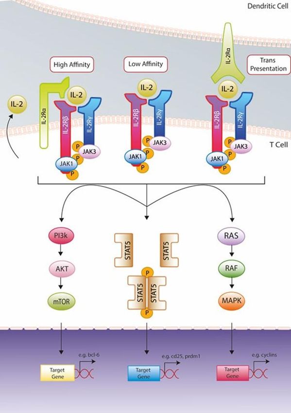
2

Abbrevations
SARS-CoV-2: Severe Acute Respiratory Syndrome Coronavirus 2; DV: Dengue Virus;
HepG2: Human Hepatoma Cell Line; RCR: Respiratory Control Ratio; Endo G: Endonuclease
G; AIF: Apoptosis-Inducing Factor; GSH: Glutathione; ROS: Reactive Oxygen Species; AA:
Amino Acid; Kb: Kilobasepair; Gb: Gigabasepair; Mb: Megabasepair; Bp: Basepair; N:
Nucleocapsid; E: Envelope; S: Spike; M: Membrane; TMPRSS2: Transmembrane Serine
Protease 2

Introduction
COVID-19 is caused by the newly discovered coronavirus SARS CoV-2, previously called
2019-nCoV. It belongs to the Coronaviridae family and is broadly distributed in humans and
other mammals. hCoV-229E, OC43, NL63 and HKU1 are some of the known coronaviruses
that cause mild respiratory diseases unlike SAR-CoV and MERS that cause severe to fatal
respiratory diseases [1].

Why is SARS-CoV-2 spreading faster than its two ancestors? Why is SARS-CoV-2 lethal?
Recent publications have shown that there are differences in their genome structure and
immunological response to SARS-CoV-2 infection. The key markers involved in these
interactions include Spike protein (S), Nucleocapsid (N), ACE-2 receptor, Furin protease in
addition to the cytokines.

Spike Protein (S): SARS-CoV2 enters the cells through the Spike mediated interaction with
the ECD domain of the ACE2 cell receptor. A recombinant fusion protein (RBD of Spike
protein and ECD of membrane protein) can a great tool to investigate this interaction (Fig. 1).

                                 Figure 1: Coronavirus with spike.
 Niknamian S | Volume 2; Issue 1 (2021) | JSRP-2(1)-013 | Mini Review

 Citation: Niknamian S. Brief Facts about COVID-19 (SARS-CoV-2). J Surg Res Prac. 2021;2(1):1-9.

 DOI: http://dx.doi.org/10.46889/JSRP.2021.2103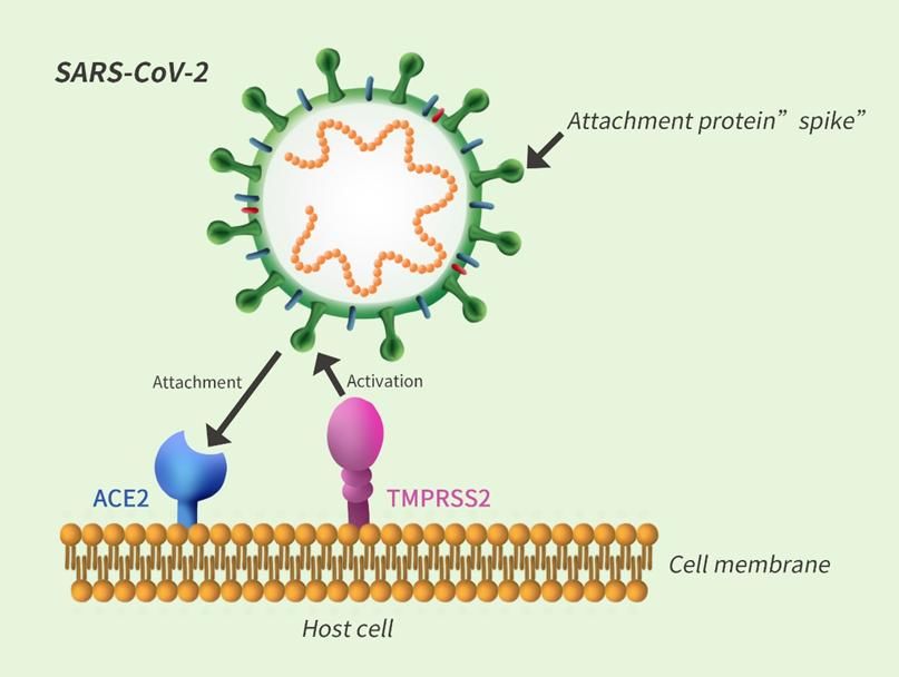
3

Transmembrane Serine Protease 2 (TMPRSS2): This is a serine protease that cleaves and
activates the viral spike glycoproteins which facilitates virus-cell membrane fusions. A recent
study showed that SARS-CoV-2 needs both ACE2 receptor and serine protease TMPRSS2 for
protein priming to enter the cell (Fig. 2) [2].

                        Figure 2: ACE2 and TMPRSS2 in Corona virus.

Nucleocapsid: The nucleocapsid phosphoprotein packages the viral genome into a helical
ribonucelocapsid, thus playing a crucial role in viral self-assembly (Fig. 3).

                                 Figure 3: Corona virus structure.
 Niknamian S | Volume 2; Issue 1 (2021) | JSRP-2(1)-013 | Mini Review

 Citation: Niknamian S. Brief Facts about COVID-19 (SARS-CoV-2). J Surg Res Prac. 2021;2(1):1-9.

 DOI: http://dx.doi.org/10.46889/JSRP.2021.2103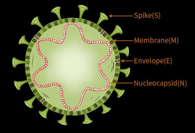
4
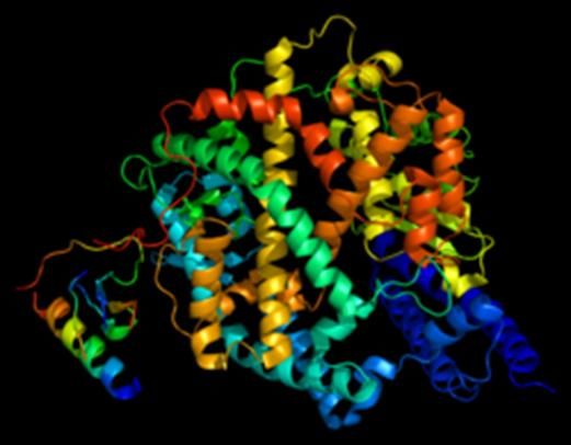
ACE-2 receptor: ACE-2 is the host cell receptor responsible for mediating infection by SARS-
CoV-2 (Fig. 4).
Figure 4: ACE-2 receptor protein structure.
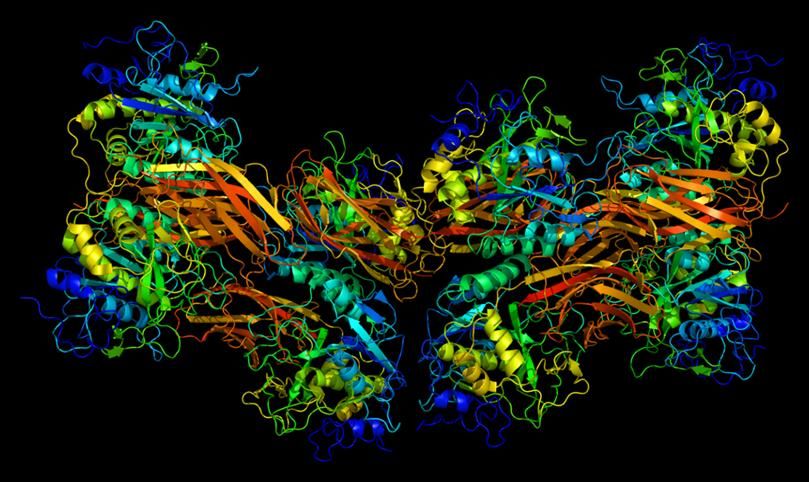
Furin: It is a protease present in many human organs that recognizes and activates a specific
site on the SARS-CoV-2 Spike protein, thus facilitating a tighter binding to the ACE-2 receptor
and might play a role in the higher infection rate (Fig. 5) [3].
Figure 5: ACE-2 receptor protein (Furin).
Niknamian S | Volume 2; Issue 1 (2021) | JSRP-2(1)-013 | Mini Review
Citation: Niknamian S. Brief Facts about COVID-19 (SARS-CoV-2). J Surg Res Prac. 2021;2(1):1-9.
DOI: http://dx.doi.org/10.46889/JSRP.2021.2103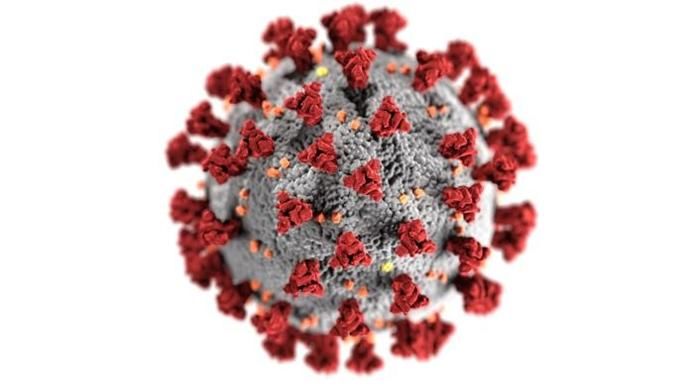
5

Cytokines: Studies have shown a strong correlation between severity of the disease and
concentrations of IL2, IL7, IL10, GCSF, MCP1 and TNF alpha (Fig. 6) [1]. Cytokines are a
category of small proteins (~5-20 kDa) that is important in cell signalling. Cytokines are
peptides and the most important thing is they cannot cross the lipid bilayer of cells to enter the
cytoplasm which means concentrating on Cytokines in COVID-19 is not so important.
Cytokines have been shown to be involved in autocrine, paracrine and endocrine signalling as
immunomodulation agents. Cytokines include chemokines, interferons, interleukins,
lymphokines and tumor necrosis factors, but generally not hormones or growth factors.
Cytokines are produced by a broad range of cells, including immune cells like macrophages,
B-lymphocytes, T-lymphocytes and mast cells, as well as endothelial cells, fibroblasts and
various stromal cells. A given cytokine may be produced by more than one type of cell. They
act through cell surface receptors and are especially important in the immune system.
Cytokines modulate the balance between humoral and cell-based immune responses and they
regulate the maturation, growth and responsiveness of particular cell populations. Some
cytokines enhance or inhibit the action of other cytokines in complex ways. They are different
from hormones, which are also important cell signalling molecules. Hormones circulate in
higher concentrations and tend to be made by specific kinds of cells. Cytokines are important
in health and disease, specifically in host immune responses to infection, inflammation, trauma,
sepsis, cancer and reproduction. In other words, Cytokines are important in immune system
functioning and when a pathogen get inside the body, immune related cells distribute these
proteins.

 Niknamian S | Volume 2; Issue 1 (2021) | JSRP-2(1)-013 | Mini Review

 Citation: Niknamian S. Brief Facts about COVID-19 (SARS-CoV-2). J Surg Res Prac. 2021;2(1):1-9.

 DOI: http://dx.doi.org/10.46889/JSRP.2021.2103
6

                                 Figure 6: Cytokines-IL2 pathway.

Discussions
COVID-19 like Influenza Virus-has 8 RNA segments which can be packaged in different ways
when they infect birds, pigs, or humans (Biology and Physics at KITP by Terry Hipsher).
Coronaviruses are large pleomorphic spherical particles with bulbous surface projections [4].
The average diameter of the virus particles is around 120 nm (0.12 μm). The diameter of the
envelope is ~80 nm (0.08 μm) and the spikes are ~20 nm (0.02 μm) long. The envelope of the
 Niknamian S | Volume 2; Issue 1 (2021) | JSRP-2(1)-013 | Mini Review

 Citation: Niknamian S. Brief Facts about COVID-19 (SARS-CoV-2). J Surg Res Prac. 2021;2(1):1-9.

 DOI: http://dx.doi.org/10.46889/JSRP.2021.2103
7 virus in electron micrographs appears as a distinct pair of electron dense shells [5-6]. The viral envelope consists of a lipid bilayer where the Membrane (M), Envelope (E) and Spike (S) structural proteins are anchored [7]. Inside the envelope, there is the nucleocapsid, which is formed from multiple copies of the Nucleocapsid (N) protein, which are bound to the positive- sense single-stranded RNA genome in a continuous beads-on-a-string type conformation [8- 9]. The lipid bilayer envelope, membrane proteins and nucleocapsid protect the virus when it is outside the host cell [10-22]. The structure of COVID-19 is so rigid and has very powerful against many antiviral and antimicrobial detergent like alcohol since the outer layer of the virus is lipid and cannot be solved in alcohol based materials. Based on our calculations, the genome size of COVID-19 as mentioned is from 26 to 32 Kilobases (basepair [bp]). 26 Kilopaepair: 26000 kilobasepair [kb], 0.026 megabasepair [Mb], 0.000026 gigabasepair [Gb], 8666.666667 amino acid [aa], 8666.666667 codon and 32 Kilobases means: 32000 kilobasepair [kb], 0.032 megabasepair [Mb], 0.000032 gigabasepair [Gb], 10666.666667 amino acid [aa], 10666.666667 codon. As you observe, the codon and amino-acids of COVID-19 are similar which is very rare in Viruses. When the Codon is similar to amino acid number it is called Codon Mismatch which we observe in COVID-19. Mitochondria are the targets of the Reactive Oxygen Species (ROS) that are produced inside a cell during viral infections and that mtDNA is a major target of these ROS [10]. Mitochondrial ATP generation requires proteins from the nuclear and mitochondrial genomes. ROS disrupt the oxidative production of ATP, which is required for normal cellular function, because damage of mtDNA disrupts the normal synthesis of proteins needed for mitochondria function and making them suitable targets for attack by ROS produced during infections by viruses and other microorganisms, although ROS also have other cellular targets. In HIV and hepatitis C virus infections, Oxidative Stress (OS) always plays a dominant pathogenic role. Peterhen and other researchers showed that almost all viruses (DNA/RNA viruses) cause cell death by generating oxidative stress in infected cells [11-13]. The OS generated during chronic hepatitis is associated with hepatic damage, a decrease in reduced Glutathione (GSH) and decrease in plasma and hepatic zinc concentration [14-15]. A loss of the MMP leads to imbalances in the membrane potentials of the IM and OM and then to arrest of normal cellular biosynthetic function and bioenergetics and finally to a “crisis” within the cell. A loss of the MMP also leads to release of several proapoptotic proteins from the IMS, such as Cyt C and Smac/DIABLO, as well as caspase independent death effectors, such as Apoptosis-Inducing Factor (AIF) and Endonuclease G (EndoG) [16], which have important roles in caspase- independent and caspase-dependent cell death [17]. The MMP transition occurs during the pathogenesis of exogenous factors (e.g., viral proteins, toxins and prooxidants [18-19]). A prolonged loss of the MMP leads to serious cell damage, from which the cell cannot recover. Therefore, in the intrinsic pathway of apoptosis, any viral factor that influences the MMP has a major impact on cell fate, either by inducing or by blocking cell death [20]. In recent years, there has been an increasing focus on the role of the MMP in disease and health. Thus, several recent models based on in-vivo and in-vitro studies explain the mechanisms underlying the Niknamian S | Volume 2; Issue 1 (2021) | JSRP-2(1)-013 | Mini Review Citation: Niknamian S. Brief Facts about COVID-19 (SARS-CoV-2). J Surg Res Prac. 2021;2(1):1-9. DOI: http://dx.doi.org/10.46889/JSRP.2021.2103
8

maintenance and loss of the MMP. A loss of the MMP by any mechanism leads to functional
and structural collapse of the mitochondria and cell death [21]. A recent study for first time has
shown that Dengue Virus (DV) infection of Human Hepatoma Cell Line (HepG2) leads
alteration in the bioenergetics function of mitochondrial morphology leading to MMP loss [22].
The alteration in respiratory properties of HepG2 cells in DV infection results due to decrease
in Respiratory Control Ratio (RCR) and ADP/O ratio, which suggest significant alteration in
mitochondrial morphology. Another additional feature observed by an increase in proton leak
termed mitochondrial uncoupling which occurs by leaking of protons through FoF1 ATP
synthase from inner membrane into matrix resulting in decrease in MMP loss. Thus, creating
an imbalance in ATP synthesis ultimately affects the bioenergetics functions of cell. The
biochemical mitochondrial damage induced in cell infected with HCV showed that E1 Protein
together with core and NS3 are responsible for ROS production. Core and NS3 induce NO
production which causes MMP loss by opening of transition pore [23]. NO could also interact
with another free radical superoxide (O2-) to form strong peroxynitrite anion (ONOO-), which
irreversibly inhibits multiple respiratory complexes (complexes I, II and IV) and aconitase and
activate proton leak and permeability transition pore [24-25]. Therefore, interfering with
energy metabolism by disrupting the ATP synthesis of cell results in modulation of
mitochondrial function [24].

Conclusion
Based on this review, a loss of the MMP in mitochondria leads to functional and structural
collapse of the mitochondria and cell death. For instance, Dengue Virus (DV) infection of
Human Hepatoma Cell Line (HepG2) leads alteration in the bioenergetic function of
mitochondrial morphology leading to MMP loss. The alteration in respiratory properties of
HepG2 cells in DV infection results due to decrease in Respiratory Control Ratio (RCR) and
ADP/O ratio, which suggest significant change in mitochondrial morphology. Another
additional feature observed by an increase in proton leak termed mitochondrial uncoupling
which occurs by leaking of protons through FoF1 ATP synthase from inner membrane into
matrix resulting in decrease in MMP loss. Therefore; creating an imbalance in ATP synthesis
ultimately affects the bioenergetics functions of cell. The biochemical mitochondrial damage
induced in cell infected with HCV showed that E1 Protein together with core and NS3 are
responsible for ROS production. Core and NS3 induce NO production which causes MMP loss
by opening of transition pore. NO could also interact with another free radical superoxide (O2-
) to form strong peroxynitrite anion (ONOO-), which irreversibly inhibits multiple respiratory
complexes (complexes I, II and IV) and aconitase and activate proton leak and permeability
transition pore. Thus, interfering with energy metabolism by disrupting the ATP synthesis of
cell results in modulation of mitochondrial function. COVID-19 like DV, is related to
mitochondrial function and ATP production of the patient. Abnormal Mitochondrial function

 Niknamian S | Volume 2; Issue 1 (2021) | JSRP-2(1)-013 | Mini Review

 Citation: Niknamian S. Brief Facts about COVID-19 (SARS-CoV-2). J Surg Res Prac. 2021;2(1):1-9.

 DOI: http://dx.doi.org/10.46889/JSRP.2021.2103
9

is an infected patient leads to the abnormal respiratory function and death of the patient. This
is the major cause of death of many people infected with SARS-CoV-19.

References
1.    Singhal T. A review of coronavirus disease-2019 (COVID-19). Ind J Pediat. 2020:1-6.
2.    Markus Hoffmann. SARS-CoV-2 cell entry depends on ACE2 and TMPRSS2 and is blocked by a clinically
      proven protease inhibitor. 2020;181(2):271-80.
3.    Wrapp D, Wang N, Corbett KS, Goldsmith JA, Hsieh CL, Abiona O, et al. Cryo-EM structure of the 2019-
      nCoV spike in the prefusion conformation. Science. 2020;367(6483):1260-3.
4.    Dr. Royal R Rife. A rare recording of Dr. Royal R Rife. 2018. Listen & Live Audio. Unabridged Audiobook.
5.    Goldsmith CS, Tatti KM, Ksiazek TG, Rollin PE, Comer JA, Lee WW, et al. Ultrastructural characterization
      of SARS coronavirus. Emer Infect Dis. 2004;10(2):320.
6.    Neuman BW, Adair BD, Yoshioka C, Quispe JD, Orca G, Kuhn P, et al. Supramolecular architecture of
      severe acute respiratory syndrome coronavirus revealed by electron cryomicroscopy. J Virol.
      2006;80(16):7918-28.
7.    Fehr AR, Coronaviruses PS. An overview of their replication and pathogenesis. Maier H, Bickerton E, Britton
      P (Eds). Coronaviruses Methods in Molecular Biology. 2015;1282.
8.    Lai MM, Cavanagh D. The molecular biology of coronaviruses. Adv in Virus Res. 1997;48:1-100.
9.    Chang CK, Hou MH, Chang CF, Hsiao CD, Huang TH. The SARS coronavirus nucleocapsid protein-forms
      and functions. Antiviral Res. 2014;103:39-50.
10.   Neuman BW, Kiss G, Kunding AH, Bhella D, Baksh MF, Connelly S, et al. A structural analysis of M protein
      in coronavirus assembly and morphology. J Structural Biol. 2011;174(1):11-22.
11.   Reshi L, Wang HV, Hui CF, Su YC, Hong JR. Anti-apoptotic genes Bcl-2 and Bcl-xL overexpression can
      block iridovirus serine/threonine kinase-induced Bax/mitochondria-mediated cell death in GF-1 cells. Fish
      and Shellfish Immunol. 2017;61:120-9.
12.   Peterhans E, Grob M, Urge TB, Zanoni R. Virus-induced formation of reactive oxygen intermediates in
      phagocytic cells. Free Radical Research Communications. 1987;3(1-5):39-46.
13.   Vierucci A, DeMartino M, Graziani E. A mechanism for liver cell injury in viral hepatitis: Effects of hepatitis
      B virus on neutrophil function in-vitro and in children with chronic active hepatitis. Pediat Res.
      1983;17(10):814-20.
14.   Muller F. Reactive oxygen intermediates and Human Immunodeficiency Virus (HIV) infection. Free Radical
      Biol Med. 1992;13(6):651-7.
15.   Boya P, Peña AD, Beloquietal O. Antioxidant status and glutathione metabolism in peripheral blood
      mononuclear cells from patients with chronic hepatitis C. J Hepatol. 1999;31(5):808-14.
16.   Bianchi GP, Marchesini G, Brizietal M. Nutritional effects of oral zinc supplementation in cirrhosis. Nutrition
      Res. 2000;20(8):1079-89.
17.   Reshi L, Su YC, Hong JR. RNA viruses: ROS-mediated cell death. Int J Cell Biol. 2014;2014:467-72.
18.   Reshi L, Wu JL, Wang HV, Hong JR. Aquatic viruses induce host cell death pathways and its application.
      Virus Res. 2016;211:133-44.
19.   Reshi L, Hong JR. Mitochondria as a favorite organelle for invading viruses. Molecular Biol. 2017;6:181.
20.   Machida K, Cheng KT, Sung VM, Lee KJ, Levine AM, Lai MM. Hepatitis C virus infection activates the
      immunologic (type II) isoform of nitric oxide synthase and thereby enhances DNA damage and mutations of
      cellular genes. J Virol. 2004;78(16):8835-43..
21.   Reshi L, Hong JR. Mitochondria as a favourite organelle for invading viruses. Molecular Biol. 2017;6:181.
22.   Meng G, Xia M, Wang D, Chen A, Wang Y, Wang H, et al. Mitophagy promotes replication of oncolytic
      Newcastle disease virus by blocking intrinsic apoptosis in lung cancer cells. Oncotarget. 2014;5(15):6365-
      74.
23.   Cytokine in John Lackie. A dictionary of biomedicine. Oxford University Press. 2010.
24.   Cytokine in Stedman’s Medical Dictionary, 28th Ed. Wolters Kluwer Health, Lippincott, Williams & Wilkins
      (2006).

 Niknamian S | Volume 2; Issue 1 (2021) | JSRP-2(1)-013 | Mini Review

 Citation: Niknamian S. Brief Facts about COVID-19 (SARS-CoV-2). J Surg Res Prac. 2021;2(1):1-9.

 DOI: http://dx.doi.org/10.46889/JSRP.2021.2103
You can also read