Differential expression of Bax and Bcl-2 in the brains of hamsters infected with 263K scrapie agent
←
→
Page content transcription
If your browser does not render page correctly, please read the page content below
MOLECULAR NEUROSCIENCE                                                                                                                NEUROREPORT

                 Differential expression of Bax and Bcl-2 in
                 the brains of hamsters infected with 263K
                                scrapie agent
Sang-Koo Park,1,2 Seung-Il Choi,1 Jae-Kwang Jin,1 Eun-Kyoung Choi,1 Jae-Il Kim,1 Richard I. Carp3 and
                                        Yong-Sun Kim1,2,CA

           1
               Institute of Environment and Life Science, Hallym Academy of Sciences, and 2 Department of Microbiology, College of Medicine,
                Hallym University, 1 Ockcheon-Dong, Chuncheon, Kangwon-Do 200-702, South Korea; 3 New York State Institute for Basic
                                           Research in Developmental Disabilities, Staten Island, NY 10314, USA

                                                         CA,1
                                                                Corresponding Author and Address

                                                  Received 28 February 2000; accepted 13 March 2000

 To study the mechanism(s) of neuronal cell death during                        Bax and Bcl-2 were found predominantly within neurons. In
 scrapie infection, we investigated the expression of Bax and                   scrapie-infected brains, the number of neuronal cells positive
 Bcl-2 in brains of hamsters infected with 263K scrapie agent.                  for Bcl-2 was signi®cantly lower in the hippocampal CA3 region
 The expression of Bcl-2 mRNA was signi®cantly decreased in                     and was decreased in the cerebral cortex, whereas the number
 the brains of 263K scrapie-infected hamsters compared with                     of neuronal cells positive for Bax was signi®cantly increased in
 controls, whereas the expression levels of Bax mRNA were                       both regions. The possibility that differential regulation of Bax
 signi®cantly increased in scrapie-infected brain. The levels of                and Bcl-2 expression may play an important role in neuronal
 Bax and Bcl-2 proteins in brains of scrapie and control animals                cell death induced by scrapie infection is discussed. NeuroReport
 re¯ected the difference in mRNA levels. Immunoreactivity for                   11:1±6 & 2000 Lippincott Williams & Wilkins.

 Key words: Bax; Bcl-2; Neurodegeneration; Neuronal cell death; Scrapie

INTRODUCTION                                                                    cell death in scrapie-infected brain could be related to
Scrapie, the archetype of a group of diseases known as                          mitochondrial dysfunction induced by oxidative stress, or
transmissible spongiform encephalopathies (TSE) or prion                        that the increase of Bax:Bax homodimers by the binding of
diseases, occurs naturally in sheep and goats [1±3]. Its                        PrP to Bcl-2 promotes apoptosis [11,12]. In order to
clinical symptoms are characterized by a long latent                            ascertain potential therapeutic targets, it is important to
period, progressive ataxia, tremor, wasting and death [4]. It                   clarify the mechanisms leading to neuronal cell death in
has been experimentally transmitted to laboratory rodents,                      TSE.
and these species have become experimental models of                               In the current study, we analyzed the levels of expres-
TSE [5,6]. Neuronal cell death is a prominent feature of                        sion of Bcl-2 and Bax by RT-PCR, Western blot, and
prion diseases; however, the pathogenesis and the molecu-                       immunohistochemistry of the brains of hamsters infected
lar basis of neuronal cell death in these diseases have not                     with the 263K scrapie agent.
been determined.
   Cell death may occur by one of two general pathways,
apoptosis or necrosis. Since many common neurodegenera-                         MATERIAL AND METHODS
tive diseases such as Alzheimer's disease, Parkinson's                          Animal injection and assessment: Six-week-old female
disease and amyotrophic lateral sclerosis are characterized                     golden Syrian hamsters were divided into control and
by a gradual loss of neurons without obvious in¯ammatory                        infected groups. Hamsters were inoculated intracerebrally
response, it has been hypothesized that cell death in these                     by stereotaxic apparatus (Stoelting Co., USA) with 20 ìl of
disorders is due to apoptosis [7]. However, controversy                         1.0% brain homogenate in 0.01 M phosphate-buffered sal-
remains over whether cell death in these diseases is                            ine (PBS). The brain homogenates were prepared from
mediated by apoptosis or necrosis. Several results suggest                      263K scrapie-infected hamster brains. Control inoculum
that neuronal cell death in TSE is due to apoptosis [8,9].                      was prepared from brains of normal hamsters (NHB). After
Apoptosis was also observed when residues 106±126 of the                        inoculation with the 263K scrapie agent, hamsters devel-
human prion protein were added to primary neuronal cell                         oped clinical symptoms (tremor, head-bobbing, ataxia) at
cultures [10]. Other studies have suggested that neuronal                       60  5 days. Animals were sacri®ced 5±7 days after the

0959-4965 & Lippincott Williams & Wilkins                                                                               Vol 11 No 8 5 June 2000   1

      Article number = 9801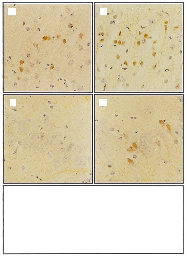
NEUROREPORT S.-K. PARK ET AL.
appearance of clinical symptoms. Hamsters injected with normal goat serum, and then incubated overnight at 48C
NHB were harvested at the same time. with primary antibody used at a 1:100 dilution for Bax
(Calbiochem, USA) and at a 1:200 dilution for Bcl-2
Preparation of brain extracts and Western blot analy- (Calbiochem, USA). After washing three times with phos-
sis: Hamsters were sacri®ced and brains were removed phate buffer, the sections were sequentially treated with
rapidly. Whole brains were homogenized with a Te¯on± biotinylated anti-rabbit immunoglobulin and avidin-biotin
glass homogenizer in 9 vol extraction buffer (pH 7.0) peroxidase complex, and developed with diaminobenzi-
containing 0.32 M sucrose, 10 mM Tris±HCl, 1 mM EDTA, dine-hydrogen peroxidase solution (0.003% 3,39-diamino-
5 mM 2-mercaptoethanol. The homogenates were centri- benzidine and 0.03% hydrogen peroxidase in 0.05 M Tris
fuged at 1000 3 g for 10 min at 48C, and the supernatants buffer), and ®nally counterstained with hematoxylin. The
were collected. After centrifugation of the supernatant at number of Bax and Bcl-2 immunolabeled neurons was
105 000 3 g for 60 min at 48C, the pellet was washed and counted in the hippocampal CA3 region and the cerebral
resuspended with extraction buffer. Protein concentration cortex in ®ve ®elds of 1 mm2 each [17].
of this fraction was measured by the method of Lowry
[13,14]. SDS-PAGE was performed by the method of Statistical analysis: The statistical differences between
Laemmli [15]. After SDS-PAGE, immunoblots were per- control and infected groups were calculated by Student's t-
formed by the method of Towbin et al. [16]. Protein test; data are shown as mean s.d. Statistically signi®cant
samples (50 ìg) were subjected to 15% SDS-PAGE. After values are shown as p , 0.05 and p , 0.01.
electrophoresis and transfer, the blotted membranes were
incubated for 1 h at room temperature with primary anti- RESULTS
body, Bax (Calbiochem, USA) and Bcl-2 (Pharmingen, Gene expression of Bax and Bcl-2: We analyzed levels of
USA) diluted (1:100) in TBST (Tris-buffered saline; 0.1% gene expression of Bax and Bcl-2 in the brains of scrapie-
Tween-20) containing 5% skim milk. The development of infected and control hamsters by RT-PCR. Ampli®cation
membranes used the chemiluminescence Western blotting using speci®c primers yielded bands of expected sizes (Bcl-
system (ECL; Amersham, USA). Results were analyzed by
scanning densitometry.
RT-PCR analysis: RT-PCR was performed from total
RNA of control and 263K scrapie-infected hamster brains.
Single strand cDNA synthesis from total RNA (2 ìg) was
performed using oligo (dT) with AMV reverse transcrip-
tase (Promega, USA). The PCR was conducted using Taq
DNA polymerase (Promega, USA). The nucleotide se-
quences of the primers were: for ampli®cation of Bax, 59-
ACCAAGAAGCTGAGCGAGTGT-C-39 (sense), 59-ACAA
AGATGGTCACGGTCTGCC-39 (antisense), generating a Fig. 1. The expression of Bax and Bcl-2 mRNA in the brains of control
374 bp fragment; Bcl-2: 59-TGCACCTGACGCCCTTCAC-39 and scrapie-infected hamsters. Each band was shown the ampli®ed
(sense), 59-AGACAGCCAGGAGAAATCAAACAG-39 (anti- products of the expressed mRNAs of Bax, Bcl-2 and â-actin in the brains
sense), generating a 260 bp fragment; â-actin: 59-ACC of 263K scrapie-infected and control hamsters. The expression of â-actin
TTCAACACCCCAGCCATG-39 (sense), 59-GGCCATCTC mRNA was used as a reference.
TTGCTCGAAGTC-39 (antisense) generating a 309 bp frag-
ment. The speci®city of each PCR product was con®rmed (a) (b)
by nucleotide DNA sequencing. Programmable tempera- 1 2 1 2
ture cycling (Techne, UK) was performed according to the
following: after an initial time of 1 min for predenaturation 26 kDa 21 kDa
at 958C, PCR was performed using 30 cycles for Bax, Bcl-2
Relative Density (%)
Relative Density (%)
and â-actin with denaturation for 60 s at 948C, annealing 125 250 **
for 45 s at 608C, and extension for 60 s at 728C. 100 * 200
75 150
Agarose gel electrophoresis: Total PCR mixtures were 50 100
subjected to electrophoresis in 1.8% agarose gels containing 25 50
ethidium bromide. Separated PCR products were visua- 0 0
Control Infection Control Infection
lized under u.v. illumination.
Fig. 2. Western blot analysis of Bax and Bcl-2 protein levels in the
Immunohistochemistry: Immunohistochemical proce- brains of control and scrapie-infected hamsters. (a) Bcl-2 immunoreactiv-
dures were carried out using the ABC kit (Vector, USA) by ity in control (lane 1) and 263K scrapie-infected brains (lane 2). The band
a modi®cation of the avidin-biotin-peroxidase method. is at the predicted molecular size (26 kDa) for Bcl-2. (b) Bax immuno-
Brie¯y, sections (6 ìm) of the brain were dewaxed with reactivity of control (lane 1) and 263K scrapie-infected brains (lane 2).
xylene and hydrated with graded ethanol, and then treated The band is at the estimated molecular size (21 kDa) for Bax. Graphs at
bottonfected brains (lane 2). The band is at the estimated molecular size
with 0.3% hydrogen peroxide in methyl alcohol for 20 min (21 kDa) for Bax. Graphs at bottom portion of the ®gure depict
to block endogenous peroxidase. After washing three times quantitative analysis of density for Bax (n 5) and Bcl-2 immunoblot
with phosphate buffer, the sections were exposed to bands. Data are expressed as mean s.d.. p , 0.05, p , 0.01.
2 Vol 11 No 8 5 June 2000DIFFERENTIAL EXPRESSION OF Bax AND Bcl-2 IN THE BRAINS OF HAMSTERS INFECTED WITH 263K SCRAPIE AGENT NEUROREPORT
(a) (b)
(c) (d)
(e)
40 * Control
Infection
30
Number of Cells
(Cells/mm2)
20
**
10
0
Cerebral Cortex Hippocampus
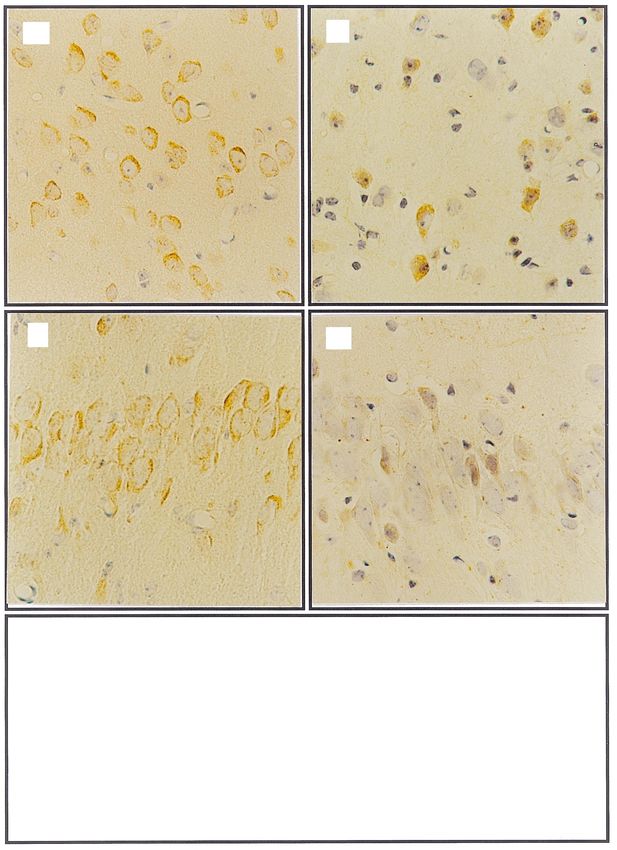
Fig. 3. Representative photomicrographs showing immunohistochemical analysis of Bax. The Bax-positive neuronal cells in cerebral cortex (a,b) and
CA3 region of hippocampus (c,d) in control (a,c) and 263K scrapie-infected brain (b,d), 3400. (e) Graphs at bottom of ®gure depict quantitative analysis
of positive neurons for Bax immunostaining in the cerebral cortex and the CA3 region of hippocampus in 263K scrapie-infected group (n 5) and
control group (n 5). Data are expressed as mean s.d., p , 0.05, p , 0.01.
Vol 11 No 8 5 June 2000 3NEUROREPORT S.-K. PARK ET AL.
(a) (b)
(c) (d)
(e)
80 Control
Infection
60
Number of Cells
(Cells/mm2)
40
20 *
0
Cerebral Cortex Hippocampus
Fig. 4. Representative photomicrographs showing immunohistochemical analysis of Bcl-2. Bcl-2 positive neuronal cells in cerebral cortex (a,b) and
CA3 region of hippocampus (c,d) in control (a,c) and 263K scrapie-infected brain (b,d), 3400. (e) Graphs at bottom of ®gure depict quantitative analysis
of positive neurons for Bcl-2 immunostaining in the cerebral cortex and the CA3 region of hippocampus in 263K scrapie-infected group (n 5) and
control group (n 5). Data are expressed as mean s.d. p , 0.05, p , 0.01.
4 Vol 11 No 8 5 June 2000DIFFERENTIAL EXPRESSION OF Bax AND Bcl-2 IN THE BRAINS OF HAMSTERS INFECTED WITH 263K SCRAPIE AGENT NEUROREPORT
2, 260 bp; Bax, 374 bp; â-actin, 309 bp). Our results showed in normal hamster brain, but following 263K scrapie
that Bcl-2 was expressed at low levels in the brains of infection the expression of Bax was signi®cantly increased.
scrapie-infected hamsters, whereas there was an increase In contrast, the expression of Bcl-2 measured by mRNA
of Bax mRNA expression in the brains of the scrapie- and protein was reduced in hamsters clinically affected by
infected group compared to the control group (Fig. 1). scrapie compared to control hamsters. The results of the in
Levels of mRNA of constitutively expressed â-actin were vitro studies were con®rmed by immunohistological ana-
virtually identical in scrapie-infected and control groups lyses that showed increased staining for Bax in neurons of
(Fig. 1). the cerebral cortex and the hippocampal CA3 region,
whereas Bcl-2 staining was reduced in these neuron
Bax and Bcl-2 protein levels: The levels of Bax and Bcl-2 populations in scrapie-infected hamsters. These ®ndings
protein were measured by Western blots. As shown in suggest that the mode of neuronal cell death observed in
Fig. 2, Bcl-2 antibody detected a band at 26 kDa, the TSE is apoptosis resulting from differential expression of
predicted mol. wt for the bcl-2 gene product. In the Bax and Bcl-2.
scrapie-infected brains, Bcl-2 protein was present at re- The cause of differential expression of Bax and Bcl-2 in
duced levels, whereas protein levels of Bax were higher the brains of scrapie-infected animals is unknown. Since
than in controls. The molecular weight for Bax was 21 kDa, p53 protein is a direct transcriptional regulator that upre-
which is the expected size. The lower portion of the ®gure gulates Bax expression and down-regulates Bcl-2 expres-
shows densitometric analysis of the Western blots. sion, we suggest that p53 protein is involved in the
differential regulation of Bax and Bcl-2 expression [21].
Immunohistochemistry of Bax and Bcl-2: Immunohisto- Although an increase of Bax and a decrease of Bcl-2
chemical methods were used to examine the distribution of expression have been detected in scrapie-infected brains,
Bax and Bcl-2 in brain tissue of control and scrapie-infected the ®nding does not prove that apoptosis is the mode of
hamsters. Immunoreactivities for Bax and Bcl-2 were found cell death. There are several factors that contribute to this
mainly within neurons in both control and scrapie-infected uncertainty. First, in addition to inhibition of apoptosis,
brains (Fig. 3, Fig. 4). Immunoreactivity for Bax increased Bcl-2 can also inhibit several forms of necrosis [22]. Second,
(Fig. 3), whereas that for Bcl-2 decreased (Fig. 4) within in a previous study we showed that neuronal cell death in
neurons of the hippocampal CA3 region and the cerebral the brains of hamsters infected with the 263K scrapie agent
cortex of the scrapie-infected group compared with the is related to structural abnormalities (swelling) of mito-
staining seen in control animals. The data in Fig. 3 and chondria, which is a major feature of necrosis [11].
Fig. 4 were quantitated by analyzing the number of cells As noted above, the current results do not distinguish
that stained positively in randomly chosen areas of 1 mm2 . whether cells die because of apoptosis or necrosis in TSE.
The results are shown in the lower portions of Fig. 3 and We suggest that neuronal cell death in these diseases may
Fig. 4. the number of stained cells revealed in scrapie- employ both mechanisms in a scenario in which cells
infected brain, Bcl-2 immunoreactivity in neuronal popula- initially undergo apoptosis and then are subject to second-
tions was reduced by 20% in the cerebral cortex and 50% ary necrosis.
in the CA3 region of the hippocampus ( p , 0.05), while the In future studies, we plan to examine signaling cascades
number of Bax-immunoreactive cells was increased in the activated before DNA fragmentation begins through analy-
cerebral cortex by 2.7-fold and in the CA3 region of the sis of expression of p53 and Fas/APO-1, transmembrane
hippocampus by 4-fold. These changes in Bax-immunor- receptors, and the relationship to differential expression of
eactive cells were signi®cant at p , 0.05 and p , 0.01, Bax and Bcl-2. We will also assess the correlation between
respectively. these values and the pathological changes in brains of
scrapie-infected animals.
DISCUSSION
Two modes of cell death can be distinguished: apoptosis CONCLUSION
and necrosis. In order to assess potential therapeutic Apoptosis is implicated in neuronal cell death in prion
targets in TSE diseases, it is important to clarify the diseases. In this study, we showed that there was an
mechanisms leading to neuronal cell death. The fact that increased expression of Bax and a reduced expression of
apoptotic cells are rapidly removed via phagocytosis com- Bcl-2 in the brains of 263K scrapie-infected hamsters; the
bined with the fact that the process often concludes in ®ndings indicate that the differential regulation of Bax and
secondary necrosis means that it is dif®cult to ascertain the Bcl-2 expression might be a decisive factor in the process
cause of initial damage to cells [18]. Nevertheless, apopto- of neuronal cell death in this scrapie strain-host model.
sis has been implicated as an important mechanism of cell
death in many neurodegenerative diseases [19]. With REFERENCES
regard to prion diseases, studies of primary neuronal 1. Dickinson AG, Fraser H, Meikle VM and Outram GW. Nature New Biol
cultures and neuronal cell lines have shown that PrP 237, 244±245 (1972).
peptides can cause apoptosis [10]. DNA cleavage, one of 2. Hope J. Arch Virol Suppl 7, 201±211 (1993).
the key features of apoptosis, has been reported in scrapie- 3. Prusiner SB. Proc Natl Acad Sci USA 95, 13363±13383 (1998).
infected sheep brains [20]. However, the sequence of 4. Carp RI, Ye X, Kascsak RJ et al. Ann NY Acad Sci 724, 221±234 (1994).
5. Kim YS, Carp RI, Callahan SM et al. Acta Neuropathol (Berl) 80, 388±392
pathological events that lead to neuronal cell death in
(1990).
scrapie-infected animals is not clear. 6. Bruce ME, McConnell I, Fraser H and Dickinson AG. J Gen Virol 72,
In the current study, the major ®ndings are as follows: 595±603 (1991).
the products of the proapoptotic gene Bax were detectable 7. Thompson CB. Science 267, 1456±1462 (1995).
Vol 11 No 8 5 June 2000 5NEUROREPORT S.-K. PARK ET AL.
8. Giese A, Groschup MH, Hess B et al. Brain Pathol 5, 213±221 (1995). 16. Towbin H, Staehelin T and Gordon J. Proc Natl Acad Sci USA 76,
9. Lucassen PJ, Williams A, Chung WCJ et al. Neurosci Lett 198, 185±188 4350±4354 (1979).
(1995). 17. Terry RD, Peck A, DeTeresa R, et al. Ann Neurol 10, 184±193 (1981).
10. Forloni G, Angretti N, Chiesa R et al. Nature 362, 543±546 (1993). 18. Hong JR, Lin TL, Hsu YL et al. Virology 250, 76±84 (1998).
11. Choi SI, Ju WK, Choi EK et al. Acta Neuropathol (Berl) 96, 279±286 (1998). 19. Haass C. Nature 399, 204±205, 207 (1999).
12. Kurschner C and Morgan JI. Brain Res Mol Brain Res 30, 165±168 (1995). 20. Fairbairn DW, Carnahan KG, Thwaits RN et al. FEMS Microbiol Lett 115,
13. Yoshihisa K, Shun S, Wataru K et al. Brain Res 780, 260±269 (1998). 341±346 (1994).
14. Lowry OH, Rosebrough NJ, Farr AL et al. J Biol Chem 193, 265±275 (1951). 21. Liu X and Zhu Xz. Neuroreport 10, 3087±3091 (1999).
15. Laemmli UK. Nature 227, 680±685 (1970). 22. Tsujimoto Y, Shimizu S et al. Leukemia (Suppl.) 3, 380±382 (1997).
Acknowledgements: We thank Hyoung-Gon Lee for his helpful comments and critical reading of the manuscript. This study was
supported by The Hallym Academy of Sciences (1999), Hallym University, Korea.
AQ: Ref 22. 3rd author please.
AQ: Check the magni®cations of your ®gures, they have been reduced.
6 Vol 11 No 8 5 June 2000You can also read