Histological Study of Effects of Saccharin on Thyroid Gland in Male White Mice
←
→
Page content transcription
If your browser does not render page correctly, please read the page content below
Annals of R.S.C.B., ISSN: 1583-6258, Vol. 25, Issue 1, 2021, Pages. 5944 - 5955
Received 15 December 2020; Accepted 05 January 2021.

Histological Study of Effects of Saccharin on Thyroid Gland in Male White
                                    Mice
                       Doaa Fareed Fawzi1a; Bassim Abdullah Jassim1b
            1
                Department ofBiology,College of Science Al-Muthanna university/Iraq
                     *Email:adoaaff913@gmail.com,bbassimabd@mu.edu.iq
Abstract:

   The present study aimed to evaluate the histological and physiological changes induced by
administration of saccharin on the thyroid gland of mice. Thirty adult male mice used in this study
were divided into two groups. The first group used as a control; the second group used as the treated
group which treated with 1 ml daily of saccharin solution orally administered for 30 days. After
ending the treatment period, blood samples were collectedto assessment of T3 and T4 levels, then the
animals were sacrificed for histological study. The results showed the thyroid gland of male after oral
administration with saccharin have significant histological differences in most tissue field of thyroid
gland, when compared with control group. The physiological results showed a significant difference
of hormonal secretions (T3 and T4). Therefore, it can be concluded that treatment of mice with
saccharin caused histological and physiological changes in the thyroid gland. So, we should be
reducing the use of saccharin as sweeteners and increase public awareness regarding artificial
sweetener consumption.

Keywords: Saccharin, Sweeteners, Thyroid gland

1.Introduction:

    In recent years in the world, market demand for low-calorie goods in the forms of widely different
sugar-free food items such as drinks powder, sweets, jams, carbonated beverages, jellies, dairy
products and canned foods has been seen observed. All sweeteners that known as a different name
like low calorie or artificial or alternative sweeteners have taste similar to sugar but it contains no
calories [1].

    Throughout recent decades, increasing quality and health of life issues have motivated people to
reduce the consumption of products that are high in sugar. Food products that contain calorie-free
alternatives have become increasingly common.Alternative sweeteners are hundreds to thousands of
times sweeter than saccharose. Each sweetener has unique features of intensity of sweetness, teeth
coating, persistence of sweet taste and aftertaste result [2]

     Sweeteners are food additives that are used to improve the taste of everyday foods. Natural
sweeteners are sweet-tasting substances that have a certain nutrient value, monoor disaccharides are
the major component in natural sweeteners. In contrast, artificial sweeteners are compounds with very
little to no nutritional worth. Artificial sweeteners are recent and are being tested and expanded every
day for their uses [3]. Artificial sweeteners which are still of concern despite common use [4].
Recently, the common assumption was that non-nutritive sweeteners (NNS) were safe replacements
for sugar, because they offer sweet taste without calories or glycemic impacts. However, evidence
from several epidemiologic studies have shown that intake of NNS is associated with increased risk of
developing obesity, metabolic syndrome and type 2 diabetes, particularly in diet sodas [5].The Food
and Drug Administration Authority of the United States has approved saccharine, acesulfame-K,
aspartame, neotame, sucralose, and stevia for human use within the appropriate daily intake limit

http://annalsofrscb.ro                                                                            5944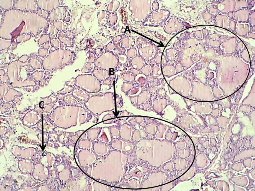
Annals of R.S.C.B., ISSN: 1583-6258, Vol. 25, Issue 1, 2021, Pages. 5944 - 5955
Received 15 December 2020; Accepted 05 January 2021.

[6].Animal studies note the impact of artificial sweeteners on the immune system. Sugar substitutes
were the culprit in the development of Hashimoto thyroiditis in patient [7].Non-nutritious sweeteners
are most widely used worldwide. Furthermore, their metabolic outcomes are still uncertain, and their
effect on the thyroid action, a main metabolism regulator, has not been studied before [8].Although
few studies are suggesting that a high dose, prolonged use of sweeteners might increase the risk of
development of certain malignancies, we have found an increased risk of thyroid cancer with artificial
sweetener consumption [9].Saccharin (benzoic sulfimide) is a very stable organic acid with chemical
formula C7H5NO3S. Saccharin is one of the oldest artificial sweeteners [10].Saccharin is used as a
tabletop sweetener in drinks, soft drinks, canned fruit, baked products, chewing gum, confectionery,
salad dressings and also in cosmetic and pharmaceutical products with considering to acceptable daily
intake (ADI) [1].Artificial sweeteners, when used in moderation are an acceptable substitute to natural
sweeteners. They appear to be a healthy and pleasant way to enrich food sweetness, taste, and texture
while keeping major health risks low [10].

   The thyroid gland and its hormones play multifaceted roles in organ development and in the
homeostatic control of fundamental physiological mechanisms such as body growth and energy
expenditure in all vertebrates [11].Thyroid hormone production is tightly regulated and clinical
manifestations of thyroid system dysfunction are well known. T3 is an important regulator [12]

2.Materials and Methods:

2.1. Experimental Animals:

   Thirty matured male albino mice with weighing range 27-30gm (three-month-old) were bought
from Drug and Healthy center in Baghdad, Iraq. Mice were kept in plastic mice cages, all cages put in
the animal house of the college of science in Al- Muthanna University under controlling of
temperature 25-28ºC, controlled humidity situation (65%) with feeding by using standard pellets for
about 15 days to adapt to their environment.

2.2. Chemical:Saccharin

2.3. Exposure dose:The dose level is 1 ml saccharin orally administered for a period of 30 days.
According to [1].

2.4. Experimental design:

Thirty male mice were divided into two (A and B) groups, Group A as a control group and group B as
treated group. 2.4.1. Control group: There were 15 mice which feed without any substance in their
food. Plenty of water was supplied using a feeding bottle.2.4.2. Treated group: There were 15 mice
which orally administered with saccharin at a dose level of 1ml daily for 30 days.

2.5. Preparation of histological slide:

   After the end of the treatment period, the animals were anesthetized by using chloroform then
sacrificed by bleeding from common carotid artery. Thyroid gland was collected carefully from the
mice by using the standard procedure. The samples washed with normal saline to remove any contest
of blood.Fixation: the tissue samples were fixated with formalin 10% for a period of (48) hour, then
washed under running tap water for one hour until completely removed of most of the formalin odder
from the tissue samples. Followed by washing, dehydration of the tissues was conducted by
immersing the tissue in a series of gradually increasing concentrations of alcohol (50%, 70%, 80%,

http://annalsofrscb.ro                                                                           5945
Annals of R.S.C.B., ISSN: 1583-6258, Vol. 25, Issue 1, 2021, Pages. 5944 - 5955
Received 15 December 2020; Accepted 05 January 2021.

90%, and absolute alcohol). Clearing it is obligatory because the used alcohol for dehydration will not
dissolve or mix with molten paraffin. Blocking means the samples embedded into paraffin wax for
making blocks. The block was to be trimmed by removing of wax from the surface of the block to
expose the tissue, cutting of the tissue was performed by using a microtome. The microtome was pre-
set to cut the tissue as thicknesses with 5 µm. Blocks Small ribbons of tissue sections were placed on
a microscopic slide with help of warm distil water containing few drops of Mayer’s albumen and
deparaffinized with xylene solution, the slide was put on the hot plate with (40°c) for overnight.
Hematoxylin and eosin yellow solution were used to stain the tissue for preparing permanent slide
[13]. Histopathological changes were observed under a light microscope and snaps were taken.

3. The Histological Results of thyroid:
3.1 The Histological Results of male Control Group:
   The histological results of thyroid gland in the male showed the parenchyma characterized have
few numbers of large follicles which have diameter (137.23±23.70 µm),(Table 1) whichdistributed
among the thyroid parenchyma; while medium and small follicles present in high aggregation among
parenchyma (88.84± 8.79µm), (45.43±4.57µm), (Table 1) respectively. The tissue section of control
thyroid gland showed the wall of follicles which lined by simple cuboidal epithelial with thickness
was (10.69±2.28 µm), the diameters of follicular cell nuclei was (41.71±8.75µm) (Table 1) fig (1).
   The thyroid gland was surrounded by connective tissue capsules, which composed of two layers;
the external layer which consist of loose collagen fibers mixed with large amount of adipose tissue
with the presence of a few amount of elastic fibers. The inner layer consists of bundles of collagenous
and elastic fibers, with few smooth muscles’ cells. The capsule of the thyroid gland was highly
vascularized with clear blood vessels supply fig (2).The connective tissue capsule sends thin
trabecular in to the parenchyma around the different types of follicles and divided the thyroid
parenchyma into poorly defined lobules.
   The thyroid parenchyma contained on the different types of follicles which consider the structural
and functional unit of thyroid gland. Three types of the thyroid follicles (large, medium, small) with
different in shape. The tissue section of male thyroid gland showed high numbers of small follicles
more than other type were peripherally distribution. The internal lumen of follicles was lining by
simple cuboidal epithelial. Two types of cells located in two locations, one of them located inside the
internal surface of thyroid follicles wall, the type one cell have dark nuclei acidophilic cytoplasm, and
second type have ovoid dark nuclei centrally location with acidophilic cytoplasm fig (1).

3.2 The Histological Result of Treated Group.

   Thyroid tissue sections of treated male mice showed some histological changes. The thyroid gland
was surrounded by prominent connective tissue capsule, also showed the wall thickness of thyroid
follicleswas (9.46±1.88µm) which have significant difference compared with the control group (Table
1). Also, the result showed the nuclei diameters of follicular cell was (33.23±6.20 µm) whichhave
significant difference when compared with the control group (Table 1).

http://annalsofrscb.ro                                                                             5946
Annals of R.S.C.B., ISSN: 1583-6258, Vol. 25, Issue 1, 2021, Pages. 5944 - 5955
Received 15 December 2020; Accepted 05 January 2021.

      The diameter of the large follicles was (151.04±27.74µm), (Table 1) which havesignificant
difference compared with the control group. The diameter of medium follicles was (79.93±17.55µm)
which have significant difference compared with the control group (Table1), the diameter of small
follicles was (49.42±4.93µm) which havesignificant difference with control group (Table 1)
respectively. This result agreement with [14] which said that the histological result of infected thyroid
gland showed the large follicles present in the most prevalent that have significantly increased the
diameter (78.8 ± 1.9 µm) compared with large follicles in control group.

      The tissue section of treated thyroid gland have abnormal cellular proliferation in the peripheral
zone of thyroid loop and between the small follicles of thyroid gland. The tissue field noted blood
hemorrhage also noted the large follicles have dominant and wide distributed among the loops of
treated thyroid gland may be due to follicles rich in colloid (fig3). This result agreed with [15] which
said that negative result from artificial sweetener use was observed. The histological result of thyroid
gland after treated time showed lobule of thyroid also showed blood congestion between different
types of thyroid follicles, noted necrotic lesion in connective tissue between lobules as shown in (fig
4).

      Tissue section of treated thyroid gland have acute necrosis and inflammatory cells between the
deferent follicles types, also showed prominent damage in wall of large follicle also noted some
abnormal aggregation of parafollicular cells, the tissue section noted disappeared the colloid secretion
from the follicles, these result agreed with [16] hypothesized that intake of artificial sweeteners may
play a role in thyroiditis (fig 5).

      The histological result appeared large follicle filled with thick colloid, the follicular epithelia that
lining the internal surface of the follicles was prominent in three types of follicles, the follicular
epithelial cells have prominent dark nuclei with acidophilic cytoplasm, also showed connective tissue
capsule, cellular proliferation between small follicles (fig 6)these result agreed with [17] which
showed in the hyperthyroid group, thyroids presented follicles of several sizes containing a large
quantity of colloid, being coated by flattened epithelium and visibly small.

      Tissue section of thyroid noted small aggregation of inflammatory cells exactly between small
follicles may be due to immune response, the tissue section showed prominent damage in the wall of
large follicles may be due to connective tissue damage on the other hand noted cellular proliferation
between small follicles and cystic dilation (fig 7).

      Tissue section of treated thyroid gland showed lobule of thyroid, abnormal cellular proliferation
and inflammatory cells aggregation agreed with [9] which suggested that the association between the
autoimmune Hashimoto’s thyroiditis with hypothyroidism and excessive consumption of beverages
containing artificial sweeteners also they found an increased risk of thyroid cancer with artificial

http://annalsofrscb.ro                                                                                  5947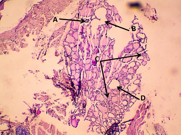
Annals of R.S.C.B., ISSN: 1583-6258, Vol. 25, Issue 1, 2021, Pages. 5944 - 5955
Received 15 December 2020; Accepted 05 January 2021.

sweetener consumption (fig 8).The tissue section showed damage in follicle wall and prominent
vacuoles in the lumen large follicle also showed irregular space in connective tissue between the
follicle, so showed parafollicular cells proliferation (fig 9)these result agreed with [18] which said that
sweeteners have induce histological abnormalities in the spleen and bladder and adverse effects on
the immune system in mice.

   The tissue section showed prominent empty cystic dilation between the follicles also showed blood
congestion between the follicles may be because of some tissue degeneration after treated time, so
noted abnormal cellular proliferation in different location beside the blood hemorrhage in thyroid
gland, also showed tissue degeneration (fig 10).This agreed with the study of [19] which showed the
histological examination of sweeteners-treated group was loss of normal architecture of the thyroid
gland. Morphometrical measurement showed many follicles were small in size and others had
disrupted wall and detached cells in their lumens, there was significant increase in the height of cells
and number of follicles with decrease in width of the cells.

        Table (1) Effect of Saccharin on Thyroid gland (mean± standard deviation).

                          Treatments
                                           Control                   Saccharin                 Sig.
parameters

wall thickness of thyroid follicles       10.69±2.28a                 9.46±1.88b               0.01

Follicular cell nucleidiameter            41.71±8.75a                33.23±6.20b               0.01

large follicle diameter                  137.23±23.70b              151.04±27.74a              0.01

medium follicle diameter                  88.84±8.79a               79.93±17.55b               0.01

small follicle diameter                   45.43±4.57b                49.42±4.93a               0.01

*Different small letters indicate the existence of significant differences between saccharin and control
at the possibility of (0.01).

4. The physiological Results of T3 and T4

A- Control group:

   The hormonal secretions of thyroid gland in control group noted in table (2) that showed the level
of T3 in serum of male control group was (2.12±0.287 nmol/l).Table (2) showed the level of T4 in
serum of male control group was (43.71±3.83 nmol/l).

B- Saccharin Treated Group:

   The physiological result showed the level of T3 in treated group noted in table (2) which have the
saccharin level in male treated group was (1.49±0.257nmol/l). This result appeared the treated group
with saccharin have significant decreased in the level of T3 compared with control group. These

http://annalsofrscb.ro                                                                                5948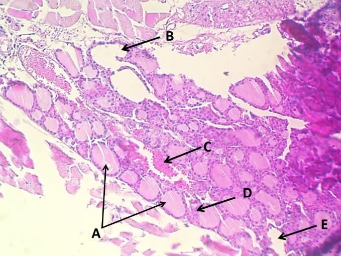
Annals of R.S.C.B., ISSN: 1583-6258, Vol. 25, Issue 1, 2021, Pages. 5944 - 5955
Received 15 December 2020; Accepted 05 January 2021.
significant difference in the level of T3 was may be due loss of food intake that effect on the
metabolism rate which lead to negative feedback mechanism on thyroid gland. This biochemical
change of T3 after treated with saccharin was agreement with [20] which demonstrated that there was
a significant decrease in T3 hormone of rats received sweeteners. This ultimately leads to the failure
of the thyroid gland. Table (2) showed the level of T4 in serum of group after treated with saccharin.
The level of T4 in male group was (34.60±4.88 nmol/l). This result noted the treatedgroup with
saccharin have significant difference compared with control group. This biochemical change of T4
after treated with saccharin was agreement with [20] which demonstrated that there was a significant
decrease in T4 hormone of rats received sweeteners.
Treatments
Control Saccharin Sig.
Parameters
T3 2.12±0.287a 1.49±0.257b 0.01
T4 43.71±3.83a 34.60±4.88b 0.01
Table (2) Effect of Saccharin onT3 and T4 in blood plasma of mice (mean± standard deviation).
*Different small letters indicate the existence of significant differences between saccharin and control
at the possibility of (0.01).
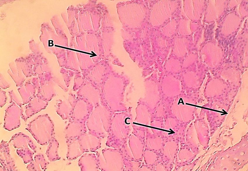
(Figure1): Tissue section of Thyroid in Control group which showed A-large follicle, B-
medium follicle, C-small follicle, D- follicular cells. H&E stain 20X
http://annalsofrscb.ro 5949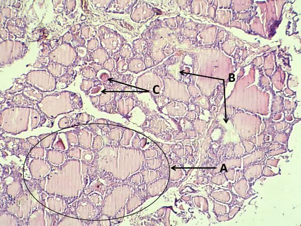
Annals of R.S.C.B., ISSN: 1583-6258, Vol. 25, Issue 1, 2021, Pages. 5944 - 5955
Received 15 December 2020; Accepted 05 January 2021.

         (Figure 2): Tissue section of Thyroid in Control group which showed A- Connective
               tissue capsule, B- Follicular cells, C- Parafollicular cells. H&E stain 10X

          (Figure 3): Tissue section of Thyroid in treated group with saccharin which showed
         A-large follicle, B- Blood hemorrhage, C-abnormal cellular proliferation. H&E stain
                                                   20X

http://annalsofrscb.ro                                                                   5950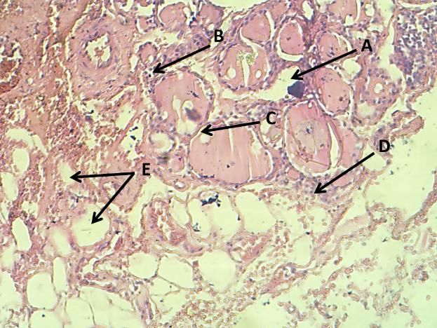
Annals of R.S.C.B., ISSN: 1583-6258, Vol. 25, Issue 1, 2021, Pages. 5944 - 5955
Received 15 December 2020; Accepted 05 January 2021.
(Figure.4): Tissue section of Thyroid in treated group with Saccharin which showed A-
lobule, B- necrotic lesion in connective tissue between lobules, C-blood congestion. H&E
stain (20) X
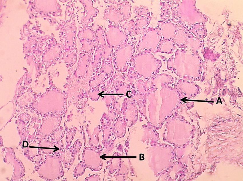
(Fig 5): Tissue section of male Thyroid in treated group with Saccharin which showed A-
acute necrosis, B- inflammatory cells, C-prominent damage in wall, D- parafollicular cells,
E- disappeared colloid. H&E stain (20)X
http://annalsofrscb.ro 5951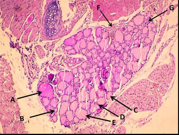
Annals of R.S.C.B., ISSN: 1583-6258, Vol. 25, Issue 1, 2021, Pages. 5944 - 5955
Received 15 December 2020; Accepted 05 January 2021.
(Fig 6): Tissue section of male Thyroid in treated group with Saccharin which showed A-
thick fluid colloid, B- small follicle, C- medium follicle, D- follicular epithelia, E- Large
follicle, F- connective tissue capsule, G- cellular proliferation. H&E stain 10X
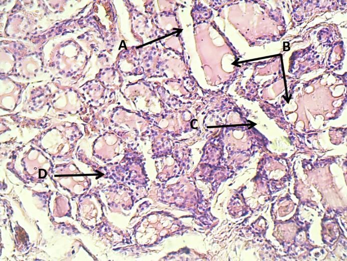
(Fig 7): Tissue section of male Thyroid in treated group with Saccharin which showed A-
inflammatory cells, B- prominent damage in follicle wall, C- cellular proliferation, D- cystic
dilation. H&E stain 10X
http://annalsofrscb.ro 5952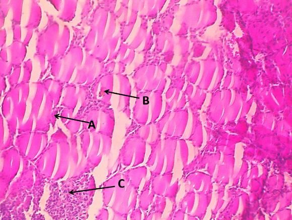
Annals of R.S.C.B., ISSN: 1583-6258, Vol. 25, Issue 1, 2021, Pages. 5944 - 5955
Received 15 December 2020; Accepted 05 January 2021.

        (Fig.8): Tissue section of male Thyroid in treated group with Saccharin which showed
        A-lobule, B- abnormal cellular proliferation, C- inflammatory cells aggregation. H&E
                                              stain (20)X

   (Fig 9): Tissue section of male Thyroid in treated group with Saccharin which showed A-
    follicle wall damage, B- vacuoles, C- irregular space, D- parafollicular cells proliferation.
                                         H&E stain (20)X

http://annalsofrscb.ro                                                                        5953
Annals of R.S.C.B., ISSN: 1583-6258, Vol. 25, Issue 1, 2021, Pages. 5944 - 5955
Received 15 December 2020; Accepted 05 January 2021.
(Fig 10): Tissue section of male Thyroid in treated group with Saccharin which showed A-
Large follicle, B- Empty cystic dilation, C- blood congestion, D- abnormal aggregation of cells,
E- tissue degeneration. H&E stain 10X
References:
1. Alsoufi, M. A., Aziz, R. A., & Hussein, Z. G. (2017). Effect of some artificial sweeteners
consumption in biochemical parameters of rats. Microbiology and Biotechnology, 5(3), 1095-
1099.
2. Lohner, S., Toews, I., &Meerpohl, J. J. (2017). Health outcomes of non-nutritive sweeteners:
analysis of the research landscape. Nutrition journal, 16(1), 55.
3. Saad, A.,Khan, F. A., Hayee, A., & Nazir, M. S. (2014). A Review on potential toxicity of
artificial sweetnersvs safety of stevia: A natural bio-sweetner. Journal of Biology, Agriculture
and Healthcare, 4(15), 1-12.
4. Piovan, S., Pavanello, A., Peixoto, G. M. L., Matiusso, C. C. I., de Moraes, A. M. P., Martins,
I. P., ... &Dacome, A. S. (2018). Stevia nonsweetener fraction displays an Insulinotropic
effect involving neurotransmission in pancreatic islets. International journal of
endocrinology, 2018.
5. Pepino, M. Y. (2015). Metabolic effects of non-nutritive sweeteners. Physiology &
behavior, 152, 450-455.
6. Saraswathy, S., Toora, B D., Pillai, M., Mishra, S. (2018). Effect of Artificial Sweeteners on
the Blood Glucose Concentration. Journal of Medical Academics. 1. 81-85. 10.5005/jp-
journals-10070-0017.
7. Sachmechi, I., Khalid, A., Awan, S. I., Malik, Z. R., &Sharifzadeh, M. (2018). Autoimmune
thyroiditis with hypothyroidism induced by sugar substitutes. Cureus, 10(9).
http://annalsofrscb.ro 5954Annals of R.S.C.B., ISSN: 1583-6258, Vol. 25, Issue 1, 2021, Pages. 5944 - 5955
Received 15 December 2020; Accepted 05 January 2021.
8. Pałkowska-Goździk, E., Bigos, A., &Rosołowska-Huszcz, D. (2018). Type of sweet flavour
carrier affects thyroid axis activity in male rats. European journal of nutrition, 57(2), 773-
782.
9. Singh, N., Lubana, S. S., Arora, S., &Sachmechi, I. (2020). A Study of Artificial Sweeteners
and Thyroid Cancer Risk. Journal of Clinical Medicine Research, 12(8), 492.
10. Sudan, P., Kaur, R., Sharma, S., & Jain, U. K. (2016). A critical review on natural and
artificial sweeteners. The Pharmaceutical and Chemical Journal, 3(1), 21-29.
11. Nilsson, M., &Fagman, H. (2017). Development of the thyroid gland. Development, 144(12),
2123-2140.
12. DesChâtelets, M. (2015). Thyroid Hormones and their Thermogenic Properties. Journal of
Restorative Medicine, 4(1), 93-99.
13. Luna LG. Manual of histologic staining methods of the Armed Forces Institute of Pathology.
1968.
14. Aboodshaher, S., &Jassim, B. A. (2020). Histological Study of Hyperthyroidism (Goiter) in
Women in Al-Muthanna Province. Indian Journal of Public Health Research &
Development, 11(4).
15. Rudenga, K. J., & Small, D. M. (2012). Amygdala response to sucrose consumption is
inversely related to artificial sweetener use. Appetite, 58(2), 504-507.
16. Ferreira, E., Silva, A. E., Serakides, R., Gomes, A. E. S., &Cassali, G. D. (2007). Model of
induction of thyroid dysfunctions in adult female mice. ArquivoBrasileiro de
MedicinaVeterinária e Zootecnia, 59(5), 1245-1249.
17. Rother, K. I., Conway, E. M., &Sylvetsky, A. C. (2018). How non-nutritive sweeteners
influence hormones and health. Trends in Endocrinology & Metabolism, 29(7), 455-467.
18. Remil, A., &Benali, M. (2020). Immunomodulatory Effect of Sodium Saccharin and
Potassium Acesulfame in BALB/C Mice in Short-Term Trials. Journal of Drug Delivery and
Therapeutics, 10(6), 1-10.
19. Amen, P. J. M., Mahmood, Z. M., &Maaruf, N. A. (2017). Effect of Aspartame on the Rat’s
Thyroid Gland: A histological and Morphometrical Study. Diyala Journal of Medicine, 12(1),
63-69.
20. Helal, E. G., Abdelaziz, M. A., Taha, N. M., & El-Gama, M. S. (2019). The influence of
acesulfame-k and aspartame on some physiological parameters in male albino rats. The
Egyptian Journal of Hospital Medicine, 75(1), 1976-1981.
http://annalsofrscb.ro 5955You can also read