Skin morphological changes in growth hormone deficiency and acromegaly
←
→
Page content transcription
If your browser does not render page correctly, please read the page content below
European Journal of Endocrinology (2001) 145 147±153 ISSN 0804-4643
CLINICAL STUDY
Skin morphological changes in growth hormone de®ciency and
acromegaly
Martin Lange, Jesper Thulesen1, Ulla Feldt-Rasmussen, Niels E Skakkebñk2, Nina Vahl3, Jens O Jùrgensen3,
Jens S Christiansen3, Steen S Poulsen1, Simone B Sneppen and Anders Juul2
Department of Endocrinology, National University Hospital (Rigshospitalet), 1Institute of Medical Anatomy, The Panum Institute, 2Department of
Growth and Reproduction, National University Hospital (Rigshospitalet), Copenhagen and 3Department of Endocrinology, Aarhus
Kommunehospital, Denmark
(Correspondence should be addressed to A Juul, Department of Growth and Reproduction, Rigshospitalet, Blegdamsvej 9, 2100 Copenhagen, Denmark;
Fax 35456054; Email: ajuul@dadlnet.dk)
Abstract
Objective: To evaluate the histomorphology of skin and its appendages, especially eccrine sweat glands,
in patients with GH disorders, because reduced sweating ability in patients with growth hormone
de®ciency (GHD) is associated with increased risk of hyperthermia under stressed conditions.
Design and methods: A skin biopsy was obtained from 17 patients with GHD treated with GH, ®ve
patients with untreated GHD, 10 patients with active acromegaly and 13 healthy controls.
Results: The sweat secretion rate (SSR) was signi®cantly decreased in both the untreated (median
41 mg/30 min, range 9±79 mg/30 min) and the GH-treated (median 98 mg/30 min, range 28±
147 mg/30 min) patients with GHD compared with that in controls (median 119 mg/30 min, range
90±189 mg/30 min; P 0:001 and 0.01 respectively). Epidermal thickness was signi®cantly
decreased in both untreated (median 39 mm, range 28±55 mm) and GH-treated patients with GHD
(median 53 mm, range 37±100 mm), compared with that in controls (median 66 mm, range 40±
111 mm; P , 0:02: A statistically non-signi®cant tendency towards thinner epidermis (median
59 mm, range 33±83 mm) was recorded in acromegalic patients P 0:08 compared with controls.
There was no signi®cant difference in the area of the sebaceous glands in the biopsies between the
three groups and the controls. The area of eccrine sweat gland glomeruli was signi®cantly decreased
in the untreated patients with GHD (median 16407 mm2, range 12758±43976 mm2) compared with
that in controls (median 29446 mm2, range 13511±128661 mm2; P 0:03; but there was no
signi®cant difference between the GH-treated patients with GHD and controls.
Conclusions: We conclude that GH, either directly or via IGF-I, may have both a structural and a
functional effect on human skin and its appendages, and that patients with GHD have
histomorphological changes in skin compared with controls. Importantly, these changes are not
fully reversed despite long-term and adequate GH treatment in patients with childhood onset GHD.
European Journal of Endocrinology 145 147±153
Introduction those with AO-GHD (4). The impaired sweating ability
is also found in patients with GH insensitivity (Laron-
The ability to perspire is of minor importance in the type dwar®sm) (5). Furthermore, most clinicians agree
maintenance of body temperature during rest. How- that patients with GHD have thin and dry skin. The
ever, in certain conditions such as exercise, infections decreased sweating ability in GHD imposes a risk of
and exposure to high environmental temperature, hyperthermia (6, 7). In contrast, patients with active
sweating becomes imperative for thermoregulation. acromegaly have thick and greasy skin and the
Sweating ability seems to be in¯uenced by sex, age prevalence of excessive sweating in such patients has
(1) and degree of physical training (2). Patients with been reported to be 65±88% (8). Furthermore, the
childhood onset (CO) growth hormone de®ciency sweat secretion rate (SSR) has been found to be
(GHD) have been shown to have impaired sweating abnormal in both active and inactive acromegaly (4).
ability (1, 3) as have men, but not women, with adult Previous studies of skin morphology in GHD have
onset (AO) GHD (4). The sweating ability has been shown decreased skin thickness with decreased collagen
shown to improve, but not normalise, in patients with content (9), whereas the opposite ®ndings are present in
CO-GHD when treated with growth hormone (GH) (3). acromegaly (8). Little, if any, attention has been paid to
A similar improvement has not been demonstrated in changes in the eccrine or sebaceous glands. It has been
q 2001 Society of the European Journal of Endocrinology Online version via http://www.eje.org
Downloaded from Bioscientifica.com at 01/11/2021 10:11:26PM
via free access148 M Lange and others EUROPEAN JOURNAL OF ENDOCRINOLOGY (2001) 145
suggested that the effect on the skin and its appendages is Japan). The area of the total biopsy, and of the eccrine
in part a direct effect of GH (9). This suggestion is sweat gland glomeruli (de®ned as adjacent eccrine
supported by identi®cation of the GH receptor in skin sweat gland tubuli) and the total area of the sebaceous
tissue, including the eccrine sweat glands (10, 11). glands were measured by means of NIH Image 1.60
The aim of this study was to determine skin (public domain image processing and analysis program
morphological changes present in GHD and acro- provided by National Institute of Health, Bethesda,
megaly, with special emphasis on identifying factors Maryland, USA) as described previously (12). Eccrine
that might explain the difference in sweating capacity sweat glands and sebaceous glands were not present in
between the two entities. all biopsies. When they were present, one to three
sebaceous glands were identi®ed in each specimen and
the sum of their areas was taken to comprise the total
Participants and methods area. After calibration, the border of the biopsy and the
Under local anaesthesia (2% lidocaine), a skin biopsy boundaries between gland tissue and surrounding
from the posterior of the right shoulder was obtained tissues were outlined by the computer cursor, allowing
from a total of 45 individuals. Five were male patients calculation of the area. The mean epidermal thickness
with CO-GHD (median age 31 years). One of them had (calculated by 10 randomly selected measurements in
not received GH treatment at all and four had been each biopsy) was determined by NIH image 1.60.
without GH treatment for a period of 7±23 years. All ®ve Serum concentrations of insulin-like growth factor I
patients had one or more additional pituitary de®cien- (IGF-I) and insulin-like growth factor binding protein 3
cies, and were receiving adequate substitution therapy (IGFBP-3) were determined in all participants, by
for the respective de®ciencies. A further 17 (12 men, ®ve radioimmunoassays.
women) were patients with GH-treated CO-GHD (median A pharmacological sweat test was performed using
age 20 years). Of these, 13 had one or more additional previously published methods: two ®lter papers (What-
pituitary de®ciencies, and were receiving adequate mann Ashless Round®lters, diameter: 2.5 cm) soaked
substitution therapy for the respective de®ciencies. in 0.2% pilocarpine were positioned on the ¯exor side
These patients had been on discontinuous GH substitu- of the distal forearm underneath two quadrangular
tion since diagnosis, for a median duration of 10 years negative electrodes (size: 3 3 cm: Iontophoresis was
(range 4±21 years). Ten of the 45 patients (six men, four performed, using a current of 2 mA for 5 min. After
women; median age 39.5 years) had active acromegaly; 5 min, the area of iontophoresis was rinsed with
®ve of them had one or more additional pituitary deionised water and 61% ethanol and thoroughly
de®ciencies, and were receiving adequate substitution dried. Sweat was collected during a 30-min period
therapy for the respective de®ciencies. The remaining 13 using three ®lter papers sealed by a plastic roundel and
participants (nine men, four women) were healthy tape to prevent evaporation. Sweat mass was measured
controls (median age 23 years). by weighing the ®lter paper (Mettler scales, precision:
The characteristics of the study participants are ^0.1 mg) before and after the sweat collection (1).
summarized in Table 1. The local Ethics Committee approved the study, and
The biopsy specimens (4 mm punch biopsies) were signed informed consent was obtained from all patients.
®xed in Lillies ®xative, dehydrated and embedded in
paraf®n. They were then cut into 4 mm sections using a
microtome and stained with haematoxylin and eosin
(H&E). The specimens were examined using a Zeiss
Statistical methods
Axiophot microscope (Carl Zeiss, Oberkocken, Germany) The results were analysed by Mann±Whitney's U-test.
connected to a high-resolution camera (Hamamatsu Correlations analyses were performed using Spear-
C2400. Hamamatsu Photonics, Hamamatsu City, man's test.
Table 1 Patient characteristics expressed as medians and ranges. Additional hormone de®ciencies are stated. Patients with additional
hormone de®ciencies were in adequate substitution therapy for the respective de®ciency. Values are absolute numbers or median
(range).
Additional pituitary hormone de®ciencies
Sex
2
Group n (M/F) Age (years) Height (cm) Weight (kg) BMI (kg/m ) ACTH TSH ADH FSH/LH
GHD untreated 5 5/0 31 (25±64) 167 (157±182) 75 (37±102) 28 (15±31) 4 5 0 5
GHD treated 17 12/5 20 (16±26) 173 (153±183) 71 (58±106) 25 (19±30) 6 13 4 9
Acromegaly 10 6/4 39.5 (26±64) 174 (165±190) 78 (64±100) 26 (21±31) 4 1 0 3
Controls 13 9/4 23 (20±28) 184 (160±190) 76 (56±93) 23 (21±26) 0 0 0 0
BMI, body mass index; ACTH, adrenocorticotrophic hormone; TSH, thyroid-stimulating hormone; ADH, antidiuretic hormone; FSH, follicle-stimulating
hormone; LH, luteinizing hormone.
www.eje.org
Downloaded from Bioscientifica.com at 01/11/2021 10:11:26PM
via free accessEUROPEAN JOURNAL OF ENDOCRINOLOGY (2001) 145 Skin changes in GH de®ciency and acromegaly 149
Results range 37±100 mm) compared with that in controls
(median 66.0 mm, range 40.0±111.0 mm) P 0:005
Skin biopsies and 0.02 respectively; Figs. 1D±F and 2B). There was a
The biopsies were very inhomogeneous between the tendency, though not signi®cant, towards reduced
groups. Those from the patients with GHD, in particular thickness in patients with acromegaly (median
the untreated patients, were characterized by a greater 59 mm, range 33±83 mm) compared with controls
content of fat compared with controls. There was a P 0:08: The epidermal thickness was, however,
tendency, though not signi®cant, towards a greater area increased in the acromegalic patients compared with
of sebaceous glands in the acromegalic patients that in untreated patients with GHD P 0:01:
compared with the other groups. In some of the biopsies
in the acromegalic patients and in some controls, the Pilocarpine iontophoresis sweat test
dermis was so thick that the subcutis was not included
in the biopsies and could therefore not be measured Pilocarpine iontophoresis tests showed a lower SSR
accurately. The dermis in the untreated patients with both in patients with untreated GHD (median 41 mg/
GHD, however, appeared thinner compared with that in 30 min, range 9±79 mg/30 min) and in those with
the other groups. The biopsies from the acromegalic GH-treated GHD (median 98 mg/30 min, range 28±
patients were furthermore characterised by the presence 147 mg/30 min) compared with the controls (median
of a dense and thick, almost membrane-like, border of 119 mg/30 min, range 90±189 mg/30 min; P
connective tissue under the epidermis that was not as 0:001 and 0.01 respectively). SSR was, however,
pronounced in the other groups (Fig. 1A±C). signi®cantly lower in patients with untreated GHD
than in patients with GH-treated GHD P 0:03: SSR
was greater in patients with acromegaly (median
Eccrine sweat glands 168 mg/30 min, range 61±260 mg/30 min) than in
the controls, but the difference did not reach statistical
Six eccrine sweat gland glomeruli were present in ®ve signi®cance P 0:26: The SSR was, however, sig-
biopsies of the untreated patients with GHD, 14 were ni®cantly greater in the acromegalic patients than in
present in 17 biopsies of the GH-treated patients with the untreated patients with GHD P 0:01 (Fig. 2C).
GHD, 13 were present in 10 biopsies of the acromegalic
patients and 18 glomeruli were present in 13 biopsies
of the controls. The area of a glomerulus was IGF-I
signi®cantly smaller in the untreated patients with IGF-I concentrations were decreased in untreated
GHD (median 16407 mm2, range 12758±43976 mm2) patients with GHD (median 43 ng/ml, range 20±
compared with that in controls (median 29446 mm2, 60 ng/ml) compared with GH-treated patients with
range 13511±128661 mm2; P 0:03: There was no GHD (median 302 ng/ml, range 89±886 ng/ml) and the
signi®cant difference between the untreated patients controls (median 244 ng/ml, range 169±636 ng/ml;
with GHD and the GH-treated patients with GHD P 0:0009 and 0.001 respectively). IGF-I concentra-
(median 33936 mm2, range 3315±105497 mm2), and tions were increased in acromegaly (median 745 ng/ml,
no signi®cant difference between the GH-treated range 523±1037 ng/ml) compared with those in con-
patients with GHD or the acromegalic patients (median trols P 0:0002: Patients with GH-treated GHD had
40421 mm2, range 9978±167971 mm2) compared similar IGF-I values as the controls (Fig. 2D).
with the controls. No difference was found between
the untreated patients with GHD and the acromegalic
patients (Figs. 1G±H and 2A). IGFBP-3
IGFBP-3 concentrations were signi®cantly lower in
Sebaceous glands untreated patients with GHD (median 1300 ng/ml,
range 562±1593 ng/ml) compared with GH-treated
Sebaceous glands were present in the biopsies of two of patients with GHD (median 3983 ng/ml, range 1638±
the ®ve untreated patients with GHD, seven of the 17 7078 ng/ml) and the controls (median 3376 ng/ml,
GH-treated patients with GHD, six of the 10 acro- range 2593±4274 ng/ml; P 0:0009 and 0.001
megalic patients and 11 of the 13 controls. There was respectively), and signi®cantly greater in the acrome-
no difference in the area of sebaceous glands in the galic patients (median 6021 ng/ml, range 5194±
three groups of patients compared with the controls. 7133 ng/ml) than in the controls P 0:00004:
There was no signi®cant difference between the
controls and the GH-treated patients with GHD.
Epidermal thickness No correlation was found between serum IGF-I and
Epidermal thickness was signi®cantly reduced in both the area of sweat gland glomeruli, epidermal thickness
patients with untreated GHD (median 39.0 mm, range and SSR respectively in any of the groups alone or in all
28±55 mm) and GH-treated GHD (median 53 mm, the groups combined.
www.eje.org
Downloaded from Bioscientifica.com at 01/11/2021 10:11:26PM
via free access150 M Lange and others EUROPEAN JOURNAL OF ENDOCRINOLOGY (2001) 145
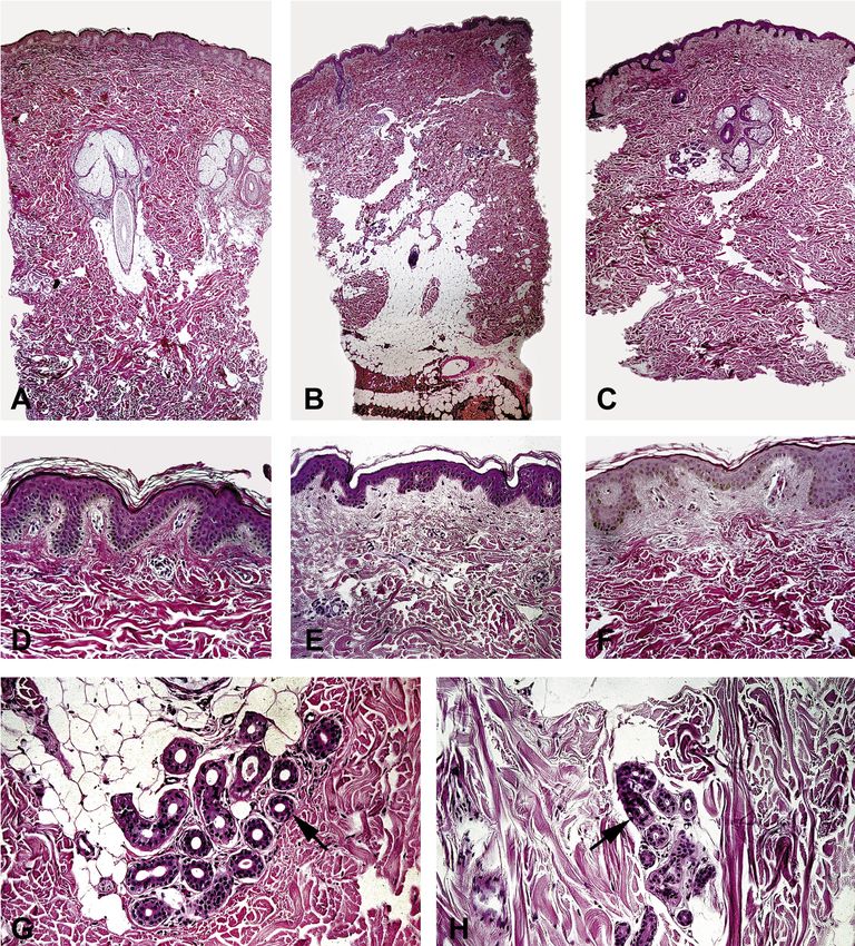
Figure 1 General views of skin biopsies from: (A) control, (B) patient with untreated GHD and (C) patient with acromegaly (original
magni®cation 20 for each). Close-ups of epidermis from: (D) control, (E) patient with untreated GHD and (F) patient with acromegaly
(original magni®cation 50 for each). Close-ups of eccrine sweat glands from: (G) control and (H) patient with untreated GHD (original
magni®cation 20 for each).
Discussion (6, 7, 13). The impact of the decreased sweating ability
is also demonstrated by the fact that some patients with
We evaluated skin histomorphology in 32 patients with GHD are poikilothermic (13). Conversely, acromegaly is
GH disorders as compared with that in 13 control associated with excess sweating and increased SSR (4,
individuals and found signi®cant changes in epidermal 8). The changes in GHD may be a result of atrophy of
thickness and eccrine sweat glands. This is in line with the eccrine sweat glands because of lack of stimulation
our hypothesis of the human skin being a GH target of either GH or IGF-I, or both. Alternatively, it could be
organ. a reduction in sweat gland function.
This hypothesis originates from the ®nding that in In this study, the ®nding of decreased SSR in the
situations of stress, impaired sweating capacity, as seen untreated GHD group was not surprising (3). The GH-
in GHD (3, 4), is associated with a risk of hyperthermia treated patients with GHD had increased SSR compared
www.eje.org
Downloaded from Bioscientifica.com at 01/11/2021 10:11:26PM
via free accessEUROPEAN JOURNAL OF ENDOCRINOLOGY (2001) 145 Skin changes in GH de®ciency and acromegaly 151
Figure 2 (A) Area of sweat gland glomeruli, (B) epidermal thickness determined as mean of 10 random measurements, (C) results of
pilocarpine sweat test expressed as sweat secretion rate and (D) serum IGF-I concentrations, in patients with untreated GHD, GH-treated
GHD, acromegalics and controls. *P , 0:03 compared with controls; #P 0:03 compared with GH-treated; ²P 0:01 compared with
acromegalic patients. Horizontal bars indicate medians.
with the untreated patients, suggesting some effect of GH age was only 10 years between the two groups, both
treatment, but they still showed a decreased SSR comprising young individuals, and the difference in
compared with the controls. This may suggest that, glomerulus area is therefore unlikely to have been due to
despite a seemingly adequate treatment (IGF-I and differences in age.
IGFBP-3 concentrations within or above normal limits), All the patients with GHD in the present study had
the treatment effect on SSR was not suf®cient. The CO-GHD. The impact of GHD in these patients may not
decreased SSR in the untreated GHD group corresponded be the same in patients with AO-GHD. In a previous
to the ®nding of a decreased area of the glomeruli of the study involving patients with AO-GHD of mean
sweat glands compared with the controls. We found no duration 8.6 years (range 2±27 years) since diagnosis,
differences in the area of the sweat gland glomeruli we found that SSR was similar to controls in female
between the GH-treated patients and the controls, patients, but lower compared with controls in male
despite differences in SSR between the two groups. One patients. Eighteen months of GH replacement therapy
explanation for these ®ndings may be that GH exerts did not effect SSR in either group (4). In the present
both a structural and a functional effect on the sweat study, only male patients were present in the untreated
glands, and that not all effects are fully restored, despite GHD group. In accordance with the ®ndings of the
long-term GH replacement in patients with GHD. Sweat previous study, reduced SSR was found in these patients
gland size is known to decrease with age (14). In the after 18 months of GH treatment (4). The GH-treated
present study, signi®cant differences were found in the patients with CO-GHD had, however, a signi®cant effect
area of sweat gland glomeruli in untreated patients with of treatment compared with the untreated patients,
GHD as compared with controls, with a difference in even though the SSR did not reach the level attained by
median size of almost 100%. The median difference in the controls. We suspect that this apparent discrepancy
www.eje.org
Downloaded from Bioscientifica.com at 01/11/2021 10:11:26PM
via free access152 M Lange and others EUROPEAN JOURNAL OF ENDOCRINOLOGY (2001) 145
between CO-GHD and AO-GHD is in part related to not signi®cant, may seem surprising, considering that
duration of treatment (10 years, range 4±21 years, in clinically these patients have very thick skin. This
the present study compared with 18 months in the ®nding has, however, also been reported by other
previous study). As in the AO-GHD study, the impact of authors using H&E staining (18). Furthermore, this
other pituitary de®ciencies on SSR and sweat gland group showed an abnormal glycosaminoglycan compo-
morphology cannot be ruled out. All patients de®cient sition in the dermis of acromegalic patients, by means
in other pituitary hormones had, however, been of tissue digestion, and increased glycosaminoglycan
receiving stable substitution therapy since diagnosis, deposition as determined by staining with colloidal iron
with comparable replacements in all groups. (18). The ®nding of increased glycosaminoglycan
The ®nding that patients with GHD (both untreated deposition may correspond to our ®nding of the
and GH-treated) also have a thin epidermis compared presence of a dense border of connective tissue under
with healthy controls supports the notion of a the epidermis as determined by H&E staining. The
permanent consequence of GHD, despite treatment tendency towards thin epidermis most probably origi-
and despite a small but non-signi®cant improvement in nates from increased cell turnover as determined by
those who are GH-treated compared with those who tritiated amino acid incorporation (19), and the clinical
are untreated (Fig. 2B). A slight, non-signi®cant, ®nding of thick skin therefore re¯ects increased dermal
increase in skin thickness as determined by skinfold thickness. Holt & Marks (19) further showed, by H&E
thickness has previously been demonstrated in normal staining, an increased size of the epidermal cells but a
elderly men treated with GH (15). We cannot exclude decreased number of cells compared with the controls.
that other pituitary de®ciencies may affect epidermal In our material the dermis in the patients with
thickness, as it is well known that treatment with high acromegaly was so thick that it extended throughout
doses of glucocorticoids may decrease skin thickness. the depth of the biopsy sample, rendering exact
This is, however, mostly caused by reduced collagen measurements impossible. In the present study, the
formation and is not in¯uencing the epidermis (16). age difference between the acromegalic patients and
Furthermore, the in¯uence of glucocorticoids on skin the controls was rather large, and the ®nding of a
thickness is most common during regular glucocorti- tendency towards thinner epidermis in the acromegalic
coid treatment and not in substitution therapy. patients may therefore simply have been a re¯ection of
Hypothyroidism and hypogonadism are also known to the age difference (17).
cause reduced skin thickness. As previously stated, The presence of the GH receptor in human skin and its
however, all hormone-de®cient patients were receiving appendages, including eccrine sweat glands, suggests a
adequate substitution therapy. Epidermal thickness has direct effect of GH (10, 11), but the exact mechanism by
been shown to decrease with age at a magnitude of which GH exerts its effect is unknown. However, studies
approximately 6±7% per decade (17). The epidermal using PCR have shown an abundance of IGF-I receptor
thickness found in the controls in the present study is mRNA in human skin biopsies (20), which could suggest
comparable to that previously reported in healthy an IGF-I-mediated action on skin.
individuals of similar age (17). In the present study, the We conclude that GH, either directly or via IGF-I may
GH-treated patients were comparable in age with the have both a structural and a functional effect on
controls. The untreated patients with GHD were older human skin and its appendages, and that patients with
compared with the controls, but the difference in GHD have histomorphological changes in skin com-
epidermal thickness was of a magnitude that could not pared with controls. Importantly, these changes are not
be explained by differences in age alone. fully reversed despite long-term and adequate GH
Increased sweating and greasy skin are well-known treatment in patients with CO-GHD.
features of acromegaly (8). In the present study
population, we found no signi®cant change in SSR,
size of sweat glands or of size of sebaceous glands in
Acknowledgements
acromegalic patients compared with the controls. The excellent technical assistance of Majbrit Kvist,
These negative ®ndings are in contrast with our Grazyna Hahn and technicians from Department of
previous ®ndings of abnormal SSR in both treated Growth and Reproduction is greatly appreciated.
and untreated acromegaly (4). This discrepancy may be
explained by the small number of patients in the References
present study and the small number of sweat glands
1 Main K, Nilsson KO & Skakkebñk NE. In¯uence of sex and
present in the histological specimens in the biopsies of growth hormone de®ciency on sweating. Scandinavian Journal of
the acromegaly group n 7: In addition, increased Clinical and Laboratory Investigation 1991 51 475±480.
sweating was found clinically only in 65±88% of 2 Buono MJ & SjoÈholm NT. Effect of physical training on peripheral
untreated acromegalic patients (8), giving a wide range sweat production. Journal of Applied Physiology: Respiratory,
Environmental and Exercise Physiology 1988 65 811±814.
of SSR in this group of patients. 3 Pedersen SA, Welling K, Michaelsen KF, Jùrgensen JOL,
The ®nding of relatively thin epidermis in the Christiansen JS & Skakkebñk NE. Reduced sweating in adults
acromegalic patients compared with the controls, albeit with growth hormone de®ciency. Lancet 1989 2 681±682.
www.eje.org
Downloaded from Bioscientifica.com at 01/11/2021 10:11:26PM
via free accessEUROPEAN JOURNAL OF ENDOCRINOLOGY (2001) 145 Skin changes in GH de®ciency and acromegaly 153
4 Sneppen SB, Main K, Juul A, Pedersen LM, Kristensen Lé, 13 Juul A & Skakkebñk NE. Growth hormone de®ciency and
Skakkebaek NE et al. Sweat secretion rates in growth hormone hyperthermia. Lancet 1991 338 887.
disorders. Clinical Endocrinology 2000 53 601±608. 14 Fenske NA & Lober CW. Structural and functional changes of
5 Main K, Kastrup KW & Skakkebñk NE. Reduced sweating in normal aging skin. Journal of the American Academy of Derma-
Laron's dwar®sm. Archives of Disease in Chilhood 1990 65 1380± tology 1986 15 571±585.
1380. 15 Rudman D, Feller AG, Nagraj HS, Gergans GA, Lalitha PY,
6 Juul A, Hjortskov N, Jepsen LT, Nielsen B, Halkjaer KJ, Vahl N Goldberg AF et al. Effects of human growth hormone in men over
et al. Growth hormone de®ciency and hyperthermia during 60 years old. New England Journal of Medicine 1990 323 1±6.
exercise: a controlled study of sixteen GH-de®cient patients. 16 Oikarinen A & Autio P. New aspects of the mechanism of
Journal of Clinical Endocrinology and Metabolism 1995 80 3335± corticosteroid-induced dermal atrophy. Clinical and Experimental
3340. Dermatology 1991 16 416±419.
7 Juul A, Behrenscheer A, Tims T, Nielsen B, Halkjñr-Kristensen J 17 Branchet MC, Boisnic S, Frances C & Robert AM. Skin thickness
& Skakkebñk NE. Impaired thermoregulation in adults with changes in normal aging skin. Gerontology 1990 36 28±35.
growth hormone de®ciency during heat exposure and exercise. 18 Matsuoka LY, Wortsman J, Kupchella CE, Eng A & Dietrich JE.
Clinical Endocrinology 1993 38 237±244. Histochemical characterization of the cutaneous involvement
8 Nabarro JDN. Acromegaly. Clinical Endocrinology 1987 26 481± of acromegaly. Archives of Internal Medicine 1982 142 1820±
512. 1823.
9 Abramovici A, Josefsberg Z, Mimoni M, Liban E & Laron Z. 19 Holt PJ & Marks R. Epidermal architecture, growth, and
Histopathological features of the skin in hypopituitarism and metabolism in acromegaly. British Medical Journal 1976 1
Laron-type dwar®sm. Israel Journal of Medical Sciences 1983 19 496±497.
515±519. 20 Tavakkol A, Elder JT, Grif®ths CE, Cooper KD, Talwar H, Fisher GJ
10 Oakes SR, Haynes KM, Waters MJ, Herington AC & Werther GA. et al. Expression of growth hormone receptor, insulin-like growth
Demonstration and localisation of growth hormone receptor in factor 1 (IGF-I) and IGF-I receptor mRNA and proteins in human
human skin and skin ®broblasts. Journal of Clinical Endocrinology skin. Journal of Investigative Dermatology 1992 99 343±349.
and Metabolism 1992 75 1368±1375.
11 Lobie PE, Breipohl W, Lincoln DT & GarcõÂa-AragoÂn J. Localization
of the growth hormone receptor/binding protein in skin. Journal
of Endocrinology 1990 126 467±472.
12 Thulesen J, Hartmann B, Nielsen C, Holst JJ & Poulsen SS.
Diabetic intestinal growth adaptation and glucagon-like peptide Received 12 December 2000
2 in the rat: effects of dietary ®bre. Gut 1999 45 672±678. Accepted 18 April 2001
www.eje.org
Downloaded from Bioscientifica.com at 01/11/2021 10:11:26PM
via free accessYou can also read