Case Report Influenza A Viral Infection with Septic Shock in Pregnancy - Semantic Scholar
←
→
Page content transcription
If your browser does not render page correctly, please read the page content below
Hindawi
Case Reports in Obstetrics and Gynecology
Volume 2019, Article ID 2470352, 4 pages
https://doi.org/10.1155/2019/2470352

Case Report
Influenza A Viral Infection with Septic Shock in Pregnancy

          Soe Lwin ,1 Myat San Yi ,1 May Shi Leong,2 Haris Suharjono,2 and Tin Moe Nwe3
          1
            Department of Obstetrics & Gynecology, Faculty of Medicine and Health Sciences, UNIMAS, Malaysia
          2
            Department of Obstetrics & Gynecology, Sarawak General Hospital, Malaysia
          3
            Department of Basic Health Sciences, Faculty of Medicine and Health Sciences, UNIMAS, Malaysia

          Correspondence should be addressed to Soe Lwin; lsoe@unimas.my

          Received 4 October 2018; Revised 31 March 2019; Accepted 10 April 2019; Published 21 April 2019

          Academic Editor: Erich Cosmi

          Copyright © 2019 Soe Lwin et al. This is an open access article distributed under the Creative Commons Attribution License, which
          permits unrestricted use, distribution, and reproduction in any medium, provided the original work is properly cited.

          The influenza virus is RNA virus and is classified into four subtypes, influenza A, influenza B, influenza C, and influenza D. One of
          the subtypes of influenza A, the H1N1 strain, also known as swine flu, is especially of high risk for development of complications
          in pregnant women. The influenza A virus infection is difficult to diagnose clinically because its presenting symptoms are similar
          to those of the common cold but are more severe, last longer, and can be potentially life-threatening. This case also presented with
          common cold symptoms but her condition worsened later. Fortunately, obstetric health providers were vigilant enough to address
          the developing infection and its related complications. It was the cooperative effort of multidisciplinary team care which resulted
          in a favourable outcome in both mother and baby.

1. Introduction                                                          development of complications of H1N1 influenza A. This
                                                                         increased risk is related to several physiological changes
Influenza virus is a single-stranded, enveloped RNA virus                during pregnancy including alterations in the cardiovascular,
from Orthomyxoviridae family. Its incubation period is 2-5               respiratory, and immune systems. The serious illness and
days and the transmission is mainly air-borne, i.e., droplet             hospitalization rates of women with influenza during preg-
inhalation or direct transmission or contact through hands               nancy have a 4- to 5-fold increase compared to nonpregnant
and fomites. It is classified into four distinct generations such        women [2, 5–8]. Qi et al. (2014) stated that pregnant women
as influenza A, B, C, and D depending on antibody responses              are particularly susceptible to severe complications from
to glycoproteins, hemagglutinin (HA), and neuraminidase                  influenza and have a greater mortality risk.
(NA) on the surface of the viruses [1].                                      Influenza A infection is diagnosed by obtaining an upper
    One of the subtypes of influenza A, influenza A (H1N1)               respiratory specimen (nasopharyngeal swab, nasal aspirate,
strain called swine flu was first identified in April 2009               or a combined nasopharyngeal swab with oropharyngeal
and the outbreak has since reached pandemic status at that               swab or throat swab) to test for novel influenza A (H1N1)
time [2, 3]. So far, there have been 4 pandemic attacks                  virus. The specimen should be placed into sterile viral
worldwide: Spain in 1918 (unknown strain but suggestive of               transport media and immediately placed on ice or cold packs
avian-like H1N1), Asian flu in 1957 by H2 N2 , in Hong Kong              or at 4∘ C (refrigerator) for transport to the laboratory and
in 1968, and Mexico in 2009. After each pandemic attack,                 the virus is identified by real-time polymerase chain reaction
healthcare personnel have become more aware of its lethal                (RT-PCR) or viral culture method [9, 10].
complications. In Malaysia, the avian influenza virus (AIV)
(H5N1) outbreaks occurred in 2004, 2006, 2007, and 2017 in               2. Case Presentation
the state of Kelantan, Perak, and Pulau Pinang [4].
    The influenza virus infection presents with fever, cough,            A 26-year-old female, period of gestation of 35 weeks and
sore throat, rhinorrhea, headache, myalgia, vomiting, and                6 days into her third pregnancy, presented with contraction
diarrhea. Pregnant women are at especially high risk of the              pain which was increased in intensity and frequency. She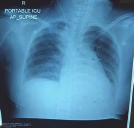
2 Case Reports in Obstetrics and Gynecology
denied any leaking liquor nor show. Fetal movements were
well felt. She claimed that she had fever, headache, sore
throat, and dry cough for one-day duration. She denied any
travelling or any contact with live poultry. She also had no
history of dysuria and frequency. Her booking visit was at 9th
week of gestation and all routine antenatal tests were negative.
Latest scan was done at 33-week follow-up and all parameters
were corresponding to gestational age.
On physical examination, she appeared alert, conscious,
with no tachypnea and no signs of dehydration. She was
afebrile, with no cervical lymph nodes and tonsillar enlarge-
ment. The lungs were clear. She had tachycardia up to
110/min and blood pressure was 105/57 mmHg. The abdom-
inal findings corresponded to gestational age, single fetus,
longitudinal lie, and cephalic presentation with good fetal Figure 1
heart rate. The estimated fetal weight was 2 to 2.2 kg. The
vaginal examination noted that os was closed. The ultrasound
examination was repeated and it corresponded to respective
gestation. The urine dip stick was suggestive of urinary
tract infection. The urine specimen was sent for culture and
sensitivity (C&S) before starting the antibiotics.
She was observed in the maternity ward. However, the
patient was noted to be febrile with temperature of 39∘ C,
tachycardia, and low blood pressure 80/50mmHg on next day
leading to septic shock. Her blood pressure was maintained
by intravenous noradrenaline infusion. A septic workup was
done and initial resuscitative measures were performed. The
blood test results came back with white cell count of 15.34 x
109 /L and C-reactive protein level of 365mg/L. She went into
active phase of labour at the same time.
Multidisciplinary input from anaesthetist for ICU back-
up, physician, and infectious disease teams was obtained, Figure 2
led by obstetric consultant shared care. The arterial blood
gas showed metabolic acidosis. Her condition worsened as
she became restless and breathless even with 2L/min oxygen phosphate) for one week. Universal precaution and isolation
under nasal prong. Four hours in labour, it was noted that of the patient were followed. The symptoms subsided with the
the progress of labour was unsatisfactory with features of fetal treatment. The patient and baby were discharged on the fifth
distress. A decision was made to deliver by caesarean section day postoperatively without complications.
in collaboration with anaesthetist and ICU team. An alive,
female, 2.61 kg baby with Apgar score of 5 in 1 minute, 7 in 3. Discussion
5 minutes and 8 in 10 minutes, was delivered and estimated
blood loss was about 700 ml. A placental swab was taken for The patient presented with a seasonal flu; however, her
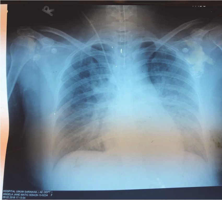
C & S. The anaesthetist noted on lungs auscultation that air conditions deteriorated quickly into septic shock with ARDS.
entry at the left lower zone and a portable chest X- ray was Despite this, early awareness of the serious symptoms and
ordered. The chest X-ray was reported as slight basal lobe a multidisciplinary team approach with appropriate and
collapse at left lobe with reactive pleural effusion (Figures aggressive treatment resulted in favourable outcome for both
1 and 2). Tamiflu was started and she was diagnosed with mother and baby. Unfortunately, the subtyping of influenza
Acute Respiratory Distress Syndrome (ARDS). The baby was A was not able to be carried out as our center lacked such
admitted to the nursery for late prematurity with presumed facility. However, it did not change the management and its
sepsis. Antibiotic treatment was given. The placenta swab good outcome.
culture result came back as sterile; still the baby needed photo There are warning signs which should alert the clinicians
therapy as neonatal jaundice arose. to the diagnosis of an influenza A infection. Early recognition
The patient was extubated after one day but continued of warning signs will lead to timely treatment and a better
support with Venturi mask 50% and 1.5L/min of oxygen. prognosis.
Once stable, she was transferred to High Dependency Warning signs for the clinician are as follows:
Unit and continued monitoring there. The throat swab (i) Difficulty breathing or shortness of breath
was reported as influenza A positive and the message was
relayed to both Infectious Diseases and Paediatric team. The (ii) Pain or pressure in the chest or abdomen
recommendation was to continue tablet Tamiflu (oseltamivir (iii) Sudden dizzinessCase Reports in Obstetrics and Gynecology 3
Table 1
Drug Treatment Chemoprophylaxis
Oseltamivir 75 mg twice a day for 5 days 75 mg once a day for 10 days
Zanamivir 5 mg inhalations twice per day for 5 days 5 mg inhalations daily for 10 days
Peramivir One time intravenously over 15-30 mins
From http://www.cdc.gov/h1n1flu/recommendation/html [12].
(iv) Confusion low-cost interventions that have been shown to have substan-
(v) Severe or persistent vomiting tial benefits for both mother and baby [14, 15].
(vi) Flu-like symptoms that improve but then return with
fever and worsening cough 8. Life Style Changes
(From flu symptoms: https://www.cdc.gov/flu/takingcare Personal hygiene is important in preventing transmission
.html [11].) of viral influenza. Measures such as hand hygiene and
wearing a face mask or N95 disposable respirator during close
contact with a sick individual should be advised to patients
4. Antiviral Medication and relatives. Household members should be monitored for
influenza-like symptoms and should be advised to contact
Pregnancy is a physiological state of relative immunosup- their healthcare provider if symptoms occur [16].
pression and all pregnant women should be assumed to be
at high risk and should be treated if the potential benefit
outweighs the theoretical risk of the fetus. Antiviral drugs 9. Conclusion
will help to relieve symptoms and reduce viral shedding,
Pregnant women are at high risk of infection with the
but therapy is more beneficial if started within 12-48 hours.
H1N1 influenza A virus at high risk of spontaneous miscar-
Neuraminidase inhibitors are used and they are as an adjunct
riage and associated neural tube defects, neonatal seizures,
to immunization. Oseltamivir is taken orally and zanamivir
encephalopathy, cerebral palsy, and neonatal death [17–19].
is administered via inhalation. Both are category C drugs
The influenza A virus infection is difficult to be diagnosed
but oseltamivir is preferred over zanamivir due to its well-
clinically because the presenting symptoms of an influenza
evidenced results from clinical experience with a guaranteed
infection are similar to those of the common cold but
systemic absorption [9, 10]. See Table 1.
they are more severe and protracted and can potentially be
life-threatening. Therefore, early recognition and awareness
5. Isolation of warning signs with appropriate, timely treatment, and
multidisciplinary support matter to achieve the best outcome.
The patients should continue to practice good hand hygiene
and cough etiquette and wear a face mask for the next
7 days. The patients with suspected N1H1 should wear a Abbreviations
face mask and be placed in an isolated room away from AIV: Avian influenza virus
providers and other hospitalized patients [9]. Surveillance ARDS: Adult respiratory distress syndrome
on the appearance of new symptoms as well as progress of CDC: Centers for Disease Control
current condition is necessary during isolation period. C& S: Culture and sensitivity
CTG: Cardiotocogram
6. Breastfeeding ICU: Intensive care unit
ID: Infectious disease
Breastfeeding is encouraged in H1N1 influenza although LSCS: Lower segment caesarean section
H1N1 transmission through breast milk is unknown; breast- O&G: Obstetrics and gynecology
feeding strengthens the neonatal immune response and RT-PCR: Real-time polymerase chain reaction
infants who are bottle fed may be prone to getting a viral US: United States
infection [13]. If the infant needs to be isolated from the WHO: World Health Organization.
infected mother, the infant should receive bottle feedings of
expressed breast milk until the mother and infant can be Consent
reunited. The patients should use a facemask and practice
strict hand hygiene and cough etiquette [9, 10]. Written informed consent was obtained from the patient for
publication of this case report and any accompanying images.
7. Immunization
Conflicts of Interest
Immunization is recommended to pregnant women espe-
cially during flu season (November–March). They are The authors declare that they have no conflicts of interest.4 Case Reports in Obstetrics and Gynecology
Acknowledgments novel Influenza A (H1N1) virus transmission. Centers for
Disease Control and Prevention, US Department of Health
We would like to thank the Head of Department of Obstetrics and Human Services, Public Health Service, Tuberculosis Con-
and Gynecology, Director of Sarawak General Hospital, and trol Division, Atlanta,” http://www.cdc.gov/h1n1flu/masks.htm
Director General of Ministry of Health for the approval to (accessed March 15, 2013), 2010.
publish this case report in a medical journal. [18] S. A. Rasmussen, D. J. Jamieson, K. MacFarlane, J. D. Cra-
gan, J. Williams, and Z. Henderson, “Pandemic influenza and
pregnant women: Summary of a meeting of experts,” American
References Journal of Public Health, vol. 99, no. 2, pp. S248–S254, 2009.
[1] G. Neumann, T. Noda, and Y. Kawaoka, “Emergence and [19] M. Rashid, R. Ara, and N. Akhter, “Novel influenza H1N1
pandemic potential of swine-origin H1N1 influenza virus,” in pregnancy: Report of a diagnosed case in Bangladesh,”
Nature, vol. 459, no. 7249, pp. 931–939, 2009. Bangladesh Journal of Obstetrics and Gynecology, vol. 24, no. 2,
pp. 75–78, 2009.
[2] F. S. Dawood, S. Jain, L. Finelli et al., “Emergence of a novel
swine-origin influenza A (H1N1) virus in humans,” The New
England Journal of Medicine, vol. 360, no. 25, pp. 2605–2615,
2009.
[3] Centers for Disease Control and Prevention, Pregnant Women
and Novel Influenza A (H1N1) Considerations for Clinicians, The
Centers, Atlanta, GA, USA, 2009.
[4] Z. Hashim and S. S. Arshad, “Avian influenza outbreaks in
Malaysia, 1980–2017,” Asia Pacific Environmental and Occupa-
tional Health Journal, vol. 12, no. 3, p. 2, 2017.
[5] D. J. Jamieson, M. A. Honein, S. A. Rasmussen et al., “H1N1
2009 influenza virus infection during pregnancy in the USA,”
The Lancet, vol. 374, no. 9688, pp. 451–458, 2009.
[6] S. A. Rasmussen, D. J. Jamieson, and J. S. Bresee, “Pandemic
influenza and pregnant women,” Emerging Infectious Diseases,
vol. 14, no. 1, pp. 95–100, 2008.
[7] T. Teke, Ü. Duran, E. Maden, K. Gezginç, M. D. Yavşan, and
K. Uzun, “A pregnant case with severe influenza a (H1N1) virus
infection-related ARDS,” European Journal of General Medicine,
vol. 8, no. 2, pp. 163–167, 2011.
[8] C. Hüzmeli, M. Saglam, A. Arıkan, B. Doner, G. Akıncı, and
F. Candan, “Infrarenal aorta thrombosis associated with H1N1
influenza A virus infection,” Case Reports in Infectious Diseases,
vol. 2016, Article ID 9567495, 3 pages, 2016.
[9] K. Trevennec, V. Grosbois, F. Roger, T. H. Ho, C. Berthouly-
Salazar, and V. Chevalier, “Evidence for freedom from swine
influenza in a remote area of Northern Vietnam,” Acta Tropica,
vol. 122, no. 1, pp. 160–163, 2012.
[10] Centers for Disease Control and Prevention (CDC), “Interim
guidance on antiviral recommendations for patients with novel
influenza A (H1N1) virus infection and their close contacts,”
http://www.cdc.gov/h1n1flu/recommendations.htm, 2009.
[11] https://www.cdc.gov/flu/takingcare.html.
[12] http://www.cdc.gov/h1n1flu/recommendation/html.
[13] Centers for Disease Control and Prevention, “Pregnant women
and novel influenza A (H1N1): considerations for clinicians,”
http://www.cdc.gov/h1n1flu/clinician pregnant.htm, 2009.
[14] Centers for Disease Control and Prevention, “Considerations
regarding novel H1N1 flu virus in obstetric settings,” 2009.
[15] K. Q. Chen, E. H. Hayles, and J. Sinn, “Flu-like symptoms in
pregnancy - A case study,” Australian Family Physician, vol. 45,
no. 5, pp. 307-308, 2016.
[16] Centers for Disease Control and Prevention, “Interim guidance
on specimen collection, processing, and testing for patients with
suspected swine-origin influenza A (H1N1) virus infection,”
http://www.cdc.gov/h1n1flu/specimencollection.htm, 2009.
[17] Centers for Disease Control and Prevention, “Interim rec-
ommendations for facemask and respirator use to reduceYou can also read