Gram-negative Cranial Bone Flap Infection Treated with Continuous Gentamicin Irrigation: A Case Report - Cureus
←
→
Page content transcription
If your browser does not render page correctly, please read the page content below
Open Access Case
Report DOI: 10.7759/cureus.4282
Gram-negative Cranial Bone Flap Infection
Treated with Continuous Gentamicin
Irrigation: A Case Report
Jamie Toms 1 , Lisa Kurczewski 1 , Robert Simonds 1 , R. Scott Graham 1 , Jason Harrison 2
1. Neurosurgery, Virginia Commonwealth University Health Systems, Richmond, USA 2. Neurosurgery,
Harbin Clinic, Rome, USA
Corresponding author: Jamie Toms, jamie.toms@vcuhealth.org
Disclosures can be found in Additional Information at the end of the article
Abstract
A 57-year-old male presented with severely altered mental status in the setting of diabetic
ketoacidosis. Neuroimaging revealed two intracranial masses. Days following surgical resection
of an olfactory groove meningioma, the patient developed Serratia marcescens bacteremia along
with an enlarging epidural and subgaleal fluid collection. Subgaleal fluid aspiration was also
positive. The patient later returned to the operating room for wound washout where purulent
collections were discovered in the subgaleal, epidural, and left subdural spaces. The wound was
evacuated and the bone flap was thoroughly cleansed with betadine and soaked in peroxide
prior to replacement. Four drains were placed (two subgaleal and two epidural) with two
serving as inlets and two as outlets. Continuous irrigation of the subgaleal and epidural spaces
with gentamicin solution was performed for five days. The bone flap was successfully salvaged
and the patient was discharged from inpatient rehab three weeks following washout.
Categories: Infectious Disease, Neurosurgery
Keywords: continuous irrigation, bone flap infection, craniotomy, subdural empyema, gentamicin,
serratia marcescens
Introduction
Although relatively rare, the incidence of bone flap infection following craniotomy is reported
to range from one to 11%. Known risk factors for such infections include: prior radiation
therapy, presence of cerebrospinal fluid leak, duration of surgery over four hours, procedures
involving the nasal sinuses or skull base, and emergency surgeries [1-3]. When infection
following craniotomy does occur, the traditional standard of care is to surgically discard the
bone flap. Unfortunately, this method subjects the patient to months of cosmetic deformity, an
Received 03/01/2019 unprotected brain, and ultimately, delayed cranioplasty (with potential need for more
Review began 03/04/2019
procedures). Therefore, whenever possible, salvage of the native bone flap is the preferred
Review ended 03/14/2019
Published 03/20/2019
option [2-4].
© Copyright 2019
Toms et al. This is an open access
We describe a case of post-craniotomy infection treated with thorough cleansing of the bone
article distributed under the terms of flap, evacuation of purulent material along with debridement, and continuous irrigation with
the Creative Commons Attribution saline containing gentamicin. This technique resulted in successful salvage of the bone flap and
License CC-BY 3.0., which permits patient recovery.
unrestricted use, distribution, and
reproduction in any medium, provided
the original author and source are
credited.
Case Presentation
Our patient, a 57-year-old African-American male with history of cerebral palsy, presented
How to cite this article
Toms J, Kurczewski L, Simonds R, et al. (March 20, 2019) Gram-negative Cranial Bone Flap Infection
Treated with Continuous Gentamicin Irrigation: A Case Report. Cureus 11(3): e4282. DOI
10.7759/cureus.4282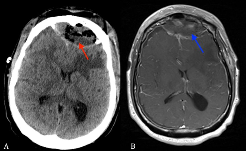
with severely altered mental status and alcohol intoxication in the setting of diabetic
ketoacidosis. His Glasgow Coma Scale (GCS) score on admission was three. Computerized
tomography (CT) scan and subsequent magnetic resonance imaging (MRI) revealed a left
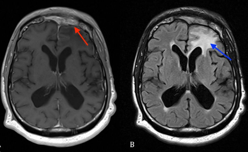
olfactory groove mass with surrounding hypodensity concerning for vasogenic edema (Figure 1)
as well as a right petroclival mass.
FIGURE 1: (A) Axial T1 MRI of brain with contrast showing left-
sided olfactory groove meningioma (red arrow). (B) Axial FLAIR
MRI of brain showing left-sided olfactory groove meningioma
and surrounding edema (blue arrow).
FLAIR: Fluid-attenuated inversion recovery; MRI: Magnetic resonance imaging.
After the patient was treated medically for his diabetic ketoacidosis and intoxication, he
underwent bifrontal craniotomy and resection of the left olfactory groove mass, cranialization
of the frontal sinus, as well as pericranial flap placement. Postoperatively, the patient was
treated with antibiotics and a dexamethasone taper. An immediate post-operative MRI
revealed good resection and expected post-operative changes. The patient did well and
remained afebrile, but it was noted that he had increased swelling at his operative site. He
developed a markedly elevated leukocytosis on postoperative day (POD) three. Urinalysis and
chest radiographs at that time were not concerning for infections. However, blood cultures
drawn grew Serratia marcescens and the patient was initiated on intravenous meropenem 2
grams every eight hours. Repeat CT imaging obtained on POD 10 revealed an enlarging epidural
fluid collection as well as a new subgaleal fluid collection as shown in Figure 2.
2019 Toms et al. Cureus 11(3): e4282. DOI 10.7759/cureus.4282 2 of 8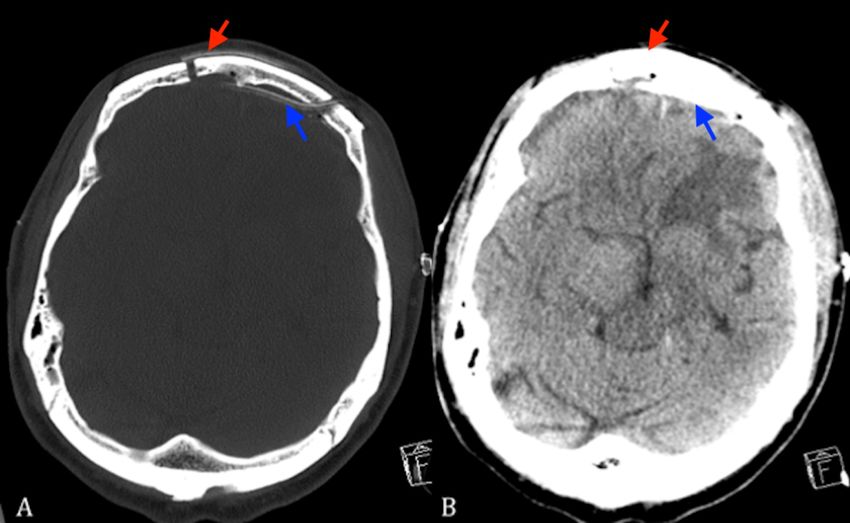
FIGURE 2: (A) Axial non-contrast CT scan showing a frontal
                                     fluid collection under the craniotomy bone flap (red arrow). (B)
                                     Axial T1 MRI with contrast showing a frontal fluid collection
                                     under the craniotomy bone flap (blue arrow).
                                     CT: Computed tomography; MRI: Magnetic resonance imaging.

                                   Subgaleal fluid aspiration taken at the time grew S. marcescens and the patient was taken back
                                   to the operating room for wound washout. Antibiotic sensitivity analysis performed on S.
                                   marcescens revealed a meropenem minimum inhibitory concentration (MIC) of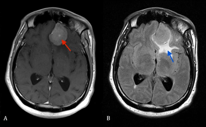
FIGURE 3: (A) Bone windowed axial non-contrast head CT. (B)
                                     Brain windowed axial non-contrasted head CT scan showing
                                     placement of subgaleal (red arrow) and epidural catheters
                                     (blue arrow).
                                     CT: Computed tomography

                                   Saline solution containing preservative-free gentamicin at a concentration of 20 mcg/mL was
                                   continuously infused through the drains at a rate of 10 mL/hour/drain for a period of five days.
                                   The patient tolerated the treatment well and was discharged from inpatient rehab three weeks
                                   following washout. The patient completed an eight-week course of intravenous meropenem
                                   followed by oral levofloxacin for a total of six months of antibiotic coverage.

                                   At five-month follow-up, a repeat MRI demonstrated no signs of infection as shown in Figure 4.
                                   The patient had completely returned to his neurologic baseline and received radiation therapy
                                   for the remaining meningioma.

2019 Toms et al. Cureus 11(3): e4282. DOI 10.7759/cureus.4282                                                                   4 of 8
FIGURE 4: (A) Axial T1 MRI with contrast showing resolution of
infection (red arrow). (B) Axial FLAIR MRI showing resolution
of infection with some residual flair changes (blue arrow).
FLAIR: Fluid-attenuated inversion recovery; MRI: Magnetic resonance imaging.
Discussion
Bone flap infection following craniotomy is a relatively rare, but troublesome complication for
the patient. The literature reports the incidence of post-craniotomy infection to range from one
to 11% [1-3]. Several studies have identified risk factors predisposing patients to post-
craniotomy infection. Known procedure-related risk factors include: prior radiation therapy,
presence of cerebrospinal fluid leak, duration of surgery longer than four hours, early
reoperation, procedures involving the nasal sinuses or skull base, and emergency surgeries.
Patient-related risk factors include: GCS score less than 10, American Society of
Anesthesiologists (ASA) score greater than two, recent antibiotic use, and recent neurosurgical
procedures prior to craniotomy [5-6].
Opinion regarding management of post-craniotomy infection varies. Traditionally, the
standard management consists of operative debridement followed by disposal of the bone flap
with delayed cranioplasty [1-3]. While proven to be a safe and effective intervention for such
infections, the process presents many issues for patients as they are left with a cranial defect
and lose protection from head trauma for an extended period of time. In some rare incidences
the consequence of large cranial defects, known as the “syndrome of the trephined”, can occur
[4-7]. Due to these undesirable factors, many have sought to identify alternative techniques
directed at salvaging the bone flap.
In 1974, Erickson et al. employed a technique involving suction and irrigation with an antibiotic
solution [8]. During the procedure, the wound was opened, and all visible sutures (as well as
necrotic tissue) were removed. Interestingly, the bone flap was left in place. Two drains were
then placed in the subgaleal space with one as an inlet and the other as an outlet connected to
a Hemovac drain. If purulent matter was seen in the epidural space, a second set of Hemovac
2019 Toms et al. Cureus 11(3): e4282. DOI 10.7759/cureus.4282 5 of 8drains was placed there as well. An antibiotic solution was then infused at a rate of 1-2
liters/day for a period of up to five days. Close monitoring of drain input and output was
necessary as clogged output drains place patients at risk for increased intracranial pressure [3].
Of note, all identified and treated pathogens were skin flora.
In 2003, Bruce and Bruce published a successful debridement strategy that resulted in salvage
of native bone flaps [3]. The bone flap was re-elevated and all visible suture and hemostatic
agents were removed. Any necrotic or purulent debris was also removed. The bone flap was
extensively scrubbed with betadine before replacement. Systemic antibiotic therapy was an
important adjunct to success. Patients typically received intravenous antibiotics for one to two
weeks followed by two to four weeks of oral therapy. The authors reported a very high degree of
success, even in patients with many undesirable risk factors for post-craniotomy infections. Of
note, the only risk factor that strongly correlated to failure and loss of bone flap was prior
procedures involving communication of the nasal sinuses [2,3]. Our patient was continued on
antibiotic therapy for a total of six months. This duration of antibiotics was chosen due to our
patient's prior bacteremia and severely uncontrolled diabetic state.
Later, Widdel and Winston implemented an approach similar to Bruce and Bruce [3], with the
added implementation of scrubbing the bone flap with iodophor or bacitracin. The bone flap
was then immersed in either povidone-iodine or bacitracin until reimplantation. This resulted
in successful salvage of all 14 bone flaps in their patient series [4].
Despite the use of antibiotic irrigation systems being described as superfluous by Bruce and
Bruce [3] based on their findings, the choice to proceed with a gentamicin irrigation was made
in our case due to the uncommon gram-negative bacilli isolated from the patient’s cultures and
persistent bacteremia with central nervous system (CNS) involvement despite administration of
appropriately dosed broad spectrum intravenous antibiotics. Additionally, Bruce and Bruce [3]
note that one of their patient complications requiring additional surgery was in a patient who
grew virulent gram-negative bacilli.
In 2006, Auguste and McDermott [2] described a “wash-in, wash-out” method that improved
upon the suction-irrigation system implemented by Erickson and Chou [8]. Important
differences in protocol included mandatory re-elevation of all bone flaps (with subsequent
debridement of the flap and soft tissues), irrigation of both the subgaleal and epidural spaces
(rather than subgaleal alone), and at least two weeks of intravenous antibiotics followed by
three months of oral therapy. Vancomycin was used for irrigation in all cases and the duration
of irrigation was typically five days. This method was less cumbersome in terms of preparation
and setup and even allowed for the patients to ambulate. The authors recommended close
patient observation as impeded output could result in fluid collection and mass effect. Their
treatment protocol resulted in the successful preservation of bone flaps in all but one patient in
a 12-patient series. Similar methods of continuous irrigation have even proven useful in the
treatment of subdural empyema [9,10]. In all cases vancomycin irrigation was used as the
majority of organisms isolated from operative cultures were predominantly skin flora. It is less
common for gram-negative bacilli to complicate surgery as opposed to skin organisms such as
Staphylococci or Propionibacterium. Gentamicin is an aminoglycoside antibiotic with broad
spectrum coverage of aerobic gram-negative bacteria. Very low cerebrospinal fluid
concentrations of the drug are achieved in patients without inflamed meningitis leading to
alternative administration routes for the treatment of CNS infections [11,12]. Utilizing a
localized route of administration minimizes systemic absorption of the drug and can avoid
common adverse effects. The use of a 10 mL/hour infusion rate decreases the risk for
complications, such as brain herniation from increased fluid volume, while keeping the
gentamicin concentration well above the organism’s reported MIC. It is impossible to measure
standard peak and trough concentrations when administering gentamicin by these means. By
providing a concentration of 20 mcg/mL of drug locally at an infusion rate of 10 mL/hour, we
2019 Toms et al. Cureus 11(3): e4282. DOI 10.7759/cureus.4282 6 of 8ensured that the amount of drug present in the subgaleal space stayed above the organism's
reported MIC of 1 mcg/mL and obtained a peak/MIC ratio > 10. This localized approach to
treatment has been documented in the literature with intraventricular gentamicin
administration for the treatment of gram-negative device infections [13-16].
By combining the meticulous surgical techniques described by Bruce and Bruce [3] and Widdel
and Winston [4] with the newer “wash-in, wash-out” irrigation system explained by Auguste
and McDermott [2], we successfully treated the patient’s infection while preserving the bone
flap with no further complications. The patient was closely monitored and could ambulate
during the five days of irrigation.
Similarly to our case, is a report published by Wada et al. describing a patient with panparanasal
sinusitis, destruction of the frontal sinus walls, and subdural fluid collection resulting in a wide
decompressive craniectomy in the bilateral frontal and left temporoparietal regions [9]. After
irrigation of the subdural space, six drainage tubes were placed and a seven-day course of
continuous antibiotic irrigation with a gentamicin solution of 20 mcg/mL in saline was
administered at a flow rate of 10 mL/hr. The patient grew pan-sensitive Streptococcus milleri,
and the patient’s infectious signs and symptoms resolved quickly without complications to the
drainage system. Extrapolating the safety data from Wada et al. [9], we used a similar dose of
gentamicin and rate of infusion over a five-day course with success. Our patient had a full
recovery and is back at his baseline.
Conclusions
The methods described above challenge the standard treatment of discarding bone flaps in the
setting of post-craniotomy infection. Although the referenced studies contain a relatively small
number of patients, they demonstrate that contaminated bone flaps can be salvaged, thus
sparing patients undesirable consequences while awaiting cranioplasty. Certainly, the decision
to pursue aggressive measures to preserve bone flaps should be based upon individual patient
characteristics. When indicated, physicians following similar methods to ours should have high
expectations of success. A gentamicin irrigation system is a valuable added therapy for gram-
negative infections not responding to broad spectrum intravenous antibiotic therapy.
Additional Information
Disclosures
Human subjects: Consent was obtained by all participants in this study. Conflicts of interest:
In compliance with the ICMJE uniform disclosure form, all authors declare the following:
Payment/services info: All authors have declared that no financial support was received from
any organization for the submitted work. Financial relationships: All authors have declared
that they have no financial relationships at present or within the previous three years with any
organizations that might have an interest in the submitted work. Other relationships: All
authors have declared that there are no other relationships or activities that could appear to
have influenced the submitted work.
References
1. Delgado-López PD, Martín-Velasco V, Castilla-Díez JM, Galacho-Harriero AM, Rodríguez-
Salazar A: Preservation of bone flap after craniotomy infection . Neurocirugia. 2009, 20:124-
131. 10.1016/S1130-1473(09)70179-4
2. Auguste KI, McDermott MW: Salvage of infected craniotomy bone flaps with the wash-in,
wash-out indwelling antibiotic irrigation system. Technical note and case series of 12
patients. J Neurosurg. 2006, 105:640-644. 10.3171/jns.2006.105.4.640
3. Bruce JN, Bruce SS: Preservation of bone flaps in patients with postcraniotomy infections . J
2019 Toms et al. Cureus 11(3): e4282. DOI 10.7759/cureus.4282 7 of 8Neurosurg. 2003, 98:1203-1207. 10.3171/jns.2003.98.6.1203
4. Widdel L, Winston KR: Pus and free bone flaps . J Neurosurg Pediatr. 2009, 4:378-382.
10.3171/2009.5.PEDS0963
5. Korinek AM: Risk factors for neurosurgical site infections after craniotomy: a prospective
multicenter study of 2944 patients. Neurosurgery. 1997, 41:1073-1081. 10.1097/00006123-
199711000-00010
6. Korinek AM, Golmard JL, Elcheick A, Bismuth R, Van Effenterre R, Coriat P, Puybasset L: Risk
factors for neurosurgical site infections after craniotomy: a critical reappraisal of antibiotic
prophylaxis on 4578 patients. Br J Neurosurg. 2005, 19:155-162. 10.1080/02688690500145639
7. Ashayeri K, Jackson EM, Huang J, Brem H, Gordon CR: Syndrome of the trephined: a
systematic review. Neurosurgery. 2016, 79:525-534. 10.1227/NEU.0000000000001366
8. Erickson DL, Seljeskog EL, Chou SN: Suction-irrigation treatment of craniotomy infections.
Technical note. J Neurosurg. 1974, 41:265-267. 10.3171/jns.1974.41.2.0265
9. Wada Y, Kubo T, Asano T, Senda N, Isono M, Kobayashi H: Fulminant subdural empyema
treated with a wide decompressive craniectomy and continuous irrigation- case report. Neurol
Med Chir (Tokyo). 2002, 42:414-416.
10. Eom KS, Kim TY: Continuous subdural irrigation and drainage for intracranial subdural
empyema in a 92-year-old woman. Minim Invasive Neurosurg. 2011, 54:87-89. 10.1055/s-
0031-1277199
11. GENTAMICIN- gentamicin injection, solution. (2016). Accessed: May 10, 2017:
https://dailymed.nlm.nih.gov/dailymed/drugInfo.cfm?setid=be5b414e-d598-4721-80ee-
5836156ad210.
12. Grayson ML, Cosgrove SE, Crowe S, et al.: Gentamicin. Kucers' the Use of Antibiotics: A
Clinical Review of Antibacterial, Antifungal, Antiparasitic and Antiviral Drugs. CRC Press,
Boca Raton, FL; 2010. 1:674-658.
13. Wen DY, Bottini AG, Hall WA, Haines SJ: Infections in neurologic surgery. The
intraventricular use of antibiotics. Neurosurg Clin N Am. 1992, 3:343-354.
14. Tängdén T, Enblad P, Ullberg M, Sjölin J: Neurosurgical gram-negative bacillary ventriculitis
and meningitis: a retrospective study evaluating the efficacy of intraventricular gentamicin
therapy in 31 consecutive cases. Clin Infect Dis. 2011, 52:1310-1316. 10.1093/cid/cir197
15. Wilkie MD, Hanson MF, Statham PF, Brennan PM: Infections of cerebrospinal fluid diversion
devices in adults: the role of intraventricular antimicrobial therapy. J Infect. 2013, 66:239-246.
10.1016/j.jinf.2012.11.006
16. Remeš F, Tomáš R, Jindrák V, Vaniš V, Šetlík M: Intraventricular and lumbar intrathecal
administration of antibiotics in postneurosurgical patients with meningitis and/or
ventriculitis in a serious clinical state. J Neurosurg. 2013, 119:1596-1602.
10.3171/2013.6.JNS122126
2019 Toms et al. Cureus 11(3): e4282. DOI 10.7759/cureus.4282 8 of 8You can also read