Therapeutic potential of anticoagulant therapy in association with cytokine storm inhibition in severe cases of COVID-19: A case report - De Gruyter
←
→
Page content transcription
If your browser does not render page correctly, please read the page content below
Open Life Sciences 2021; 16: 809–814
Case Report
Qiancheng Xu#, Tao Wang#, Weihua Lu*
Therapeutic potential of anticoagulant therapy in
association with cytokine storm inhibition in
severe cases of COVID-19: A case report
https://doi.org/10.1515/biol-2021-0088
received February 02, 2021; accepted July 28, 2021
1 Background
Abstract: Inflammation and coagulation are considered Coronavirus disease 2019 (COVID-19) has been a global
to the development of Coronavirus disease 2019 (COVID- epidemic for nearly 2 years since its emergence in China
19)-related hypoxemia. However, this is still controver- in late 2019 [1]. However, it is still unclear if inflammation
sial, which brings challenges to clinical treatment. and coagulation are relevant to COVID-19-related hypox-
Here, we reviewed the levels of interleukin-6 (IL-6), coa- emia [2,3], which brings challenges to clinical treatment.
gulation indexes, and clinical manifestations of a patient Tocilizumab, an IL-6 receptor blocker, is considered to be
with severe COVID-19 after Tocilizumab administration. an effective treatment for COVID-19, but after recent ran-
In this case, the patient’s body temperature quickly domized controlled trials, no definitive conclusions have
dropped to normal after using Tocilizumab, while C been drawn [4,5]. However, IL-6 initiates the systemic
reactive protein progressively decreased and stabilized pro-inflammatory response and inhibits inflammation,
at a lower level. However, IL-6 and D-dimers increased thus promoting cell proliferation and tissue repair [6].
and were accompanied by a continuous decrease of the Moreover, inappropriate routine use of Tocilizumab to
oxygenation index. After anticoagulant therapy with treat COVID-19 should not be encouraged until there
heparin, D-dimer decreased slowly, gradually improving is insufficient evidence. Therefore, we measured IL-6,
the oxygenation index and disease remission. This case coagulation indexes, and clinical manifestations in a
suggests that the formation of microthrombus might be severe COVID-19 patient after the use of Tocilizumab (in
the main reason for COVID-19-derived hypoxemia. However, accordance with the regulations of the National Health
the mechanism of hypoxemia and the role of Tocilizumab in Commission of China) [7]. This study aimed to better
COVID-19 need further research. Nevertheless, these find- understand the pathophysiology of COVID-19-related hypox-
ings might still assist medical workers in formulating timely emia and improve the clinical strategy for the treatment of
treatment strategies for similar severe patients. this disease.
Keywords: COVID-19, SARS-CoV-2, tocilizumab, cytokine
storm, inflammation, coagulation
2 Case report
On February 27, 2020, a 65-year-old woman was admitted
to our fever clinic with symptoms of fever, cough, fatigue,
# These authors contributed equally to this work.
and shortness of breath. She had developed fever 6 days
back with a maximum temperature of 37.8°C. She did not
* Corresponding author: Weihua Lu, Department of Critical Care present cold and chills before the fever. The cough started 4
Medicine, The First Affiliated Hospital of Wannan Medical College days before with a small amount of white phlegm (mucus),
(Yijishan Hospital of Wannan Medical College), No. 2, West Road of no chest tightness, chest pain, or dyspnea. Apparent chest
Zheshan, Jinghu District, Wuhu, Anhui, 241000, China, tightness and dyspnea began to appear on February 27,
tel: +86 0553-5739731, e-mail: lwh683@126.com
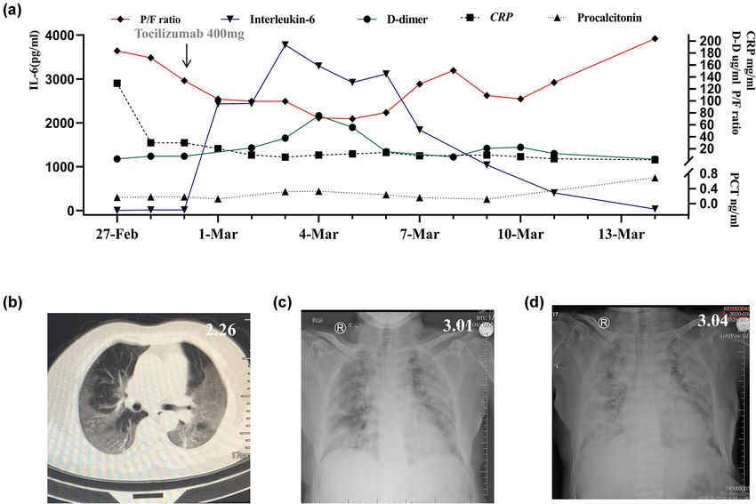
2020, with SpO2 of 80%; Chest CT showed multiple shadows
Qiancheng Xu, Tao Wang: Department of Critical Care Medicine,
The First Affiliated Hospital of Wannan Medical College (Yijishan
in both lungs (Figure 1b); leukocyte count was 6.58 × 10⁹/L,
Hospital of Wannan Medical College), No. 2, West Road of Zheshan, absolute lymphocyte count was 0.44 × 10⁹/L and C reac-
Jinghu District, Wuhu, Anhui, 241000, China tive protein (CRP) was 97.3 mg/L. The patient denied that
Open Access. © 2021 Qiancheng Xu et al., published by De Gruyter. This work is licensed under the Creative Commons Attribution 4.0
International License.810 Qiancheng Xu et al.
Figure 1: Changes in FiO2, P/F ratio (a), IL-6, CRP, D-dimer, PCT in a severe COVID-19 patient (a). Chest CT showed multiple shadows in both
lungs on February 26 (b). X-ray showed multiple infiltrations in both lungs on March 01 (c). X-ray showed more severe infiltration 3 days
before on March 04 (d). FiO2: fraction of inspiration O2; P/F ratio: SpO2/FiO2 ratio; IL-6: interleukin-6; CRP: C reactive protein; PCT:
procalcitonin.
she had been to Wuhan but had a history of contact with The patient’s oxygenation index decreased gradually
asymptomatic people who had returned from the COVID- between the 1st and 3rd days of hospitalization. On
19 epidemic area in Wuhan. February 29, 2020, the patient presented with a high-
The patient was diagnosed with suspected COVID-19 flow nasal cannula (HFNC) oxygen therapy (60% concen-
virus infection and was immediately admitted to the iso- tration, flow rate: 40 L/min) and partial pressure of oxygen
lation ward and kept at the prone position combined with of 80.3 mm Hg. A chest X-ray showed progressive infil-
high-flow oxygen therapy. Interferon α-2b (5 million units trate and diffuse gridding shadows in both lungs (Figure 1c
twice daily, atomization inhalation) and Arbidol (200 mg and d). Tocilizumab 400 mg was administered intrave-
twice daily, orally) were prescribed as antiviral therapy, nously. The oxygenation index between the 3rd and
and moxifloxacin (0.4 g once daily, intravenously) was 8th days of hospitalization progressively decreased, while
used to prevent secondary bacterial infections. Methylpred- IL-6 rapidly increased to 3,775 pg/mL, and then started to
nisolone (40 mg twice) was administered to attenuate the slowly decrease. CRP and procalcitonin (PCT) remained at a
lung inflammation and a throat swab sample was taken relatively low level, but the D-dimer gradually increased,
(Table 1). Laboratory test results are listed in Table 2. showing the same trend as the decrease of the oxygenation
On February 28, 2020 (Day 8 after the beginning of index. Heparin anticoagulation therapy was given on the
symptoms and second day of hospitalization), COVID-19 3rd day of hospitalization (Table 1), D-dimer gradually
was confirmed by reverse transcription PCR performed decreased on the 7th day of hospitalization, and the oxy-
by the Anhui Provincial Centers for Disease Control genation index gradually improved on the 8th day of hos-
(CDC). pitalization. A throat swab nucleic acid detection wasAnticoagulant therapy in severe COVID-19 811
Table 1: Timeline of disease course according to days from initial presentation of illness and days from hospital admission
In the local Hospital
Home
hospital Day 1~2 Day 3 Day 4 Day 5 Day Day 7 Day 8~9 Day Day 11 Day 12 Day 13 Day 14
Day of illness 1-4 5-6 7-8 9 10 11 12 13 14-15 16 17 18 19 20
Cough
Shortness of breath
Fever (°C) Subjective 37.8 37.6~38.1 37.4 37.6 37.7 37.2 36.5 36.8~36.3 36.6 36.8 36.7 36.5 36.5
Fatigue
PP duration per day (h) 16 18 22 22 20 6
HFNC HFNC
LMWH 5000U i.h Q12h 5000U i.h Q12h
UFH( Continuous pumping) 5-15U/kg.h
Arbidol Arbidol tablets
Interferon alfa-2b Interferon alfa-2b physicochemical inhalation
Convalescent Plasma(ml) 300 200
Tocilizumab(mg) 400
Methylprednisolon(mg/d) 40
Rehabilitation Therapy - - Rehabilitation Therapy
Nucleic acid testing Positive Negative Negative
21~22-Feb 23~26-Feb 27~28-Feb 29-Feb 1-Mar 2-Mar 3-Mar 4-Mar 5~6-Mar 7-Mar 8-Mar 9-Mar 10-Mar 11-Mar
PP: Prone position; HFNC: High flow nasal cannulae; LMWH: Low molecular weight heparin; UFH: Unfraction heparin
negative for COVID-19 on the 9th day of hospitalization antibody against the IL-6 receptor (IL-6R). At present,
(Figure 1a). On March 9, the patient was transferred to a this drug has been successfully used to treat a variety
local hospital. of chronic inflammatory diseases, such as rheumatoid
arthritis, systemic and polyarticular juvenile idiopathic
Informed consent: Informed consent has been obtained arthritis, and Castleman’s disease [9]. However, so far,
from all individuals included in this study. there is no definitive evidence that Tocilizumab is effec-
tive against severe cases of COVID-19, while the relevance
Ethical approval: The research related to human use has of the inflammatory cytokine storm in COVID-19 is still con-
been complied with all the relevant national regulations, troversial [2,3,10].
institutional policies and in accordance with the tenets In the case herein presented, the patient’s body
of the Helsinki Declaration, and has been approved by temperature quickly dropped to normal after the use of
the authors’ institutional review board or equivalent tocilizumab; however, the respiratory function did not
committee. improve. The oxygenation index continued to decrease
until March 5 before slowly increasing. Chest CT showed
that bilateral lung exudation was still progressing gradu-
ally. Levels of IL-6 (pg/mL) and D-dimer (μg/mL) were
3 Discussion increased later (from baseline of 3.1 and 3.0 to 3,114.0 and
73.70, respectively). This is similar to two cases pre-
The dysregulation of host immune response character- viously reported in Chest. After tocilizumab use, IL-6
ized by a cytokine storm and lymphocyte depletion is (pg/mL) and D-dimer (ng/mL) increased from 74.3 and
a prominent manifestation of SARS-CoV-2 infection. 982 to 345 and 30,233, respectively [11].
Critically ill patients with COVID-19 are often accompa- Qin et al. [12] reported a COVID-19 patient with an IL-6
nied by a significant increase in IL-6 and inflammatory of 25 pg/mL and an acute respiratory distress syndrome
cytokine storm, which are considered key factors that (ARDS) of up to 1,618 pg/mL, which is ten times higher
lead to a rapid progression of the disease and culminating than that of other reported cases of COVID-19 [13].
in death [8]. China’s “Diagnosis and Treatment Protocol Another study published in JAMA came to the same
for Novel Coronavirus Pneumonia (Trial Version 7)” sug- conclusion [14]. Besides, there are obvious differences
gests that tocilizumab can be used for severe patients with traditional ARDS in lung compliance and ventila-
with increased IL-6 [7]. Tocilizumab is a monoclonal tion blood flow [10]. Therefore, it is suggested that the812 Qiancheng Xu et al.
Table 2: Clinical laboratory tests
Hospital Day Day 1 Day 3 Day 4 Day 6 Day 8 Day 10 Day 11 Fever
clinic
Illness Day 7 9 10 12 14 16 17 36
Measure Reference range Date
27-Feb 29-Feb 1-Mar 3-Mar 5-Mar 7-Mar 8-Mar 29-Mar
Complete blood count
White-cell count (×109/L) 4–10 2.9 7 5.4 5.3 9.3 10.3 11 8.7
Absolute neutrophil count (×109/L) 2.0–7.5 2.4 6.4 4.2 4.6 8.2 8.4 8.5 6.3
Absolute Lymphocyte count (×109/L) 2.0–7.5 0.4 0.4 1.1 0.6 0.7 0.8 0.9 2
Absolute monocyte count (×109/L) 0.12–0.8 0.1 0.1 0.1 0.6 0.3 0.3 0.3 0.4
Red-cell count (×1012/L) 3.5–5.0 3.76 3.73 3.58 3.75 3.57 3.76 3.99 3.62
Hemoglobin (g/L) 110–150 118 116 110 116 113 121 123 119
Platelet count (×109/L) 100–300 168 207 215 217 212 218 221 280
Biochemical test
Total protein (g/L) 65.0–85.0 56.8 73.4 69.9 73.5 75.6 74.3 73.9 64.1
Albumin (g/L) 40.0–55.0 29.8 33.5 32 33.9 41.1 40.6 39.7 32.1
Prealbumin (mg/L)
Alanine aminotransferase (ALT) (U/L) 7–40 44 78 55 43 31 36 32 19
Aspartate aminotransferase (AST) (U/L) 13–35 76 50 35 30 39 41 37 16
Lactate dehydrogenase (LDH) (U/L) 135–225 438 343 328 388 559 561 511 213
Urea (mmol/L) 2.3–7.1 3.5 6.8 6.4 5.9 4.6 4.9 4.8 2.28
Creatinine (µmol/L) 40–130 43.4 49.8 52.3 39 43.9 44.6 44.3 41.8
Sodium (mmol/L) 135–149 139.2 143.7 142.8 140.2 140 141 140.7 139.4
Potassium (mmol/L) 3.5–5.3 3.87 3.53 3.22 3.36 3.84 3.76 4.02 4.64
Chloride (mmol/L) 99–110 101.9 101.8 102.9 102 97.8 102.1 101.5 102
Arterial blood gas (ABG) analysis
pH 7.350–7.450 7.443 7.452 7.5 7.477 7.499 7.456 7.433
Pressure of oxygen in arterial 83–108 76.4 80.3 61.8 79.4 63.1 89.7 98
blood (mm Hg)
Pressure of carbon dioxide in arterial 35–45 29.4 33.6 33.2 29.6 34.7 38.9 44.7
blood (mm Hg)
Base excess (mmol/L) −3 to 3 −3.6 −0.4 2.6 −1.5 3.6 3.3 5.2
Coagulation profile
Prothrombin time (s) 11–14.5 12.8 12.5 12.3 11.2 13.1 12.1 12.6
International normalized ratio 0.8–1.25 1.11 1.08 1.06 0.96 1.13 1.04 1.09
Fibrinogen (g/L) 1.8–4.0 6.04 4 3.24 1.99 2.43 4.57 4.33
formation of microthrombus was the main reason for important strategy to improve the prognosis of patients
the decrease of oxygenation index in our reported case. with COVID-19 [2,17,18]. In our reported case, D-dimer
A growing number of studies have shown that the continued to increase and was accompanied by a con-
main cause of hypoxemia in COVID-19 patients is due tinuous decrease of the oxygenation index after the
to vasodilation that occurs upon the activation of vas- application of tocilizumab. After anticoagulant therapy
cular endothelial cells. This may result in pulmonary with heparin, D-dimer decreased slowly, and the oxy-
microvascular embolism; however, the effect of inflam- genation index gradually improved. D-dimer had a sig-
matory factors is not obvious [7,11]. An autopsy result nificant negative correlation with the oxygenation index
confirmed that there was no significant inflammatory of the patient, suggesting that the activation of vascular
cell infiltration in the renal interstitium despite the acti- endothelial cells and the formation of microthrombus
vation of endothelial cells and the detection of virus par- were the main causes for the decrease of oxygenation
ticles [15]. Zheng et al. [16] suggested that D-dimer is an index in this patient.
important predictor of the prognosis of COVID-19 patients. In this case, it was not clear whether there is a causal
This conclusion has been confirmed in other studies [6]. relationship between the use of tocilizumab and the
Therefore, anticoagulant therapy is considered to be an increase of D-dimer. Previous studies have shown thatAnticoagulant therapy in severe COVID-19 813
IL-6 can exert different activities (pro-inflammatory and [2] Jose RJ, Manuel A. COVID-19 cytokine storm: the interplay
anti-inflammatory) through classic- and trans-signaling between inflammation and coagulation. Lancet Respir Med.
pathways [9,19]. Under steady-state conditions, the IL-6 2020;8(6):e46–7.
[3] Leisman DE, Deutschman CS, Legrand M. Facing COVID-19 in
buffer in the blood can prevent systemic IL-6 activity.
the ICU: vascular dysfunction, thrombosis, and dysregulated
Only when the blood level of IL-6 is strongly elevated inflammation. Intens Care Med. 2020;46(6):1105–8.
and exceeds that of soluble IL-6 receptor (sIL-6R) can [4] Rosas IO, Bräu N, Waters M, Go RC, Hunter BD, Bhagani S,
the trans-signaling pathway of IL-6 and systemic inflam- et al. Tocilizumab in hospitalized patients with severe Covid-
mation be activated [20,21]. Previous studies have shown 19 pneumonia. New Engl J Med. 2021;384(16):1503–16.
[5] Stone JH, Frigault MJ, Serling-Boyd NJ, Fernandes AD, Harvey L,
that genetic deletion of the IL-6 receptor does not improve
Foulkes AS, et al. Efficacy of tocilizumab in patients hospita-
the prognosis of septic mice, while the application of lized with Covid-19. New Engl J Med. 2020;383(24):2333–44.
soluble gp130-Fc can significantly reduce the survival [6] Ji H, Zhao R, Matalon S, Matthay MA. Elevated plasmin(ogen)
rate of CLP mice, decreasing apoptosis of endothelial cells as a common risk factor for COVID-19 susceptibility. Physiol
[22]. In addition, the cytokine storm contains a variety of Rev. 2020;100(3):1065–75.
[7] Wei PF. Diagnosis and treatment protocol for novel coronavirus
other inflammatory factors (IL-8, IL-10, IFN-γ and MCP-1,
pneumonia (trial version 7). Chin Med J. 2020;133(9):1087–95.
TNF-α, IL-1, and IL-3) [23]. Blocking IL-6R with tocili-
[8] Mehta P, McAuley DF, Brown M, Sanchez E, Tattersall RS,
zumab alone may be insufficient to improve the prognosis Manson JJ. COVID-19: consider cytokine storm syndromes and
of COVID-19 patients. immunosuppression. Lancet. 2020;395(10229):1033–4.
The reported case suggests that the formation of [9] Kang S, Tanaka T, Kishimoto T. Therapeutic uses of anti-
microthrombus might be the main reason for hypoxemia; interleukin-6 receptor antibody. Int Immunol. 2014;27(1):21–9.
[10] Gattinoni L, Coppola S, Cressoni M, Busana M, Rossi S,
however, the mechanism of hypoxemia and the role of toci-
Chiumello D. COVID-19 does not lead to a “typical” acute
lizumab in COVID-19 need further research. Nevertheless, respiratory distress syndrome. Am J Resp Crit Care.
these findings may still assist medical workers to formulate 2020;201(10):1299–300.
timely treatment strategies for similarly severe patients. [11] Radbel J, Narayanan N, Bhatt PJ. Use of tocilizumab for COVID-
19-induced cytokine release syndrome. Chest.
Funding information: This work was supported, in part, by 2020;158(1):e15–9.
[12] Qin C, Zhou L, Hu Z, Zhang S, Yang S, Tao Y, et al.
the Anhui Provincial Special Project of Central Government
Dysregulation of immune response in patients with corona-
Guiding Local Science and Technology Development of virus 2019 (COVID-19) in Wuhan, China. Clin Infect Dis.
China (201907d07050001). 2020;71(15):762–8.
[13] Sinha P, Delucchi KL, McAuley DF, O’Kane CM, Matthay MA,
Author contributions: W.H.L., the corresponding author, Calfee CS. Development and validation of parsimonious algo-
rithms to classify acute respiratory distress syndrome pheno-
was responsible for the conceptualization of the study
types: a secondary analysis of randomised controlled trials.
and the revision and approval of this manuscript. Q.C.X. Lancet Respir Med. 2020;8(3):247–57.
and T.W. participated in the design and drafted the manu- [14] Kox M, Waalders NJB, Kooistra EJ, Gerretsen J, Pickkers P.
script, collected data, and were responsible for its accuracy. Cytokine levels in critically Ill patients with COVID-19 and other
All authors contributed to the data analysis and interpreta- conditions. JAMA. 2020;324(15):1565.
tion. All authors read and approved the final manuscript. [15] Su H, Yang M, Wan C, Yi L, Tang F, Zhu H, et al. Renal histo-
pathological analysis of 26 postmortem findings of patients
with COVID-19 in China. Kidney Int. 2020;98(1):219–27.
Conflict of interest: The authors state no conflict of [16] Zheng Z, Peng F, Xu B, Zhao J, Liu H, Peng J, et al. Risk factors
interest. of critical and mortal COVID-19 cases: a systematic literature
review and meta-analysis. J Infect. 2020;81(2):e16–25.
Data availability statement: The datasets generated dur- [17] Bikdeli B, Madhavan MV, Jimenez D, Chuich T, Dreyfus I,
Driggin E, et al. COVID-19 and thrombotic or thromboembolic
ing and/or analyzed during the current study are available
disease: implications for prevention, antithrombotic therapy,
from the corresponding author on reasonable request. and follow-up. J Am Coll Cardiol. 2020;75(23):2950–73.
[18] Vivas D, Roldán V, Esteve-Pastor MA, Roldán I, Tello-
Montoliu A, Ruiz-Nodar JM, et al. Recomendaciones sobre el
tratamiento antitrombótico durante la pandemia COVID-19.
Posicionamiento del grupo de Trabajo de trombosis cardio-
References vascular de la sociedad española de cardiología. Rev Esp
Cardiol. 2020;73(9):749–57.
[1] World Health Organization. Coronavirus disease (COVID-19) [19] Schaper F, Rose-John S. Interleukin-6: biology, signaling and
pandemic; 2020. https://www.who.int/emergencies/ strategies of blockade. Cytokine Growth Factor Rev.
diseases/novel-coronavirus-2019 2015;26(5):475–87.814 Qiancheng Xu et al.
[20] Jones SA, Scheller J, Rose-John S. Therapeutic strategies for interleukin-6 trans-signaling improves survival in a murine
the clinical blockade of IL-6/gp130 signaling. J Clin Invest. polymicrobial sepsis model. Crit Care Med.
2011;121(9):3375–83. 2011;39(6):1407–13.
[21] Scheller J, Chalaris A, Schmidt-Arras D, Rose-John S. The pro- [23] Price LC, Garfield B, Bleakley C, Keeling A, Mcfadyen C,
and anti-inflammatory properties of the cytokine interleukin-6. McCabe C, et al. Rescue therapy with thrombolysis in
Biochim Biophys Acta Mol Cell Res. 2011;1813(5):878–88. patients with severe COVID-19-associated acute res-
[22] Barkhausen T, Tschernig T, Rosenstiel P, van Griensven M, piratory distress syndrome. Pulm Circ.
Vonberg R, Dorsch M, et al. Selective blockade of 2020;10(4):765573806.You can also read