Papillon-Lefèvre syndrome of Iranian girl: Case report and review of the literature
←
→
Page content transcription
If your browser does not render page correctly, please read the page content below
2128         International Journal of Collaborative Research on Internal Medicine & Public Health

       Papillon-Lefèvre syndrome of Iranian girl: Case report
                    and review of the literature
                           Jalaleddin Hamissi 1*, Hesameddin Hamissi 2

         1
           D.M.D,M.D.S (Perio),PG.Dip.Perio (UK).Associate Professor; Department of Periodontics
       &Preventive Dentistry, Faculty of Dentistry, Qazvin University of Medical Sciences, Qazvin, Iran
         2
           Dental Student; Faculty of Dentistry, Qazvin University of Medical Sciences, Qazvin, Iran

  * Corresponding Author: Associate Prof Dr. Jalaleddin Hamissi
  Department of Periodontics & Preventive Dentistry
  College of DentistryQazvin University of Medical Science
  Shaheed Bahonar Blv, Qazvin, 34197-59811., I. R. Iran
  Mobile: +989121812543 | Emails: jhamissi@qums.ac.ir, jhamissi@gmail.com

  Abstract
  Papillon- Lefevre syndrome (PLS) is one of rare autosomal recessive disorder which
  characterized by hyperkeratosis of palms and soles and diagnosed in both sexes and it may
  have severe destructive periodontal disease affecting the primary and permanent teeth. The
  exact patho-mechanism of these clinical syndrome events mainly remains speculative. This
  is transmitted as a recessive autosomal condition and consanguinity of parents has apparent
  in about one-third of all cases .This paper describes classic clinical features and briefly
  reviews the relevant current literature.
  An 11 years old female presented with keratotic plaques over the skin of her palms and
  soles extending on the dorsal surface and swollen gums since the age of 4 with subsequent
  loss of most of his permanent dentition.

  Key words: palmar-plantar keratoderma, Papillon-Lefevre syndrome (PLS), periodontitis

  Introduction
  The first Papillion–Lefebvre Syndrome (PLS) was described by two French physicians,
  Papillon and Lefevre in 1924.1 It is an extremely rare genodermatosis inherited as an
  autosomal recessive trait that is mainly ascertained by dentists because of the severe
  perioodontitis that afflicts patients.2 PLS varies from mild psoriasiform scaly skin to overt
  hyperkeratosis, typically develops within the first 3 years of life. Most patients display both
  periodontitis and hyperkeratosis.3 This syndrome (PLS) is one of rare autosomal recessive
  disorder of keratinization and locus has been mapped to chromosome 11q14-q21. The
  prevalence of PLS has been reported as 1 to 4 per million population both sexes are equally

                                                                                Vol. 4 No. 12 (2012)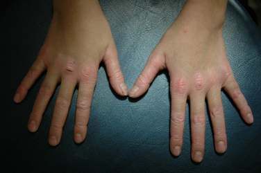
2129 International Journal of Collaborative Research on Internal Medicine & Public Health
affected with a carrier rate of 2–4 per 1000.The likely mechanisms relating genetics and
periodontal disease include virulent infection, immune response and underlying tissue
pathology.4,5
It may manifests “between” 1-5 year of life. Another component of this syndrome is
asymptomatic ectopic calcification in choroid plexus and tentorium. Although this has been
taken as a cardinal feature, but being inconsistent it is not considered important for the
diagnosis. About 20% of these patients also show an increased susceptibility to infections
due to some dyes-function of lymphocytes and leukocytes.6 The diagnosis is mainly
clinical. We describe here case of PLS with classic clinical features.
Case Report
An 11-year-old girl reported to the department of Periodontics for periodontal assessment
and discomfort in chewing. The family history revealed to marriage of the parents. The
parents and other family members were not affected. Pregnancy period and delivery were
normal. The mother had noticed of skin lesions on the palms and soles of the child when
they were five months old. On general examination, the patient had overall normal physical
and mental development. Routine hematological examination revealed Hb was 10.0 g/dl,
total white blood cell was 9200 and ESR was 20 mm/hour. On dermatological examination
there were multiple fissured erythematous hyperkeratotic plaques on the soles. Xerosis and
erythema on the palms and dispersed erythematous papules on the trunk.
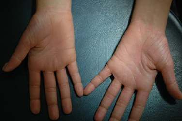
In extra oral examination were seen hyperkeratosis of the palms, soles, and the knees of
both the limbs; the affected skin was well demarcate from adjacent normal skin but the hair
or nail anomaly can be observe. (Fig 1-4).
Intraoral examination
On oral cavity examination the patient had permanent dentition with no primary teeth,
normal gingival and periodontum, mild deposits of plaque and calculus, were present
(Figure 5).
Orthopantomograph examination
Showed no destruction of alveolar bone. Laboratory investigation assessment was carried
out, which included hematological and biochemical test. The results were within normal
limits. In view of the findings, the cases were diagnosed as PLS. Treatment was planned to
restore masticatory function with space maintainer to keep the teeth in correct position and
to restore masticatory function.
Vol. 4 No. 12 (2012)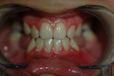
2130    International Journal of Collaborative Research on Internal Medicine & Public Health

  Discussion
  The Papillon-Lefèvre syndrome, described by two French physicians Papillon and Lefèvere
  for first time in 1924,1 Papillon-Lefèvre syndrome (PLS) is an autosomal recessive
  disorder29,and extremely rare genodermatosis inherited as an autosomal recessive trait, its
  affect children between the ages 1-4 years.7,8 It has a prevalence of 1-4 cases per million
  persons.9 Both sexes are affected equally and there is no racial predominance.10 Disorder is
  characterized by diffuse palmoplantar keratoderma and typically has its onset between age
  1 and 4 years. It may have premature loss of both deciduous and permanent teeth years.11
  PLS involve the entire surface of the palms and soles and may be extending on to the dorsal
  surfaces of the hands and feet.12 Hyperhidrosis of the palms and soles may result in a foul-
  smelling odor.8 Well-demarcated psoriasiform plaques occur on the elbows and knees.12
  The clinical findings may worsen in winter and be associated with painful fissures. Severe
  periodontitis is second major feature of PLS, which starts at age 3 or 4 years. 12 The
  eruption of the deciduous teeth proceeds normally, but sometimes the process is associated
  with appearance of gingival inflammation and subsequent rapid destruction of the
  periodontium.13,4 The level of periodontal infection may not be related to degree of
  dermatologic involvement.15 In advanced cases nail changes may appear and in this case,
  manifested by transverse grooving and fissuring.16 In addition to the skin and oral findings,
  decreased neutrophil, lymphocyte, or monocyte functions and an increased susceptibility to
  bacteria, of the patients may have associated with recurrent pyogenic infections of the
  skin.17 One of complication of PLS is associated with impairment of the immune system
  and pyogenic liver abscess is increasingly recognized. 17 Another feature of this syndrome
  may be radiographic evidence of intracranial calcification.18

  In the literature histopathological findings of affected skin have not been well described.
  But those reported findings have consisted of hyperkeratosis, occasional patches of
  parakeratosis, acanthosis, and a slight perivascular inflammatory infiltrate.19 PLS causes is
  not well understood, but recently, 2 research investigator have reported that loss of function
  mutations affecting both the alleles of the cathepsin-C gene, located on chromosome
  11q14.1-q14.3, were associated with this syndrome.13,20 Prevalence increase of parental
  consanguinity has been reported in PLS patients.20 The palmoplantar keratodermas (PPKs)
  are a heterogeneous group and more than 40 different types of PPK have been described.21
  PLS, HMS has been described as allelic variants of prepubertal periodontitis. And
  described consider only HMS and premature periodontitis in the differential diagnosis of
  this syndrome. Haim Munk syndrome has been mentioned as an autosomal-recessive
  genodermatosis characterized by congenital palmoplantar keratoderma. In 1965 Haim and
  Munk first reported in Jewish families from Cochin, India on the Malabar Coast. 23 In
  Haim-Munk syndromes affecting a highly conserved amino-acid residue, and
  demonstrating that PLS and are allelic disorders .The cutaneous findings in HMS have been
  described to be more severe and extensive. 24

                                                                        Vol. 4 No. 12 (2012)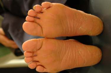
2131    International Journal of Collaborative Research on Internal Medicine & Public Health

  An important multidisciplinary approach for the care of patients with PLS. The skin
  manifestations of PPK are usually treated with emollients.12 It may add Salicylic acid and
  urea to enhance their effects.17 Treatment might be started during the eruption and
  maintained during the development of the permanent teeth.22,25 The periodontitis usually
  PLS is usually difficult to control. Effective treatment are includes extraction of the
  primary teeth combined treatment with oral antibiotics and professional teeth cleaning.17,26
  Antibiotics tried to control the active periodontitis in an effort to preserve the teeth and to
  prevent bacteremia and subsequently pyogenic liver abscess.17 Noack et al in their studies
  mentioned that PLS should be considered in all children suffering from severe aggressive
  periodontitis, particularly in the deciduous dentition, although the type and location of
  Cathepsin C (CTSC) mutations do not predict the severity, progression or therapy outcome
  of the disease.27

  Conclusion
  We have described an 11 years Old Iranian girl diagnosed as having highly suspected PLS
  and slight palmoplanter keratosis of hands and soles, together with knee pigmentation.

  Conflict of Interest: None declared.

  References
  1- Papillon M M, Lefèvre P. Deux cas de keratodermie palmaire et plantaire symmetrique
  famiale (maladie de Meleda) chez le frère et la soeur: coéxistance dans les deux cas
  d'altérations dentaires graves. Bull Soc Derma Symp 1924; 31:82-7.

  2- Shahbaz A, Amor MD. Papillon – Lefevre syndrome: Case report and review of
  literature. Dermatology Online Journal 2004; 10:13.

  3- Carmel Toomes, Jacquiline James, Joseph Wood. Loss-of function mutations in the
  cathepsin C gene result in periodontal disease and palmoplantor keratosis. Nature Genetics
  2003; 23: 421-424.

  4-Griffiths WAD, Judge MR, Leigh IM. Disorders of keratinization In: Champion RH,
  Burton JL, Burns DA, Breathnach SM eds. Textbook of Dermatology, 6th Ed. Oxford:
  Blackwell Scientific Publications, 1998; 1569-1571.

  5- Bas AY, Yilmaz G. Papillon-Lefevre syndrome: a case report. International Pediatrics
  2004; 19:224-225.

                                                                         Vol. 4 No. 12 (2012)
2132    International Journal of Collaborative Research on Internal Medicine & Public Health

  6- Bergman R, Friedman-Birnbaum R. Papillon-Lefevre syndrome a study of the long term
  clinical course of recurrent pyogenic infections and the effects of etretinate treatment. Br J
  Dermatol. 1998; 119: 131-136.

  7- Hart TC,Shapira L. Papillon-Lefèvre syndrome.Periodontol 2000;1994 Oct; 6:88- 100.

  8- Gorlin RJ, Sedano H, Anderson VE. The syndrome of palmar-plantar hyperkeratosis and
  premature periodontal destruction of the teeth. J Pediatr. 1964;65:895-908.

  9- Cury VF, Costa JE, Gomez RS, Boson WL, Loures CG, De ML.A novel mutation of the
  cathepsin C gene in Papillon-Lefèvre syndrome. J Periodontol. 2002 Mar;73(3):307-12.

  10-Bach J N,Levan N E.Papillon-Lefèvre syndrome. Arch Dermatol. 1968; Feb;97(2):154-
  8.

  11- Siragusa M, Romano C, Batticane N, Batolo D, Schepis C. A new family with
  Papillon-Lefèvre syndrome: effectiveness of etretinate treatment. Cutis. 2000; Mar;65
  (3):151-5.

  12- Hart TC, Hart PS, Bowden DW, Michalec MD, Callison SA, Walker SJ, Zhang Y,
  Firatli E. Mutations of the cathepsin C gene is responsible for Papillon-Lefèvre syndrome.J
  Med Genet. 1999; Dec 36(12):881-7.

  13- Haneke E. The Papillon-Lefèvre syndrome: keratosis palmoplantaris with
  periodontopathy. Report of a case and review of the cases in the literature. Hum Genet.
  1979; 51 (1):1-35.

  14- Ullbro C, Crossner CG, Nederfors T, Alfadley A, Thestrup-Pedersen K. Dermatologic
  and oral findings in a cohort of 47 patients with Papillon-Lefèvre syndrome. J Am Acad
  Dermatol. 2003 Mar; 48 (3):345-51.

  15- Giansanti JS, Hrabak RP, Waldron CA.Palmar-plantar hyperkeratosis and concomitant
  periodontal destruction (papillon-Lefèvre syndrome). Oral Surg Oral Med Oral Pathol.
  1973 Jul; 36(1):40-8.

  16- Almuneef M, Al Khenaizan S, Al Ajaji S, Al-Anazi A. Pyogenic liver abscess and
  Papillon-Lefèvre syndrome: not a rare association. Pediatrics. 2003 Jan; 111 (1):e85-8.

  17- Reyes VO, King-Ismael D, Abad-Venida L. Papillon-Lefèvre syndrome. Int J
  Dermatol. 1998 Apr; 37(4):268-70.

  18- Angel TA, Hsu S, Kornbleuth SI, Kornbleuth J, Kramer EM.Papillon-Lefèvre
  syndrome: A case report of four affected siblings. J Am Acad Dermatol. 2002 Feb; 46(2
  SupplCaseReports):S8-10.

                                                                        Vol. 4 No. 12 (2012)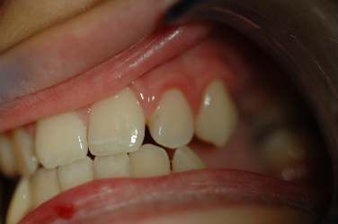
2133    International Journal of Collaborative Research on Internal Medicine & Public Health

  19- Toomes C, James J, Wood AJ, Wu CL, McCormick D, Lench N, Hewitt C, et. al. Loss-
  of-function mutations in the cathepsin C gene result in periodontal disease and
  palmoplantar keratosis. Nat Genet. 1999 Dec; 23 (4):421-4.

  20- Pilger U, Hennies HC, Truschnegg A, Aberer E. Late-onset Papillon-Lefevre syndrome
  without alteration of the cathepsin C gene. J Am Acad Dermatol. 2003 Nov; 49(5
  Suppl):240-3.

  21- Itin PH. Classification of autosomal dominant palmoplantar keratoderma: past-present-
  future. Dermatology. 1992;185:163-165.

  22- Haim S, Munk J. Keratosis palmo-plantaris congenital, with periodontosis,
  arachnodactyly, and peculiar deformity of the terminal phalanges. Br J Dermatol. 1965;
  77:42-54.

  23- Hart TC, Hart PS, Michalec MD, Zhang Y, Firatli E, et. al. Haim-Munk syndrome and
  Papillon-Lefèvre syndrome are allelic mutations in cathepsin C. J Med Genet. 2000 Feb;
  37(2):88-94.

  24- Blanchet-Bardon C, Nazzaro V, Rognin C, Geiger JM, Puissant A. Acitretin in the
  treatment of severe disorders of keratinization.Results of an open study.J Am Acad
  Dermatol.1991Jun ;24 (6Pt1):982-6.

  25- Ishikawa I, Umeda M, Laosrisin N. Clinical, bacteriological, and immunological
  examinations and the treatment process of two Papillon-Lefèvre syndrome patients. J
  Periodontol.1994, Apr; 65(4):364-71.

  26- Galmetti C, Nazzaro V, Cerri D, Fracasso L. Long-term preservation of permanent
  teeth in a patient with Papillon-Lefèvre syndrome treated with etretinate. Pediatr Dermatol.
  1989 Sep; 6(3):222-5.

  27-Noack B, Gorgens H, Schacher B , Puklo M, Eickholz P, et al. Functional Cathepsin C
  mutations cause different Papillion–Lefebvre syndrome phenotypes. J Clin Periodontol.
  2008 doi: 10.1111/j.1600-051X.2008.01201.

                                                                       Vol. 4 No. 12 (2012)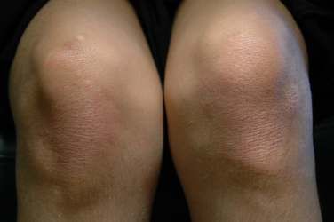
2134   International Journal of Collaborative Research on Internal Medicine & Public Health

            Figure 1: Absence of first and 2nd premolar in upper and lower jaw

                                                                   Vol. 4 No. 12 (2012)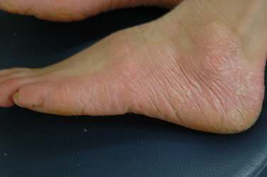
2135   International Journal of Collaborative Research on Internal Medicine & Public Health

                  Figure 2: Yellow colored hyperkeratotic areas on sole

                                                                  Vol. 4 No. 12 (2012)
2136 International Journal of Collaborative Research on Internal Medicine & Public Health
Figure 3: Photograph showing hyperkeratosis of the knees, which is well demarcated from
adjacent normal skin
Vol. 4 No. 12 (2012)2137 International Journal of Collaborative Research on Internal Medicine & Public Health
Figure 4: Photograph showing hyperkeratosis of the palms
Vol. 4 No. 12 (2012)2138 International Journal of Collaborative Research on Internal Medicine & Public Health
Figure 5: Orthopantomograph showing missing of first and 2nd premolars in mandible and
maxilla
Vol. 4 No. 12 (2012)You can also read